The National Preconception Care Clinical Toolkit (“PCC Toolkit”)

Advanced Care Toolkit

OverviewPatients that survive an out of hospital cardiac arrest (OHCA) get admitted to hospitals. In hospital care is a critical link to the chain of survival for all the patients of OHCA that has been resuscitated or are brought in the hospital with on going CPR. The majority of deaths occur either at the scene or within the first 24 hours from admission to the hospital.
Fundamentally, in-hospital care can make a significant difference in the overall mortality and morbidity as well as long term quality of life.
We will focus on two important issues that have substantial evidence behind them to establish them as best practices and standard of care.
1) In hospital early access to therapeutic hypothermia (TH)2) Early access to cardiac catheterization laboratories (CCL) in patients
that present with VF/VT or AED shockable rhythms.
Leadership. Organization of the EMS providers to deliver patients to participating hospitals that provide high quality therapeutic hypothermia programs and participate in early cardiac catheterization laboratory access is a very important task. Smooth operation from delivery of the patient to the emergency department, access to CCL, early initiation of TH either en route to the hospital or by ED or cath lab physicians (depending on individual hospital practices) is critical to shave of time from providing potentially lifesaving treatments.
To provide superior care a close working relation needs to be established between paramedics, emergency physicians, cardiologists and intensive care unit doctors. The need for a champion to take over the task of multidisciplinary dialogue and writing protocols that transcend and cross between traditional boundaries of medical care cannot be underscored enough.
Program ProtocolsBelow there are samples of successful implementation protocols on therapeutic hypothermia and access to cardiac catheterization laboratory for resuscitated patients from VF /VT with out-of-hospital cardiac arrest
Advanced Care
1
1)Therapeutic Hypothermia and Post Cardiac Arrest Care Pathway Implementation Sample Protocol
Courtesy of the University of Pennsylvania TH Program Special acknowledgement goes to Drs Abella and Becker.
Similar programs are established in all the participating hospital of the Minnesota Resuscitation Consortium.
I. EQUIPMENT LIST
All equipment is available in ED, MICU & CCU.
If protocol will be initiated in an ICU, have all equipment available and assembled at bedside before transfer to unit.
1. Arterial line kits (both radial and femoral).
2. PreSep central venous oximetry catheter (Edwards Lifesciences).
3. Two one liter bags of cold 0.9% saline (stored in ED and ICU refrigerators).
4. Gaymar Medi-Therm III 7900 external cooling system (available in ED and ICU):
a. Gaymar Rapr-Round cooling pads: one torso and two thigh cooling pads, sized appropriately for patient.
b. Weight of Gaymar wraps when filled:
1. Large Torso: 3.0 lbs
2. Medium/Small Torso: 2.5 lbs
3. Each Leg: 2.0 lbs
5. Temperature probe foley catheter with appropriate adapter for cooling device.
Bard Temperature Sensing Foley 400 Series (product #90911616).
(Gaymar Medi-Therm III requires 1/8 inch to 1/4 inch converter)
6. Neuromuscular blockade equipment (not required for ED):
Advanced Care
2
a. Peripheral nerve stimulator
b. BIS monitor and sensor
7. Ensure fluid warmer is available in case need arises after cooling.
II. LOGISTICAL PEARLS REGARDING THERAPEUTIC HYPOTHERMIA
1. Ensure appropriate supervisory staff is notified:
A: Resuscitation Consult Team: add a phone number
*If you can not reach the Resuscitation Consult Team consider notifying any of following:
additional members to provide support in case of emergency.
2. Medical patients most appropriate for MICU or CCU should be admitted to either of those units if possible, rather than to a ready-bed on an alternative unit.
Surgical patients should be admitted to the appropriate SICU, NTSICU or CTSICU.
3. ED personnel should continue to implement TARGETED TEMPERATURE MANAGEMENT
protocol until ICU bed is available and all equipment is ready for patient.
4. Place arterial line while initiating cooling (may be very difficult to place once the patient is at target temperature).
5. Paralyze patient (after sedation), before initiating cooling. Maintain paralysis until after re-warming is complete (36°C).
If using the Blue-faced Gaymar Medi-Therm III:o Set device to Automatic mode, Rapid with target temp 33°C. (Goal
is target temp within 4 hours.) o Rewarming is begun 24 hours after target temperature is reached.
Set in Automatic mode, moderate, with target temperature of 37°C (this will rewarm patient @ 0.33°C/hr, [1°C/3hrs]). Maintain sedation and paralysis until temperature reaches 36°C to avoid shivering and rapid rewarming.
If using the Gray-faced Gaymar III:o Set device to Automatic mode, Rapid cooling with set point of 34ºC.
Once the patient reaches 34ºC set to Gradual mode at 33ºC.
Advanced Care
3

o Rewarming is begun 24 hours after target temperature is reached. Set in Manual mode and manually increase the blanket temperature 0.5ºC every 2 hours until the patient temperature reaches 36ºC.
6. Notify neurology team ASAP, by paging xxxxx, to arrange for continuous EEG monitoring within 6 hrs and no later than 12 hrs after onset of cooling (EEG techs are not available between midnight and 7:30 am). EEG is monitored for 24 hours after the patient has reached normothermia. EEG should not be discontinued until the patient is re-warmed and paralytics discontinued.
a. For issues or concerns, or if you cannot get a hold of the EEG fellow, please page the Neurology on call resident.
III. PURPOSE: To provide a guideline to optimize the care of comatose cardiac arrest survivors.
IV. BACKGROUND
A. TARGETED TEMPERATURE MANAGEMENT (TTM)Brain temperature during the first 24 hours after resuscitation from cardiac arrest has a significant effect on survival and neurological recovery.
Fever (T°max) during the first 48 hours is associated with a decreased chance of good neurological recovery (OR 2.26 [1.24, 4.12] for each 1°C over 37°C)1. Two RCTs provided the best evidence to support the use of TH in the appropriate comatose survivors of cardiac arrest. In the first, cooling to 32-34°C for 24 hours was associated with a decreased mortality (OR 0.74 [0.58, 0.95]) and increased likelihood of good neurological recovery (OR 1.40 [1.08,1.81])2. In the second study, cooling to 32-34°C for 12 hours increased the likelihood of good neurological recovery (OR 2.65 [1.02, 6.88])3.
B. Early Coronary RevascularizationOut-of-hospital cardiac arrest patients have a high incidence of acute coronary syndrome and early coronary revascularization may improve survival (OR 5.2 [1.1, 24.5])4.
C. Early Goal-Directed TherapyPost-resuscitation syndrome has many pathophysiological features in common with acute sepsis5. Early goal-directed therapy has been demonstrated to decrease in-hospital mortality of patients suffering from severe sepsis with an elevated lactate or septic shock (OR 0.58 [0.38-0.87])6. A similar approach may have the same beneficial effects in post-resuscitation syndrome.
D. Glycemic Control
Advanced Care
4
Avoiding hyper- and hypo-glycemia (goal 110-150) is a reasonable goal during post-arrest resuscitation and TTM. This should be accomplished using ICU specific insulin protocols. 7, 8.
E. Management of Adrenal InsufficiencyAcute adrenal insufficiency is a well-documented component of post-resuscitation syndrome. In patients with fluid and vasopressor refractory septic shock, treatment with “stress dose” corticosteroids significantly reduces mortality (OR 0.67 [0.47-0.95])9. Thus, in post-cardiac arrest patients with hemodynamic instability associated with vasodilation, diagnosis and treatment of acute adrenal insufficiency should be considered.
F. PrognosisThe neurologic prognosis of the comatose cardiac arrest survivor cannot be reliably predicted until at least 72 hours after resuscitation10. In addition, the reliability of the routinely utilized neurologic prognostic parameters has not been evaluated in patients treated with TTH, Therefore DNAR status should not be established and care should not be withdrawn based on neurologic prognosis before 72 hours after rewarming.
Advanced Care
5
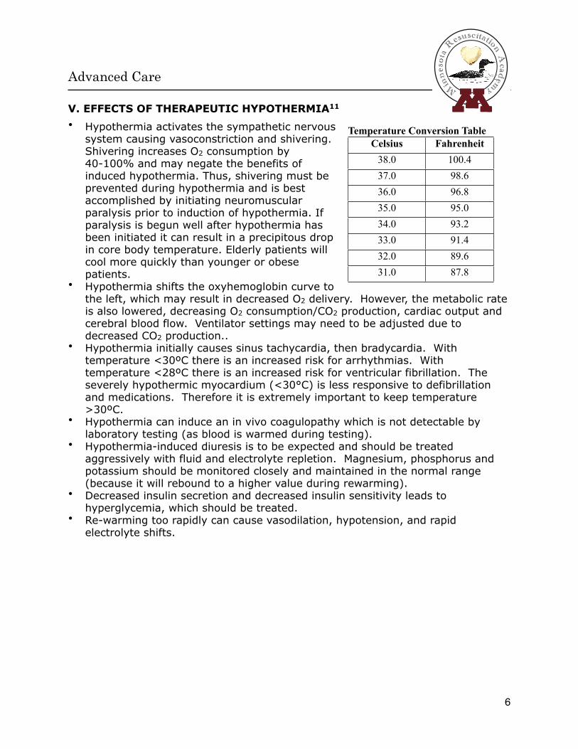
V. EFFECTS OF THERAPEUTIC HYPOTHERMIA11
• Hypothermia activates the sympathetic nervous system causing vasoconstriction and shivering. Shivering increases O2 consumption by 40-100% and may negate the benefits of induced hypothermia. Thus, shivering must be prevented during hypothermia and is best accomplished by initiating neuromuscular paralysis prior to induction of hypothermia. If paralysis is begun well after hypothermia has been initiated it can result in a precipitous drop in core body temperature. Elderly patients will cool more quickly than younger or obese patients.
• Hypothermia shifts the oxyhemoglobin curve to the left, which may result in decreased O2 delivery. However, the metabolic rate is also lowered, decreasing O2 consumption/CO2 production, cardiac output and cerebral blood flow. Ventilator settings may need to be adjusted due to decreased CO2 production..
• Hypothermia initially causes sinus tachycardia, then bradycardia. With temperature <30ºC there is an increased risk for arrhythmias. With temperature <28ºC there is an increased risk for ventricular fibrillation. The severely hypothermic myocardium (<30°C) is less responsive to defibrillation and medications. Therefore it is extremely important to keep temperature >30ºC.
• Hypothermia can induce an in vivo coagulopathy which is not detectable by laboratory testing (as blood is warmed during testing).
• Hypothermia-induced diuresis is to be expected and should be treated aggressively with fluid and electrolyte repletion. Magnesium, phosphorus and potassium should be monitored closely and maintained in the normal range (because it will rebound to a higher value during rewarming).
• Decreased insulin secretion and decreased insulin sensitivity leads to hyperglycemia, which should be treated.
• Re-warming too rapidly can cause vasodilation, hypotension, and rapid electrolyte shifts.
Advanced Care
6
Temperature Conversion TableCelsius Fahrenheit
38.0 100.437.0 98.636.0 96.835.0 95.034.0 93.233.0 91.432.0 89.631.0 87.8

POTENTIAL LABORATORY ABNORMALITIES ASSOCIATED WITH HYPOTHERMIA:
Potential Lab Abnormality TreatmentIncreased amylase No intervention unless persistent after rewarmingIncreased LFTs No intervention unless persistent after rewarmingIncreased serum glucose Follow Insulin protocolDecreased K+, Mg, Phos, Ca Correct as neededIncreased lactate Optimize oxygen deliveryMetabolic acidosis Optimize oxygen deliveryThrombocytopenia Correct if active bleedingLeukopenia No intervention unless persistent after rewarmingIncreased PT/PTT Correct if active bleeding
Advanced Care
7
VI. ELIGIBILITY CRITERIA FOR POST-CARDIAC ARREST CARE PATHWAY
Post-cardiac arrest, defined as a period of absent pulses requiring chest compressions, regardless of location or presenting rhythm followed by return of spontaneous circulation (ROSC).
Not DNAR-B or C or DNI status prior to Cardiac Arrest.
Pre-arrest cognitive status not severely impaired (i.e. performed ADL independently).
VII. ELIGIBILITY CRITERIA FOR POST-CARDIAC ARREST TARGETED TEMPERATURE MANAGEMENT
Meets eligibility criteria for Post-Cardiac Arrest Care Pathway.
Full Code or DNAR-A
Comatose at enrollment with a Glasgow Coma Motor Score <6 pre-sedation (i.e. does not follow commands – two thumbs up, squeeze and release).
No other obvious reasons for coma.
No uncontrolled bleeding.
Absence of multi-organ dysfunction syndrome, severe sepsis, or a comorbidity associated with minimal chance of meaningful survival independent of neurological status.
Less than 12 hours since ROSC. Data support cooling patients as soon as possible post-arrest.
VIII. RELATIVE CONTRAINDICATIONS FOR TARGETED TEMPERATURE MANAGEMENT:
Pregnancy (as per one case report, TTM can be performed safely on a pregnant female14)
- Consult Maternal Fetal Medicine service if pregnant and TTM instituted.
Advanced Care
8
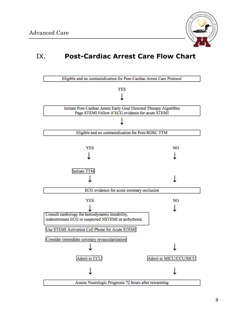
IX. Post-Cardiac Arrest Care Flow Chart
Advanced Care
9

X. POST-CARDIAC ARREST CARE PATHWAY
NOTE: Data Gathering (Section A), Monitoring (Section B), and Interventions (Section C) are all initiated immediately and carried out simultaneously when feasible.
A. Initial Data Gathering (after ABCs are stabilized)
1. History:
a. Review eligibility, contraindications, advance directives and overall prognosis
b. Discuss issues with health care proxy, if available
2. Physical: Baseline Neurological Evaluation
a. Exclude other causes of coma (mass lesions, metabolic coma, seizures etc)
b. Document Glasgow Motor Score and Glasgow Coma Score
3. Initial laboratories:
a. ABG with iCa+, Mg+
b. CBC / PT / PTT/INR, Fibrinogen
c. P7, plus iCa / Mg / Phos
d. Lactate/CPK-MB/CK/Troponin
e. Cortisol level (As Indicated)
f. Urinanalysis
g. Blood Cultures, Urine Culture, and Sputum Culture (if appropriate)
h. Toxicology screen if appropriate
1. Amylase, Lipase
j. Beta HCG on all women of child-bearing age
k. Co-oximetry (Central)
4. Serial laboratories:
a. Co-oximetry q1-2 hrs for first 6 hours until re-warmed and then q 6 hrs if PreSEP not used
b. Lactate q 6 hrs x 2 days
c. Repeat CPK-MB/CK/Troponin q 6 hrs
d. CBC / PT / PTT/INR, P7 / iCa / Mg / Phos q 6 hours
Advanced Care
10
e. ABG q6hr and PRN
f. P7, plus iCa / Mg / Phos
5. CXR
6. Head CT: To rule out intracranial hemorrhage, or other cause for coma, as deemed medically necessary
7. SSEP: Order day 3 (72 hours-post-ROSC). Place order for SSEP in Sunrise with a note that states “post-cardiac arrest” or for questions call the lab at 215-662-2661.
8. Consult:
a. Cardiology for:- hemodynamic instability, indeterminate ECG or suspected Non-STEMI,
or arrhythmia (use Cardiology Consult pager)- Acute STEMI (Use STEMI Activation Cell Phone)
Note: If cardiac catheterization is indicated, TTM should not be delayed.
Echocardiogram: To r/o regional wall motion abnormality and severe contractile dysfunction. EF used for selection of vasoactive drugs.
-Consider early Echo if requested by Cardiology Consult, or in the case of any of the following: -Ongoing hypotension with possible cardiac etiology -Ongoing arrhythmias -Indeterminate ECG
Or, obtain routine Echocardiogram within first 24-48 hours of protocol
b. Maternal Fetal Medicine if +B-HCG (while initiating TTM).
c. Nutrition support on day 3.
d. Neurology
e. EEG fellow 215-404-6771. If you cannot get a hold of the EEG fellow, please page the Neurology resident on-call.
f. Resuscitation team 267-253-9035.
B. Establish Appropriate Monitoring Immediately:
1. Cardiovascular:
a. ECG after initial stabilization and repeat q 8 hours x 2 and prn to r/o acute coronary syndrome.
Advanced Care
11
b. Arterial-line for continuous arterial blood pressure monitoring (essential prior to initiating hypothermia). Attempt radial artery x 1 and then proceed to femoral artery if necessary.
c. Temperature monitoring Foley (Bard Temperature Sensing Foley 400 Series - product #90911616 – no minimum urine output required for use) for continuous urine output and temperature monitoring. If bladder monitoring is not an option or temperature is inaccurate consider an alternative site for temperature measurement such as an esophageal temperature probe. NOTE: If bladder pressure being monitored, it is recommended that an esophageal probe be used for temperature monitoring.
d. PreSep catheter or other CVC (MAC) for CVP & Scv02 (RIJ or SCV site preferred) though don't delay initiation of hypothermia to perform this.
2. Pulmonary: Continuous SaO2 probe, frequent ABGs.
3. Temperature: Foley with temperature probe. (Bard Temperature Sensing Foley 400 Series - product #90911616 – no minimum urine output required for use) for continuous urine output and temperature monitoring. If bladder monitoring is not an option or temperature is inaccurate consider an alternative site for temperature measurement such as an esophageal temperature probe.
4. Neurologic:
a. Continuous EEG monitoring beginning ASAP while paralyzed. EEG must be initiated within 6-12 hours of TTM. EEG should not be discontinued until the patient is re-warmed and paralytics discontinued.
-For issues or concerns, or if you cannot get a hold of the EEG fellow, please page the Neurology on call resident.
b. Once in ICU, consider using BIS monitor to titrate sedation (with goal of 40-60).
c. Neuro checks q 2 hrs (while paralyzed follow pupils and titrate paralysis per NMB Nursing Policy).
d. Neuroprognostication should not take place until at least 72 hours after re-warming.
e. SSEP performed on day 3 (72 hours post-ROSC) or later. Place order for SSEP in Sunrise with a note that states “post-cardiac arrest” .
Advanced Care
12
5. Additional monitoring and follow-up studies:
a. If net fluid balance is > 5 liters in 24 hrs, monitor intrabdominal pressure (IAP) via Foley catheter after cooling device has been discontinued (call medical resident if IAP is ≥ 20 mmHg).
b. Consider repeat echocardiogram 24 hours after ROSC or as clinically indicated.
c. Repeat CXR in AM and after 72 hours to rule out aspiration pneumonia.
C. Initiate Appropriate Interventions
NOTE: Interventions should be carried out simultaneously when appropriate and feasible
1. Post-Cardiac Arrest Early Goal-Directed Therapy (See appendix or laminated algorithm)
a. Initial Fluid Infusion: Use NSS for first two liters (use 4°C NSS if initiating TTM) of IVF then change to LR unless hyperkalemia or hepatic insufficiency. If Acute Coronary Syndrome suspected first transfuse blood to Hgb ≥10. Change IVF back to NSS immediately prior to rewarming (to avoid rebound hyperkalemia).
b. IVF Resuscitation and CVP goals: Titrate IVF to ensure volume repletion using CVP as a guide. A minimum CVP ≥ 8 mm Hg is a reasonable target goal in most patients. Continue fluid boluses to reach MAP target, unless CHF, CVP>15, or > 5 liters; then consider right heart catheterization (RHC). If MAP target is reached, but shock is present (↓Scv02, particularly if oliguric or acidotic (elevated lactate), bolus IVF to CVP > 8, providing no CHF. If CHF, CVP>15, high dose vasopressors, or >5 liters positive fluid balance, proceed with RHC. If no hypotension or shock, no need to give fluid regardless of the CVP (i.e. even if < 8) if adequate urine output (>1mL/kg/hr).
c. Vasoactive Drug Use in the Volume Repleted Patient:
1. HTN: If MAP > 100 mmHg, titrate IV nitroglycerin, starting @ 10 mcg/min, to MAP goal. (See algorithm for additional detail.) If tachycardic or acute ischemia/MI without significant LV dysfunction (based on ECHO, absence of CHF, or venous desaturation) consider Esmolol infusion.
2. Hypotension: Goal MAP is 80mmHg, providing no evidence of ACS, CHF, or shock. If ACS, CHF, or shock consider lower MAP range depending on degree of myocardial ischemia/dysfunction.
Advanced Care
13

a. If EF is normal (>/ 50%), use Norepinephrine (Begin at 2-4 mcg/min, titrate to maintain MAP >70-80 mmHg) to reach MAP goal.
b. If EF is reduced (<50%):
• MAP Titration: Use dobutamine (2.5-20 mcg/kg/min) to MAP> 80 mmHg and/or ScvO2 >65%. If MAP falls, add Norepinephrine, Epinephrine, or place Intra-Aortic Balloon Pump (IABP) if severe.
• Scv02 Titration: Regardless of MAP, if Scv02 is low (< 65%), particularly if other signs of shock are present, consider PRBC to HgB >10 gram/dL and increase Dobutamine as tolerated. If MAP falls, add Norepinephrine, Epinephrine, or place Intra-Aortic Balloon Pump (IABP) if severe. NOTE: When rewarming, anticipate vasodilation and volume depletion (↓CVP and ↓Scv02+/- ↓MAP), and treat with IVF boluses based on same algorithm.
c. Institute appropriate critical care protocols for sepsis, GI /DVT/ VAP prophylaxis, low stretch protocol, etc.
2. Initiate TTM if indicated
a. Goal: Achieve target temp of 33°C (range 32°-34 °C) within 4 hrs and maintain for 24 hours from time cooling target temperature is reached.
b. Induction: Sedative and paralytic medications are begun prior to inducing hypothermia and are continued until patient is rewarmed to 36°C. If patients temp is ≤ 34° on presentation, maintain temp at 32-34°C with cooling blanket. Hold paralysis unless shivering is evident or temp rises to > 34°C despite cooling measures.
1. Use an HME for vent humidification.
2. Initiate sedation and analgesia with fentanyl (50 to 100 mcg IV bolus followed by 50 mcg/hour infusion) and/or propofol (5-10 mcg/kg/min infusion). Note: Propofol interferes with measurement of aPTTs. Please consider this when ordering propofol on patients already on heparin and use Factor Xa levels to monitor anticoagulation Lorazepam (2mg/ml) can be used as alternative to propofol. Consider BIS monitor to titrate sedatives (ICU) to 40-60.
Advanced Care
14
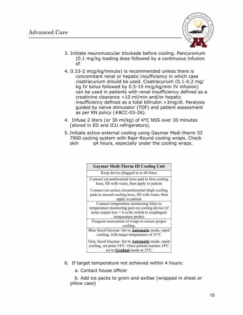
3. Initiate neuromuscular blockade before cooling. Pancuronium (0.1 mg/kg loading dose followed by a continuous infusion of
4. 0.33-2 mcg/kg/minute) is recommended unless there is concomitant renal or hepatic insufficiency in which case cisatracurium should be used. Cisatracurium (0.1-0.2 mg/kg IV bolus followed by 0.5-10 mcg/kg/min IV infusion) can be used in patients with renal insufficiency defined as a creatinine clearance <10 ml/min and/or hepatic insufficiency defined as a total bilirubin >3mg/dl. Paralysis guided by nerve stimulator (TOF) and patient assessment as per RN policy (#BCC-03-26).
4. Infuse 2 liters (or 30 ml/kg) of 4°C NSS over 30 minutes (stored in ED and ICU refrigerators).
5. Initiate active external cooling using Gaymar Medi-therm III 7900 cooling system with Rapr-Round cooling wraps. Check skin q4 hours, especially under the cooling wraps.
Gaymar Medi-Therm III Cooling UnitKeep device plugged in at all times
Connect circumferential torso pad to first cooling hose, fill with water, then apply to patient
Connect (in series) circumferential thigh cooling pads to second cooling hose, fill with water, then
apply to patientConnect temperature monitoring foley to
temperature monitoring port on cooling device (if urine output less < 4 cc/hr switch to esophageal
temperature probe)Frequent assessment of wraps to ensure proper
cooling. Blue faced Gaymar: Set to Automatic mode, rapid
cooling, with target temperature of 33°C
Gray faced Gaymar: Set to Automatic mode, rapid cooling, set point 34ºC. Once patient reaches 34ºC
set to Gradual mode at 33ºC
6. If target temperature not achieved within 4 hours:
a. Contact house officer
b. Add ice packs to groin and axillae (wrapped in sheet or pillow case)
Advanced Care
15
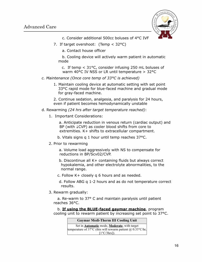
c. Consider additional 500cc boluses of 4°C IVF
7. If target overshoot: (Temp < 32°C)
a. Contact house officer
b. Cooling device will actively warm patient in automatic mode
c. If temp < 31°C, consider infusing 250 mL boluses of warm 40°C IV NSS or LR until temperature > 32°C
c. Maintenance (Once core temp of 33°C is achieved)
1. Maintain cooling device at automatic setting with set point 33°C rapid mode for blue-faced machine and gradual mode for gray-faced machine.
2. Continue sedation, analgesia, and paralysis for 24 hours, even if patient becomes hemodynamically unstable
d. Rewarming (24 hrs after target temperature reached):
1. Important Considerations:
a. Anticipate reduction in venous return (cardiac output) and BP (with ↓CVP) as cooler blood shifts from core to extremities. K+ shifts to extracellular compartment.
b. Vitals signs q 1 hour until temp reaches 37°C.
2. Prior to rewarming
a. Volume load aggressively with NS to compensate for reductions in BP/Scv02/CVP.
b. Discontinue all K+ containing fluids but always correct hypokalemia, and other electrolyte abnormalities, to the normal range.
c. Follow K+ closely q 6 hours and as needed.
d. Follow ABG q 1-2 hours and as do not temperature correct results.
3. Rewarm gradually:
a. Re-warm to 37° C and maintain paralysis until patient reaches 36°C.
b. If using the BLUE-faced gaymar machine, program cooling unit to rewarm patient by increasing set point to 37°C.
Gaymar Medi-Therm III Cooling UnitSet in Automatic mode, Moderate, with target
temperature of 37°C (this will rewarm patient @ 0.33°C/hr, [1°C/3hrs]).
Advanced Care
16
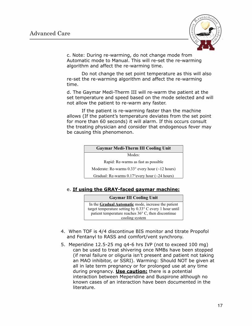
c. Note: During re-warming, do not change mode from Automatic mode to Manual. This will re-set the re-warming algorithm and affect the re-warming time.
Do not change the set point temperature as this will also re-set the re-warming algorithm and affect the re-warming time.
d. The Gaymar Medi-Therm III will re-warm the patient at the set temperature and speed based on the mode selected and will not allow the patient to re-warm any faster.
If the patient is re-warming faster than the machine allows (If the patient’s temperature deviates from the set point for more than 60 seconds) it will alarm. If this occurs consult the treating physician and consider that endogenous fever may be causing this phenomenon.
Gaymar Medi-Therm III Cooling UnitModes:
Rapid: Re-warms as fast as possible
Moderate: Re-warms 0.33° every hour (~12 hours)
Gradual: Re-warms 0.17°every hour (~24 hours)
e. If using the GRAY-faced gaymar machine:
Gaymar III Cooling UnitIn the Gradual Automatic mode, increase the patient
target temperature setting by 0.33° C every 1 hour until patient temperature reaches 36° C, then discontinue
cooling system
4. When TOF is 4/4 discontinue BIS monitor and titrate Propofol and Fentanyl to RASS and comfort/vent synchrony.
5. Meperidine 12.5-25 mg q4-6 hrs IVP (not to exceed 100 mg) can be used to treat shivering once NMBs have been stopped (if renal failure or oliguria isn’t present and patient not taking an MAO inhibitor, or SSRI). Warming: Should NOT be given at all in late term pregnancy or for prolonged use at any time during pregnancy. Use caution: there is a potential interaction between Meperidine and Buspirone although no known cases of an interaction have been documented in the literature.
Advanced Care
17

6. Discontinue active re-warming and remove Gaymar external cooling system and wraps when patient reaches temperature of 37°C.
3. Treat acute coronary syndrome
a. Treat everyone with single dose Aspirin per rectum (300 mg suppository) or OG (325mg Tab), unless contraindicated (allergy or active bleeding).
b. If ST segment MI or new LBBB, and no prolonged arrest time, cardiology may perform early cath. NOTE: Patients may receive cardiac interventions as needed while hypothermic.
4. Treat hyperglycemia Use ICU Insulin Infusion Protocol
5. Fever prophylaxis Acetaminophen 1 gram per rectum or per NGT, then 500 mg q 6 hours. If fever develops in the 48 hours following rewarming, aggressively treat with acetaminophen and cooling blanket to target temperature of 37 degrees Celsius as needed.
6. Other ICU protocols
a. If bilateral pulmonary infiltrates, use low stretch protocol based on Predicated Body Weight (obtain patient height) 13.
b. Pneumonia prevention with mouth care protocol and HOB > 30° at all times (unless hypotensive).
c. GI and DVT prophylaxis with ranitidine and SQ Heparin & Intermittent Compression Stockings.
d. NPO.
XI. Assessment of Neurologic Prognosis (Determined 72 hours after ROSC)
1. Determination of neurological prognosis is unreliable before 72 hours after ROSC.
2. Recommended criteria for initiating DNR status and/or withdrawal of care10:
Brain Region Test
72 hours POST-ROSC
Specificity for poor outcome
95% CI
Cortical and brainstem Brain Death Protocol 100%Brain stem Absence of pupillary light reflex 100% 88 to 100%
Cortical Absence of motor response to pain 100% 93 to 100%Cortical Bilateral absence of early cortical SSEPs 100% 98 to 100%
Advanced Care
18
XII. References:1. Zeiner A, Holzer M, Sterz F, et al. Hyperthermia after cardiac arrest is associated with
an unfavorable neurologic outcome. Arch Intern Med. Sep 10 2001; 161(16):2007-2012.
2. Hypothermia after Cardiac Arrest Study G. Mild therapeutic hypothermia to improve the neurologic outcome after cardiac arrest. [see comment][erratum appears in N Engl J Med 2002 May 30; 346(22):1756]. New England Journal of Medicine. Feb 21 2002; 346(8):549-556.
3. Bernard SA, Gray TW, Buist MD, et al. Treatment of comatose survivors of out-of-hospital cardiac arrest with induced hypothermia.[see comment]. New England Journal of Medicine. Feb 21 2002; 346(8):557-563.
4. Spaulding CM, Joly LM, Rosenberg A, et al. Immediate coronary angiography in survivors of out-of-hospital cardiac arrest. [see comment]. New England Journal of Medicine. Jun 5 1997; 336(23):1629-1633.
5. Adrie C, Laurent I, Monchi M, et al. Postresuscitation disease after cardiac arrest: a sepsis-like syndrome? Curr Opin Crit Care. Jun 2004; 10(3):208-212.
6. Rivers E, Nguyen B, Havstad S, et al. Early goal-directed therapy in the treatment of severe sepsis and septic shock. [see comment]. New England Journal of Medicine. 2001; 345(19):1368-1377.
7. van den Berghe G, Wouters P, Weekers F, et al. Intensive insulin therapy in the critically ill patients. [see comment]. New England Journal of Medicine. Nov 8 2001; 345(19):1359-1367.
8. Van den Berghe G, Wouters PJ, Bouillon R, et al. Outcome benefit of intensive insulin therapy in the critically ill: Insulin dose versus glycemic control.[see comment]. Crit Care Med. Feb 2003; 31(2):359-366.
9. Annane D, Sebille V, Charpentier C, et al. Effect of treatment with low doses of hydrocortisone and fludrocortisone on mortality in patients with septic shock.[see comment]. JAMA. 2002; 288(7):862-871.
10. Zandbergen EG, de Haan RJ, Stoutenbeek CP, et al. Systematic review of early prediction of poor outcome in anoxic-ischaemic coma. [see comment]. Lancet. Dec 5 1998; 352(9143):1808-1812.
11. Rello J. Risk factors for developing pneumonia within 48 hours of intubation. Am J Respir Crit Care Med. 1999; 159:1742-1746.
12. Sirvent JM. Protective effect of intravenously administered cefuroxime against nosocomial pneumonia in patients with structural coma. Am J Respir Crit Care Med. 1997; 155:1729-1734.
13. ARDSnet. Ventilation with lower tidal volumes as compared with traditional tidal volumes for acute lung injury and the acute respiratory distress syndrome. The Acute Respiratory Distress Syndrome Network. [see comment]. New England Journal of Medicine. 2000; 342(18):1301-1308.
14. Rittenberger JC et al. Successful outcome utilizing hypothermia after cardiac arrest in pregnancy: a case report. Critical Care Med 2008; 36(4):1354-6.
Advanced Care
19
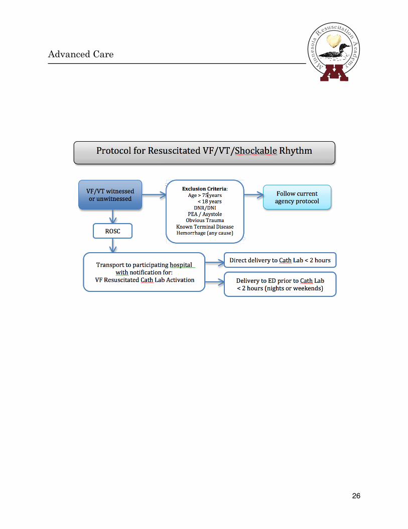
2. A quality improvement project for patients resuscitated from ventricular fibrillation in the Twin Cities.
The cardiac catheterization laboratory initiative.
The purpose of this initiative is to promote a system wide quality improvement protocol in all participating hospitals of the Minnesota Resuscitation Consortium (MRC). Starting in mid July 2012 all participating hospital will provide access to a cardiac catheterization laboratory (CCL) with PCI capabilities within 2 hours from successful field resuscitation of an initially recorded VF/VT arrest.
In patients with initial presenting rhythm of ventricular fibrillation or ventricular tachycardia or who were shocked by an AED, who are successfully resuscitated in the field, there is >70% chance of thrombotic and/or flow limiting lesions.1 There are multiple published case series showing that bringing these patients to a CCL is safe and leads to early diagnosis of the underlying etiology resulting in improved outcomes.2, 3 In addition, patients with witnessed refractory or recurrent VF arrest have a very high likelihood of a culprit thrombotic coronary lesion.
In the Twin Cities, the MRC data from the last six months of 2011 show that 33/40 (>80%) patients that received angiography early on (<6 hours) after resuscitated VF arrest, survived with good neurological function and a total of 36/40 survived to hospital discharge. By comparison, patients that did not get access to cardiac catheterization laboratories had a 33% hospital discharge rate and only 27% with good neurological function.
We anticipate that early access to CCL after resuscitated ventricular fibrillation cardiac arrest will improve survival and hospital discharge rates with good neurological function compared to our one year aggregate baseline data recorded by the MRC in 2011.
Consent and IRB process: This is a quality improvement project based on published best evidence and as such the protocol implementation is exempt from informed consent. Data will be monitored closely from the CARES database in the Twin Cities and close monitoring and patients tracking from CCL coordinators in each hospital. Project coordinators will be paid by the MRC and Heart Rescue Funds as well as from additional grant money/ funds from the University of Minnesota that are currently available. Grants are going to be available for up to 10% FTE salary support for data tracking and record keeping through the MRC.
Advanced Care
20
Implementation strategy: Starting July 6th2012 participating hospitals will start receiving patients from the major emergency medical services in the Twin Cities.
Inclusion criteria: Resuscitated VF/VT/AED shockable first recorded rhythm arrest, witnessed or unwitnessed, age >18 and <75, presumed cardiac etiology cardiac arrest, emergency medicine physician verification of the presenting initial rhythm and verification of the inclusion criteria, comatose or conscious patients.
Exclusion criteria: PEA or asystole as first recorded rhythm, known DNR/DNI, obvious non cardiac etiology, obvious GI bleeding, terminal illness (e.g. terminal cancer) and known acute intra cranial hemorrhage of various etiologies.
After inclusion criteria are met, patients will go to the CCL within two hours of ED arrival by utilizing established ST elevation myocardial infarction protocols.(NOTICE: ST-elevation on the ECG is not needed to be included in the protocol!).
Implementation strategy: Hospitals that will participate will agree to implement the new strategy of early access to the CCL within 2 hours from arrival to the ED. How the participating centers choose to evaluate these patients is left to the discretion of individual groups. Each center will have to implement a protocol that will clearly state if they want those patients held in the ED first and under which circumstances direct access to the CCL will be implemented. For example, during the day at the UMMC, the emergency department will be bypassed and patients will be treated as STEMIs brought directly into the CCL, but during the evening or night hours the patients will be held in the ED until CCL staff arrives for patient safety purposes. The rest of the centers will have to provide the MRC the exact strategy that they will follow so that the EMS directors have a clear picture of the available options. The only protocol obligation for the cardiology groups is that they will have to provide access to the CCL within two hours from the ED arrival with a goal to optimize and expedite this time.
Based on best practice guidelines we also ask the participating groups to collect the following data for the patients received in their centers:
Data Collection for Catheterization Lab Initiative as part of QA/QI Monitoring1. Standard CARES data
2. Troponin at 24 hours post arrest
3. In hospital hypothermia times
Advanced Care
21
a. Time to target temperature
b. Duration at target therapy
4. CCL times
a. 911 call to CCL door
b. ED door to balloon if angioplasty
5. CCL data (including entire catheterization report)
a. Number of vessels affected
b. Stents placed # and location
6. Neurological evaluation report at discharge (implement a standard evaluation of this patients with a neurological assessment at discharge with clear documentation of functional evaluation)
a. CPC
b. MRS
7. At least one left ventricular function ejection fraction evaluation before hospital discharge or at least 3 days after the arrest.
8. Was pt readmitted within 6 months of discharge, and why?
Responsibility for data acquisition by participating centers:1. Every 3 months a list of the patients should be reported to Lucinda
Klann ([email protected]) by the CCL medical directors to be identified in CARES so that a full record of all the patients is kept centrally securely and verified.
2. All data will be sent to MRC for aggregation of the complete data set and once data are verified de-identification will occur as per protocol under CARES.
Minnesota Resuscitation Consortium data management obligations.The MRC under the auspices of Department of Health will centrally manage the data as per standard protocol for every other arrest report in CARES. All data will be kept securely as per MRC protocols for current data sharing. Aggregate data will be kept by the MRC. No information will be made public or be available for anyone except for biannual reports that will provide data on outcomes for each center individually (and not to the group) and their comparison to the average MRC outcomes.
Privacy/PermissionsThe intention of this initiative is to improve care for patients post resuscitated VF/VT. After one year of operation, the collective results will be published to report the experience and outcomes in the twin cities to share with the rest of medical community.
Advanced Care
22
If a trend is identified in regards to timing of CCL and survival, we will apply as a consortium for funding to NIH to perform a randomized controlled trial that will be run through the MRC infrastructure and its participating centers.
Scientific Background for the proposed initiative: The American Heart Association (AHA) has recently issued a Policy Statement calling for the establishment of regional systems of care to manage out-of-hospital cardiac arrest (OHCA) patients.4 The proposed model is patterned after similar successful programs for major trauma, ST-segment elevation myocardial infarction (STEMI), and stroke. The Policy Statement recommends a comprehensive, regionalized approach to post-resuscitation care that includes therapeutic hypothermia, goal-directed management of critical physiological functions (i.e., organ perfusion, ventilation, glucose levels), and early coronary angiography/percutaneous intervention (PCI) when indicated.
Up to 71% of OHCA patients have coronary artery disease and nearly half have an acute coronary occlusion regardless of the post resuscitation ECG findings. 1, 5-18 Even under the best reported circumstances, mortality in the overall post arrest population who have been initially resuscitated is approximately 50%. An aggressive revascularization approach, has been shown to result in improved survival from OHCA.1, 2, 6-11, 13-21
The role of PCI in OHCA patientsTwenty-five percent of patients who survive OHCA to hospital
admission have evidence of STEMI (including new left bundle branch block) on their presenting ECG. The American Heart Association 2010 Guidelines on Cardiopulmonary Resuscitation and Emergency Cardiac Care state that "aggressive treatment of ST elevation myocardial infarction (STEMI) on the presenting ECG should begin as in non-cardiac arrest patients, regardless of coma or induced hypothermia". They also state that "because of the high incidence of acute coronary ischemia, consideration of emergent coronary angiography may be reasonable even in the absence of STEMI.”22 The basis for this recommendation is the recognized role of acute coronary ischemia as a dominant mechanism in the setting of OHCA. The potential contribution of coronary ischemia in OHCA was initially observed in a post mortem case series as well as in angiography data obtained in survivors of sudden cardiac death.5, 23
Coronary angiography followed by successful coronary intervention has been shown to be an independent predictor for survival and improved neurological outcomes for patients with OHCA irrespective of the presence or absence of STEMI on the presenting ECG. The supportive data for emergent coronary angiography is most compelling among patients with manifest ST elevation on the surface ECG as the benefits of emergent reperfusion of the infarct related artery is well established in this setting. In a retrospective study limited to cardiac arrest cases, Garot et al.20 reported on outcomes in 186 consecutive patients over a 10 year period undergoing immediate
Advanced Care
23
percutaneous coronary intervention (PCI) after successful resuscitation for cardiac arrest complicating acute myocardial infarction with STEMI on the initial ECG. PCI was successful in 87%. Survival at 6 months was 54% and survival free of neurological sequelae at 6 months was 46%.
Bendz et al. 24 compared 40 patients treated with primary PCI after OHCA with a reference group of 325 patients without CA also treated with primary PCI in the same period. In-hospital mortality was 27.5% in the CA group and 4.9% in the STEMI without CA group. The two year mortality rate was unchanged in the CA group and rose to 7.1% in the non-CA STEMI group. Gorjup et al. 16 reported on 135 STEMI-patients between 2000 and 2005 resuscitated after CA. Catheterization was performed in all patients, of whom 64% remained comatose during the initial evaluation. In comatose patients, primary PCI was performed in 79% with a procedural success rate of 82% demonstrating that successful PCI can be urgently accomplished in comatose post arrest patients. Survival to hospital was 51% for comatose post arrest patients, 100% for non-comatose post arrest patients, and 95% for patients with STEMI and no CA.
In an angiographic analysis of 84 consecutive patients with OHCA aged 30-75 years by Spaulding et al.1, 60 subjects were noted to have a severe coronary stenosis and 40 (48%) had an occluded coronary artery, irrespective of the presence or absence of STEMI on the initial ECG. On multivariate logistic-regression analysis successful angioplasty was noted to be an independent predictor of survival (odds ratio, 5.2; 95 percent confidence interval, 1.1to 24.5; p= 0.04). The same group of investigators confirmed their findings in the larger PROCAT Registry which performed urgent angiography in 435 of 714 patients with OHCA without an obvious extra cardiac cause.25 Immediate coronary angiography in this population revealed a significant stenosis in 96% (128/134) of subjects with manifest ST elevation on surface ECG after return of spontaneous circulation (ROSC) and in 58%, (176/301) of patients without ST elevation. As in the prior study, multivariable analysis showed successful coronary angioplasty to be an independent predictive factor of survival, regardless of the post resuscitation ECG pattern (odds ratio, 2.06; 95% CI, 1.16 to 3.66). This cohort of patients 70% of the patients resuscitated had VF/VT as presenting rhythm.
Strote et al. 26 evaluated a retrospective cohort of 240 patients with OHCA with ventricular fibrillation (VF) or pulseless ventricular tachycardia (pVT) as the presenting rhythm. Survival was greater in those patients undergoing coronary angiography within 6 hours of presentation compared to later than 6 hours or not at all (72% vs. 49%, p = 0.001). Seventy-five percent of patients in the <6 hour group had STEMI on the presenting ECG, leading these authors to conclude that their data support the idea that all patients resuscitated from OHCA caused by VF or pVT should receive early cardiac catheterization and PCI if indicated. In this cohort 100% of patients had VF/VT as presenting rhythm.
Advanced Care
24
Ventricular Fibrillation/ Ventricular Tachycardia population:In the above referenced studies, the majority of patients included in the reported cohorts (varying from 70-100%) were resuscitated victims that presented with initial rhythm of VF/VT. Therefore the evidence best supports implementation of the proposed strategy in this population.
In summary, there is strong data supporting the Level I AHA guideline for taking all post arrest patients with STEMI to the catheterization laboratory immediately irrespective of the presence or absence of coma, and there is increasing evidence that patients without evidence of STEMI on presenting ECG resuscitated from VF/VT may also benefit from early intervention.
Impact on number of patients treated: Based on two large randomized trials with CPR including a total of 14,000 patients,27, 28 it is apparent that the presence of VF/VT is the single most predictive variable of MRS status at hospital discharge, present in about 33% of all cases, but accounting for approximately 90% of subjects with MRS≤3. Therefore any significant increase in the survival rates of the VF population would translate to an overall survival for OHCA patients with a factor of 0.9.Currently the overall survival in Minnesota from VF arrest (witnessed and unwitnessed) is 36% (for witnessed VF is 45%). In all the published literature the average survival of patients that get early access to CCL varies from 60-80%. Of course the true effect would be smaller since these are highly selected patients and there is physician bias at play. Assuming that the true effect would be smaller and overall survival would be closer to 55-60% we could still be close to doubling the overall survival of that group of patients (VF/VT presenting rhythm) and OHCA population as a whole.
Minnesota goal: Currently in Minnesota there are 300-400 patients/year that are treated conservatively after VF/VT arrest. From those, a small proportion (<20%) gains access early to cardiac catheterization laboratories (within 2 hours), mostly patients that present with STEMI on the post arrest ECG. We anticipate that the new protocol would increase the proportion to 80-90% of the patients and significantly increase the overall survival rates for MN.
Current participating hospitals include: University of Minnesota Medical Center, Abbot Northwestern, Fairview Southdale, Mercy, St Paul Heart, North Memorial and Minneapolis VAMC .
On behalf of the Minnesota Resuscitation Consortium and the EMS directors,
Demetris Yannopoulos M.D.Associate Professor of MedicineMedical Director, Minnesota Resuscitation ConsortiumDirector of Research, Interventional Cardiology SectionDivision of Cardiovascular Medicine, University of Minnesota
Advanced Care
25
Advanced Care
26

References:
1. Spaulding CM, Joly LM, Rosenberg A, Monchi M, Weber SN, Dhainaut JF, Carli P. Immediate coronary angiography in survivors of out-of-hospital cardiac arrest. N Engl J Med. 1997;336:1629-1633
2. Cronier P, Vignon P, Bouferrache K, Aegerter P, Charron C, Templier F, Castro S, El Mahmoud R, Lory C, Pichon N, Dubourg O, Vieillard-Baron A. Impact of routine percutaneous coronary intervention after out-of-hospital cardiac arrest due to ventricular fibrillation. Crit Care. 2011;15:R122
3. Kern KB. 'cooling and cathing' the post-resuscitated. Crit Care. 2011;15:178
4. Nichol G, Aufderheide TP, Eigel B, Neumar RW, Lurie KG, Bufalino VJ, Callaway CW, Menon V, Bass RR, Abella BS, Sayre M, Dougherty CM, Racht EM, Kleinman ME, O'Connor RE, Reilly JP, Ossmann EW, Peterson E. Regional systems of care for out-of-hospital cardiac arrest: A policy statement from the american heart association. Circulation. 2010;121:709-729
5. Lo YS, Cutler JE, Blake K, Wright AM, Kron J, Swerdlow CD. Angiographic coronary morphology in survivors of cardiac arrest. Am Heart J. 1988;115:781-785
6. Kern KB, Rahman O. Emergent percutaneous coronary intervention for resuscitated victims of out-of-hospital cardiac arrest. Catheter Cardiovasc Interv. 2010;75:616-624
7. Reynolds JC, Callaway CW, El Khoudary SR, Moore CG, Alvarez RJ, Rittenberger JC. Coronary angiography predicts improved outcome following cardiac arrest: Propensity-adjusted analysis. J Intensive Care Med. 2009;24:179-186
8. Hosmane VR, Mustafa NG, Reddy VK, Reese CLt, DiSabatino A, Kolm P, Hopkins JT, Weintraub WS, Rahman E. Survival and neurologic recovery in patients with st-segment elevation myocardial infarction resuscitated from cardiac arrest. J Am Coll Cardiol. 2009;53:409-415
9. Anyfantakis ZA, Baron G, Aubry P, Himbert D, Feldman LJ, Juliard JM, Ricard-Hibon A, Burnod A, Cokkinos DV, Steg PG. Acute coronary angiographic findings in survivors of out-of-hospital cardiac arrest. Am Heart J. 2009;157:312-318
10. Tadel-Kocjancic S, Zorman S, Jazbec A, Gorjup V, Zorman D, Noc M. Effectiveness of primary percutaneous coronary intervention for acute st-elevation myocardial infarction from a 5-year single-center experience. Am J Cardiol. 2008;101:162-168
11. Pleskot M, Babu A, Hazukova R, Stritecky J, Bis J, Matejka J, Cermakova E. Out-of-hospital cardiac arrests in patients with acute st elevation myocardial infarctions in the east bohemian region over the period 2002-2004. Cardiology. 2008;109:41-51
Advanced Care
27
12. Merchant RM, Abella BS, Khan M, Huang KN, Beiser DG, Neumar RW, Carr BG, Becker LB, Vanden Hoek TL. Cardiac catheterization is underutilized after in-hospital cardiac arrest. Resuscitation. 2008;79:398-403
13. Werling M, Thoren AB, Axelsson C, Herlitz J. Treatment and outcome in post-resuscitation care after out-of-hospital cardiac arrest when a modern therapeutic approach was introduced. Resuscitation. 2007;73:40-45
14. Sunde K, Pytte M, Jacobsen D, Mangschau A, Jensen LP, Smedsrud C, Draegni T, Steen PA. Implementation of a standardised treatment protocol for post resuscitation care after out-of-hospital cardiac arrest. Resuscitation. 2007;73:29-39
15. Marcusohn E, Roguin A, Sebbag A, Aronson D, Dragu R, Amikam S, Boulus M, Grenadier E, Kerner A, Nikolsky E, Markiewicz W, Hammerman H, Kapeliovich M. Primary percutaneous coronary intervention after out-of-hospital cardiac arrest: Patients and outcomes. Isr Med Assoc J. 2007;9:257-259
16. Gorjup V, Radsel P, Kocjancic ST, Erzen D, Noc M. Acute st-elevation myocardial infarction after successful cardiopulmonary resuscitation. Resuscitation. 2007;72:379-385
17. Azman KJ, Gorjup V, Noc M. Rescue percutaneous coronary intervention during cardiopulmonary resuscitation. Resuscitation. 2004;61:231-236
18. Borger van der Burg AE, Bax JJ, Boersma E, Bootsma M, van Erven L, van der Wall EE, Schalij MJ. Impact of percutaneous coronary intervention or coronary artery bypass grafting on outcome after nonfatal cardiac arrest outside the hospital. Am J Cardiol. 2003;91:785-789
19. O'Connor RE, Brady W, Brooks SC, Diercks D, Egan J, Ghaemmaghami C, Menon V, O'Neil BJ, Travers AH, Yannopoulos D. Part 10: Acute coronary syndromes: 2010 american heart association guidelines for cardiopulmonary resuscitation and emergency cardiovascular care. Circulation. 2010;122:S787-817
20. Garot P, Lefevre T, Eltchaninoff H, Morice MC, Tamion F, Abry B, Lesault PF, Le Tarnec JY, Pouges C, Margenet A, Monchi M, Laurent I, Dumas P, Garot J, Louvard Y. Six-month outcome of emergency percutaneous coronary intervention in resuscitated patients after cardiac arrest complicating st-elevation myocardial infarction. Circulation. 2007;115:1354-1362
21. Lettieri C, Savonitto S, De Servi S, Guagliumi G, Belli G, Repetto A, Piccaluga E, Politi A, Ettori F, Castiglioni B, Fabbiocchi F, De Cesare N, Sangiorgi G, Musumeci G, Onofri M, D'Urbano M, Pirelli S, Zanini R, Klugmann S. Emergency percutaneous coronary intervention in patients with st-elevation myocardial infarction complicated by out-of-hospital cardiac arrest: Early and medium-term outcome. Am Heart J. 2009;157:569-575 e561
Advanced Care
28
22. Neumar RW, Barnhart JM, Berg RA, Chan PS, Geocadin RG, Luepker RV, Newby LK, Sayre MR, Nichol G. Implementation strategies for improving survival after out-of-hospital cardiac arrest in the united states: Consensus recommendations from the 2009 american heart association cardiac arrest survival summit. Circulation.123:2898-2910
23. Davies MJ. Anatomic features in victims of sudden coronary death. Coronary artery pathology. Circulation. 1992;85:I19-24
24. Bendz B, Eritsland J, Nakstad AR, Brekke M, Klow NE, Steen PA, Mangschau A. Long-term prognosis after out-of-hospital cardiac arrest and primary percutaneous coronary intervention. Resuscitation. 2004;63:49-53
25. Dumas F, Cariou A, Manzo-Silberman Sp, Grimaldi D, Vivien Bt, Rosencher J, Empana J-P, Carli P, Mira J-P, Jouven X, Spaulding C. Immediate percutaneous coronary intervention is associated with better survival after out-of-hospital cardiac arrest / clinical perspective. Circulation: Cardiovascular Interventions.3:200-207
26. Strote JA, Maynard C, Olsufka M, Nichol G, Copass MK, Cobb LA, Kim F. Comparison of role of early (less than six hours) to later (more than six hours) or no cardiac catheterization after resuscitation from out-of-hospital cardiac arrest. Am J Cardiol. 2012;109:451-454
27. Aufderheide TP, Nichol G, Rea TD, Brown SP, Leroux BG, Pepe PE, Kudenchuk PJ, Christenson J, Daya MR, Dorian P, Callaway CW, Idris AH, Andrusiek D, Stephens SW, Hostler D, Davis DP, Dunford JV, Pirrallo RG, Stiell IG, Clement CM, Craig A, Van Ottingham L, Schmidt TA, Wang HE, Weisfeldt ML, Ornato JP, Sopko G. A trial of an impedance threshold device in out-of-hospital cardiac arrest. N Engl J Med. 2011;365:798-806
28. Aufderheide TP, Frascone RJ, Wayne MA, Mahoney BD, Swor RA, Domeier RM, Olinger ML, Holcomb RG, Tupper DE, Yannopoulos D, Lurie KG. Standard cardiopulmonary resuscitation versus active compression-decompression cardiopulmonary resuscitation with augmentation of negative intrathoracic pressure for out-of-hospital cardiac arrest: A randomised trial. Lancet. 2011;377:301-311
29. Nichol G, Thomas E, Callaway CW, Hedges J, Powell JL, Aufderheide TP, Rea T, Lowe R, Brown T, Dreyer J, Davis D, Idris A, Stiell I. Regional variation in out-of-hospital cardiac arrest incidence and outcome. JAMA. 2008;300:1423-1431
Advanced Care
29