
Adv. Biores., Vol 11 (6) November 2020: 188-196 Advances ...
9
ABR Vol 11 [6] November 2020 188 | P age © 2020 Society of Education, India Advances in Bioresearch Adv. Biores., Vol 11 (6) November 2020: 188-196 ©2020 Society of Education, India Print ISSN 0976-4585; Online ISSN 2277-1573 Journal’s URL:http://www.soeagra.com/abr.html CODEN: ABRDC3 DOI: 10.15515/abr.0976-4585.11.6.188196 ORIGINAL ARTICLE Bio-molecular dynamic analysis of Elastic network models predict the crystal structure of the snake (Bothrops asper) venom metalloproteinase Inhibitor binding Prarthana Prabudhan 1 ,Charumathy M 2 ,Vipin Kumar Sharma 3 ,Anooj.E.S 4 ,Vibala.B.V 4 * 1 Department of Biotechnology & Biochemical Engineering, Sree Buddha College of Engineering,Pattoor,Kerala, 690529 2 Department of Biochemistry,MarudharKesari Jain College for Women, Vaniyambadi, Tamil Nadu 635751 3 Department of Biochemistry, Central University of Haryana, JantPali, Mahendergarh, Haryana,123031 4 *Xcellogen Biotech India Pvt Ltd, Thiruvananthapuram, Kerala-695035 Email: [email protected] ABSTRACT Bothrops asper is a one of the highest poisonous snake species family of viperidae. This snake venom toxins are proteins. It causes severe tissue necrosis in human. These Biomolecule contain some pathologic effects such as bleeding, inflammation, cardio toxic, cytotoxic, hemorrhage, myonecrosis, dernamonicrosis, blistering, and edema tissue damaging activities. Most of the snake venom toxins are still uncharacterized. Modern bioinformatics tools have been recently developed for these toxins some computational technique used to Biomolecule properties are analyzed. The crystal structure of snake venom metalloproteinase complex structure (2W14) retrieved from protein databank. Dynomics 1.0 is an online tool used to the protein elastic network models was predicted. One is Gaussian network model and another one is anisotropic network model. Those protein network (2W14) models construct and analysis the protein functional sites, residues, effectors, sensors, mean square fluctuation and B factors domain separation, domain movement, biological assemblies, homologous structure, sequence conservation, evolution properties, drug ability, inter node distance fluctuation, correlation, deformation energy all properties are calculated. The metalloproteinase protein contains Metzincins this drug cure various diseases in human kind. The metalloproteinase inhibitor main challenges for clinical studies. The snake bite pathological effects are caused few inhibitors are try to clinical trials. The (2W14) inhibitor was try to structurally whole properties are analyzed and they further research studies will focus on drug designing and molecular modeling areas. Keywords: Bothrops asper, 2W14, Metzincins Received 11.08.2020 Revised 06.10.2020 Accepted 01.11.2020 How to cite this article: P Prabudhan, Charumathy M, V K Sharma, Anooj.E.S, Vibala.B.V. Bio-molecular dynamic analysis of Elastic network models predict the crystal structure of the snake (Bothrops asper) venom metalloproteinase Inhibitor binding. Adv. Biores., Vol 11 (6) November 2020: 188-196 INTRODUCTION Snake venom is combined of peptides and mixture of protein. It contains several medical and therapeutic applications [1, 2,]. In modern science different types of molecules are derived from snake toxins are used in clinical development [3, 4, 5]. Mostly snake venom toxins are proteins. The bioactive diverse such as various pathologic effects are caused in human kind [6, 7, 8, 9, 10]. In modern bioinformatics study online resources of tools have been used to the molecules are identified and the targets are predicted and properties are analyzed [11, 12, 13, 14]. Highly venomous pit viber species of Bothrops asper mainly found in South America and Mexico [15, 16, 17]. It is a very rare species contain high toxic effects in the venom [18, 19]. Computational and Insilco method used to the molecule structure was predicted and deposited in protein databank (PDB-High resolution crystal structure of snake venom metalloproteinase with peptidomimetric insights into inhibitor (2WI4).The molecular target was applied to the drug target and further molecular docking studies[20,21,22,23,24]. Advances in Bioresearch
Transcript of Adv. Biores., Vol 11 (6) November 2020: 188-196 Advances ...
ABR Vol 11 [6] November 2020 188 | P a g e © 2020 Society of Education, India
Advances in Bioresearch Adv. Biores., Vol 11 (6) November 2020: 188-196 ©2020 Society of Education, India Print ISSN 0976-4585; Online ISSN 2277-1573 Journal’s URL:http://www.soeagra.com/abr.html CODEN: ABRDC3 DOI: 10.15515/abr.0976-4585.11.6.188196
ORIGINAL ARTICLE
Bio-molecular dynamic analysis of Elastic network models predict the crystal structure of the snake (Bothrops asper) venom
metalloproteinase Inhibitor binding
Prarthana Prabudhan1,Charumathy M2,Vipin Kumar Sharma3,Anooj.E.S4,Vibala.B.V4* 1Department of Biotechnology & Biochemical Engineering, Sree Buddha College of
Engineering,Pattoor,Kerala, 690529 2Department of Biochemistry,MarudharKesari Jain College for Women, Vaniyambadi, Tamil Nadu 635751
3Department of Biochemistry, Central University of Haryana, JantPali, Mahendergarh, Haryana,123031 4*Xcellogen Biotech India Pvt Ltd, Thiruvananthapuram, Kerala-695035
Email: [email protected]
ABSTRACT Bothrops asper is a one of the highest poisonous snake species family of viperidae. This snake venom toxins are proteins. It causes severe tissue necrosis in human. These Biomolecule contain some pathologic effects such as bleeding, inflammation, cardio toxic, cytotoxic, hemorrhage, myonecrosis, dernamonicrosis, blistering, and edema tissue damaging activities. Most of the snake venom toxins are still uncharacterized. Modern bioinformatics tools have been recently developed for these toxins some computational technique used to Biomolecule properties are analyzed. The crystal structure of snake venom metalloproteinase complex structure (2W14) retrieved from protein databank. Dynomics 1.0 is an online tool used to the protein elastic network models was predicted. One is Gaussian network model and another one is anisotropic network model. Those protein network (2W14) models construct and analysis the protein functional sites, residues, effectors, sensors, mean square fluctuation and B factors domain separation, domain movement, biological assemblies, homologous structure, sequence conservation, evolution properties, drug ability, inter node distance fluctuation, correlation, deformation energy all properties are calculated. The metalloproteinase protein contains Metzincins this drug cure various diseases in human kind. The metalloproteinase inhibitor main challenges for clinical studies. The snake bite pathological effects are caused few inhibitors are try to clinical trials. The (2W14) inhibitor was try to structurally whole properties are analyzed and they further research studies will focus on drug designing and molecular modeling areas. Keywords: Bothrops asper, 2W14, Metzincins Received 11.08.2020 Revised 06.10.2020 Accepted 01.11.2020 How to cite this article: P Prabudhan, Charumathy M, V K Sharma, Anooj.E.S, Vibala.B.V. Bio-molecular dynamic analysis of Elastic network models predict the crystal structure of the snake (Bothrops asper) venom metalloproteinase Inhibitor binding. Adv. Biores., Vol 11 (6) November 2020: 188-196
INTRODUCTION Snake venom is combined of peptides and mixture of protein. It contains several medical and therapeutic applications [1, 2,]. In modern science different types of molecules are derived from snake toxins are used in clinical development [3, 4, 5]. Mostly snake venom toxins are proteins. The bioactive diverse such as various pathologic effects are caused in human kind [6, 7, 8, 9, 10]. In modern bioinformatics study online resources of tools have been used to the molecules are identified and the targets are predicted and properties are analyzed [11, 12, 13, 14]. Highly venomous pit viber species of Bothrops asper mainly found in South America and Mexico [15, 16, 17]. It is a very rare species contain high toxic effects in the venom [18, 19]. Computational and Insilco method used to the molecule structure was predicted and deposited in protein databank (PDB-High resolution crystal structure of snake venom metalloproteinase with peptidomimetric insights into inhibitor (2WI4).The molecular target was applied to the drug target and further molecular docking studies[20,21,22,23,24].
AAddvvaanncceess iinn
BBiioorreesseeaarrcchh
ABR Vol 11 [6] November 2020 189 | P a g e © 2020 Society of Education, India
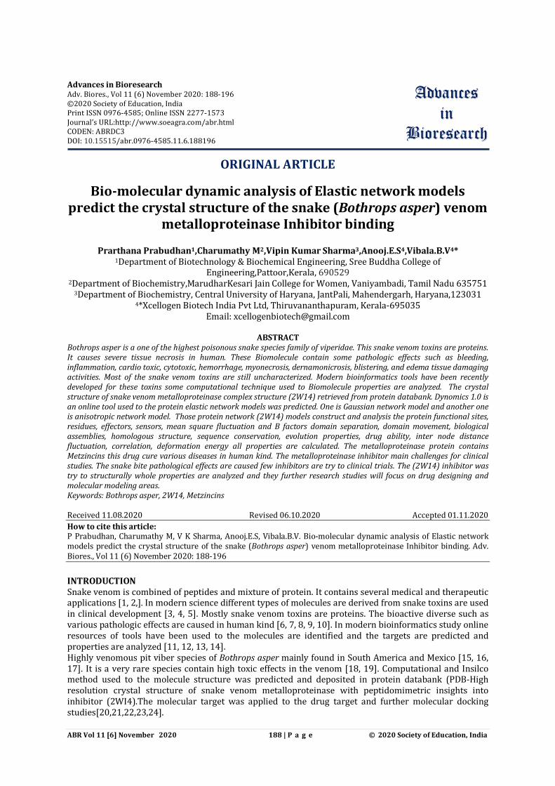
Our present work is carry on the molecule (2WI4) target was constructed by elastic network models (ENM) and combined with Gaussian network model (GNM) and anisotropic network model (ANM).The Biomolecule protein dynamics and functions whole properties are analyzed in the Dynamics 1.0 software. Material and methodology Protein retrived NCBI The Biomolecule High resolution crystal structure of snake venom metalloproteinase with peptidomimetric insights into inhibitor (2WI4) Bap1 complex insights into inhibitor binding protein PDB coordinate file format was retrived from protein data bank. Network prediction in dynamics Dynomics 1.0 online access tool contain Elastic network models (ENM) based predict two different network one is Gaussian network model (GNM) and anisotropic network model (ANM).These two networks are used to construct the protein was further analyzed via properties. Query dynamics Enter the PDB 2WI4 and click submit button a query with default option. The biological assembly structure will be selected. Then advance option method used to select the network models. Protein dynamics The Biomolecule (2WI4) properties of molecular fluctuation, Mean square fluctuation of residues, Residue cross correlation between residue fluctuation, Inter residue map and Gaussian network model (GNM) mode spectrums are analyzed. Prediction of functional site based protein dynamics The protein Biomolecule (2WI4) functional site based the domain are separated. Protein functional site sensor effectors, signaling communication sites are predicted.
Prabudhan et al
ABR Vol 11 [6] November 2020 190 | P a g e © 2020 Society of Education, India
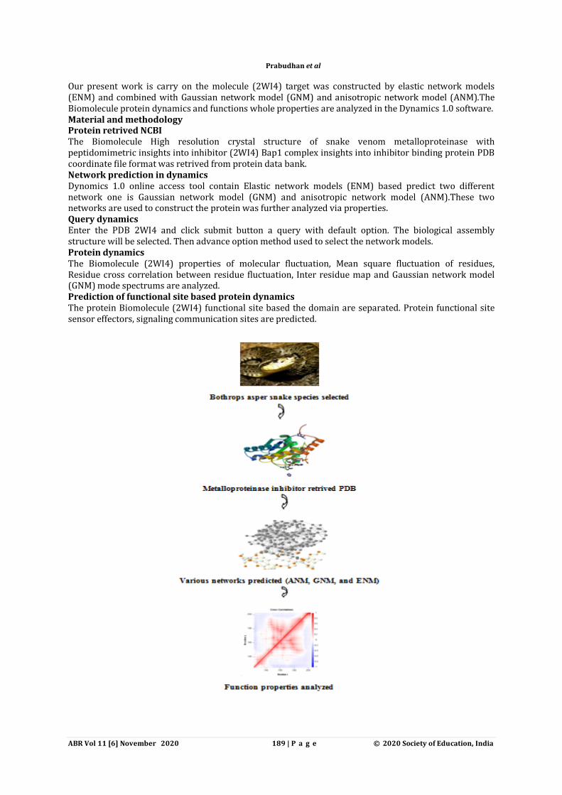
RESULTS Molecular motion
Fig 1: shows that a snapshot from molecular motions webpage generated by ENM 1.0.A snapshot from the animation generated for 2WI4.slowest mode shown in figure. The protein is in ENM representation color coded based on the size of motion (red-most mobile, blue-most rigid) user download full atomic conformers after selecting the RMSD from the pdb structure. Mean square fluctuation of residues
Fig 2: shows that Correlation between observed and predicted fluctuation Fig a and b.In this 3D Jsmol window the structure was modeled as cartoon and colouir coded by GNM defined theoretical fluctuation(left) X-ray experimental factor (right).The colors are defined by the mobility of the residues/nodes. Rigid residues are blue and mobile residues are red. Theoretical and experimental B factors Plotted
Fig 3: shows that The 2 d profiles of B-factor (y) as a function of residue (x) are plotted using the interactive graph. The plot will display the plotted series with Theoretical chain A, ASER156, (11.594) residue information corresponding B factors.
ABR Vol 11 [6] November 2020 191 | P a g e © 2020 Society of Education, India
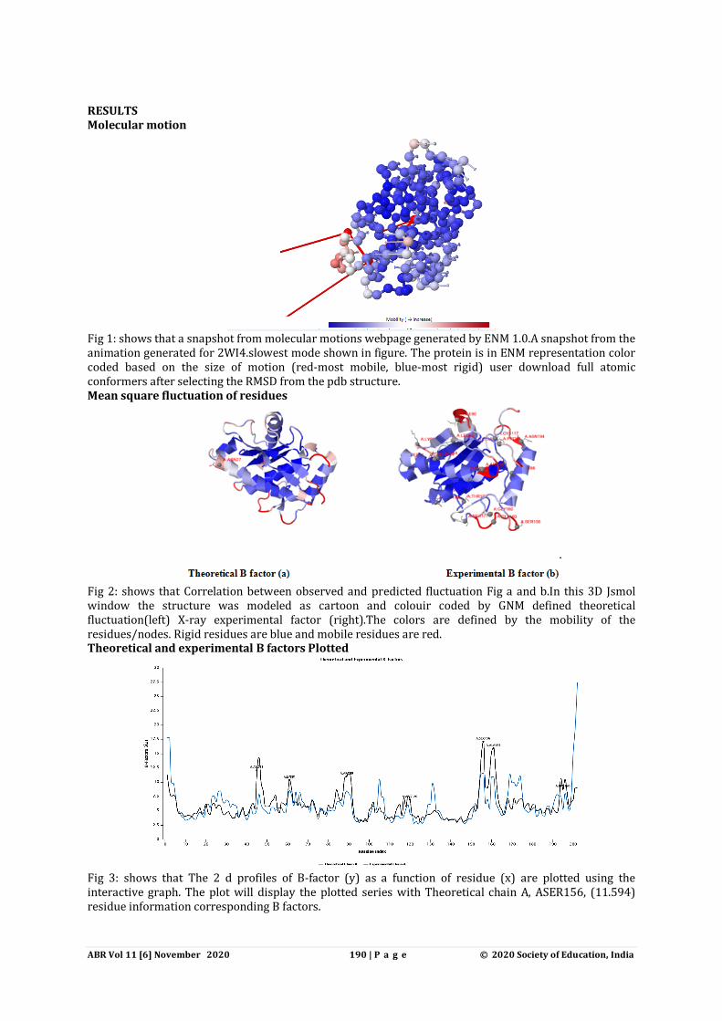
Nodes selection
Fig 4: shows that the 3D structure of 2WI4 shows in Jsmol window is colour coded based on the mobility of the residues in particular node. The colour spectrum varies from blue (most rigid) to white to red (most mobile). Graph mode/Residue index graph
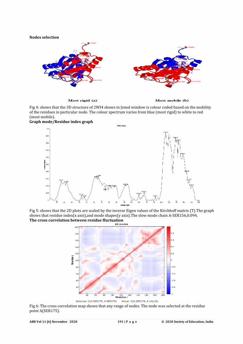
Fig 5: shows that the 2D plots are scaled by the inverse Eigen values of the Kirchhoff matrix (T).The graph shows that residue index(x axis),and mode shapes(y axis).The slow mode chain A-SER156,0.094. The cross correlation between residue fluctuation
Fig 6: The cross correlation map shows that any range of nodes. The node was selected at the residue point A(SER175).
ABR Vol 11 [6] November 2020 192 | P a g e © 2020 Society of Education, India
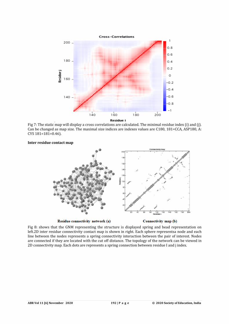
Fig 7: The static map will display a cross correlations are calculated. The minimal residue index (i) and (j). Can be changed as map size. The maximal size indices are indexes values are C180, 181=CCA, ASP180, A: CYS 181=181=0.46). Inter residue contact map
Fig 8: shows that the GNM representing the structure is displayed spring and bead representation on left.2D inter residue connectivity contact map is shown in right. Each sphere representsa node and each line between the nodes represents a spring connectivity interaction between the pair of interest. Nodes are connected if they are located with the cut off distance. The topology of the network can be viewed in 2D connectivity map. Each dots are represents a spring connection between residue I and j index.
ABR Vol 11 [6] November 2020 193 | P a g e © 2020 Society of Education, India
Properties of GNM mode of spectrum
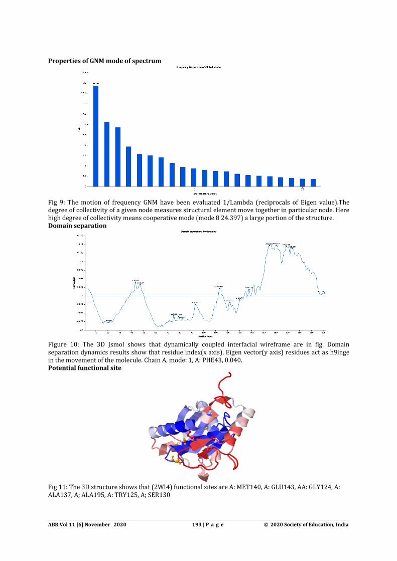
Fig 9: The motion of frequency GNM have been evaluated 1/Lambda (reciprocals of Eigen value).The degree of collectivity of a given node measures structural element move together in particular node. Here high degree of collectivity means cooperative mode (mode 8 24.397) a large portion of the structure. Domain separation
Figure 10: The 3D Jsmol shows that dynamically coupled interfacial wireframe are in fig. Domain separation dynamics results show that residue index(x axis), Eigen vector(y axis) residues act as h9inge in the movement of the molecule. Chain A, mode: 1, A: PHE43, 0.040. Potential functional site
Fig 11: The 3D structure shows that (2WI4) functional sites are A: MET140, A: GLU143, AA: GLY124, A: ALA137, A; ALA195, A: TRY125, A; SER130
ABR Vol 11 [6] November 2020 194 | P a g e © 2020 Society of Education, India
Sensor and effectors
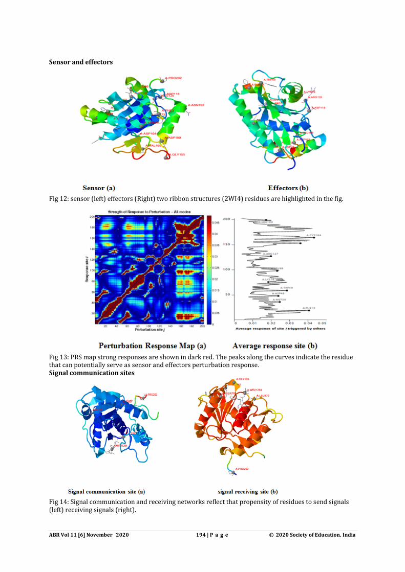
Fig 12: sensor (left) effectors (Right) two ribbon structures (2WI4) residues are highlighted in the fig.
Fig 13: PRS map strong responses are shown in dark red. The peaks along the curves indicate the residue that can potentially serve as sensor and effectors perturbation response. Signal communication sites
Fig 14: Signal communication and receiving networks reflect that propensity of residues to send signals (left) receiving signals (right).
ABR Vol 11 [6] November 2020 195 | P a g e © 2020 Society of Education, India
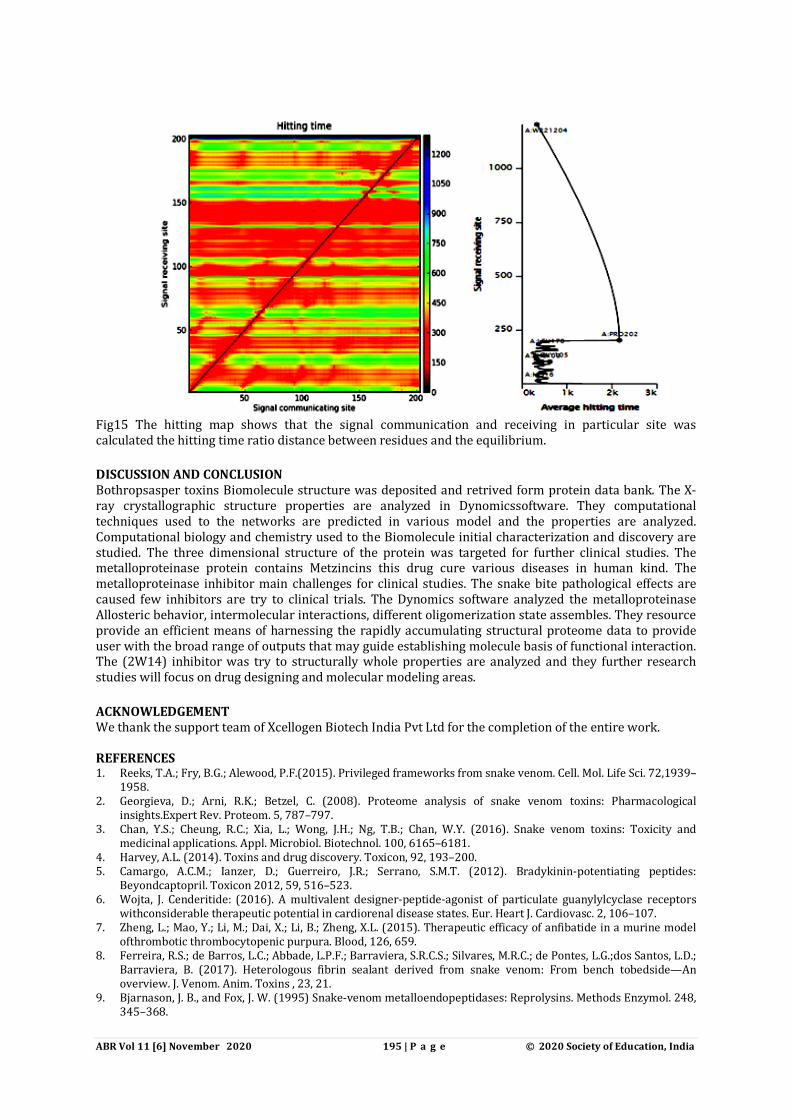
Fig15 The hitting map shows that the signal communication and receiving in particular site was calculated the hitting time ratio distance between residues and the equilibrium. DISCUSSION AND CONCLUSION Bothropsasper toxins Biomolecule structure was deposited and retrived form protein data bank. The X-ray crystallographic structure properties are analyzed in Dynomicssoftware. They computational techniques used to the networks are predicted in various model and the properties are analyzed. Computational biology and chemistry used to the Biomolecule initial characterization and discovery are studied. The three dimensional structure of the protein was targeted for further clinical studies. The metalloproteinase protein contains Metzincins this drug cure various diseases in human kind. The metalloproteinase inhibitor main challenges for clinical studies. The snake bite pathological effects are caused few inhibitors are try to clinical trials. The Dynomics software analyzed the metalloproteinase Allosteric behavior, intermolecular interactions, different oligomerization state assembles. They resource provide an efficient means of harnessing the rapidly accumulating structural proteome data to provide user with the broad range of outputs that may guide establishing molecule basis of functional interaction. The (2W14) inhibitor was try to structurally whole properties are analyzed and they further research studies will focus on drug designing and molecular modeling areas. ACKNOWLEDGEMENT We thank the support team of Xcellogen Biotech India Pvt Ltd for the completion of the entire work. REFERENCES 1. Reeks, T.A.; Fry, B.G.; Alewood, P.F.(2015). Privileged frameworks from snake venom. Cell. Mol. Life Sci. 72,1939–
1958. 2. Georgieva, D.; Arni, R.K.; Betzel, C. (2008). Proteome analysis of snake venom toxins: Pharmacological
insights.Expert Rev. Proteom. 5, 787–797. 3. Chan, Y.S.; Cheung, R.C.; Xia, L.; Wong, J.H.; Ng, T.B.; Chan, W.Y. (2016). Snake venom toxins: Toxicity and
medicinal applications. Appl. Microbiol. Biotechnol. 100, 6165–6181. 4. Harvey, A.L. (2014). Toxins and drug discovery. Toxicon, 92, 193–200. 5. Camargo, A.C.M.; Ianzer, D.; Guerreiro, J.R.; Serrano, S.M.T. (2012). Bradykinin-potentiating peptides:
Beyondcaptopril. Toxicon 2012, 59, 516–523. 6. Wojta, J. Cenderitide: (2016). A multivalent designer-peptide-agonist of particulate guanylylcyclase receptors
withconsiderable therapeutic potential in cardiorenal disease states. Eur. Heart J. Cardiovasc. 2, 106–107. 7. Zheng, L.; Mao, Y.; Li, M.; Dai, X.; Li, B.; Zheng, X.L. (2015). Therapeutic efficacy of anfibatide in a murine model
ofthrombotic thrombocytopenic purpura. Blood, 126, 659. 8. Ferreira, R.S.; de Barros, L.C.; Abbade, L.P.F.; Barraviera, S.R.C.S.; Silvares, M.R.C.; de Pontes, L.G.;dos Santos, L.D.;
Barraviera, B. (2017). Heterologous fibrin sealant derived from snake venom: From bench tobedside—An overview. J. Venom. Anim. Toxins , 23, 21.
9. Bjarnason, J. B., and Fox, J. W. (1995) Snake-venom metalloendopeptidases: Reprolysins. Methods Enzymol. 248, 345–368.
ABR Vol 11 [6] November 2020 196 | P a g e © 2020 Society of Education, India
10. Bode, W., Gomis-Ruth, F. X., and Stocker, W. (1993) Astacins, serralysins, snake-venom and matrix metalloproteinases exhibit identical zinc-binding environments (HExxHxxGxxH and Metturn) and topologies and should be grouped into a common family, the metzincins. FEBS Lett. 331, 134–140.
11. Gomis-Ruth, F. X. (2003) Structural aspects of the metzincin clan of metalloendopeptidases. Mol. Biotechnol. 24, 157–202.
12. Ramos, O. H. P., and Selistre-De-Araujo, H. S. (2006) Snake venom metalloproteases: Structure and function of catalytic and disintegrin domains. Comp. Biochem. Phys., Part C: Pharmacol.,Toxicol. Endocrinol. 142, 328–346.
13. Stocker, W., Grams, F., Baumann, U., Reinemer, P., Gomis-Ruth, F. X., Mckay, D. B., and Bode, W. (1995) Themetzincins: Topological and sequential relations between the astacins, adamalysins, serralysins, and matrixins (collagenases) define a superfamily of zinc-peptidases. Protein Sci. 4, 823–840.
14. Gomis-Ruth, F. X. (2009) Catalytic domain architecture of metzincinmetalloproteases. J. Biol. Chem. 284, 15353–15357.
15. Haliloglu,T. and Bahar,I. (2015) Adaptability of protein structures to enable functional interactions and evolutionary implications. Curr.Opin. Struct. Biol., 35, 17–23.
16. Yang,L.W. and Bahar,I. (2005) Coupling between catalytic site and collective dynamics: a requirement for mechanochemical activity of enzymes. Structure, 13, 893–904.
17. Yang,L.W., Eyal,E., Bahar,I. and Kitao,A. (2009) Principal component analysis of native ensembles of biomolecular structures (PCA NEST): insights into functional dynamics. Bioinformatics, 25, 606–614.
18. Chandrasekaran,A., Chan,J., Lim,C. and Yang,L.W. (2016) Protein dynamics and contact topology reveal protein–DNA binding orientation. J. Chem. Theory Comput.,12, 5269–5277.
19. Li,H., Sakuraba,S., Chandrasekaran,A. and Yang,L.W. (2014) Molecular binding sites are located near the interface of intrinsic dynamics domains (IDDs). J. Chem. Inf. Model., 54, 2275–2285.
20. Chennubhotla,C. and Bahar,I. (2007) Signal propagation in proteins and relation to equilibrium fluctuations. PLoSComput. Biol., 3, 1716–1726.
21. Atilgan,C. and Atilgan,A.R. (2009) Perturbation-response scanning reveals ligand entry-exit mechanisms of ferric binding protein. PLoSnComput. Biol., 5, e1000544.
22. General,I.J., Liu,Y., Blackburn,M.E., Mao,W., Gierasch,L.M. and Bahar,I. (2014) ATPase subdomain IA is a mediator of interdomainallostery in Hsp70 molecular chaperones. PLoSComput. Biol., 10, e1003624.
23. Bahar,I., Cheng,M.H., Lee,J.Y., Kaya,C. and Zhang,S. (2015) Structure-encoded global motions and their role in mediating protein-substrate interactions. Biophys. J., 109, 1101–1109.
24. Krebs,W.G., Alexandrov,V.,Wilson,C.A., Echols,N., Yu,H. and Gerstein,M. (2002) Normal mode analysis of macromolecular motions in a database framework: developing mode concentration as a useful classifying statistic. Proteins, 48, 682–695.
Copyright: © 2020 Society of Education. This is an open access article distributed under the Creative Commons Attribution License, which permits unrestricted use, distribution, and reproduction in any medium, provided the original work is properly cited.
![Advances CODEN: ABRDC3 Bioresearchsoeagra.com/abr/abrjan2015/4f.pdf · ABR Vol 6 [1] January 2015 19 | Page ©2015 Society of Education, India Advances in Bioresearch Adv. Biores.,](https://static.fdocuments.net/doc/165x107/5f31fd6c3b7a543e696cdd91/advances-coden-abrdc3-abr-vol-6-1-january-2015-19-page-2015-society-of-education.jpg)


![Advances CODEN: ABRDC3 Bioresearchsoeagra.com/abr/abrmay2015/14.pdfABR Vol 6 [3] May2015 78 | Page ©2015 Society of Education, India Advances in Bioresearch Adv. Biores., Vol 6 (3)](https://static.fdocuments.net/doc/165x107/5f5fc77831ca133d8821743b/advances-coden-abrdc3-abr-vol-6-3-may2015-78-page-2015-society-of-education.jpg)


