Acute pancreatitis ca pancreas naulo_lmc
31
Acute pancreatitis and Carcinoma pancreas By Naulo pokhrel LMC, Palpa, Nepal
-
Upload
naulo-pkrl -
Category
Education
-
view
42 -
download
2
Transcript of Acute pancreatitis ca pancreas naulo_lmc

Acute pancreatitis and Carcinoma pancreas
By Naulo pokhrelLMC, Palpa, Nepal
Acute PancreatitisPancreatitis: inflammation in the pancreas associated with injury to exocrine parenchyma
Acute pancreatitis Chronic pancreatitis

Acute PancreatitisReversible pancreatic
parenchymal injury associated with inflammation
Relatively commonCAUSE : Alcoholism, Gall bladder
disease accounts for 80%Gall stones are present in about
35 to 60 % of cases and 5% of patients with gall stones develop pancreatitis
`
Male : female ratio – 1:3 for gall bladder disease and 6:1 for alcoholism
OTHER CAUSES : Obstruction in pancreatic duct
system : periampullary neoplasm, biliary sludge , parasites esp. ascaris
Medication : Furosemide , azathioprine , Estrogen, sulphonamides
Infection including mumps Trauma : Blunt injury to the abdomen
Ischemic injury from shock, embolism, vasculitis
Metabolic disorders- hypertriglyceridaemia, hyperparathyoidism
Germline mutation in cationic trypsinogen and trypsin inhibitor genes

Hereditary PancreatitisCharacterized by recurrent attack
of severe pancreatitis esp. beginning in childhood
Germline (inherited) mutation in cationic trypsinogen gene
Trypsin becomes resistance to cleavage
It can activate other digestive enzymes giving rise to pancreatitis
Autosomal dominant mode of inheritance

Serine protease inhibitor kazal type I (SPINK1) gene codes for Pancreatic secretory trypsin inhibitor
Leads to increased trypsin levelAnd pancreatitisThis mode is autosomal recessive
Pathogenesis
Autodigestion of pancreatic substance by inappropriately activated pancreatic enzymes
inappropriately activated pancreatic enzymes activates other proenzymes prophospholipase(degrade fat cells) , proelastase (damage elastic fibres of blood vessels)
Activates kinin system and by activation fo Hagmen factor; clotting system as well
Leads to inflammation and thromboses
Mechanism of inappropriate activation of pancreatic enzymes
Pancreatic duct obstruction : gall stones, biliary sludge in ampulla of vatar obstruction
Accumulation of enzyme rich fluid in the interstitium including activated lipase
Fat necrosisInjury to tissuesRelease of inf. Cytokines IL-1B, IL-6, TNF,
PAF
Initiation of inflammationEdemaVascular compromiseIschemic injury to acinar cells

Primary acinar injuryBY viruses(mumps)DrugsDirect traumaIschemiashock
Defective intracellular transport proenzymes within the acinar cells
Pancreatic proenzymes and lysosomal hydrolases packed together
Alcohol consumptionIncreased release of protein rich
pancreatic fluidDeposition of inspissated protein
plugObstruction of small pancreatic
ductsMay result in transient
contraction of sphincter of oddi (direct toxic effect on acinar cells)
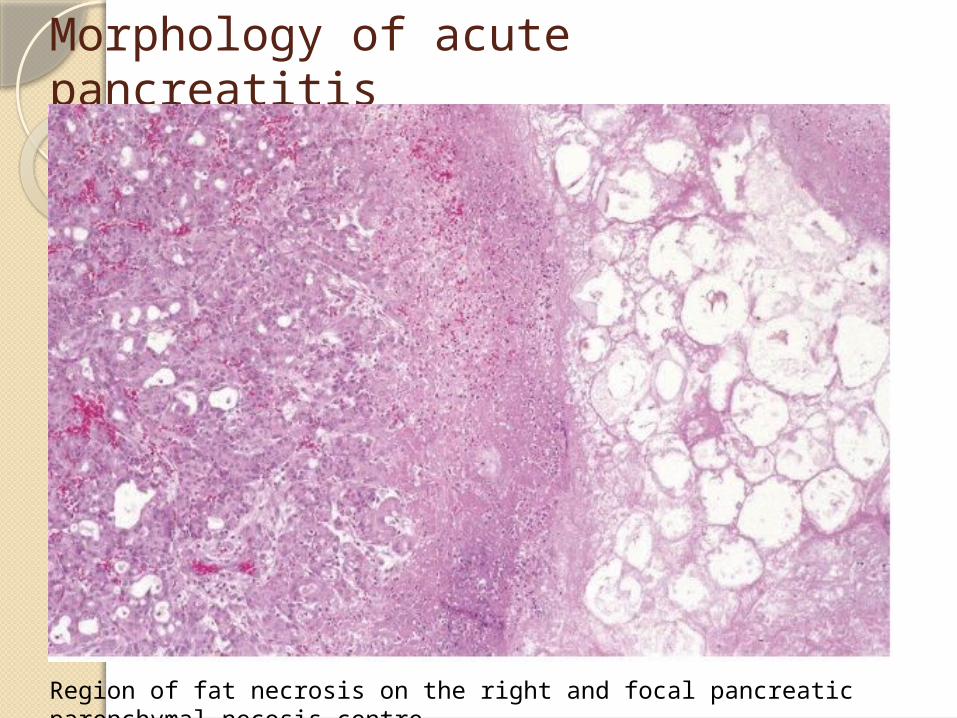
Morphology of acute pancreatitis
Region of fat necrosis on the right and focal pancreatic parenchymal necosis centre
Morphology of acute pancreatitisBasic alterations are:Microvascular likage causing
edemaNecrosis of fat by lipolytic
enzymesAcute inflammationProteolytic destruction of
pancreatic parenchymaDestruction of blood vessels and
haemorrhage
In milder formEdemaFocal areas of fat necrosisMild inflammation
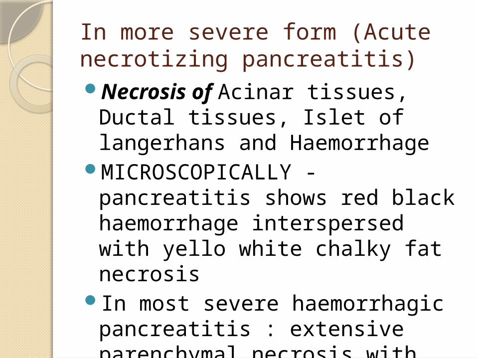
In more severe form (Acute necrotizing pancreatitis)Necrosis of Acinar tissues,
Ductal tissues, Islet of langerhans and Haemorrhage
MICROSCOPICALLY - pancreatitis shows red black haemorrhage interspersed with yello white chalky fat necrosis
In most severe haemorrhagic pancreatitis : extensive parenchymal necrosis with haemorrhage
Clinical featureAbdominal pain referred to upper
back and sometimes to left shoulder accompained by anorexia , nausea , vomitting
Lab findings : increased serum amylase, glycosuria, hypocalcaemia(d/t precepatation of Ca soaps in necrotic fat),
Direct visualisation of inflamed pancres by radiography
Key management is resting of pancreas by total restriction of oral intake and supportive therapy by iv fluids and analgesia
5% of patients of severe acute pancreatitis die from shock in first week of illness
Complications : Acute respiratory distress syndrome , aute renal failure.
Carcinoma of pancreas4th leading cause of death (lung,
colon, breast)Infiltrating ductal adenocarcinoma of
pancreasProgression from non neoplastic
epithelium to histologically well-defined non invasive leisons in ducts and ductules to invasive carcinoma
Precursor leisons : Pancreatic Intraepithelial Neoplasias (PanINs)
Location in pancreas60% in head15% in body5% in tail20% diffuseMost commonly seen in elder
patients (>60 years)Highly invasive carcinoma5 year survival rate is less than
5%

PathogenesisFrom inherited and acquired
mutationsProgressive accumulation of
genetic changesPrecursor leisons are Pancreatic
Intraepithelial Neoplasias (PanINs)
Molecular alterations in pancreatic carcinogenesis – KRAS, p16, SMAD4,and P53

K-RAS gene(chromosome 12p) is most frequently altered;point mutation leads to activation: in 80 to 90% of cases
P 16 tumor supressor gene inactivated 90% cases
SMAD4 tumor supressor gene inactivated in 55% of pancreatic cancers
P 53 tumor supressor gene inactivated in 50 to 70% of cases
Carcinomaa of pancreas more common in black than in white
Smoking doubles the riskChronic pancreatitis and dibetes
mellitus are also risk factors

Morphology
MorphologySite: 60% in head ,15% in body ,5% in
tail ,20% diffuse carcinomas are hard , stellate, gray white,
poorely defined massesMajority are ductal adeno-carcinoma2 charactreistic feature : highly invasive,
and elicts intense non-neoplastic host reaction composed of fibroblasts , lymphocytes and extracellular matrix(desmoplastic response)
Carcinomas in head of pancreas obstruct the distal common bile duct leads to jaundice
Often extend through retroperitonial space
May invade adrenals, spleen, transverse colon, stomach
Liver is often enlarged because of metastatic deposits
Distant metastasis to LUNGS and BONES
Microscopically
Moderately to poorely differentiated adenocarcinoma forming abortive tubular structures or cell clusters and exhibiting an aggressive , deeply infiltrative growth pattern
Deep stromal fibrosisLess comm varients includes acinar cell
carcinomas showing prominent acinar cells with zymogen granules and enzyme production
Adenosquamous carcinomaUndifferentiated carcinomas with osteoclast
like giant cell
Clinical featuresPainObstructive jaundiceWeight loss, anorexiaMigratory thrombophlebitis
(Trousseau syndrome) in 1o% of patients; elaboration of PAF and procoagulants from tumors or its necrotic contents
Elevated serum level of carcinoembryonic and CA19 antigens