ACTIONS OF DENDROTOXI ONN K CHANNEL ANDS …jeb.biologists.org/content/jexbio/147/1/21.full.pdf ·...
21
V. exp. Biol. 147, 21-41 (1989) 21 'Primed in Great Britain © The Company of Biologists Limited 1989 ACTIONS OF DENDROTOXIN ON K + CHANNELS AND NEUROMUSCULAR TRANSMISSION IN DROSOPHILA MELANOGASTER, AND ITS EFFECTS IN SYNERGY WITH K + CHANNEL-SPECIFIC DRUGS AND MUTATIONS BY CHUN-FANG WU 1 ' 2 , MING-CHENG TSAI 1 , MAI-LEI CHEN 1 , YI ZHONG 2 , SATPAL SINGH 2 AND C. Y. LEE 1 1 Department of Pharmacology, College of Medicine, National Taiwan University, Taipei, Taiwan, ROC and 2 Department of Biology, University of Iowa, Iowa City, Iowa 52242, USA Accepted 30 May 1989 Summary The blockade of K + channels and enhancement of neuromuscular transmission by dendrotoxin (DTX), a convulsant peptide from mamba snake venom, were examined in normal and mutant larval preparations of Drosophila. Two- microelectrode voltage-clamp experiments showed that DTX reduced the tran- sient K + current, I A , in muscle membrane. This effect was suppressed by raising the Mg 2 " 1 " concentration or by lowering the temperature. The interaction of DTX with Mg 24 " was further analyzed at a low cation concentration, at which DTX reduced both I A and the delayed rectifier I K . These results were correlated with the action of DTX on the neuromuscular junction. Its facilitatory effect on excitatory junctional potentials (EJPs) was relatively mild but the effect was drastically enhanced when combined with certain mutations and K + channel blocking drugs, leading to repetitive or prolonged giant EJPs. Only the mutations or drugs that reduced I K or the Ca 2+ -dependent K + current, ICF, could yield these synergistic effects with DTX. In contrast, the abnormal EJPs caused by the mutation or drug that blocked I A were not further enhanced by DTX, indicating that DTX also affects I A at the neuromuscular junction. Thus, the A-type K + channels in muscle and nerve terminals appeared very similar in their sensitivity to the specific toxin, drugs and mutations examined here. Introduction A diversity of K + channels gives rise to a large repertoire of firing patterns in nervous systems, and a definitive classification of different K + channels is not yet possible (Latorre & Miller, 1983; Wu & Ganetzky, 1986; Rudy, 1988). Specific toxins, drugs and mutations thus provide useful tools to compare the types and properties of the channels in different excitable membranes. Key words: dendrotoxin, Drosophila mutants, presynaptic terminals, muscle, K + channel diversity, quinidine, 4-aminopyridine.
Transcript of ACTIONS OF DENDROTOXI ONN K CHANNEL ANDS …jeb.biologists.org/content/jexbio/147/1/21.full.pdf ·...

V. exp. Biol. 147, 21-41 (1989) 2 1'Primed in Great Britain © The Company of Biologists Limited 1989
ACTIONS OF DENDROTOXIN ON K+ CHANNELS ANDNEUROMUSCULAR TRANSMISSION IN DROSOPHILA
MELANOGASTER, AND ITS EFFECTS IN SYNERGY WITHK+ CHANNEL-SPECIFIC DRUGS AND MUTATIONS
BY CHUN-FANG WU1'2, MING-CHENG TSAI1, MAI-LEI CHEN1,YI ZHONG2, SATPAL SINGH2 AND C. Y. LEE1
1 Department of Pharmacology, College of Medicine,National Taiwan University, Taipei, Taiwan, ROC and2 Department of Biology,
University of Iowa, Iowa City, Iowa 52242, USA
Accepted 30 May 1989
Summary
The blockade of K+ channels and enhancement of neuromuscular transmissionby dendrotoxin (DTX), a convulsant peptide from mamba snake venom, wereexamined in normal and mutant larval preparations of Drosophila. Two-microelectrode voltage-clamp experiments showed that DTX reduced the tran-sient K+ current, IA, in muscle membrane. This effect was suppressed by raisingthe Mg2"1" concentration or by lowering the temperature. The interaction of DTXwith Mg24" was further analyzed at a low cation concentration, at which DTXreduced both IA and the delayed rectifier IK. These results were correlated withthe action of DTX on the neuromuscular junction. Its facilitatory effect onexcitatory junctional potentials (EJPs) was relatively mild but the effect wasdrastically enhanced when combined with certain mutations and K+ channelblocking drugs, leading to repetitive or prolonged giant EJPs. Only the mutationsor drugs that reduced IK or the Ca2+-dependent K+ current, ICF, could yield thesesynergistic effects with DTX. In contrast, the abnormal EJPs caused by themutation or drug that blocked IA were not further enhanced by DTX, indicatingthat DTX also affects IA at the neuromuscular junction. Thus, the A-type K+
channels in muscle and nerve terminals appeared very similar in their sensitivity tothe specific toxin, drugs and mutations examined here.
Introduction
A diversity of K+ channels gives rise to a large repertoire of firing patterns innervous systems, and a definitive classification of different K+ channels is not yetpossible (Latorre & Miller, 1983; Wu & Ganetzky, 1986; Rudy, 1988). Specifictoxins, drugs and mutations thus provide useful tools to compare the types andproperties of the channels in different excitable membranes.
Key words: dendrotoxin, Drosophila mutants, presynaptic terminals, muscle, K+ channeldiversity, quinidine, 4-aminopyridine.

22 C.-F. W u AND OTHERS
Dendrotoxin (DTX), a non-enzymatic venom protein from the mamba snakeDendroaspis angusticeps, is a potent convulsant neurotoxin when injected into thebrain. DTX is also the first snake toxin found to facilitate neurotransmitter releaseat vertebrate neuromuscular junctions (Harvey & Karlsson, 1980; Harvey et al.1984; Harvey, 1987) and central synapses (Dolly et al. 1984). Its action has beencharacterized in several vertebrate species by voltage-clamp experiments and areduction in K+ conductances is correlated with the facilitatory effect of the toxin(Dolly etal. 1984; Wellere/a/. 1985; Benoit & Dubois, 1986; Halliwellef al. 1986).However, the specific types of K+ conductances that are affected by this toxin varyin different preparations. In frog peripheral nerve nodes of Ranvier, a componentof slowly inactivating K+ current is decreased by DTX (Weller et al. 1985; Benoit& Dubois, 1986), whereas in the cell body of central hippocampal neurons atransient K+ current, IA, is suppressed (Dolly et al. 1984; Halliwell et al. 1986).However, the transient K+ current in rat sympathetic ganglion neurons is reportedto be insensitive to DTX (Dolly et al. 1987). The endplate potential (EPP) in thefrog neuromuscular junction is enhanced by increased quantal content and bymultiple firing of the motor axon, but the type of K+ channels affected by DTX isunknown (Harvey & Anderson, 1985; Harvey, 1987). Although these contrastingresults may be attributed to species differences, a more intriguing possibility is thatthey reflect distinctions and similarities among different K+ channels.
The generality of a DTX blocking action on K+ channels has not been wellestablished since only vertebrate species have been studied. In the present study,we examined the effects of DTX on neuromuscular transmission and on muscleK+ channels of an arthropod species, Drosophila melanogaster. Both the wild-type and K+ channel mutants were studied to examine whether DTX can be usedas a probe for characterization of various K+ channels. The genetics of membraneexcitability in Drosophila has been well studied. Several mutations that eliminateor alter specific types of K+ currents are available (Ganetzky & Wu, 1986; Wu &Ganetzky, 1986).
The larval neuromuscular preparation of Drosophila is suitable for electro-physiological studies because the properties of neuromuscular transmission and ofvarious ion channels in the muscle membrane have been characterized in detail(Jan & Jan, 1976a,b; Jan et al. 1977; Wu et al. 1978, 1983; Ganetzky & Wu, 1982,1983,1985; Wu & Haugland, 1985; Gho & Mallart, 1986; Haugland, 1987; Singh &Wu, 1989; F. N. Haugland & C.-F. Wu, in preparation). Voltage-clamp studies ofthese larval muscle fibers have demonstrated that mutations of three genesseparately affect different types of K+ channels. The transient IA is missing inShaker (Sh) mutants (Wu et al. 1983; Wu & Haugland, 1985); the delayed rectifier,IK, is reduced to about 50 % in an ether a go-go (eag) mutant (Wu et al. 1983); anda Ca2+-dependent K+ current, IcF, is eliminated in the slowpoke (slo) mutant(Elkins etal. 1986; Singh & Wu, 1987,1989). We used these mutants in conjunctionwith other K+ channel blocking agents to characterize the effect of DTX onneuromuscular transmission. The synergism between the effects of DTX and!various mutations was studied to infer the types of presynaptic K+ channels

Dendrotoxin and K+ currents in Drosophila 23
affected by DTX. The action of DTX on muscle membrane currents was alsodirectly monitored by the voltage-clamp technique. These experiments usingDrosophila mutants not only extend our understanding of the action of DTX oninvertebrate K+ channels but also offer a unique opportunity for comparing thetypes and properties of the K+ channels present in nerve terminals and musclefibers.
Materials and methods
Mutants
The wild-type strain, Canton-S, of Drosophila melanogaster was used forcharacterization of normal physiology. Among available mutant alleles of the Shand eag loci (Ganetzky & Wu, 1986), ShKS133 and eag1 were chosen for this studybecause they have been described in the most detail. They are also the mostextreme alleles of these two loci; ShKS133 completely blocks IA and eag1 removesabout 50% of IK (WU et al. 1983; Wu & Haugland, 1985; Y. Zhong & C.-F. Wu,unpublished results). The slo locus is represented by a single allele (Elkins et al.1986), which was used in this study.
Electrophysiological recording
For intracellular recording of EJPs, the larval neuromuscular preparation (Jan& Jan, 1976a; Wu et al. 1978) was used. Briefly, a late third-instar larva was pinnedout in saline, cut along the dorsal midline and eviscerated. The nerves innervatingthe body wall muscles in the abdominal segments were severed near the ventralganglion. The cut end of a segmental nerve was stimulated via a suction electrodeand EJPs were recorded in the ventral longitudinal muscle fibers. Experimentswere carried out in Drosophila saline containing 128mmoll~1 NaCl, 2mmoll~1
KC1, SSmmoir1 sucrose, and buffered at p H 7 1 with Smmoll"1 Hepes. Theconcentrations of Ca2+ (0-1, 0-2 or l-SmmolT1) and Mg24" (0-5, 2, 4 orMmmoll"1) were varied as specified in each experiment. Other details of theexperimental protocol have been described elsewhere (Jan & Jan, 1976a; Wu et al.1978).
An identical preparation was used for conventional two-microelectrode voltage-clamp measurements of muscle membrane currents. We followed the establishedparadigms and protocols (Wu & Haugland, 1985; Haugland, 1987;F. N. Haugland & C.-F. Wu, in preparation) and only a brief description is givenhere. Measurements of IA and IK were made in Ca2+-free saline containing(Mmmoi r 1 EGTA. The concentration of MgCl2 varied in each experiment, asspecified. The composition of the Ca2+-free saline was otherwise identical to thatused for EJP recordings (see above). The large differences in inactivation andrecovery kinetics between IA and IK were utilized to separate these two currents.At a holding potential (VH) of — 80 mV, step depolarizations elicited IA, whichfceaked rapidly and then inactivated, and IK, which rose to a sustained plateauwith a delay (Figs 6, 8 and 10). When a depolarizing prepulse (to -20 mV)

24 C.-F. W u AND OTHERS
preceded the test pulse, IA was inactivated, leaving IK alone. Digital subtraction ofthe two measurements (with or without the prepulse) yielded the inactivating IA inisolation from IK (Figs 7 and 9). The duration of the prepulse (2 s) and the intervalbetween the prepulse and test pulse at 5°C (10 ms) and 16 °C (5 ms) were chosenfrom previously established values to maximize the separation of these twocurrents (Haugland, 1987; F. N. Haugland & C.-F. Wu, in preparation;F. N. Haugland, Y. Zhong & C.-F. Wu, unpublished data).
When Ca2+ currents and Ca2+-dependent K+ currents were examined,20mmoll~1 CaCl2, 4mmoll~1 MgCl2 and no EGTA were present in saline. Allvoltage-clamp measurements were stored digitally and analyzed by a microcom-puter (Macintosh, Apple Computer, Cupertino, CA). After subtraction of linearleakage currents, the density of active currents was determined by adjustment forthe cell membrane area of the fiber and expressed in /xA/xF"1 using membranecapacitance as an indicator. For each experiment, data collected from a number offibers were averaged as shown in the traces and current-voltage (I-V) relation-ships in Figs 6-10. The numbers of fibers and larvae used in each experiment arespecified in the legend of the figure.
Measured amounts of DTX from D. angusticeps, 4-aminopyridine (4-AP) andquinidine stock solutions were added to the bathing chamber (volume, lml) toachieve the final concentrations. Solutions were carefully mixed and enough timewas allowed for equilibration. Quinidine and 4-AP were acquired from SigmaChemical Co. (St Louis, MO) and DTX was a gift from Drs F. J. Joubert and F. F.H. Carlsson.
Intracellular recordings of EJPs were performed at 25-29°C, except wherestated otherwise. The results in each figure (Figs 1-5) were based on observationson 5-11 fibers in 3-8 larvae. In voltage-clamp experiments, preparations werecooled to either 5 or 16°C. At higher temperatures, the rapid kinetics of themembrane currents made it difficult to achieve satisfactory control of voltagejumps (Wu & Haugland, 1985).
Results
DTX effects on transmitter release at normal neuromuscidar junctions
The ventral longitudinal body-wall muscle fibers in the abdominal segments ofDrosophila larvae are each innervated by two motor neurons; and only excitatoryjunctional potentials (EJPs), but not inhibitory potentials, can be evoked by nervestimuli (Jan & Jan, 1976a). As in other arthropods, glutamate is the putativetransmitter at the neuromuscular junction (Jan & Jan, 19766; Johansen etal. 1989)and there are Ca2+ channels and various types of K+ channels, but no Na+
channels, in the muscle membrane (Wu & Haugland, 1985; Haugland, 1987; Singh& Wu, 1989). Normally, only graded potentials are evoked in these muscle fibers,although all-or-none Ca2+ action potentials may be generated when the K^|channels are blocked (Suzuki & Kano, 1977).

Dendrotoxin and K+ currents in Drosophila 25
It has been reported that the activity, but not binding, of DTX is suppressed byincreased Mg2"1" concentrations (3 mmol I"1) in isolated vertebrate nerve-musclepreparations (Harvey & Anderson, 1985). When this phenomenon was investi-gated in larval muscles, we found that the effects of DTX on neuromusculartransmission in Drosophila depended on Ca2+ as well as Mg2"1" concentrations inthe saline. Even at the normal Ca2+ concentration (1-8 mmol I"1), the action ofDTX was apparently inhibited since EJPs were not significantly altered in thepresence of 4-2/anoll"1 (30/igml"1) DTX. This was true for preparations bathedin salines containing either normal (4 mmol I"1, data not shown) or reduced(2 mmol I"1, Fig. 1A) concentrations of Mg2"1".
The action of DTX became apparent when the external Ca2+ concentration waslowered (Fig. 1B,C). At O-lmmoll"1 Ca2+, EJPs showed quantal fluctuations insize and miniature EJPs (mEJPs) were the only spontaneous activity observed,consistent with previous reports (Jan et al. Wll; Ganetzky & Wu, 1982). Uponaddition of DTX, the amplitude of EJPs increased appreciably (on average to164 % of the control value in two fibers) and repetitive EJPs were occasionallyevoked by a single nerve stimulus (Fig. IB). The DTX effects were furtherenhanced if the Mg2"1" concentration was also reduced from 4 to 2 mmol I"1. At lowMg2+ and Ca2+ concentrations the neuromuscular junction became more excit-able, often leading to more than one, and sometimes up to five, EJPs in responseto a single stimulus (Fig. 1C). In the presence of DTX, prolonged trains ofrepetitive EJPs (up to 30) of increased size occurred spontaneously or in responseto stimulation (Fig. 1C).
Further experiments showed that at very low cation concentrations(0-2 mmol I"1 Ca2+ and O-Smmoll"1 Mg2"1"), DTX caused clearly enhancedneuromuscular transmission at either high (20°C) or low (8°C) temperature. Thiseffect was completely suppressed by 14 mmol I"1 Mg2"1" (data not shown). Thesuppressive effect of Mg2"1" is apparently on the activity rather than on the bindingof DTX to its target site, as previously shown in vertebrate preparations (Harvey& Anderson, 1985). Application of DTX (2-8 jmioir1) in high-Mg2+ saline (4 or14 mmol I"1 Mg2* and 0-2 mmol I"1 Ca2+) did not change larval EJPs at 20°C.Nevertheless, following a rapid wash with low-Mg2"1" saline (containing 0-5 or1 mmol I"1 Mg2"1" and 0-2 mmol I"1 Ca2+, but no DTX), a single nerve stimulusevoked repetitive EJPs with increased amplitude. These facilitated EJPs weresuppressed again by high-Mg2"1" saline (without DTX) and reappeared in low-Mg2"1" saline (data not shown).
Calcium ions may suppress the effect of DTX on EJPs by a mechanism similar tothat of magnesium ions. But at least part of the suppression at high external Ca2+
concentrations can be attributed to the activation of a Ca2+-dependent K+
conductance in the nerve terminals (Jan etal. 1977; Ganetzky & Wu, 1982, 1983).This notion was supported by the results obtained in slo mutants, in which a Ca2+-dependent K+ current, ICF, is eliminated (Elkins et al. 1986; Singh & Wu, 1987,1989). In saline containing 1-8 mmol P 1 Ca2+ and 4-0 mmol I"1 Mg2+, EJPs in slolarvae were nearly normal. Following treatment with 4-2/zmolP1 DTX, the

26 C. -F . WU AND OTHERS
Control DTX
20 mV
40 ms
Ca2 +
Mg24
0-14-0
A.10 mV
0-ls
4mV
40 ms
Fig. 1. Effects of dendrotoxin (DTX) on excitatory junctional potentials (EJPs)recorded intracellularly from ventral longitudinal muscle fibers of normal larvae at theCa2+ and Mg2"1" concentrations indicated. (A) Addition of 4-2/miolP1 (30/igmT1)DTX did not result in any apparent changes in standard saline containing 1-8 mmol P 1
Ca2+ and 4 mmol P 1 Mg2"1". Even when the Mg2"1" concentration was lowered to2 mmol I"1, no differences in EJPs were observed 30 min after the application of DTX.(B) At a low concentration (0-1 mmolP1) of external Ca2+, only small EJPs and somespontaneous miniature EJPs were seen. Treatment with DTX (4-2/anolP1, 3 min)caused an increase in EJP amplitude and repetitive discharges of EJPs. (C) When theexternal Mg2+ concentration was lowered from 4 to 2 mmol P 1 , the motor nerveterminals became more excitable at 0-2 mmol P 1 Ca2+, leading to occasional multipleEJPs. After adding 4-2/miolP1 DTX to saline, long trains of repetitive EJPs ofincreased size were elicited by nerve stimuli or occurred spontaneously. A nervestimulus failed to elicit additional EJPs during a spontaneous discharge (lower trace).In this and the following four figures, stimulus artifacts are indicated by arrows.Recordings were performed at 25-29 °C.
duration of EJPs showed an approximately twofold increase (three fibers indifferent larvae, data not shown). Thus, DTX might affect some conductancesother than ICF and its effects become more obvious when ICF, and preferably allother K+ currents that are not affected by DTX, are removed. In the followingexperiments, EJP data were collected at low Ca2+ concentrations (0-1 o
,2+0-2 mmol 1 ) to minimize the complications introduced by the Ca -dependenK+ currents.
I
Dendrotoxin and K currents in Drosophila 27
Combined effects of DTX with a drug and a mutation that block IA
The effects of DTX on larval neuromuscular junctions were relatively minor incomparison with the changes induced by the K+ channel blocker 4-AP or the Shmutation. Voltage-clamp measurements in larval muscle have shown that the Shmutation eliminates IA and that 4-AP at 0-1 mmol I"1 blocks nearly all IA with aminimal reduction in IK (Haugland, 1987; Haugland & Wu, 1987). In addition,quinidine at 0-1 mmol I"1 suppresses IK without affecting other K+ currents (Singh& Wu, 1987, 1989).
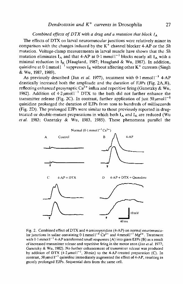
As previously described (Jan et al. 1977), treatment with 0 1 mmol I"1 4-APdrastically increased both the amplitude and the duration of EJPs (Fig. 2A,B),reflecting enhanced presynaptic Ca2+ influx and repetitive firing (Ganetzky & Wu,1982). Addition of 4-2/anoll"1 DTX to the bath did not further enhance thetransmitter release (Fig. 2C). In contrast, further application of just 50/anoi r 1
quinidine prolonged the duration of EJPs from tens to hundreds of milliseconds(Fig. 2D). The prolonged EJPs were similar to those previously reported in drug-treated or double-mutant preparations in which both IA and IK are reduced (Wuetal. 1983; Ganetzky & Wu, 1983, 1985). These phenomena parallel the
Normal (01 mmol P ' Ca2 +)
Control B 4-AP
4-AP + DTX D 4-AP + DTX + Quinidine
lOmV
40 ms
Fig. 2. Combined effect of DTX and 4-aminopyridine (4-AP) on normal neuromuscu-lar junctions in saline containing 0-1 mmol I"1 Ca2+ and4mmoll~' Mg2+. Treatmentwith 0-1 mmol I"1 4-AP transformed small responses (A) into giant EJPs (B) as a resultof increased transmitter release and repetitive firing in the motor axon (Jan et al. 1977;Ganetzky & Wu, 1982). No further enhancement of transmitter release was producedby addition of DTX (4-2/unoir1, 20min) to the 4-AP-treated preparation (C). Incontrast, 50/anolP1 quinidine immediately augmented the effect of 4-AP, resulting ingreatly prolonged EJPs. Sequential data from the same cell.

28 C.-F. W u AND OTHERS
Sh
A Control B DTX DTX + Quinic
10 mV
40 ms 40 ms 0 1 s
Fig. 3. Combined effects of DTX and a mutation eliminating IA in saline containingO^mmoir1 Ca2+ and 4mmoll"1 Mg2"1". (A) EJPs in Sh larvae resembled those in4-AP-treated normal larvae. (B) The presence of 4-2/anoH"1 DTX did not furtherenhance the transmitter release in Sh larvae. (C) Quinidine (50/imoll"1) generatedgreatly prolonged EJPs in the same preparation. Note the different time scale in C.Sequential recordings from the same cell. The resting potential had decreased from—47 mV in A and B to — 35 mV in C owing to muscle contractions, resulting in a smalleramplitude of EJPs in C.
observations in Sh larvae. Identical results were obtained by adding quinidine(50/zmolP1) alone to Sh larvae with or without DTX pre-treatment. The Shphenotype, i.e. raised and prolonged transmitter release (Fig. 3A, also see Jan etal. 1977), was further enhanced by quinidine but not by DTX (Fig. 3B,C).
Synergistic effects of DTX with a drug and a mutation that reduce IK
The above results raised the possibility that DTX acted mainly to reduce IA innerve terminals and thus was not able to exert additional effects when IA wasblocked by 4-AP or the Sh mutation. This interpretation was confirmed in adifferent set of experiments employing quinidine and the eag mutation, whichblocks about 50 % of IK in larval muscle (Wu et al. 1983).
Although the effects of DTX on transmitter release are relatively mild in normallarvae (Fig. 1), it produced strong enhancement of neuromuscular transmission ineag larvae or in normal preparations pre-treated with quinidine. A low dose(50//moll"1) of quinidine alone only slightly increased the amplitude of EJPs(Fig. 4A,B) and sometimes produced repetitive discharges of motor axons (datanot shown). Application of 4-2^moll~1 DTX qualitatively altered the EJPs,increasing their amplitude and duration several times (Fig. 4C). A further increasein the concentration of DTX (8-4 jmioll"1) enhanced transmitter release to aneven greater extent. As shown in Fig. 4D, this resulted in large, prolonged EJPssimilar to those caused by diminishing both IA and IK (see Figs 2D and 3C).
Consistent with the above finding, our experiments on eag larvae further

Dendrotoxin and K+ currents in Drosophila 29
Normal (0-2mmoir'Ca2+)
A Control B Quinidine
-TV-A.
|4mV
40 ms
C Quinidine + DTX
(4-2/nnoir1)D Quinidine + DTX
(8-4/fltioll"1)
Fig. 4. Synergistic interaction between DTX and quinidine in normal larvae. The bathsolution contained O^mmoll"1 Ca2+ and 4mmoll~1 Mg2"1". Treatments with50/anolP1 quinidine only slightly facilitated neuromuscular transmission (compare Aand B). Adding 4-2/imoll"1 DTX to the preparation clearly enhanced the quinidineeffect (C). Adding another 4-2^moll~1 DTX produced prolonged giant EJPs (D).Sequential recordings from the same cell. Note the different voltage and timecalibrations in D.
demonstrated the facilitatory effect derived from the synergistic interaction ofDTX with a defect in IK. The EJPs in eag muscles were not substantially greaterthan normal (Fig. 5A, also see Ganetzky & Wu, 1983, 1985; Wu et al. 1983).Application of 4-2/zmoll"1 DTX immediately increased the size of EJPs from2-4 mV to 5-10 mV (Fig. 5B) and in a few minutes caused repetitive discharges ofEJPs of varying sizes (Fig. 5C), some reaching 30 mV. Adding 0-1 mmoll"1 4-APto the preparation transformed these repetitive EJPs into the typical prolongedgiant EJPs (data not shown) observed when both IA and IK are blocked (seeFigs 2D and 3C). In sum, our findings indicate that DTX has an effect on theneuromuscular junction similar to, but smaller than, those caused by the Shmutation or 4-AP treatment.
Voltage-clamp analysis of DTX actions on IA and IK in muscle membrane
Direct evidence for alterations in K+ currents caused by DTX requiresmeasurements by voltage-clamp experiments. The Drosophila presynaptic nerveterminals are not easily accessible to this type of analysis and such measurements
Pvere performed in larval body-wall muscles. These large, identifiable musclefibers meet isopotential conditions, are enveloped by very little connective tissue

30 C . - F . WU AND OTHERS
A Control B /y DTX(2min)
4mV
40 ms
C I DTX(4min)
4mV L0-2s
Fig. 5. Synergistic interaction between DTX and a mutation reducing IK- (A) In salinecontaining O^mmoll"1 Ca2+ and 4mmoll~1 Mg2"1", the size of EJPs in eag larvae wasnot substantially greater than normal. (B) Within 2min of the addition of 4-2janoll~1
DTX, EJPs increased in size. (C) As time progressed (another 2min elapsed),repetitive discharges of giant EJPs appeared. Sequential data from the same cell.
and are directly exposed to the external medium (Wu & Haugland, 1985).Experiments were first carried out in Ca2+-free saline to eliminate Ca2+ and Ca2+-dependent K+ currents. Under this condition only IA and IK remain, and they canbe physiologically separated by their differences in inactivation properties (Wu &Haugland, 1985; Haugland, 1987; N. H. Haugland & C.-F. Wu, in preparation).The current-voltage (I-V) relationships in Figs 6-9 illustrate IA and IK, extractedas described in Materials and methods, and the effects of DTX at different Mg2+
concentrations and temperatures. The time courses of the total active currents (IA
plus IK) are shown in Figs 6 and 8 and the extracted IA currents are shown in Figs 7and 9.
Previous studies of larval muscle currents were conducted at low temperature(4-15 °C) for the ease of voltage-clamp control and, to improve membranestability, a high concentration of Mg2"1" (10-15 mmoll"1) was usually present in theCa2+-free saline (Salkoff & Wyman, 1981; Wu & Haugland, 1985). The EJPexperiments (Fig. 1) suggest that the DTX effect is suppressed by high Mg2"^concentration. We found that, in the presence of MmmoU"1 Mg2"1", DTX

Dendrotoxin and K+ currents in Drosophila 31
(1-4/mioll ) did not exert any significant effect on either IA or IK at 5°C (Figs 6and 7, 14 mmol I"1 Mg2"1" data). Subsequently, the concentration of Mg2+ waslowered to examine the possible inhibitory effect of Mg2"1" on DTX activity. At lowtemperature (5°C), we still could not detect a significant effect of DTX at thephysiological concentration (4 mmol I"1) of Mg2+ (Figs 8 and 9, 5°C data). Notuntil the Mg2"1" concentration was further lowered to a non-physiological level(0-5 mmol I"1) could we observe a clear effect of DTX at 5°C. Long-termtreatment (30min) with DTX reduced both IA and IK (Figs 6 and 7, 0-5 mmol I"1
Mg2"1" data). The effect of DTX at low temperature and low Mg2"1" concentration
5°C Mmmoir'Mg2"1"
O 0/rniolP1 DTX • l-4;rnioir' DTX+40
+20
t 16O
£ 12
< 8
D
5°C 0-5 mmol l"'Mg2+
DTX
1-4/mioir' DTX
16^A^F
12
0 160 320ms 0 160 320ms / , - . .
-4k—o-- 8 0 - 6 0 - 4 0 - 2 0 0 20 40 mV
Fig. 6. Action of DTX on K+ currents in normal larval muscle fibers measured by thetwo-microelectrode voltage-clamp technique at 5°C. Inward Ca2+ current and Ca2+-dependent outward currents were abolished by the use of Ca2+-free saline in this andthe following figures. The effects of 1-4/anoir1 DTX at 14 and 0-5mmol I"1 Mg2"*"were determined from 6-14 fibers in 3-7 larvae for each experiment. Superimposedtraces averaged from the fibers show the outward K+ currents elicited by voltage steps(not shown) to -40, -20, 0, +20 and +40 mV from a holding potential (VH) of—80 mV. The outward currents contain the transient IA, which peaks rapidly and theninactivates, and the delayed IK, which rises gradually to a plateau. The active currentdensity was determined by subtraction of leakage currents and adjustment formembrane area (expressed in JIA/JF'1 by using membrane capacitance as anindicator). The current-voltage (I-V) relationships show the mean±s.E.M. of theamplitude of the remaining IK after subtraction of the inactivating IA (measured at theend of the 400 ms pulses to different voltages). There was a decrease in IK in low-Mg2"1"saline.

32 C.-F. W u AND OTHERS
5°C 14mmoir1Mg2+
/" \O 0/mioir1 DTX " ^ • 1 -4/OTioir1 DTX
«—+40
5°C 0-5mmoll"'Mg2+
D 0/anoir1 DTX • l-4/rnioll"1 DTX
16 32 ms
-60 -40 -20 20 40 mV
Fig. 7. The effect of DTX on IA extracted from the data shown in Fig. 6. Tracesaveraged from different fibers represent the inactivating IA determined by subtractingfrom the total current (Fig. 6 traces) the remaining non-inactivating current attainablefollowing a prepulse to inactivate IA (see Materials and methods). The I-Vrelationships show the mean ± S.E.M. of the peak current density of IA. At 5°C, theeffect of DTX was apparent in saline containing 0-5 mmol P 1 Mg2"1" but was suppressedby 14 mmol r 1 Mg2"".
was not reversed by washing and could not be suppressed again if the Mg2"1"concentration was raised (data not shown).
It should be noted that at Ommoir1 Ca2+ and O-SmmolT1 Mg2+ themembrane underwent a characteristic sequence of changes even in the absence ofDTX (see Discussion and Fig. 10 for details). At 5°C, the changes in membranecurrents took about 50-70 min to reach a final steady state. Addition of DTX tosaline at this time again initiated a similar sequence of events (see Discussion andFig. 10) before reaching a new steady state 30 min later. The above results (Figs 6and 7, 0-5 mmol I"1 Mg2"1" data) include only steady-state data.
The activity of DTX is known to be enhanced at high temperature in chickbiventer cervicis and mouse diaphragm preparations (Harvey & Anderson, 1985).The effect of temperature on the action of DTX was therefore examined in salinecontaining a physiological concentration (4 mmol I"1) of Mg2"1". Raising thetemperature to 16°C revealed a readily demonstrable effect of DTX. Aspreviously reported (Wu & Haugland, 1985; Haugland, 1987), both IA and I) |approximately doubled in size for an increase in temperature of about 10 °C (see

Dendrotoxin and K+ currents in Drosophila 33
5°C and 16°C data in Figs 8 and 9). At 16°C, DTX reduced the amplitude of IA
without any obvious change in its kinetics (Fig. 9). In contrast, IK was largelyunaltered (Fig. 8). The same experiment was performed at lower DTX concen-trations. We found that at 470 nmol 1 DTX had nearly reached a plateau level inits inhibitory effect on IA, with a reduction in IA almost identical to that caused by1-4/anoir1 DTX. By extrapolation, the blockade of IA by DTX might be larger atroom temperature and could account for the observed changes in EJPs (Figs 1-5)at the physiological concentration of Mg2"1" (4mmoll~1).
The same type of voltage-clamp analysis was carried out in the presence ofZOmmoir1 Ca2+ at both 5 and 16°C. Under these conditions, the inward Ca2+
current and outward Ca2+-dependent K+ currents could be examined (Singh &Wu, 1987, 1989) but, in more than 10 fibers, no apparent effects of DTX(1-4/imolP1) on these currents could be demonstrated (data not shown). Eventhough this result is consistent with previous reports on several vertebratepreparations (Halliwell et al. 1986; Harvey, 1987), we cannot rule out the
16°C 4mmoll"1Mg2+
4a.
40
30
20
10
0
O 0/imoll"1 DTX 1-4/imoir1 DTX
32 64 ms 0 32 64 ms
5°C 4mmoll"1Mg2+
DO/anoir'DTX • 1-4/anoir1 DTX
-80 -60 -40 40 mV
Fig. 8. Temperature dependence of the effect of DTX at a physiological concentration(4mmol r 1 ) of Mg2"1". Same experimental protocol as in Fig. 6. In each experiment 5-8fibers from three larvae were used. Superimposed traces show the total active currentdensity from the mean of different fibers. The I-V relationships show themean ± S.E.M. of IK measured at the end of voltage pulses. DTX did not significantlyaffect IK in saline containing 4mmoll~1 Mg2"1" at either 5 or 16°C.

34 C.-F. WU AND OTHERS
fcu
I
16°C 4mmoir'Mg2+
I O O/unoir1 DTX • 1-4/anoll"1 DTX
0 16 32ms 0 16 32ms
40 mV
Fig. 9. Temperature dependence of the effect of DTX on IA. Traces represent themean density of IA extracted from the data shown in Fig. 8. The I-V relationshipshows the mean±s.E.M. of the peak IA. In saline containing 4mmolP1 Mg2"*", asignificant reduction in IA was observed at 16 CC but not at 5°C.
possibility of a minor effect on these channels in Drosophila. Since the Ca2+-dependent currents are intrinsically more variable than IA and IK amongDrosophila muscle fibers (Singh & Wu, 1989), a small quantitative change in theiramplitude would be difficult to detect.
Discussion
Effects of DTX on K+ channels
On the basis of electrophysiological and pharmacological criteria, a variety offunctional types of K+ channels have been identified in a large number ofvertebrate and invertebrate species, but there is no information about theirmolecular distinctions and similarities (Latorre & Miller, 1983; Rudy, 1988).Mutations that alter K+ currents can provide insight into this problem (Wu &Ganetzky, 1986). A combination of genetic and pharmacological techniques mayprove to be a powerful approach to the study of the functioning and mechanisms ofthis class of membrane channels.
So far, only a few specific, high-affinity toxins for K+ channels are known. ThusjDTX represents an especially valuable probe for functional and biochemical

Dendrotoxin and K+ currents in Drosophila 35
analyses of these channels (Dolly et al. 1984, 1987; Rehm & Lazdunski, 1988).Previous voltage-clamp studies on vertebrate preparations have indicated that thesensitivity of K+ channels to DTX in different excitable cells does not necessarilyreflect similarity in channel function. DTX suppresses the non-inactivating orslowly inactivating K+ currents in the node of Ranvier (Weller et al. 1985; Benoit& Dubois, 1986), in nodose ganglion neurons (Dolly et al. 1987), in dorsal rootganglion neurons (Penner et al. 1986) and in visceral sensory neurons (Stansfeldet al. 1986) but acts on the rapidly inactivating transient IA in hippocampal neurons(Halliwell et al. 1986). However, a rapidly inactivating variant of the transient K+
current in superior cervical ganglion neurons is reportedly not sensitive to DTX(Dolly et al. 1987). Recent toxin binding studies using DTXt (Rehm & Lazdunski,1988) and molecular genetic analyses of the Sh gene in Drosophila (Kamb et al.1988; Pongs et al. 1988; Schwarz et al. 1988) have suggested the possibility ofmultiple subtypes of closely related K+ channel proteins.
Our experiments extended the study of DTX to an invertebrate preparation.The action of toxin on muscle membrane was analyzed by voltage-clampexperiments. The results showed that, at physiological concentrations of Mg2"1",DTX affects mainly IA without apparent effects on other currents. Although directvoltage-clamp measurements of the DTX effects at higher temperatures are notavailable for technical reasons (see Materials and methods), our data at 16°C and4mmoll~1 Mg2+ (Figs 8 and 9) could be extrapolated to infer a similar, or agreater, DTX effect on IA at 25-29°C to account for the observed changes in EJPs(Figs 1-5). At low divalent cation concentrations IK, in addition to IA, wasreduced (Fig. 6).
The exact mechanisms of DTX actions on membrane channels remain un-known. It has been previously reported that the toxin potency of enhancingneuromuscular transmission decreases drastically when temperature is lowered orwhen Mg2"1" concentration is raised (Harvey & Anderson, 1985). In Drosophila,the effect of DTX also critically depends on temperature and the concentration ofMg2"1", and possibly Ca2+, in the saline. It is worth pointing out the intriguinginteractions between DTX and Mg2"1", as suggested by the experiments in Ca2+-free saline at 5°C (Fig. 10). Decreasing the Mg2"1" concentration to O-SmmolF1
initiated a sequence of changes in the amplitude and kinetics of membranecurrents. Before reaching a steady state in 70min, the voltage dependence of thetotal current was strikingly different (Fig. 10A). Outward currents larger thannormal were elicited by strong depolarizing steps but the current turned inwards atsome intermediate levels of depolarization (Fig. 10AH, arrow). At the steadystate, the extracted IA was reduced in amplitude whereas IK retained nearlynormal amplitude but seemed to rise more rapidly (Figs 6 and 7, O/zmoll"1 DTXdata, and Fig. lOBi). Interestingly, when DTX was added at this time, themembrane currents again underwent characteristic changes similar to the abovesequence of events (Fig. 10Bi,ii). At the new steady state there was a decrease in
poth IA and IK (Figs 6 and 7,0-5 mmol I"1 Mg2+ data, and Fig. lOBiii). These non-physiological changes induced by low Mg2+ concentration were prevented by the

36 C.-F . WU AND OTHERS
Aii
4/zA/iF" •L160 ms
Biii
40 min ;from Aii , 10 min 20min
Ci Cii Ciii
Mmmoir'Mg2"1".
0-5 mmol r 'ioir '30min 40 min
^ V * W t * * * * »
Fig. 10. Interaction of DTX and Mg2"1" on membrane currents in Ca2+-free saline.Superimposed traces represent active currents elicited by voltage steps to —40, —20, 0and +20mV from VH (-80 mV) at 5°C. Lowering the Mg2"1" concentration fromMmmoir 1 to 0-5 mmol I"1 initiated a transient change in the voltage dependence ofthe membrane currents (Ai and Aii). Note that, at an intermediate level ofdepolarization, the total current was inward (arrow in Aii). At the steady state (Bi),both IA and IK regained nearly normal voltage dependence, in spite of the reducedamplitude of IA and apparently more rapid kinetics of IK (compare Ai and Bi, also seeFigs 6 and 7, 0/anoll DTX data). Addition of 1-4/anoll"1 DTX at this time againinduced a similar transient change (Bii, arrow). Eventually, both IA and IK weredecreased and the membrane reached a new steady state (Biii). If DTX (1-4/anoll"1)was also present in the low-Mg2"1" (0-5 mmol I"1) saline during the solution replace-ment, no transient changes in the voltage dependence or kinetics of IA and IK wereinduced (Ci and Cii). IA and IK gradually decreased and no alterations in kinetics wereapparent (Ciii). Sequential data from the same cell are shown in A and B with leakagecurrent subtracted. Data from a different cell are shown in C.
presence of DTX. Replacing 14 mmol 1 l Mg2"1" saline by 0-5 mmol 1 1 Mg2+ salinecontaining DTX caused only a gradual reduction in the amplitude of IA and IK
without evident alterations in their kinetics (Fig. 10C).The basis of the non-physiological events initiated by reducing Mg2"1" concen-
tration (Fig. 10A) is not understood. Divalent cations can change the local electricpotential profile in the membrane by interaction with fixed negative charges on itsouter surface (Hille, 1984). Divalent cations may also directly affect the gating ofcertain ion channels (Dingledine, 1986; Armstrong & Lopez-Barneo, 1987; Steri^et al. 1987). One possibility is that an inward current was induced transiently after

Dendrotoxin and K+ currents in Drosophila 37
reducing the Mg2"1" concentration in the Ca2+-free saline (Fig. lOAii, Bii, arrows).Permeation of Na+ through muscle Ca2+ channels in Ca2+-free saline has beenobserved in one insect species (Yamamoto, 1987) and some vertebrate species(McCleskey et al. 1986) but not in another insect species (Ashcroft & Stanfield,1982). Whether permeation of Na+ is responsible for the phenomenon inDrosophila muscle at very low cation concentrations must await further investi-gation. No matter what exact mechanism underlies these changes, the surprisingeffect of DTX, preventing their initiation (Fig. IOC) and reiterating the sequenceof changes once they are induced by Mg2"1" reduction (Fig. 10B), must beconsidered in future physicochemical approaches to the mechanism of DTXactions.
The effects of several presynaptic snake toxins, such as /3-bungarotoxin (yS-BTX)and related toxins, are known to show a similar temperature and cationdependence (Chang, 1985). More interestingly, DTX, /3-BTX and another toxin,the mast cell degTanulating peptide, have been shown to bind to common proteintargets and inhibit K+ currents (Petersen et al. 1986; Bidard et al. 1987; Stansfeldet al. 1987). The structures of these toxins and DTX have been completely orpartially determined (Lee, 1979; Joubert & Taljaard, 1980; Bidard et al. 1987).Such structural information may be important in designing further biophysical andbiochemical experiments to study the nature of the observed temperature andcation dependence, which may in turn give clues to the mechanism of interactionof toxins with relevant membrane sites.
Effects of DTX on neuromuscular transmission
Direct measurements of the various ion currents in presynaptic nerve terminalsby voltage-clamp are technically difficult. Therefore, Drosophila mutants of thegenes that encode specific types of ion channels provide a unique opportunity toinfer a defective current underlying the mutant phenotypes in nerve terminals.The altered currents can be directly analyzed by the voltage-clamp method inlarger cells, such as muscle fibers.
It is worth noting that the DTX effect is specific but relatively weak comparedwith the actions of other channel-specific toxins or drugs. As in Drosophila larvalmuscle, DTX reduces but does not eliminate the K+ currents in differentvertebrate preparations (Weller et al. 1985; Halliwell ef a/. 1986; Penneref al. 1986;Stansfeld et al. 1986). At mouse and frog neuromuscular junctions, DTX enhancestransmission by a mild increase in the quantal content of EPPs and the occasionalappearance of repetitive EPPs (Anderson & Harvey, 1985; Harvey & Anderson,1985; Harvey, 1987). As in vertebrate preparations, the effect of DTX atDrosophila neuromuscular junctions is relatively mild (Fig. 1). Similar but moreextreme effects are observed when IA is eliminated by Sh mutations or treatmentwith 4-AP (Figs 2 and 3). Previous extracellular nerve recordings and focal EJPrecordings in Sh and eag larvae and in normal larvae treated with 4-AP orletraethylammonium show that the abnormal EJPs reflect predominantly pre-synaptic defects rather than a failure of postsynaptic membrane repolarization.
38 C.-F. WU AND OTHERS
The nerve terminals fail to repolarize properly, resulting in repetitive firing of theaxon and release of an abnormally large amount of transmitter (Jan et al. 1977;Ganetzky & Wu, 1982). The results of the present study indicate that DTXfacilitates larval neuromuscular transmission, probably by reducing IA but notother K+ currents in the nerve terminal.
We found that the relatively mild action of DTX on synaptic transmission can,nevertheless, be enhanced and clearly demonstrated through its synergisticinteractions with mutations or drugs that reduce different K+ currents (Figs 4 and5). The synergistic effect of DTX on Sh, eag and slo mutants could be readilystudied to obtain information regarding the type of K+ currents (i.e. IA, IK andICF) in the nerve terminal affected by DTX. The results were also corroborated byexamining the synergism of DTX with drugs blocking IA or IK. At the neuro-muscular junction, DTX effectively augmented the effect of quinidine or the eagmutation but did not further increase the effect of 4-AP or the Sh mutation.
Two conclusions may be drawn from these results. First, the IA channels in thenerve terminal and muscle membrane may be very similar or identical in theirproperties, as suggested by their similar sensitivity to DTX as well as to 4-AP andthe Sh mutation. Second, the regulation of synaptic transmission by interplay ofdifferent K+ currents is intricate and can produce results in a wide dynamic range.Blocking IK produced a relatively smaller effect on EJPs than blocking IA
(compare Figs 2B and 3 A with 4B). Partial blockade of IA by DTX resulted in onlymild effects on EJPs at physiological concentrations of Mg2"1" (Fig. 1A,B) butcould lead to explosive discharges of repetitive or prolonged giant EJPs (Figs 4Dand 5C) when IK was also blocked or reduced. Since multiple K+ conductances arealso present in vertebrate nerve terminals (David & Yaari, 1985; Mallart, 1985),our understanding of the ionic mechanisms underlying the generation of repetitivenerve spikes and increased synaptic activity in certain human pathologicalconditions (Dichter & Ayala, 1987) may be improved by further studies of thefunctional consequences of K+ channel-specific toxins and mutations.
We thank Drs F. J. Joubert and F. H. H. Carlsson (National Chemical ResearchLaboratory, CSIR, Pretoria, South Africa) for kindly providing DTX, Mr C.-Y.Han for technical assistance, and Mr P. Taft, Mrs K. Howard and Mrs G. Folkinsfor helping with the preparation of this manuscript. This work was supported by agrant (NSC-76-0412-B002-103) from the National Science Council, ROC, and USPublic Health Service grants NS00675, NS18500 and NS15797.
ReferencesANDERSON, A. J. & HARVEY, A. L. (1985). Electrophysiological actions of facilitatory toxins
from mamba venoms on mammalian neuromuscular junctions. Br. J. Pharmac. 86, 588P.ARMSTRONG, C. M. & LOPEZ-BARNEO, J. (1987). External calcium ions are required for
potassium channel gating in squid neurons. Science 236, 712-714.ASHCROFT, F. M. & STANFIELD, P. R. (1982). Calcium and potassium currents in muscle fibers of
an insect (Carausius morosus). J. Physiol., Lond. 323, 93-115. \BENOIT, E. & DUBOIS, J. M. (1986). Toxin I from the snake Dendroaspis polylepis polylepis:

Dendrotoxin and K+ currents in Drosophila 39
a highly specific blocker of one type of potassium channel in myelinated nerve fibre. Brain Res.377, 374-377.
BIDARD, J.-N., MOURRE, C. & LAZDUNSKI, M. (1987). Two potent central convulsant peptides, abee venom toxin, the MCD peptide, and a snake venom toxin, dendrotoxin I, known to blockK+ channels, have interacting receptor sites. Biochem. biophys. Res. Commun. 143,383-389.
CHANG, C. C. (1985). Neurotoxins with phospholipase A2 activity in snake venoms. Proc. natn.Sci. CouncilR.O.C. B9, 126-142.
DAVID, G. & YAARI, Y. (1985). Several potassium conductances modulate the excitability of frogmotor nerve terminals. In Calcium, Neuronal Function and Transmitter Release (ed.R. Rahaminoff & B. Katz), PP- 563-574. Boston: M. Nijhoff.
DICHTER, M. A. & AYALA, G. F. (1987). Cellular mechanisms of epilepsy: a status report.Science 237, 157-164.
DINGLEDINE, R. (1986). NMDA receptors: What do they do? Trends Neurosci. 9, 47^19.DOLLY, J. O., HALLIWELL, J. V., BLACK, J. D., WILLIAMS, R. S., PELCHEN-MATTHEWS, A.,
BREEZE, A. L., MEHRABAN, F., OTHMAN, I. B. & BLACK, A. R. (1984). Botulinum toxin anddendrotoxin as probes for studies on transmitter release. J. Physiol, Paris 79, 280-303.
DOLLY, J. O., STANSFELD, C. E., BREEZE, A. L., PELCHEN-MATTHEWS, A., MARSH, S. J. &BROWN, D. A. (1987). Neuronal acceptor sub-types for dendrotoxin and their relation to K+
channels. In Neurotoxins and Their Pharmacological Implications (ed. P. Jenner &B. Pharm), pp. 81-%. New York: Raven Press.
ELKJNS, T. B., GANETZKY, B. & Wu, C.-F. (1986). A Drosophila mutation that eliminates acalcium-dependent potassium current. Proc. natn. Acad. Sci. U.S.A. 83, 8415-8419.
GANETZKY, B. & Wu, C.-F. (1982). Drosophila mutants with opposing effects on nerveexcitability: Genetic and spatial interactions in repetitive firing. J. Neurophysiol. 47, 501-514.
GANETZKY, B. & Wu, C.-F. (1983). Neurogenetic analysis of potassium currents in Drosophila:synergistic effects on neuromuscular transmission in double mutants. J. Neurogenetics 1,17-28.
GANETZKY, B. & Wu, C.-F. (1985). Genes and membrane excitability in Drosophila. TrendsNeurosci. 8, 322-326.
GANETZKY, B. & Wu, C.-F. (1986). Neurogenetics of membrane excitability in Drosophila.A. Rev. Genetics 20, 13-44.
GHO, M. & MALLART, A. (1986). Two distinct calcium-activated potassium currents in larvalmuscle fibers of Drosophila melanogaster. Pflugers Arch. ges. Physiol. 407, 526-533.
HALLIWELL, J. V., OTHMAN, I. B., PELCHEN-MATTHEWS, A. & DOLLY, J. O. (1986). Centralaction of dendrotoxin: selective reduction of a transient potassium conductance inhippocampus and binding to localized acceptors. Proc. natn. Acad. Sci. U.S.A. 83, 493-497.
HARVEY, A. L. (1987). Facilitation of neuromuscular transmission by dendrotoxins. Asia PacificJ. Pharmac. 2, 187-194.
HARVEY, A. L. & ANDERSON, A. J. (1985). Dendrotoxin: snake toxins that block potassiumchannels and facilitate neurotransmitter release. Pharmac. Ther. 31, 33-55.
HARVEY, A. L., ANDERSON, A. J. & KARLSSON, E. (1984). Facilitation of transmitter release byneurotoxins from snake venoms. J. Physiol., Paris 79, 722-227.
HARVEY, A. L. & KARLSSON, E. (1980). Dendrotoxin from the venom of the green mamba,Dendroaspis angusticeps. A neurotoxin that enhances acetylcholine release at neuromuscularjunctions. Naunyn Schmiedeberg's Arch. Pharmac. 312, 1-6.
HAUGLAND, F. N. (1987). A voltage-clamp analysis of membrane potassium currents in larvalmuscle fibers of the Shaker mutants of Drosophila. PhD thesis, The University of Iowa, IowaCity, Iowa.
HAUGLAND, F. N. & Wu, C.-F. (1987). Concomitant alteration of potassium channel gating andpharmacology in a Shaker mutant of Drosophila. Abstr. Soc. Neurosci. 13, 530.
HILLE, B. (1984). Ionic Channels of Excitable Membranes. Sunderland, MA: SinauerAssociates, Inc., pp. 316-324.
JAN, L. Y. & JAN, Y. N. (1976a). Properties of the larval neuromuscular junction in Drosophilamelanogaster. J. Physiol., Lond. 262, 189-214.N, L. Y. & JAN, Y. N. (1976b). L-Glutamate as an excitatory transmitter at the Drosophilalarval neuromuscular junction. J. Physiol., Lond. 262, 215-236.

40 C.-F. WU AND OTHERS
JAN, Y. N., JAN, L. Y. & DENNIS, M. J. (1977). Two mutations of synaptic transmission inDrosophila. Proc. R. Soc. B 198, 87-108.
JOHANSEN, J., HALPERN, M. E., JOHANSEN, K. M. & KESHISHIAN, H. (1989). Stereotypicmorphology of glutamatergic synapses on identified muscle cells of Drosophila larvae./. Neurosci. 9, 710-725.
JOUBERT, F. J. & TALJAARD, N. (1980). The amino acid sequence of two proteinase inhibitorhomologues from Dendroaspis angusticeps venom. Hoppe-Seylers Z. Physiol. Chem. 361,661-674.
KAMB, A., TSENG-CRANK, J. & TANOUYE, M. A. (1988). Multiple products of the DrosophilaShaker gene may contribute to potassium channel diversity. Neuron 1, 421-430.
LATORRE, R. & MILLER, C. (1983). Conduction and selectivity in potassium channels. J. Membr.Biol. 71, 11-30.
LEE, C. Y. (1979). Recent advances in chemistry and pharmacology of snake toxins. InAdvances in Cy to pharmacology, vol. 3 (ed. B. Ceocarelli & F. Clementi), pp. 1-16. NewYork: Raven Press.
MALLART, A. (1985). A calcium-activated potassium current in motor nerve terminals of themouse. /. Physiol., Lond. 368, 577-591.
MCCLESKEY, E. W., PALADE, P. T. & ALMERS, W. (1986). The mechanism of ion selectivity incalcium channels of skeletal muscle. In Ion Channels in Neural Membranes (Neurol.Neurobiol. vol. 20) (ed. J. M. Ritchie, R. D. Keynes & L. Bolis), pp. 193-204. New York:Alan R. Liss, Inc.
PENNER, R., PETERSEN, M., PIERAU, F.-K. & DREYER, F. (1986). Dendrotoxin: a selectiveblocker of a non-inactivating potassium current in guinea-pig dorsal root ganglion neurones.Pflugers Arch. ges. Physiol. 407, 365-369.
PETERSEN, M., PENNER, R., PIERAU, F.-K. & DRYER, F. (1986). /J-Bungarotoxin inhibits a non-inactivating potassium current in guinea-pig dorsal root ganglion neurones. Neurosci. Lett. 68,141-145.
PONGS, O., KECSKEMETHY, N., MOLLER, R., KRAH-JENTGENS, I., BAUMANN, A., KILTZ, H. H.,CANAL, I., LLAMAZARES, S. & FERRUS, A. (1988). Shaker encodes a family of putativepotassium channel proteins in the nervous system of Drosophila. EMBO J. 7, 1087-10%.
REHM, H. & LAZDUNSKI, M. (1988). Existence of different populations of dendrotoxin I bindingprotein associated with neuronal K+ channels. Biochem. biophys. Res. Commun. 153,231-240.
RUDY, B. (1988). Diversity and ubiquity of K channels. Neuroscience 25, 729-749.SALKOFF, L. & WYMAN, R. (1981). Genetic modification of potassium channels in Drosophila
Shaker mutants. Nature, Lond. 293, 228-230.SCHWARZ, T. L., TEMPEL, B. L., PAPAZIAN, D. M., JAN, Y. N. & JAN, L. Y. (1988). Multiple
potassium-channel components are produced by alternative splicing at the Shaker locus inDrosophila. Nature, Lond. 331, 137-142.
SINGH, S. & Wu, C.-F. (1987). Genetic and pharmacological separation of four potassiumcurrents in Drosophila larvae. Soc. Neurosci. Abstr. 13, 579.
SINGH, S. & Wu, C.-F. (1989). Complete separation of four potassium currents in Drosophila.Neuron 2, 1325-1329.
STANSFELD, C. E., MARSH, S. J., HALUWELL, J. V. & BROWN, D. A. (1986). 4-Aminopyridineand dendrotoxin induce repetitive firing in rat visceral sensory neurones by blocking a slowlyinactivating outward current. Neurosci. Lett. 64, 299-304.
STANSFELD, C. E., MARSH, S. J., PARCEJ, D. N., DOLLY, J. O. & BROWN, D. A. (1987). Mast celldegranulating peptide and dendrotoxin selectively inhibit a fast-activating potassium currentand bind to common neuronal proteins. Neuroscience 23, 893-902.
STERN, J. H., KNUTSSON, H. & MACLEISH, P. R. (1987). Divalent cations directly affect theconductance of excised patches of rod photoreceptor membrane. Science 236, 1674-1678.
SUZUKI, N. & KANO, M. (1977). Development of action potential in larval muscle fibers inDrosophila melanogaster. J. Cell Physiol. 93, 383-388.
WELLER, U., BERNHARDT, U., SIEMEN, D., DREYER, F., VOGEL, W. & HABERMAN, E. (1985).Electrophysiological and neurobiochemical evidence for the blockade of a potassium channaiby dendrotoxin. Naunyn-Schmiedeberg's Arch. Pharmac. 330, 77-83. ™
Wu, C.-F. & GANETZKY, B. (1986). Genes and ionic channels in Drosophila. In Ion Channels in

Dendrotoxin and K+ currents in Drosophila 41
Neural Membranes (Neurol. Neurobiol. vol. 20) (ed. J. M. Ritchie, R. D. Keynes & L. Bolis),pp. 407-423. New York: Alan R. Liss, Inc.
Wu, C.-F., GANETZKY, B., HAUGLAND, F. & Liu, A.-X. (1983). Potassium currents inDrosophila: Different components affected by mutations of two genes. Science 220,1076-1078.
Wu, C.-F., GANETZKY, B., JAN, L. Y., JAN, Y.-N. & BENZER, S. (1978). A Drosophila mutantwith a temperature-sensitive block in nerve conduction. Proc. natn. Acad. Sci. U.S.A. 75,4047-4051.
Wu, C.-F. & HAUGLAND, F. (1985). Voltage-clamp analysis of membrane currents in larvalmuscle fibers of Drosophila: Alteration of potassium currents in Shaker mutants. J. Neurosci.5, 2626-2640.
YAMAMOTO, D. (1987). Sodium inward currents through calcium channels in mealworm musclefibers. Archs Insect Biochem. Physiol. 5, 227-231.