ABSTRACT Bioremediation with White-Rot Fungi at ... responsible for making and maintaining the...
88
ABSTRACT Bioremediation with White-Rot Fungi at Fisherville Mill: Analyses of Gene Expression and Number 6 Fuel Oil Degradation Darcy Young Extracellular enzymes that white-rot fungi secrete during lignin decay have been proposed as promising agents for oxidizing pollutants. We investigated the abilities of the white-rot fungi Punctularia strigosozonata, Irpex lacteus, Trichaptum biforme, Phlebia radiata, Trametes versicolor, and Pleurotus ostreatus to degrade Number 6 fuel oil in wood sawdust cultures. Our goals are to advise bioremediation efforts at a brownfield redevelopment site on the Blackstone River in Grafton, Massachusetts and to contribute to the understanding of decay mechanisms in white-rot fungi. All species tested degraded a C10 alkane. When cultivated for 6 months, Irpex lacteus, T. biforme, P. radiata, T. versicolor and P. ostreatus also degraded a C14 alkane and the polycyclic aromatic hydrocarbon phenanthrene. Gene expression analyses of P. strigosozonata indicate differential gene expression in the presence of Number 6 oil and on pine and aspen sawdust. David Hibbett, Ph.D. Chief Instructor Deborah Robertson, Ph.D. Associate Professor Heather Wiatrowski, Ph.D. Assistant Professor
Transcript of ABSTRACT Bioremediation with White-Rot Fungi at ... responsible for making and maintaining the...

ABSTRACT
Bioremediation with White-Rot Fungi at Fisherville Mill: Analyses of Gene
Expression and Number 6 Fuel Oil Degradation
Darcy Young
Extracellular enzymes that white-rot fungi secrete during lignin decay have been proposed as
promising agents for oxidizing pollutants. We investigated the abilities of the white-rot fungi
Punctularia strigosozonata, Irpex lacteus, Trichaptum biforme, Phlebia radiata, Trametes versicolor,
and Pleurotus ostreatus to degrade Number 6 fuel oil in wood sawdust cultures. Our goals are to advise
bioremediation efforts at a brownfield redevelopment site on the Blackstone River in Grafton,
Massachusetts and to contribute to the understanding of decay mechanisms in white-rot fungi. All
species tested degraded a C10 alkane. When cultivated for 6 months, Irpex lacteus, T. biforme, P.
radiata, T. versicolor and P. ostreatus also degraded a C14 alkane and the polycyclic aromatic
hydrocarbon phenanthrene. Gene expression analyses of P. strigosozonata indicate differential gene
expression in the presence of Number 6 oil and on pine and aspen sawdust.
David Hibbett, Ph.D.
Chief Instructor
Deborah Robertson, Ph.D.
Associate Professor
Heather Wiatrowski, Ph.D.
Assistant Professor

ACADEMIC HISTORY
Name: Darcy Young Date: October 2012
Baccalaureate Degree: B.A. Biology
Source: Clark University Date: May 2011
Other Degrees:
Occupation and Academic Connection: Clark University Biology Department M.A.
candidate June 2011 – May 2012

ACKNOWLEDGEMENTS
I would like to thank my adviser, David Hibbett, for his perpetual patience, humor,
and guidance. I truly appreciate the support and advice of my thesis committee members
Deb Robertson and Heather Wiatrowski. My fellow members of the Hibbett lab provided
invaluable knowledge and friendship. I am especially grateful to Dimitris Floudas, who
supervised and encouraged me during every phase of this thesis, and Rachael Martin, who
was responsible for making and maintaining the cultures involved in the comparative
screen. Thank you also to Gene Bernat of Fisherville Redevelopment LLC and the
Mosakowski Institute at Clark University, whose generous financial support and scientific
curiosity made this project possible. I am thankful for the counsel and expert contributions
of Jim Rice in the School of Engineering at Brown University and Erika Lindquist and Igor
Grigoriev at the U.S. Department of Energy Joint Genome Institute. The work conducted
by the U.S. Department of Energy Joint Genome Institute is supported by the Office of
Science of the U.S. Department of Energy under Contract No. DE-AC02-05CH11231.
Thanks, finally, to the Clark University Biology Department.
iv

TABLE OF CONTENTS
List of Tables vi
List of Figures vii
Introduction 1
Literature Review 6
Materials and Methods 32
Results 43
Discussion 49
Tables 55
Figures 70
Literature Cited 73
v

LIST OF TABLES
Table 1: Studies showing degradation of petroleum components by white-rot fungi 55
Table 2: Hydrocarbon degradation ratios of species grown on Bunker C oil 57
Table 3: Quantity and quality of LiCl-purified total RNA 57
Table 4: Illumina RNA-Seq summary 58
Table 5: Pearson correlations (r) between biological replicates 58
Table 6: P. strigosozonata genes overexpressed ≥ 5-fold in aspen spawn with
Bunker C oil relative to aspen spawn without Bunker C oil 59
Table 7: P. strigosozonata genes overexpressed ≥ 5-fold in pine spawn with
Bunker C oil relative to pine spawn without Bunker C oil 61
Table 8: P. strigosozonata genes overexpressed ≥ 5-fold in pine spawn without
Bunker C oil relative to aspen spawn without Bunker C oil 62
Table 9: P. strigosozonata genes overexpressed ≥ 5-fold in aspen spawn without
Bunker C oil relative to pine spawn without Bunker C oil 64
Table 10: Genes overexpressed ≥ 2-fold 68
Table 11: Counts and average lengths of uncharacterized genes overexpressed
≥ 5-fold. 68
Table 12: P. strigosozonata genes overexpressed ≥ 5-fold in pine and aspen spawn
with Bunker C oil relative to pine and aspen spawn without Bunker C oil. 69
vi

LIST OF FIGURES
Figure 1: Flame ionization detector chromatogram of terpene in control samples 70
Figure 2: Flame ionization detector chromatogram of phenanthrene in control
compounds 71
Figure 3: Flame ionization detector chromatogram of unidentified C10 and C14
alkanes in control compounds 72
vii

1
Introduction
Fisherville Mill cleanup employs bioremediation to diminish industrial
pollutants
Humans have adopted fungi for use in applications ranging from fermentation to
biofuel production (Moore et al. 2011). One biological function that has garnered
substantial interest is the degradation of environmental pollutants such as polycyclic
aromatic hydrocarbons (PAHs), explosives, polychlorinated biphenyls (PCBs), and
organochlorine pesticides (Kanaly and Hur 2006). The high cost of traditional methods for
removal of these contaminants has motivated research into alternative biological systems
that rely on or supplement native organisms that convert contaminants into less toxic
derivatives (Head 1998). Diverse bacterial, plant, and fungal species have been assayed for
their abilities to oxidize pollutants (Pouli and Agathos 2011). The success of
bioremediation efforts hinges on developing a clearer understanding of the metabolic
pathways of pollutant degradation in different taxa so that the organisms most suited to a
given task and environmental conditions may be engaged.
We became interested in bioremediation using fungal systems through the
redevelopment efforts at Fisherville Mill, a brownfield site on the Blackstone River in
Grafton, Massachusetts. Originally a manufacturing plant for furniture, textiles, and
machine parts, Fisherville Mill burned down in 1999, leaving soil and groundwater at the
site contaminated with petroleum, chlorinated volatile organic compounds, asbestos, and
heavy metals (Ollila et al. 2008). Immediately following the fire, the Environmental
Protection Agency (EPA) and the Massachusetts Department of Environmental Protection

2
(DEP) removed asbestos-contaminated building debris and oil to prevent contamination of
nearby drinking water. However, the soils and groundwater at the site remain contaminated
with chlorinated volatile organic compounds (VOCs) and petroleum. Number 6 oil, a
viscous and dense fuel oil, is particularly persistent in soils, sediment, and surface water
(Ollila et al. 2008).
The redevelopment project ongoing at Fisherville Mill offers the opportunity to test
the large-scale applicability of bioremediation of No. 6 oil. Part of the project includes
building an “Eco-Machine” that uses the water-filtration capabilities of various organisms
to degrade the oil that remains in the water column (Adams et al. 2007). The design of the
EcoMachine includes the use of fungal cultures to oxidize the oil into less toxic products.
The goals of our investigation were to identify native fungal species with oil-degrading
potential and to identify the genes that are differentially expressed in the presence of No. 6
oil. With this information, we hoped to make a recommendation for the fungal species that
could be used in the EcoMachine.
Irpex lacteus FD-9 was incorporated into the EcoMachine at Fisherville Mill on
5/4/12. Bags of millet grain inoculated with I. lacteus and other strains at Fungi Perfecti
(Olympia, Washington, USA) were mixed with black birch wood chips in large plastic
bins. Water from the Blackstone Canal will be diverted to flow through several of these
bins into large aquatic systems containing plants that are meant to further purify the water
before it is redirected back to the canal. As of 5/11/12, I. lacteus had fully colonized the
wood chips with no visible evidence of fungal or bacterial contamination.

3
Bioremediation is a feasible alternative to industrial cleanup of pollutants
Traditional approaches to cleaning polluted soils and water involve some kind of
physical processing of the contaminated material. In situ treatments such as soil aeration
attempt to address the problem where it occurred without moving the contaminated
substance while ex situ approaches like soil excavation isolate the polluted material either
to dispose of it or to further treat it (Sims 1990). These approaches are the norm today
because the methods are relatively well-established (Head 1998). However, many groups
are interested in alternative methods of pollutant remediation because traditional
approaches are expensive and laborious (Barr and Aust 1994). Furthermore, traditional
treatments are under scrutiny because they do not catalyze the degradation or
detoxification of pollutants. Instead, contaminated material is usually sequestered in situ or
removed to a landfill (Head 1998).
Bioremediation, defined as the application of biological processes to the
management of environmental pollution, has been proposed as an attractive alternative to
traditional pollution remediation methods (Gadd 2001). Proponents of bioremediation
argue that it offers the possibility of the complete mineralization of various persistent
pollutants at a lower cost than physical processing (Head 1998). The desired metabolic
processes are usually controlled through the application of one or a succession of
microorganisms that have been observed or hypothesized to be able to degrade or detoxify
a group of pollutants.

4
Similar to industrial approaches, bioremediation can be carried out in situ or ex situ.
The choice between these two scenarios depends on the growth requirements for the
remediation organisms. While in situ methods are sometimes preferred because they are
non-invasive, ex-situ methods may be more successful because the growth environment for
the remediating organism can be engineered for efficient degradation. This may be
accomplished by maintaining a different temperature, pH, or moisture content than is
present at the site of pollution or by physically processing the polluted material to make it
more bioavailable to the remediating organisms (Head 1998).
Several obstacles prevent the broad-scale adoption of bioremediation efforts. While
traditional physical treatments do not usually break down persistent pollutants, they are
effective in the short-term and may therefore garner more support than the gradual process
of bioremediation (Head 1998). Physical methods also have the advantage of being
understood by the professionals who are responsible for their implementation, including
scientists, engineers, and environmental policy advocates. By comparison, bioremediation
is poorly understood. Many of the scientific hurdles that remain to be cleared are questions
of basic biology – how do organisms degrade and detoxify pollutants? Which organisms
are most effective on which pollutants? What are the growth parameters necessary for
organisms to oxidize pollutants? Other unknowns are related to engineering. Before an
organism can be applied to a polluted site, it must be grown in large volumes. Sometimes
the growth conditions must be optimized at the site by adding nutrients to the substrate or
altering the pH or temperature of the substrate (Pouli and Agathos 2011). Addressing these

5
issues will help garner the support that is necessary for implementing field trials of
candidate bioremediation organisms.
White-rot fungi show promise for bioremediation
White-rot fungi are particularly promising candidates for bioremediation of
petroleum hydrocarbons, polycyclic aromatic hydrocarbons (PAHs), explosives,
polychlorinated biphenyls (PCBs), and organochlorine pesticides because they possess a
suite of extracellular enzymes that oxidize several, or in some cases all, of the components
of woody substrates (Boddy 1992; Kanaly and Hur 2006). Many researchers have
hypothesized the same enzymes that these fungi secrete to oxidize woody plant material
may be able to oxidize other aromatic compounds (Bezalel et al. 1996a; Bezalel et al.
1997; Bogan et al. 1996b; 1996c; Levin et al. 2003; Novotny et al. 1999).
The two most common types of fungal wood decay are white-rot, which results
from the degradation of all of the polysaccharides in woody substrates, and brown-rot,
which is produced when cellulose and hemicellulose but not lignin are attacked (Boddy
1992). The enzyme systems that produce both types of rot have been studied for their
bioremediation potentials, but the enzymes involved in white-rot have been most
rigorously analyzed because they attack lignin, the plant polymer that has aromatic ring
substructures that resemble several environmental pollutants (Harvey and Thurston 2001).
The regulation of the genes that encode these enzymes during fungal growth on different
substrates remains somewhat obscure (Chigu et al. 2010; Pozdnyakova et al. 2011a; Syed
et al. 2010). Identifying genes that are differentially regulated during growth in the

6
presence of different environmental pollutants could help identify organisms that best
degrade various pollutants. We were interested in screening for genes that may be
differentially regulated in white-rot fungi during growth on No. 6 oil.
Current study
Here, we investigated the abilities of six species of white-rot fungi to degrade
number 6 oil using total petroleum hydrocarbon analyses. One species, Punctularia
strigosozonata, was also subjected to transcriptome analyses to identify genes that are
differentially regulated in the presence of oil on two different wood substrates. The
sequencing of the P. strigosozonata genome enabled these anlyses (Floudas et al. 2012).
A better understanding of the decay mechanisms of P. strigosozonata, a member of
the order Corticiales, could inform the selection of other species from this order in
bioremediation. Our goals are to advise the bioremediation efforts underway at Fisherville
Mill and to contribute to a broader understanding of the mechanisms of decay in white-rot
fungi.
Literature Review
Biology and phylogeny of white-rot fungi and their ligninolytic enzymes.
Lignin is a plant polymer that provides strength and rigidity to secondary cell walls
(Evans and Hedger 2001). Its composition of alcohol monomers varies among plant types,
but all of these phenylpropanoid alcohols are essentially methoxylated benzene derivatives.
The phenylpropanoid monomers in lignin are linked by random C-C and C-O linkages

7
with the chiral carbons in both L and D configurations. In the secondary cell walls of
woody plants, lignin is densely crosslinked to cellulose and hemicellulose, making a
substance that is impermeable to large molecules. Secondary cell walls in woody plants are
resistant to degradation by most microbes because the lignin polymers that are crosslinked
to cellulose and hemicellulose provide protection from all but a few microbes (ten Have
and Teunissen 2001). Although some bacterial species can degrade simple synthetic
lignins, the ability to attack natural lignin has so far been observed solely in fungal species
from the Basidiomycota and Ascomycota (Evans and Hedger 2001). However, these fungi
cannot use lignin as a sole carbon source. Instead, they degrade it in order to gain access to
cellulose and hemicellulose (ten Have and Teunissen 2001).
White-rot and brown-rot fungi in the Agaricomycetes (Basidiomycota) are
responsible for decaying the vast majority of the lignocellulose produced annually in
temperate ecosystems (Hibbett and Donoghue 2001). Unlike brown rotters, white rot fungi
secrete extracellular enzymes that catalyze the degradation of lignin polymers. Initiation of
lignin decay probably involves small molecules produced by these enzymes that disrupt
lignin structure and render it more permeable to the enzymes themselves (Harvey and
Thurston 2001). White-rot fungi also digest cellulose and hemicellulose either
simultaneously with lignin or after the lignin has been degraded (Moore et al. 2011).
Mineralizing lignin to CO2 and H2O allows cellulases and hemicellulases gain access to
these polysaccharides (Evans and Hedger 2001).

8
Studies of the ligninolytic extracellular enzyme system of white-rot fungi have
been ongoing since the first enzyme with observed ligninolytic activity was isolated in
1984 (Tien and Kirk 1984). Scientific understanding of lignin degradation is incomplete,
but research on secreted fungal enzymes in the presence of natural and synthetic lignin has
advanced enough to implicate laccases and class II fungal peroxidases as the types of
enzymes typically involved in white-rot.(Moore et al. 2011).
The array of ligninolytic enzymes that a fungus secretes determines that organism’s
ability to decay lignocellulose and other recalcitrant materials because each type of enzyme
possesses different oxidative capabilities (Asgher et al. 2008). These enzymes exist in
diverse organisms, but distinct types with unique roles evolved in fungi (ten Have and
Teunissen 2001). The ligninolytic enzyme system associated with white-rot fungi evolved
multiple times in the Agaricomycetes and extant white-rot species are present in
approximately ten orders in this group (Floudas et al. 2012). Phylogenetic studies suggest
that white-rot is a plesiomorphic character and that there were multiple origins of brown
rot (Garcia-Sandoval et al. 2011). Laccases are the most broadly-distributed ligninolytic
enzymes in fungi while class II fungal peroxidases are specific to basidiomycetous taxa
(Baldrian 2006; Morgenstern et al. 2008).
Laccase
Laccases are copper-containing phenol oxidase enzymes found in plants and fungi
that convert mono- and polyphenols to quinone radicals and then quinones in the presence
of oxygen (Thurston 1994). Phenols are organic molecules consisting of an aromatic
9
hydrocarbon with a bound hydroxyl group that are common in lignin and other plant
polymers (Tyman 1996). Since the phenolic component of lignin accounts for only about
10% of the overall polymer, laccases cannot attack the majority of lignin (Hammel 1997).
Some artificial auxiliary substrates such as Remazol Blue and 2,2'-azinobis(3-
ethylbenzthiazoline-6-sulphonate) (ABTS) extend laccase activity to non-phenolic
substrates but it has not been determined if there are natural forms of these substances that
function similarly in vivo (Bourbonnais and Paice 1990).
Laccases catalyze the oxidation of diverse phenolic substrates, making them ideal
candidates for biotechnological applications (Baldrian 2006). Laccases secreted by
filamentous fungi produce radicals that are believed to play roles in soil humus synthesis,
morphogenesis, pathogenesis, spore pigmentation, degradation of lignocellulose, and the
oxidation of several xenobiotics (Mougin et al. 2003). The diversity of oxidation substrates
is due to the enzyme’s redox potential, which is low compared to other ligninolytic
enzymes but high enough to be capable of oxidizing synthetic dyes (Zille et al. 2005),
pesticides (Torres-Duarte et al. 2009), chlorinated phenolics (Schultz et al. 2001), and
PAHs (Pozdnyakova et al. 2004). Laccases have been identified in members of the
Ascomycota and Basidiomycota, including the majority of white-rot fungi, but their
presence in the Zygomycota and Chitridiomycota is not well-studied (Baldrian 2006). Most
laccases of wood-rotting fungi are extracellular enzymes, but some are found
intracellularly. These enzymes are expected to have different roles than their extracellular
counterparts, but their exact functions are not well understood (Baldrian 2006).

10
Fungal laccases catalyze the one-electron oxidation of phenolic compounds to
radicals that participate in various further reactions that depend on the substituent groups
of the radical and the reaction conditions (Harvey and Thurston 2001). The phenolic
compounds in these reactions serve as hydrogen donors for the reduction of O2 to H2O
(Baldrian 2006). Many fungal species produce more than one laccase isoenzyme,
suggesting that laccases have evolved minute differences that enable them to oxidize a
wide range of substrates (Salas et al. 1995). For example, Pleurotus ostreatus produces at
least eight laccase isoenzymes (Baldrian 2006; Palmieri et al. 2000). Natural mediators
such as 4-hydroxybenzoic acid may enable laccases to oxidize the non-phenolic
components of lignin as well as the phenolic ones (ten Have and Teunissen 2001). In
addition, laccases may catalyze both the polymerization and depolymerization of lignin
and lignin-like aromatic substances (Harvey and Thurston 2001). Depolymerization can
occur via aromatic ring breakage due to alkyl-arene cleavage. The opened ring, a fatty
acid, can then be absorbed into a normal metabolic pathway. The oxidation of aromatic,
lignin-like phenolics can lead to carbon-carbon and carbon-oxygen couplings between
radicals, resulting in products with higher molecular weights than the original radicals
(Gierer and Opara 1973).
Despite extensive research into their oxidative abilities, laccases have historically
had a questionable role in lignin degradation because they are not present in all ligninolytic
fungi and the fungi that do have them do not necessarily use them to degrade lignin
(Harvey and Thurston 2001). These findings once led many researchers to conclude that

11
laccases are not involved in lignin degradation. Strains of Phanerochaete chrysosporium
that do not synthesize laccase at all are still capable of ligninolysis (Thurston 1994). Those
fungi that do synthesize laccases appear to differ in their requirements for laccases to affect
lignin degradation. Laccase-minus mutants of the laccase-synthesizing Sporotrichum
pulverulentum exhibited a decreased ability to degrade lignin that was recovered in
laccase-plus strains (Ander and Eriksson 1976). However, lignin degradation by Coriolus
versicolor was unaffected by a specific antibody inhibition of laccase activity (Evans
1985). Since fungal laccases vary widely in their abilities to oxidize substrates of different
redox potentials, some fungi may use laccases to degrade lignin while other fungi may use
different extracellular enzymes depending on the isoenzymes available to the fungus (Xu
1996). Despite these conflicting reports, most researchers support a role for laccase in
lignin mineralization (Thurston 1994).
Peroxidases
The superfamily of plant and microbial peroxidases is composed of three classes of
enzymes that use hydrogen peroxide as an electron acceptor for the oxidation of a variety
of substrates (Morgenstern et al. 2008). Class II contains the secretory fungal peroxidases
– manganese peroxidase (MnP), lignin peroxidase (LiP), and versatile peroxidase (VP). All
of these are glycosylated heme proteins that couple the reduction of hydrogen peroxide to
water with the oxidation of a variety of substrates. Heme proteins are bound to a group
containing Fe2+
ion in the center of a porphyrin ring (Martinez 2002).
12
Class II peroxidases appear to be monophyletic and restricted to the basidiomycetes
(Morgenstern et al. 2008). The individual enzymes in this class are not equally distributed
among fungi. MnPs are the most widespread ligninolytic peroxidases, occurring in the
Agaricomycetes, Polyporales, Agaricales, Corticiales, Hymenochaetales, and Russulales.
There are at least five copies of MnPs in each of the white-rot genomes sequenced to date
(Floudas et al. 2012). Conversely, LiPs are monophyletic and restricted to the Polyporales,
a group that contains many, but not all, wood-decaying fungi. So far, VP sequences have
only been found in members of the Agaricales and Polyporales, suggesting that these
enzymes arose independently at least twice. MnPs appear to be the oldest of these enzymes
as they diversified and duplicated before the major lineages of Agaricomycetes split
(Floudas et al. 2012).
Evidence for peroxidase involvement in lignin degradation is stronger than that for
laccase involvement. In 1983, Faison and Kirk (1983) demonstrated that lignin degradation
is inhibited by catalase, which interferes with peroxidase activity by decomposing
hydrogen peroxide, the essential starting substrate for peroxidases. A year later, the first
study describing isolation of an enzyme with lignin-degrading activity, lignin peroxidase,
was published (Tien and Kirk 1984). Later studies showed that the secreted fungal
peroxidases have mechanistic similarities but differ in the substrates that they can oxidize
(Camarero et al. 1999; Martinez 2002; Morgenstern et al. 2008).

13
Manganese peroxidase
MnP uses H2O2 to oxidize Mn2+
to Mn3+
-chelate, a non-specific oxidant that acts
upon phenolic compounds and monomers, phenolic lignin dimers (Wariishi et al. 1989),
and synthetic lignin (Wariishi et al. 1991). Although the enzyme is structurally very
similar to other class II peroxidases, it has three specific binding sites for Mn2+
that are
replaced by neutral residues in other peroxidases (Harvey and Thurston 2001). Thus, the
enzyme itself is very specific, but the radicals it produces are not. MnP does not produce
strong enough oxidants to degrade non-phenolic lignin in the absence of unsaturated lipids
(Bao et al. 1994; Hammel 1997).
Lignin peroxidase
LiP is oxidized by H2O2, producing the Fe4+
-containing active state of the enzyme
and a porphyrin cation radical (Aust 1995). In its activated state, LiP oxidizes small
dimethoxylated non-phenolic aromatics like veratryl alcohol or 1,4-dimethoxybenzene.
Oxidation of these substances produces radical cations that can cause cleavage of carbon-
carbon and Cβ-O-ether side chains in lignin, leading to degradation of the phenolic and
non-phenolic forms of the polymer (Harvey and Thurston 2001). Current opinion favors
lignin oxidation via a long-range electron transfer pathway extending from the LiP heme
cofactor to the surface of the enzyme (Pérez-Boada et al. 2005).
Versatile peroxidase
The action of VPs has been described as a combination of the catalytic attributes of
LiP and MnP (Camarero et al. 1999). They have both the long-range electron transfer

14
pathway initiating from an exposed tryptophan residue that is characteristic of LiPs and the
three Mn-binding residues that are characteristic of MnPs. VPs can catalyze the oxidation
of Mn2+
to Mn3+
by H2O2 like MnP and oxidize nonphenolic aromatic substrates with
aromatic radicals like LiP (Pérez-Boada et al. 2005).
Hypothesized activities of fungal enzymes on petroleum hydrocarbons
Lignin is a heterogeneous polymer with a variety of bonds and hydrophobic
substructures that preclude any one enzyme from developing specificity for the polymer as
a whole (Evans and Hedger 2001). In order to explain the mechanism of lignin degradation
by fungal extracellular enzymes, researchers have proposed that free radicals formed by
these enzymes under ligninolytic (low-nutrient) conditions cleave C-C and C-O bonds in
lignin regardless of the chiral conformations of the bonds (Fernando and Aust 1994).
These radical intermediates are very reactive and can quickly give up or extract an electron
from structurally diverse materials (Buettner 1993). The structural similarity of many
environmental pollutants to lignin has prompted hypotheses that ligninolytic enzymes may
be able to oxidize compounds such as aromatic hydrocarbons, pesticides, and
polychlorinated biphenyls (Harvey and Thurston 2001). The mechanistic roles that specific
fungal enzymes play in pollutant degradation have not been completely elucidated, but it is
evident from numerous studies that white-rot fungi do possess mechanisms for oxidizing
several of the basic components of petroleum (Table 1).
Crude oils contain numerous hydrocarbon substructures that are plausible
substrates for fungal enzymes (Jobson et al. 1972). Alkanes are composed of carbon and
15
hydrogen atoms in single-bond configurations and, in cycloalkanes, rings. Lighter alkanes
with five to eight carbon atoms are usually refined into light fuel oils while heavier alkanes
are left for heavy diesel and jet fuel oils (Leahy and Colwell 1990). Aromatics include a
diverse array of hydrocarbons with alternating double and single bonds between carbon
atoms. Many aromatics are derivatives of benzene, the simplest kind of aromatic ring,
containing six carbon atoms with one hydrogen atom bonded to each carbon. Polycyclic
aromatic hydrocarbons (PAHs) are stable, water-insoluble compounds with two or more
fused benzene rings. Like other aromatics, these compounds are naturally-occurring in oil,
but the most widespread and hazardous PAHs are produced and released into the
atmosphere by burning fossil fuels, coal mining, and oil drilling (Fernando and Aust 1994).
Many PAHs are mutagenic and carcinogenic (Cerniglia and Sutherland 2001). Finally,
asphaltenes are complex and heavy molecules with nitrogen, sulfur, and oxygen
heteroatoms that are present in high proportions in low-grade crude oil extractions (Aske et
al. 2002).
Oil fractions that are separated by fractional distillation contain different
proportions of these hydrocarbons. Light fractions that contain high percentages of
paraffins and cycloalkanes are usually used to make gasoline and other light fuel oils.
Heavy fractions that contain larger portions of high molecular weight alkanes and
aromatics are blended into diesel and heating oils. The heaviest fractions, which contain
high proportions of asphaltenes, are incorporated into asphalt and tar (Aske et al. 2002).
Number 6 fuel oil is a heavy and viscous oil that is used in industrial burners and as bunker

16
fuel for ships. As a complex residue from the refining of lighter oils, No. 6 oil contains a
mixture of heavy paraffins and cycloalkanes, highly fused PAHs, and asphaltenes (Hess).
The four major types of hydrocarbons are differentially susceptible to microbial
degradation and display a general pattern of decay rates. Saturated alkanes tend to undergo
complete degradation fairly quickly. Light aromatic compounds are slightly more
recalcitrant, but high molecular weight aromatics and asphaltenes show very low rates of
degradation (Leahy and Colwell 1990). Metabolism of these molecules varies among taxa.
For example, many fungi are able to attack high molecular weight aromatic compounds
that are generally unavailable to bacteria (Cerniglia and Sutherland 2006). Rates of
degradation also vary by experimental conditions and the presence of co-pollutants.
Walker et al. (1976) found that a crude oil high in saturated alkanes and low in sulfurous
compounds was susceptible to degradation by a suite of marine bacteria while No. 6 oil,
high in sulfur and aromatic compounds, was much more recalcitrant. Likewise, Jobson et
al. (1972) demonstrated that high-quality crude oil is more readily biodegradable than oils
containing high levels of sulfur, aromatics, resins, and asphaltenes. Components of
pollutant mixtures that are easily degradable may encourage co-oxidation of recalcitrant
components because microorganisms can grow on the degradable substances (Horvath
1972).
Oil components are degradable by fungi
Most of the work on fungal degradation of alkanes has been done with yeasts
(Komagata et al. 1964; Markovetz et al. 1968). These studies have demonstrated that fungi

17
perform mono- or diterminal oxidation of alkanes to the corresponding alcohols that are
then modified to aldehydes and fatty acids (Ahearn and Crow 1986). Fatty acids can then
be oxidized to acetates or incorporated into cellular lipids (Singer and Finnerty 1984).
Fungi can grow directly on linear alkanes of various chain lengths (Markovetz et al. 1968).
Some fungi oxidize alkanes to secondary alcohols and then to ketones (Rehm and Reiff
1981). Cycloalkanes appear to be less yielding to microbial degradation. A screen of 498
yeast strains found that none could oxidize cyclopentane (Komagata et al. 1964).
Cycloalkanes may be attacked in mixtures via co-oxidation with more easily-degradable
hydrocarbon components (Ahearn and Crow 1986). Other researchers have postulated that
cycloalkane degradation may proceed most efficiently when multiple microbial species are
applied to mixtures in which cycloalkanes are one of several components (Bartha 1986).
White-rot fungi also degrade the components of benzene-toluene-ethylbenzene-xylene
(BTEX) compounds individually and in mixtures (Buswell 2001; Yadav and Reddy 1993).
Little evidence exists for microbial degradation of asphaltenes. Crude oils that have
been weathered by biodegrading bacteria often contain high percentages of asphaltenes
because these compounds are unavailable to bacteria (Aske et al. 2002). Fungi appear to be
similarly averse to degrading asphaltic substances (Ogbo and Okhuoya 2008; Pozdnyakova
et al. 2011a). The inability of organisms with otherwise excellent biodegrading capabilities
to oxidize asphaltic substances should be taken into account when designing goals for
bioremediation projects. Asphaltenes may be more bioavailable to these organisms in
certain pollutant mixtures. Several studies have shown that high-molecular-weight PAHs

18
are more bioavailable when the polluted substance is augmented with mediators like
Tween 80 and 2,2'-azino-bis(3-ethylbenzothiazoline-6-sulphonic acid) (ABTS) (Johannes
and Majcherczyk 2000; Pozdnyakova et al. 2004; Torres-Duarte et al. 2009). Research into
substances that mediate asphaltene degradation could increase the efficacy of
bioremediation efforts.
Evidence for fungal oxidation of aromatic compounds is substantial (Table 1).
Generally, polycyclic aromatic compounds with 2-3 rings are more easily and quickly
degradable than those with 4+ rings because high-MW PAHs have low water solubility
and substantial resonance energies (Cerniglia 1992). Opinions vary on which fungal
enzymes are involved in PAH oxidation. Some implicate oxidiation by the radical species
produced by extracellular ligninolytic enzymes (Cerniglia and Sutherland 2001; Harvey
and Thurston 2001) while others advocate for the role of intracellular enzymes (Bezalel et
al. 1997; Sutherland et al. 1991). Still others propose that PAHs may be co-oxidized
alongside a lipid peroxidation reaction (Bogan and Lamar 1995; Moen and Hammel 1994).
Research on this topic is ongoing so the following section is a discussion of the existing
evidence for each mechanism of oxidation.
Are ligninolytic enzymes involved in PAH degradation?
The structural similarities of aromatic pollutants to components of lignin have
invited speculation that fungal extracellular ligninolytic enzymes oxidize pollutants.
Researchers who support the role of ligninolytic enzymes in pollutant biodegradation are
divided: Some think that laccases and peroxidases produce free radicals that oxidize

19
aromatic pollutants while others think that pollutant degradation is a co-oxidation with
MnII-dependent lipid peroxidation (Hammel 1995).
Free radical oxidation
The proposed mechanism of extracellular enzyme-catalyzed degradation of PAHs
mirrors that of lignin degradation because both lignin and PAHs are water-insoluble and
stereoirregular (Harvey and Thurston 2001). The diffusible free radicals that ligninolytic
enzymes produce could degrade PAH molecules that are too large and hydrophobic to pass
through cell walls. The small size of free radical oxidants could also allow them to attack
PAHs regardless of their variable stereochemistry. Researchers that support ligninolytic
enzyme oxidative activity on PAHs posit that free radicals oxidize the aromatic rings,
creating PAH-quinones that may then be mineralized to CO2 following ring fission of the
resulting phthalic acids (Bamforth and Singleton 2005; Cerniglia 1992; Cerniglia 1997;
Cerniglia and Sutherland 2001; Cerniglia and Sutherland 2006; Harvey and Thurston 2001;
Reddy and Mathew 2001; Steffen et al. 2003).
Laccases catalyze the reduction of O2 to H2O using various phenolic compounds as
hydrogen donors, resulting in the single-electron oxidation of the hydrogen donor
(Baldrian 2006). Phenolic compounds are oxidized to cation radicals that participate in
number of reactions. Carbon-carbon and carbon-oxygen coupling reactions between
radicals are common, as are depolymerization reactions from alkyl-arene cleavage. Both of
these reactions have the potential to detoxify phenolics (Harvey and Thurston 2001).
Laccases have the potential to oxidize a range of phenolics because their binding pockets

20
for hydrogen-donating substrates are shallow and relatively non-stereospecific compared to
their highly specialized pockets for oxygen (Harvey and Thurston 2001). However,
laccases can only directly oxidize compounds with ionization potentials under the fairly
low ceiling of 7.45 eV (Baldrian 2006).
The class II peroxidases have higher redox potentials than laccases. MnP catalyzes
the oxidation of Mn2+
to Mn3+
, which is released from the enzyme complexed to various
chelators as an MnIII
-chelate (Asgher et al. 2008). LiP can produce radical cations from the
one-electron oxidation of veratryl alcohol (VA) and 1,4-dimethoxybenzene, non-phenolic
fungal metabolites with high redox potentials (Harvey and Thurston 2001). LiP mediated
by VA has been implicated in the oxidation of compounds with ionization potentials up to
approximately 7.6 eV (Bogan and Lamar 1996; Haemmerli et al. 1986; Hammel 1995).
Versatile peroxidase should be expected to have a fairly high redox potential because it can
oxidize veratryl alcohol as well as Mn2+
and phenolic compounds (Asgher et al. 2008;
Pérez-Boada et al. 2005). VP is able to decolorize several indicator dyes that are only
available to LiP in the presence of the mediator veratryl alcohol (Pérez-Boada et al. 2005).
Free radical oxidation experimental evidence
Much of the experimental evidence for ligninolytic enzyme oxidative activity on
PAHs comes from studies that report correlations between levels of ligninolytic enzymes
and rates of PAH degradation. Bogan et al. (1996b) reported LiP transcripts in cultures of
P. chrysosporium during anthracene degradation over 25 days of monitoring. MnP was the
predominant enzyme produced during anthracene and benzo[a]pyrene degradation by
21
Phanerochaete laevis (Bogan and Lamar 1996). Gramss et al. (1999) documented high
rates of PAH conversion by whole cultures of Pleurotus ostreatus when peroxidases were
identified but stagnant rates of conversion when peroxidases could not be found.
Kotterman et al. (1994) found a correlation between anthracene disappearance and
anthraquinone appearance in Bjerkandera sp. strain BOS55, suggesting that anthracene
was degraded by a free-radical generating enzyme system. Anthracene disappearance was
also correlated with decolorization of the ligninolytic indicator dye Poly R-478, indicating
that anthracene degradation by this strain occurs during high production of ligninolytic
peroxidases or MnIII
. Lang et al. (1996) observed that Pleurotus sp. degraded pyrene most
rapidly during a phase when laccase activity had declined but MnP activity was high.
Levin et al. (2003) observed high and stable levels of laccase and lower levels of MnP
during a 24-day trial of nitrobenzene and anthracene degradation by Trametes trogii.
Mixtures of PAHs also stimulate LiP, MnP, and VP in Irpex lacteus (Cajthaml et al. 2008).
Phenanthrene, anthracene, fluoranthene, pyrene, chrysene, and some derivatives of these
PAHs induced high levels of P. ostreatus laccase and VP activity in liquid culture
(Pozdnyakova et al. 2011b; Pozdnyakova et al. 2010). Laccase activity peaked after 3 days
of cultivation while VP activity peaked after 10 days, leading the authors to propose that
laccases are involved in the first stages of PAH degradation while peroxidases may oxidize
the downstream degradation products.
Other evidence for PAH conversion by lignin-modifying enzymes comes from
studies that purify extracellular enzymes and incubate them with PAHs in liquid cultures.

22
Benzo[a]pyrene was oxidized to three different quinones by crude and purified forms of
LiP from P. chrysosporium (Haemmerli et al. 1986). Addition of veratryl alcohol, an LiP
mediator, increased the rate of reaction. Hammel et al. (1992) observed that ligninolytic
cultures of P. chrysosporium oxidized phenanthrene to the ring-fission product 2,2’-
diphenic acid. The authors proposed an LiP- and cytochrome P450-independent system for
the oxidation of phenanthrene because purified LiP was unable to oxidize phenanthrene in
vitro and phenanthrene-trans-9,10-dihydrodiol, the expected product of cytochrome P450
monooxygenase activity, was undetectable. Whole cultures of P. chrysosporium and
purified LiP from these cultures converted pyrene to two different quinones (Hammel et al.
1986). Field et al. (1992) observed that anthracene was converted to anthraquinone in
centrifuged culture fluids from Bjerkandera sp.. LiP was not found in the culture fluid,
leading the authors to conclude that MnP or laccase may have been responsible for the
conversion. Collins et al. (1996) found that crude laccase preparations from T. versicolor
oxidized anthracene to anthraquinone with or without the mediator 2,2'-azino-bis(3-
ethylbenzothiazoline-6-sulphonic acid) (ABTS) while the mediator was essential for
oxidation of benzo[a]pyrene by the purified enzyme. Neither the crude laccase preparations
nor the purified laccases could oxidize phenanthrene or fluorene, both of which have
ionization potential values above the threshold of laccase ( > 7.45 eV). Johannes &
Majcherczyk (2000) found that a number of other natural mediator compounds, including
phenol, aniline, 4-hydroxybenzoic acid, and 4-hydroxybenzyl alcohol, were similarly
effective in mediating T. versicolor laccase oxidation of the PAHs acenaphthene,

23
acenaphthylene, anthracene, and fluorene. Pickard et al. (1999) found that purified laccases
from Coriolopsis gallica and P. ostreatus separately oxidized benzo[a]pyrene, 9-
methylanthracene, 2-methylanthracene, anthracene, biphenylene, acenaphthene, and
phenanthrene when incubated with the mediators ABTS and 1-hydroxybenzotriazole
(HBT). This result is surprising because anthracene, biphenylene, acenaphthene, and
phenanthrene all have ionization potentials above the reported threshold for laccase (7.45
eV). Further, no relationship was found between PAH ionization potential and its rate
constant for oxidation by laccase.
MnP-dependent lipid co-peroxidation
Researchers were puzzled by studies demonstrating the abilities of white-rot fungi
and their ligninolytic enzymes to degrade PAHs whose ionization potentials were too high
to be accessible to the free radicals produced by LiP, MnP, VP, and laccase. Moen &
Hammel (1994) were the first to demonstrate that in vitro, white-rot fungi that secrete MnP
co-oxidize PAHs with high ionization potentials alongside the peroxidation of unsaturated
lipids by MnP and Mn3+
. They proposed that the same mechanisms work in vivo and that
they involve a one-electron oxidant that is stronger than either LiP or Mn3+
.
Multiple radical species have been proposed as potential one-electron PAH
oxidants. The cascade of reactions that catalyzes lipid peroxidation produces several
radical species. Peroxyl radicals (ROO·) and carbon-centered lipid radicals (R·) are
intermediates in the pathway to the formation of lipid hydroperoxides (ROOH). Alkoxyl
radicals (RO·), stronger oxidants than either ROO· or R·, are produced by homolytic

24
cleavage of ROOH (Buettner 1993). LiP, MnP, and cytochrome P-450 monooxygenase all
catalyze lipid hydroperoxide cleavage and produce alkoxyl radicals that could be involved
in PAH degradation. Oxidation of PAHs by peroxyl radicals proceeds by hydrogen
abstraction and addition reactions at olefinic bonds (Bogan and Lamar 1995).
Lipid co-peroxidation experimental evidence
Most studies supporting the role of MnP-dependent lipid peroxidation reactions in
PAH degradation consider PAHs with high ( > 7.6 eV) IPs that render them inaccessible to
LiP. Collins and Dobson (1996) demonstrated the ability of T. versicolor to oxidize
fluorene (IP 7.78-8.52) in liquid cultures that did not produce LiP. Liquid cultures of P.
chrysosporium oxidized fluorene to 9-hydroxyfluorene via the ketone 9-fluorenone (Bogan
et al. 1996a). Fluorene is not a substrate for LiP, but it was a substrate for an in vitro
reaction of Mn2+
, unsaturated fatty acids, and either crude P. chrysosporium peroxidases or
purified recombinant MnP. This reaction was inhibited by a free-radical scavenger, but not
by an LiP inhibitor, suggesting that PAH degradation by MnP-dependent lipid
peroxidation systems is driven by the action of free radicals. Reactions with Mn3+
-chelates,
the products of normal MnP turnover, were unable to oxidize fluorene, whose IP (7.78-
8.52 eV) is outside the range available to Mn3+
. Expression of three genes encoding MnPs
in soil cultures of P. chrysosporium coincided with high rates of fluorene oxidation and
chrysene disappearance (Bogan et al. 1996c). An in vitro lipid peroxidation system
containing MnP and Mn3+
from P. chrysosporium catalyzed the oxidative cleavage of
phenanthrene to diphenic acid (Moen and Hammel 1994). Diphenic acid is also a product
25
of phenanthrene degradation by whole cultures of P. chrysosporium (Hammel et al. 1992).
Bogan & Lamar (1995) reported the disappearance of creosote-range PAHs with three to
six rings from intact cultures of P. chrysosporium and from the in vitro reaction of an
MnP-dependent lipid peroxidation system. Compounds with lower IPs were degraded
more readily than those with high IPs. In vitro reactions of unsaturated lipids, Mn2+
, and
MnP extracted from Phanerochaete laevis transformed the high-IP PAHs phenanthrene,
benz[a]anthracene, and benzo[a]pyrene to polar compounds that were assumed to be ring-
cleavage products (Bogan and Lamar 1996). Intact cultures of P. laevis incubated with
PAHs did not produce LiP and accumulated lower amounts of quinone products than P.
chrysosporium did under similar culture conditions (Field et al. 1992), suggesting that
mechanisms for PAH degradation vary widely among species of white-rot fungi.
Intracellular metabolism
The same types of intracellular enzymes that are part of detoxification pathways in
many eukaryotes and some bacteria may function in non-ligninolytic and ligninolytic fungi
to modify xenobiotics (Cerniglia and Sutherland 2001; Cerniglia and Sutherland 2006).
Metabolism and conjugation reactions catalyzed by cytochrome P450 monooxygenases,
epoxide hydrolases, and various transferase enzymes transform xenobiotics into
eliminatable water-soluble forms (Doddapaneni and Yadav 2005). Cytochrome P450
monooxygenases catalyze the first step of PAH metabolism by incorporating one atom of
molecular oxygen into the PAH, creating an arene oxide (Bamforth and Singleton 2005).
Epoxide hydrolases catalyze the hydration of arene oxides to trans-dihydrodiols (Bezalel et

26
al. 1997). Arene oxides that are not modified by epoxide hydrolases may rearrange non-
enzymatically into phenols (Bamforth and Singleton 2005). The conjugation enzymes
glutathione S-transferase, aryl sulfotransferase, UDP-glucuronosyltransferase, and UDP-
glucosyltransferase may add glutathione, sulfate, or sugar residues to phenols (Leahy and
Colwell 1990). The resulting glucuronides, sulfates, methoxyls, xylosides, and glucosides
are considerably less toxic than their parent PAHs (Bamforth and Singleton 2005;
Cerniglia 1997; Cerniglia and Sutherland 2006).
The oxidation pathway initiated by cytochrome P450 monooxygenases can also
result in toxic products. Arene oxides that are hydrated to trans-dihydrodiols by epoxide
hydrolase instead of being modified by conjugation enzymes following rearrangement to
phenols can be further metabolized by cytochrome P450 monooxygenases to form
dihydrodiol-epoxides (Cerniglia 1997). These toxic electrophilic species can bind to
nucleophilic groups on DNA, RNA, and proteins (Cerniglia 1997; Cerniglia and
Sutherland 2001). The potential to generate toxic end-products highlights the necessity of
understanding the mechanisms of xenobiotic degradation by candidate bioremediation
organisms.
Intracellular metabolism experimental evidence
Amidst the evidence demonstrating the roles of ligninolytic enzymes in PAH
degradation were a few disputing studies that questioned the ability of extracellular
enzymes to carry out the steps of PAH oxidation. Several of these studies simply showed
that PAH disappearance occurred under conditions in which ligninolytic enzymes are not

27
usually produced while others found that wild-type and peroxidase-lacking mutant cultures
were equally capable of degrading PAHs. Gramss et al. (1999) determined that cultures of
non-ligninolytic fungi degraded PAHs with 3-5 rings. Pleurotus. ostreatus, which does not
produce LiP, degraded various PAHs without correlated MnP or laccase activity (Bezalel
et al. 1996a). Dhawale et al. (1992) found that an isolate of P. chrysosporium that lacked
LiP and MnP activities mineralized phenanthrene equally as well as an isolate that
produced both of these enzymes. This pattern occurred in ligninolytic and non-ligninolytic
cultures.
There is also evidence that extracellular enzymes may not be involved in degrading
other petroleum components. In a study of BTEX oxidation by P. chrysosporium,
degradation was favored under non-ligninolytic conditions (Yadav and Reddy 1993).
41.4% of the ethylbenzene incubated with mycelial pellets that contained no extracellular
enzymes disappeared while no disappearance was observed under incubation with
extracellular fluid from P. chrysosporium cultures. Ethylbenzene degradation by wild-type
P. chrysosporium and the per mutant, which lacks LiP and MnP, was statistically identical.
Kanaly and Hur (2006) found that a cytochrome P450 inhibitor decreased the formation of
metabolites from the middle fraction of diesel fuel.
These results spurred researchers to think about other enzyme systems that could be
involved in PAH degradation either in place of or alongside extracellular ligninolytic
enzymes. Many studies focused on identifying the PAH metabolites formed by various
fungi under ligninolytic and non-ligninolytic conditions. Sutherland et al. (1991)

28
demonstrated that after 7 days of growth under non-ligninolytic conditions, P.
chrysosporium converted phenanthrene to phenols and trans-dihydrodiols, the expected
products of cytochrome P450 and epoxide hydrolase activity. The authors looked for but
did not find LiP or phenanthrene 9,10-quinone, the expected product of peroxidase
activity. Sack et al. (1997) observed that phenanthrene and pyrene degradation by the
white-rot species T. versicolor, Flammulina velutipes, Agrocybe aegerita, and
Kuehneromyces mutabilis produced trans-dihydrodiols as major metabolites.
A series of studies by Bezalel et al. reported on PAH metabolites produced by P.
ostreatus. Similarly to P. chrysosporium, P. ostreatus oxidized PAHs to dihydrodiols
(Bezalel et al. 1997; Bezalel et al. 1996b; 1996c). After 11 days of incubation with P.
ostreatus, phenanthrene was converted to phenanthrene trans-9,10-dihydrodiol, 2,2’-
diphenic acid, and CO2 (Bezalel et al. 1996c). The authors hypothesized that all of these
products could be metabolites from a P450 monooxygenase- and epoxide hydrolase-
catalyzed pathway. P450 monooxygenase inhibitors decreased the formation of
phenanthrene trans-9,10-dihydrodiol. A subsequent study by these authors showed that
cytosolic and microsomal cell fractions isolated from P. ostreatus that contained P450
monooxygenases and epoxide hydrolases oxidized phenanthrene to phenanthrene trans-
9,10-dihydrodiol (Bezalel et al. 1997). Again, P450 monooxygenase and epoxide
hydrolase inhibitors decreased the formation of phenanthrene trans-9,10-dihydrodiol in
both cell fractions. However, no conjugation products were detected even though
conjugation enzymes were present in the cell fractions. Degradation of pyrene and

29
anthracene by P. ostreatus also resulted in dihydrodiol metabolites, but fluorene and
dibenzophiothene were oxidized at aliphatic bridges instead of aromatic rings (Bezalel et
al. 1996b). These results demonstrate that P. ostreatus has multiple mechanisms for
catalyzing PAH degradation. Some of these mechanisms may be common to ligninolytic
and non-ligninolytic fungi, evidenced by the fact that Cunninghamella elegans, a soil
zygomycete, also modifies PAHs to dihydrodiols (Cerniglia and Yang 1984). Bezalel et al.
propose that the phase I metabolism enzymes that are common to many eukaryotes are
likely responsible for the first steps of phenanthrene degradation (Bezalel et al. 1997).
Two recent studies used microarray technology to look into the regulation of P450
monooxygenase expression in the presence of PAHs. Syed et al. (2010) identified six P450
monooxygenase genes from a P. chrysosporium microarray that were induced three-fold
by the PAHs naphthalene, phenanthrene, pyrene, and benzo[a]pyrene. The authors cloned
cDNAs from each of the six upregulated genes into the yeast Pichia pastoris. All of the
recombinant P450 monooxygenases showed catalytic activity against PAHs with 3-5 rings
while the empty control vector yeast was not catalytically active. Each of the six P450
monooxygenases exhibited substrate specificity for PAHs with different ring sizes,
suggesting that these genes may be differentially regulated depending on what kinds of
PAHs are present. Chigu et al. (2010) used a microarray and a functional screen with
Saccharomyces cerevisiae to determine that five cytochrome P450 monooxygenases that
are catalytically active against anthracene are also upregulated at least twofold in response
to anthracene. The functional screen in S. cerevisiae demonstrated that P. chrysosporium

30
P450 monooxygenases catalyzed the conversion of anthracene to anthraquinone via the
intermediate formation of anthrone. These metabolites are more similar to those produced
by peroxides than the phenols and dihydrodiols that other authors reported from oxidation
of PAHs by cytochrome P450 monooxygenases. These results indicate that the mechanism
of PAH conversion by intracellular enzymes may be sensitive to culture conditions.
It is likely that both extracellular and intracellular enzymes play roles in aromatic
pollutant degradation. Cytochrome P450 monooxygenases could perform the initial
modifications of PAHs while ligninolytic enzymes could attack the resulting hydroxylated
and epoxidated PAHs (Bamforth and Singleton 2005; Bezalel et al. 1996a; Bezalel et al.
1997; Bezalel et al. 1996b; 1996c; Chigu et al. 2010; Gramss et al. 1999; Levin et al.
2003; Sack et al. 1997). It is thought that these intermediate metabolites have structures
that resemble those of oxygenated lignins, which extracellular ligninolytic enzymes attack
readily (Bezalel et al. 1996a). After P450 monooxygenases oxidized the PAHs,
ligninolytic enzymes could open the aromatic rings, leading to mineralization of the
intermediate compound to CO2 (Bezalel et al. 1997). This hypothesis is promising, but the
identification of both intracellular and extracellular enzymes in fungal cultures that are able
to mineralize PAHs to CO2 has yet to be achieved.
Overall, there is little consensus on the roles of intracellular and extracellular
white-rot enzymes in degradation of aromatic pollutants. It is evident that white-rot fungi
possess a unique enzyme system that degrades aromatic pollutants, but researchers
disagree on which enzymes are responsible for mineralization and what kind of metabolites

31
these enzymes produce. It is likely that enzyme production in response to pollutants is
species- and substrate-specific, with different species being appropriate for different
conditions. Thus, degradation studies with taxonomically diverse organisms will be
important for informing bioremediation efforts.
Summary
Numerous studies have demonstrated that white-rot fungi degrade a range of
petroleum hydrocarbons, including a number of those that are present in Bunker C oil. The
mechanisms of degradation are not yet conclusively mapped but it is likely that
intracellular and extracellular enzymes are responsible for oxidizing different pollutants
under different conditions. A clearer understanding of these mechanisms will inform the
field-scale applications of fungi as bioremediation organisms. The fungal species we chose
to study possess the extracellular and intracellular enzymes that form the basis of the
discussion of fungal mechanisms for pollutant degradation. All of the species except for P.
strigosozonata have been previously studied for their bioremediation potentials.
Our goals were to evaluate the abilities of six species of white-rot fungi to degrade
Bunker C oil and to assess the extent of differential gene expression in P. strigosozonata in
the presence and absence of Bunker C oil on two different wood substrates. The species we
tested were able to degrade several components of Bunker C oil. We documented
differential gene expression of Punctularia strigosozonata during growth on Bunker C oil
and on pine and aspen sawdust substrates.
32
Materials and Methods
Study organisms
All study organisms are members of the Agaricomycetes, a class of the
Basidiomycota (Kirk and Ainsworth 2008).
Irpex lacteus
Irpex lacteus is a cosmopolitan member of the Polyporales (Kirk and Ainsworth
2008) that causes white-rot on fallen hardwood and rarely on conifer logs (Gilbertson and
Ryvarden 1986b). Whole cultures and isolated enzymes degrade PAHs with 3-6 rings
(Cajthaml et al. 2008), TNT (Kim and Song 2003), synthetic dyes (Shin et al. 2005;
Svobodová et al. 2008), and numerous other pollutants (Novotny et al. 2009; Novotny et
al. 2000). Laccases and peroxidases from I. lacteus have been described (Novotny et al.
2009; Rothschild et al. 2002; Shin et al. 2005; Svobodová et al. 2008).
Phlebia radiata
Phlebia radiata is a member of the Polyporales (Kirk and Ainsworth 2008) that
causes white rot on fallen angiosperm and sometimes gymnosperm logs throughout North
America (Ginns and Lefebvre 1993). The thin corticioid basidiocarp fruits on dead bark
and wood (Ginns and Lefebvre 1993). Phlebia radiata degrades TNT (Van Aken et al.
1999) and its extracellular peroxidases and laccases have been experimentally stimulated
with aromatic compounds (Rogalski et al. 1991). Its laccases and peroxidases have been
described (Niku-Paavola et al. 1988; Saloheimo et al. 1991; Vares et al. 1995).

33
Pleurotus ostreatus
Pleurotus ostreatus is a member of the Agaricales (Kirk and Ainsworth 2008) that
causes white rot on standing and fallen hardwood and sometimes conifer logs (Arora
1986). Whole cultures and isolated enzymes degrade PAHs (Bezalel et al. 1996a), crude
oil (Pozdnyakova et al. 2008), bisphenol A (Hirano et al. 2000), and phenolic compounds
in olive oil wastewater (Aggelis et al. 2003). Its laccases and peroxidases have been
described (Novotny et al. 1999; Palmieri et al. 2000).
Punctularia strigosozonata
Punctularia strigosozonata is a member of the Corticiales (Kirk and Ainsworth
2008) that has been recorded throughout North America causing a white-rot on multiple
angiosperms (Ginns and Lefebvre 1993). Like P. radiata, P. strigosozonata has a corticoid
fruiting body morphology (Ginns and Lefebvre 1993). In the sequenced genome (in press),
there are 11 annotated peroxidase sequences, 13 annotated multicopper oxidase sequences
(which include laccases) and 144 annotated cytochrome P450 monooxygenase sequences
(Floudas et al. 2012). To our knowledge, P. strigosozonata has not yet been evaluated for
its bioremediation potential.
Trametes versicolor
Trametes versicolor is a cosmopolitan member of the Polyporales (Kirk and
Ainsworth 2008) that is commonly found on decaying hardwood and sometimes conifer
wood (Gilbertson and Ryvarden 1986a). Whole cultures and isolated enzymes from this
species degrade PAHs (Collins and Dobson 1996; Collins et al. 1996; Field et al. 1992;

34
Johannes and Majcherczyk 2000; Novotny et al. 1999; Sack et al. 1997). In the sequenced
T. versicolor genome, there are 26 annotated peroxidase genes, 10 annotated multicopper
oxidase genes, and 190 annotated cytochrome P450 monooxygenase genes (Floudas et al.
2012).
Trichaptum biforme
Trichaptum biforme is a cosmopolitan member of the Polyporales (Kirk and
Ainsworth 2008) that causes a white-rot of dead hardwoods of numerous genera and rarely
of dead conifers (Gilbertson and Ryvarden 1986a). Whole cultures decolorize various dyes
with aromatic structures, suggesting that this species has bioremediation potential (Eshghi
et al. 2011; Freitag and Morrell 1992). In addition, it possesses laccase and peroxidase
activies (Elisashvili et al. 2008; Okino et al. 2000).
Sources
Bunker C oil was obtained from the Blackstone Canal at Fisherville Mill in
Grafton, MA. Before mixing the oil into spawn, vials of oil were autoclaved three times at
121°C for 1 hour and 14 minutes with one day in between each cycle
Ball-milled aspen sawdust was provided by the Forest Products Laboratory in
Madison, Wisconsin, USA. Pine sawdust was provided by a mill in Canaan, Maine, USA.
Irpex lacteus strain FD-9 was isolated on 8/30/08 from pieces of unidentified wood
in a mixed forest at Rutland State Park, Rutland, MA 01543. Trametes versicolor strain
FD-61 was isolated from this location on 8/9/2008. Phlebia radiata strain FD-121 and

35
Pleurotus ostreatus strain FD-119 were isolated on 10/5/2008 from unidentified pieces of
wood in a mixed forest at Wachusett Mountain State Reservation, Princeton, MA 01541.
Trichaptum biforme strain FD-177 was isolated from an unidentified piece of wood on
6/23/09 in a mixed forest at Harvard Forest Long-Term Ecological Research Site,
Petersham, MA 01366. Punctularia strigosozonata strain HHB-11173 SS-5 was isolated in
1981 in Minnesota. The same monokaryon used for gene expression analyses in this study
was used to generate the P. strigosozonata genome. Trametes versicolor strain FP-101664
SS-1 was isolated in 1978 in Wisconsin and was used to generate the T. versicolor
genome. Strains were maintained on malt yeast agar (MYA) plates at 27°C. MYA was
made by dissolving 20g granulated agar (Becton Dickinson), 20g malt extract (EMD
Chemicals), 0.5g yeast extract (EM Science) in 1L distilled water and autoclaved at 121°C
for 1 hour and 14 minutes.
Preliminary screen
Inocula of the following white-rot fungi were placed on malt extract agar (MEA)
plates with a drop of autoclaved Bunker C oil to determine fungal responses to the oil in
the growth environment: T. versicolor FD-61, P. radiata FD-121, I. lacteus FD-9, and T.
biforme FD-177. MEA was made by dissolving 15g granulated agar (Becton Dickinson)
and 15g malt extract (EMD Chemicals) in 1L distilled water and autoclaved at 121°C for 1
hour and 14 minutes. Plates were stored at room temperature for 8 months.

36
Culture conditions
Comparative screen
The comparative hydrocarbon degradation screen tested the white-rot strains T.
biforme FD-177, P. radiata FD-121, T. versicolor FP-101664 SS-1, I. lacteus FD-9, and P.
ostreatus FD-119. Pine spawn for comparative culture screening was made with 600 mL
wheat bran, 2400 mL pine sawdust, and 1400 mL water. This recipe was halved according
to weight and 20 g of Bunker C oil was mixed into one half. Approximately 25 g of spawn
was weighed into each glass petri dish. To inoculate spawn, .5 cm² cubes of new growth
were taken from agar plates and placed in the center of each spawn dish. Dishes were
sealed with Parafilm and incubated at 27°C for 6 months. After 6 months, dishes were
stored at -20°C to prevent further growth.
Transcriptome screen
Cultures in four different experimental conditions (pine, pine with Bunker C oil,
aspen, and aspen with Bunker C oil) were made in triplicate (replicates A, B, and C) to
analyze gene expression by the P. strigosozonata monokaryon HHB-11173 under these
conditions. Pine spawn for the transcriptome cultures was made with 250mL bran,
1000mL pine sawdust, and 500mL water. Aspen spawn for the transcriptome cultures were
made with 125mL bran, 500mL aspen sawdust, and 500mL water. 65 g of Bunker C was
mixed into 650 g of each type of spawn. Approximately 27.5 g of spawn was weighed into
each glass petri dish. Two dishes were kept as spawn controls without fungal inoculation
for the degradation analysis. Replicates were kept for degradation tests and RNA

37
extraction. To inoculate spawn with the P. strigosozonata HHB-11173, .5 cm2 cubes were
taken from agar plates at the newest edges of mycelial growth and placed in the center of
each spawn dish. Dishes were sealed with Parafilm and incubated at 27°C for 20 days.
After 20 days, dishes were stored at -80°C to prevent further growth.
Hydrocarbon degradation analysis
Experimental and control cultures were analyzed by gas chromatrography – mass
spectrometry (GC-MS) at Brown University in Providence, RI. The mixtures of oil,
sawdust, and fungi could not be injected into a GC-MS system. Hence, accelerated solvent
extraction (ASE) was used to extract oil and potentially other unrelated compounds (e.g.,
soluble components of the aspen or pine sawdust) from the sawdust into the organic
solvent methylene chloride.
Prior to extraction and subsequent analytical analysis, all sample and control
materials were dried for 48 hours in a freeze dry system (LabConco, Model 77530-10) to
remove any water. Once dried, roughly 1 gram of experimental material was transferred
from each petri dish to a separate, clean, 34 mL, stainless steel ASE capsule. Any capsule
volume void of sample material was filled with clean, inert sand to reduce the total amount
of solvent used during extraction. The filled capsules were sealed and placed onto the ASE
system (Dionex, Model ASE 200). The automated extraction process consisted of five 10
min static cycles at ~1600 psi and 373°K. Methylene chloride was pumped into each
capsule, which was subsequently held at the above temperature and pressure for the given
time, and this “static cycle” was repeated five times. Liquid effluent (methylene chloride +

38
extracted compounds) from the above extraction process was collected in 40 mL glass
vials and dried to remove all methylene chloride using an evaporator system (Zymark,
TurboVap LV). The oil residues that remained in each vial were then redissolved in
methylene chloride to a total volume of 10 mL. The composition of these analytes was
then acquired by GC-MS (Agilent, Models GC: HP6890; MS: 5973) using a modified U.S.
EPA 8270 method.
GC-MS chromatograms were analyzed by calculating the ratios of biodegradable
compounds to non-biodegradable compounds. A terpene (mass to charge ratio m/z = 191,
retention time = 38.3 min), which existed in the original oil, was chosen as the non-
biodegradable compound due to its complex structure, indicated by its high retention time.
Two unidentified alkanes (m/z = 57, retention time = 10.1 and 20.4 min) and phenanthrene
(m/z = 178, retention time = 20.0 min), a polycyclic aromatic hydrocarbon, were chosen to
represent the degradable organic compounds. Using Agilent instrumentation software,
chromatographic peaks representing each of the above compounds were identified and
integrated to calculate the area of the peak. The area of each peak represents the amount of
that compound in a given sample. The ratios from the original oil were then compared to
those of the potentially degraded oil to quantitatively describe any biodegredation. A ratio
below that of the original oil is a likely indicator of biodegradation.
Total RNA isolation
Benches and materials were pretreated with RNase-free spray. Samples weighing
between 0.87-0.93g were taken from P. strigosozonata cultures by cutting triangle-shaped

39
slices that transected the plates from center to edge. New growth was assumed to be on the
edges of the cultures and old growth at the center of the cultures. Slices were ground with
pre-baked (260°C, 5 hours) mortars and pestles and mixed with 4mL of Buffer RLT
(Qiagen) and 40 µl 14.3M β-mercaptoethanol. The resulting mixture was centrifuged for 1
minute at 2000 rpm and filtered twice through 20G 1 ½ needles (Becton Dickinson) into
new tubes using plastic syringes according to instructions included in the Qiagen RNeasy
Midi Kit. The filtered material was centrifuged for 10 minutes at 5000 rpm. Lysate was
decanted from each tube into separate RNeasy Midi Kit extraction columns. The remainder
of the extraction proceeded according to the manufacturer’s manual. Each sample was
treated with RNase-free DNase (Qiagen) to prevent DNA contamination. Samples were
eluted first with 150 µl TE buffer. An additional 100 µl of TE buffer was used to elute
after centrifuging the first 150 µl. Eluted total RNA was stored at -80°C.
RNA quantification
Individual extractions were quantified using an MWGt LambdaScan 200 or a
Shimadzu UV-1650PC UV-visible spectrophotometer. Each sample was measured at 230,
260, and 280 nm. RNA concentration was calculated using the Beer-Lambert law, where
sample concentration is expected to change linearly with absorbance such that an A260
reading of 1.0 corresponds to 40 µg/mL of single-stranded RNA molecules (Sambrook and
Russell 2001). RNA purity was assessed by calculating A260/A280 and A260/A230 absorbance
ratios. For each extraction, a value of 1.7-2.0 for the A260/A280 ratio and a value of greater

40
than 1.8 for the A260/A230 ratio were the standards for pure RNA (Sambrook and Russell
2001). Samples were stored at -80°C.
RNA purification
Extractions from samples that were taken from identical culture plates were
combined before RNA purification to meet the concentration standards for Illumina
sequencing (1 µg/µl total RNA). This resulted in three pooled replicate total RNA samples
(A, B, and C) for each condition that correlated to the original culture replicates. Combined
sample volumes were adjusted to 500 µl with TE buffer. 500 µl 4M LiCl was added to
each tube and the tubes were shaken vigorously and then placed at -20°C for one hour.
Samples were centrifuged at 4°C at 16 000 x g for 20 minutes. The supernatant from each
tube was drawn off and aliquoted into new tubes. After adding 1 volume of isopropanol to
each, the new tubes containing the supernatant from the first centrifuge step were kept at -
20°C during processing of the pellets in the original tubes. Each pellet was washed twice
with 1 mL of 70% ethanol. The tubes were centrifuged at 4°C at 16 000 x g for 10 minutes.
The ethanol was drawn off from each tube and the pellets were left to air dry. When dry,
the pellets were resuspended in enough TE to bring the RNA concentration in each tube to
approximately 1 µg/µl.
The supernatant and 70% ethanol mixture from the first centrifuge step was
removed from the freezer and centrifuged at 4°C at 16 000 x g for 20 minutes. The
supernatant was removed and discarded. Each resulting pellet was washed twice with 1 mL
of 70% ethanol. The tubes were centrifuged at 4°C at 16 000 x g for 10 minutes. The
41
supernatant was removed and discarded and the pellets were air dried. Each pellet was
resuspended in the same TE buffer containing the resuspended pellets from the first
centrifuge step. Clean samples were requantified and then stored at -80°C.
RNA gel electrophoresis
RNA electrophoresis was carried out under denaturing conditions. Samples were
run on an agarose gel (1.2g agarose, 10 mL 10X formaldehyde gel buffer (MOPS), 90 mL
RNase-free water, 1.8 mL 12.3 M formaldehyde, 2 µl 10 mg/mL ethidium bromide) with
formaldehyde loading dye in 1X MOPS (100 mL 10X MOPS, 20 mL 12.3 M
formaldehyde, 880 mL RNase-free water) at 110V for approximately 1 hour.
RNAseq
LiCl-purified total RNA was sent to the Department of Energy Joint Genome
Institute (JGI) in Walnut Creek, CA for unamplified stranded Illumina TruSeq RNA-Seq.
Two biological replicates from each condition were chosen for sequencing based on RNA
quantity and quality. cDNA libraries of the mRNA components of the submitted samples
were prepared at JGI. Total RNA from the four separate P. strigosozonata conditions were
used to generate single-stranded RNASeq libraries. mRNA was purified from total RNA
using the Absolutely mRNA™ purification kit (Stratagene). The mRNA samples were then
chemically fragmented to the size range 200-250 bp using 1x fragmentation solution for 5
minutes at 70°C (RNA Fragmentation Reagents, AM8740 – Zn, Ambion). First strand
cDNA was synthesized using Superscript II Reverse Transcriptase (Invitrogen) and
random hexamers. The second strand was synthesized using E.coli RnaseH, DNA ligase,

42
and DNA polymerase I for nick translation. The dscDNA was then cleaned using a double
SPRI bead selection (Agencourt Ampure beads). The TruSeq Sample Prep kit (Illumina)
was used for RNASeq library creation using the dscDNA and the manufacturer’s
instructions. Briefly, dscDNA was end repaired and ligated to Illumina adaptors. The
second strand was removed by AmpErase UNG (Applied Biosystems) similar to the
method described by Parkhomchuk et al (Parkhomchuk et al. 2009). Paired end 100 bp
reads were generated by sequencing using the Illumina HiSeq 2000 instrument.
The number of RNA-Seq reads which align to each gene model has been shown to
be an accurate estimate of gene expression levels. In order to obtain these counts, the reads
from each RNA-Seq sample are aligned to the reference gene models using the Burrows-
Wheeler Aligner (BWA) (Li and Durbin 2010). The reads are first trimmed to 36bp to
ensure that reads with a low quality 3' end will still align. Using the alignment, the number
of reads which aligned to each gene is counted for each sample. The counts were
normalized by gene length and sample size due to variations in library construction and
RNA quality. The Pearson correlation coefficient (r) was calculated for each pair of
biological replicates.
A small number of the reads generated were used to predict what percentage of the
total reads would map to the P. strigosozonata genome and transcriptome.

43
Transcriptome analysis
Transcript counts from biological replicates were pooled into counts for each
condition. Differentially expressed transcripts were identified by calculating ratios of
transcript counts for the following comparisons: 1) aspen spawn with Bunker C oil relative
to aspen spawn without Bunker C oil, 2) pine spawn with Bunker C oil relative to pine
spawn without Bunker C oil, 3) pine spawn without Bunker C oil compared to aspen
spawn without Bunker C oil, and 4) aspen spawn without Bunker C oil compared to pine
spawn without Bunker C oil. Transcripts overexpressed at least five-fold in each of these
comparisons were searched against the P. strigosozonata genome
(http://genome.jgi.doe.gov/pages/search-for-genes.jsf?organism=Punst1) and a cluster of
the 31 genomes from the Saprotrophic Agaricomycotina Project (SAP)
(http://genome.jgi.doe.gov/clustering/pages/cluster/clusters.jsf?runId=1775) to identify
PFAM domains (http://pfam.sanger.ac.uk/).
Results
Preliminary screen
All strains tested grew through the Bunker C oil spots on the MEA plates. No color
change of the oil was observed.
Hydrocarbon degradation analysis
Figure 1 shows that a terpene (retention time = 38.3 mins) in the Bunker C oil
control was not present in noteworthy amounts in the uninoculated spawn controls. The

44
phenanthrene peak in the oil control sample (retention time = 19.9 mins) is detectable in
the uninoculated spawn controls (Figure 2). The C14 alkane (retention time = 20.2
minutes) peak is detectable in the oil control and the uninoculated spawn controls (Figure
3). The C10 alkane peak (retention time = 10 minutes) was detectable in the oil sample but
not in the uninoculated spawn controls. The amounts of these compounds attributable to
the wood were not subtracted from the amounts attributed to the oil. Therefore, the actual
amount of phenanthrene and C14 alkane degradation is likely being underestimated.
The phenanthrene/terpene, C14 alkane/terpene, and C10 alkane/terpene compound
ratios in the Bunker C oil control were 3.9, 2.87, and 1.00 respectively (Table 2). Under
the assumption that the terpene would not be degraded by any fungus, we interpreted ratios
with lower values than those in the oil control to be indicators of degradation. Irpex lacteus
reduced the compound ratios of phenanthrene (0.9), the C14 alkane (2.87), and the C10
alkane (0.02) (Table 2). Phlebia radiata reduced the compound ratios of phenanthrene
(0.4, 1.3), the C14 alkane (1.11, 1.54), and the C10 alkane (0.01, 0.01) (Table 2). Pleurotus
ostreatus reduced the compound ratios of phenanthrene (0.6, 0.2), the C14 alkane (1.85,
1.49), and the C10 alkane (0.03, 0.02) (Table 2). Punctularia strigosozonata grown on pine
spawn and aspen spawn for 20 days reduced the compound ratio of the C10 alkane (pine
0.01, aspen 0.01) but not the C14 alkane (pine 3.02, aspen 3.07) or phenanthrene (pine 4.0,
aspen 3.7) (Table 2). Trametes versicolor reduced the compound ratios of phenanthrene
(0.6, 2.2), the C14 alkane (1.54, 1.88), and the C10 alkane (0.02, 0.04) (Table 2).

45
Trichaptum biforme reduced the compound ratios of phenanthrene (1.0, 1.1), the C14
alkane (1.30, 1.25) and the C10 alkane (0.01, 0.01).
Each species reduced the C10 alkane/terpene ratio from 1.00 in the Bunker C oil
sample to less than or equal to 0.04 regardless of culturing duration (Table 2). This result
indicates that white-rot fungi degrade small-chain alkanes in 20 days or less. All of the
species that were grown for 6 months also reduced the C14 alkane/terpene and
phenanthrene/terpene ratios (Table 2). Punctularia strigosozonata was unable to reduce the
C14 alkane/terpene ratio (pine 3.02, aspen 3.07) or the phenanthrene/terpene ratio (pine
4.0, aspen 3.7) relative to those ratios in the Bunker C oil sample within a 20-day
timeframe.
Degradation by biological replicates varied. In some replicate pairs, one member of
the pair consistently reduced the compound ratios more than the other replicate. This was
evident in P. ostreatus (phenanthrene/terpene 0.6, C14 alkane/terpene 1.85, C10
alkane/terpene 0.03 compared to 0.2, 1.49, and 0.02), P. radiata (phenanthrene/terpene 0.4,
alkane/terpene 1.11, C10 alkane/terpene 0.01 compared to 1.3, 1.54, and 0.01), and T.
versicolor (phenanthrene/terpene 0.6, C14 alkane/terpene 1.54, C10 alkane/terpene 0.02
compared to 2.2, 1.88, and 0.04) (Table 2). In the T. biforme replicate pair
(phenanthrene/terpene 1.0, 1.1; C14 alkane/terpene 1.30, 1.25; C10 alkane 0.01, 0.01),
degradation success varied by compound. Biological replicates were unavailable for I.
lacteus, P. strigosozonata grown on pine spawn, and P. strigosozonata grown on aspen
spawn.

46
RNA isolation and quantification
Total P. strigosozonata RNA yield, A260/A280 and A260/A230 quality ratios, and
concentration values varied among the combined and LiCl-purified RNA extractions
(Table 3). Aspen without oil replicates B and C, aspen with oil replicates A, B, and C, pine
without oil replicates A and C, and pine with oil replicates B and C were used to construct
cDNA libraries for Illumina RNA-Seq (Table 3). The samples from biological replicate A
of the aspen without oil plates were lost, so there were only two biological replicates
available for RNA extraction from this condition.
RNAseq
100-bp RNA-Seq reads were generated for aspen without oil replicates B
(89,421,640 reads) and C (121,720,712 reads), aspen with oil replicates A (45,205,066
reads), B (92,155,066 reads), and C (316,975,014 reads), pine without oil replicates A
(72,936,232 reads) and C (62,199,846 reads), and pine with oil replicates B (62,287,744
reads) and C (64,422,628 reads) (Table 4). These transcripts corresponded to 11,540 genes
from the P. strigosozonata transcriptome. A small sample of the reads generated predicted
that ≥ 91.2% of the total reads from the two replicates sequenced for each condition would
map to the P. strigosozonata genome and ≥ 77.3% of the total reads would map to the
transcriptome (Table 4). Genome and transcriptome mapping data were unavailable for the
aspen with oil replicate C.
Biological replicates A and C from the pine condition (r = 0.85) and replicates B
and C from the pine with Bunker C oil condition (r = 0.98) were relatively well correlated

47
to each other (Table 5). The third biological replicate (C) from the aspen with Bunker C oil
condition was sequenced because the correlation between the biological replicates that
were sequenced first (A and B) was unacceptably low (r = 0.27). The correlation between
replicates A and C was higher (r = 0.73), so replicate C was used in downstream analyses.
The correlation between replicates B and C of the aspen spawn without Bunker C oil was
acceptable (r = 0.76).
Transcriptome analysis
356 gene models were overexpressed at least 2-fold in aspen with oil relative to
aspen without oil, representing 3.08% of the 11,540 total P. strigosozonata gene models
(Table 10). The 69 genes overexpressed at least 5-fold in this comparison are listed with
putative PFAM domain characterizations in Table 6. 235 gene models were overexpressed
at least 2-fold on pine with oil relative to pine without oil, representing 2.04% of P.
strigosozonata gene models (Table 10). The 11 genes overexpressed at least 5-fold in this
comparison are listed with putative PFAM domain characterizations in Table 7. 314 gene
models were overexpressed at least 2-fold on pine without oil relative to aspen without oil,
representing 2.72% of P. strigosozonata gene models (Table 10). The 53 genes
overexpressed at least 5-fold in this comparison are listed with putative PFAM domain
characterizations in Table 8. 448 gene models were overexpressed at least 2-fold in aspen
without oil relative to pine without oil, representing 3.88% of P. strigosozonata gene
models (Table 10). The 98 genes overexpressed at least 5-fold in this comparison are listed
with putative PFAM domain characterizations in Table 9.

48
The comparison of P. strigosozonata growth on aspen spawn without oil relative to
pine spawn without oil returned the highest percentage (3.88%) of genes overexpressed at
least 2-fold while the that of P. strigosozonata growth on pine spawn with oil relative to
pine spawn without oil returned the lowest percentage (2.04%).
Uncharacterized genes were identified in each comparison. 37 of the 69 genes
overexpressed at least 5-fold in aspen with oil relative to aspen without oil did not have a
PFAM domain associated with the P. strigosozonata gene model (Table 11). 7 of the 11
genes overexpressed at least 5-fold in pine with oil relative to pine without oil were not
associated with PFAM domains (Table 11). 21 of the 53 genes overexpressed at least 5-
fold in pine without oil relative to aspen without oil were not associated with PFAM
domains (Table 11). 34 of the 98 genes overexpressed at least 5-fold in aspen without oil
relative to pine without oil were not associated with PFAM domains (Table 11). In all of
the comparisons, the average size in basepairs of the uncharacterized transcripts expressed
≥ 5-fold was smaller than the average size of all transcripts expressed ≥ 5-fold (Table 11).
Genes potentially involved in oil degradation
Six genes were overexpressed at least five-fold in both aspen spawn with Bunker C
oil and pine spawn with Bunker C oil relative to both types of spawn without Bunker C oil
(Table 12). Two of these genes were small (87 and 145 aa) heat shock proteins, suggesting
that P. strigosozonata was under stress from the presence of Bunker C oil in the spawn
despite its ability to grow in the medium. Both of these proteins were predicted to be
present in 19 of the 31 SAP genomes (Table 12). This insinuates that a stress response that

49
is shared among many agaricomycete fungi may be upregulated in the presence of a toxic
substrate such as Bunker C oil.
One of the upregulated genes in the oil-containing spawn was a small (113 aa)
hydrophobin that has been identified in 18 SAP genomes (Table 12). These proteins form
hydrophobic layers on the surfaces of several fungal structures and may be responsible for
the adherence of hyphae to each other and to other hydrophobic surfaces (Wessels 1996).
Three of the upregulated genes did not have PFAM domain predictions (Table 12).
Two of the uncharacterized transcripts returned relatively large predicted proteins (> 300
aa). One of the uncharacterized proteins (Transcript ID 50726) was identified in 17 of the
SAP genomes while the other uncharacterized protein (Transcript ID 42048) was only
identified in two (Table 12). The small (111 aa) uncharacterized protein (Transcript ID
63469) was identified in 22 SAP genomes, more than any other protein overexpressed at
least 5-fold in spawn with oil relative to spawn without oil.
Discussion
Preliminary screen
The lack of responses to oil in the growth environment suggested that Bunker C oil
was not toxic to the study organisms.

50
Hydrocarbon degradation
Compound degradation patterns appeared to be sensitive to growth duration but not
species. The simple C10 alkane was degradable by all species regardless of growth time
(Table 2). This result is consistent with other studies that report short-chain alkane
degradation by white-rot fungi (Kanaly and Hur 2006; Markovetz et al. 1968; Ogbo and
Okhuoya 2008; Pozdnyakova et al. 2011a; Pozdnyakova et al. 2008). The C14 alkane and
the polycyclic aromatic hydrocarbon phenanthrene required more time to degrade than the
20 days allotted for P. strigosozonata (Table 2). Previous studies have found that complex
molecules, especially PAHs, are more recalcitrant to biodegradation and require more time
to be oxidized (Pozdnyakova et al. 2008). However, the inability to degrade complex
molecules could also be specific to P. strigosozonata regardless of growth duration. This
discrepancy would be best addressed by cultivating all of the species tested for 20 days and
6 months and assessing differences in hydrocarbon degradation. Degradation comparisons
would also be more rigorous if the spawn composition and oil concentration were identical
among all cultures. Methods for evenly mixing Bunker C oil into wood sawdust spawn and
other substrates should be explored.
These results indicate that white rot fungi may secrete enzymes capable of
degrading simple alkanes relatively early in substrate colonization while those enzymes
responsible for oxidizing more recalcitrant substances may be produced later in
colonization. This study did not consider a time-course of degradation or gene expression
levels, but it is evident that after 20 days, oxidative enzymes such as peroxidases,

51
multicopper oxidases, and cytochrome P450 monooxygenases are already expressed in
cultures with and without oil (Tables 6-9). Whether the levels of expression of these
putatively pollutant-oxidizing enzymes vary before and after 20 days of growth is a
question for further study.
Degradation of phenanthrene and the C14 alkane differed between biological
replicates of all of the species tested. This demonstrates the importance of testing many
biological replicates before making conclusions about the relative abilities of different
species to degrade recalcitrant compounds.
The presence of phenanthrene and the C14 alkane in both types of wood spawn
lends evidence to the assertion that it is important to carefully consider the basal growth
substrate when measuring hydrocarbon degradation. We plan to subtract from the
degradation ratios the amounts of these compounds attributable to the wood spawn in order
to more accurately estimate Bunker C oil degradation.
Transcriptome analysis
Illumina RNA-Seq returned between 45,205,066 (aspen with oil replicate A) and
316,975,014 (aspen with oil replicate C) 100-bp reads from each of the P. strigosozonata
conditions tested, and these reads were mappable to the P. strigosozonata genome ( ≥
91.2% of reads) and transcriptome ( ≥ 77.3% of reads) (Table 4). The replicates that were
used in the final condition comparisons were acceptably well-correlated (r ≥ 0.73) to each

52
other to pool reads into four conditions of P. strigosozonata growth: aspen spawn with and
without Bunker C oil and pine spawn with and without Bunker C oil.
The two heat shock proteins that were overexpressed at least five-fold in both pine
and aspen spawn with Bunker C oil may have been stress responses to the Bunker C oil. A
90-kDa heat shock protein plays a role in determining dioxin receptor function in the yeast
Saccharomyces cerevisiae (Whitelaw et al. 1995). Heat shock proteins were induced in rat
hepatocytes by arsenite, cadmium, and zinc (Bauman et al. 1993). Since heat shock
proteins were not found to be overexpressed in either of the comparisons of spawn without
Bunker C oil (Tables 8 and 9), it is plausible that they were upregulated as stress responses
to the oil. Thresholds for fungal responses to hydrocarbons have not been established. The
concentration of oil in the spawn may have been high enough to induce stress responses
and possibly even to repress the activity of some genes. Our study did not address
downregulation in response to oil.
The high proportions of uncharacterized genes expressed at least 5-fold in each
condition was expected (Table 11). Despite the availability of the P. strigosozonata
genome, many of its genes remain uncharacterized or linked to putative domains with little
evidence. Thus, we assigned PFAM domains and functions conservatively (Tables 6-9).
Full characterizations of these genes will require genome and biochemistry studies.
However, studies like this one remain important because they can identify genes of interest
even though they are relatively uninformative about gene function. The three
uncharacterized genes that were upregulated at least 5-fold in pine and aspen spawn with
53
Bunker C oil may be involved in P. strigosozonata stress response and Bunker C oil
degradation. That these genes were identified by automatic and manual annotations in
several other sequenced Agaricomycete genomes indicates that other fungi may have
similar mechanisms for responding to a petroleum growth substrate (Table 12). Our
ongoing characterization of genes overexpressed at least two-fold may identify more genes
that are putatively upregulated in response to Bunker C oil and differentially regulated
during growth on aspen and pine spawn without Bunker C oil.
It would be valuable to consider a time-course of gene expression on the test
substrates. The results of the hydrocarbon degradation analysis indicated that white-rot
fungi may require several weeks to several months before they begin to degrade
recalcitrant compounds like PAHs and long-chain alkanes. It is likely that quantifing gene
expression after a month or two would reveal significant shifts in overexpressed genes in
the oil-containing spawn relative to the plain spawn and the pine spawn relative to the
aspen spawn. Therefore, the genes identified as overexpressed in the presence of Bunker C
oil in this study may be representative of only the early stages of the P. strigosozonata
response to oil.
Conclusions
Six species of white-rot fungi degraded components of Bunker C oil. The species
that were cultivated on oil-containing spawn for six months degraded a short-chain alkane,
a long-chain alkane, and the polycyclic aromatic hydrocarbon phenanthrene while P.
strigosozonata, which was cultivated for 20 days, was only capable of appreciably

54
degrading the short-chain alkane. Transcriptome analyses suggest that several genes are
overexpressed at least five-fold in the presence of Bunker C oil. Two of the identified
genes are heat shock proteins that likely function in stress response. Three of the genes
encode uncharacterized proteins that have also been identified in other Agaricomycete
genomes. The results of this study have direct implications for field studies of
bioremediation. One of the species tested in the comparative degradation screen was
incorporated into an active bioremediation project on the Blackstone Canal in Grafton,
MA, USA. Ongoing analyses of gene expression in P. strigosozonata will identify genes
that were significantly expressed at least two-fold in response to Bunker C oil and two
different wood substrates.

55
Tables
TABLE 1: Studies showing degradation of petroleum components by white-rot fungi.
Pollutant Type Organisms (references)
Alkanes Pleurotus ostreatus (Pozdnyakova et al. 2011a;
Pozdnyakova et al. 2008), Phanerochaete chrysosporium
(Kanaly and Hur 2006), Pleurotus tuber-regium (Ogbo and
Okhuoya 2008), Polyporus circinata (Markovetz et al.
1968)
Aromatics 2-3 rings Agrocybe aegerita (Sack et al. 1997), Bjerkandera adusta
(Field et al. 1992; Gramss et al. 1999; Novotny et al. 2000;
Pickard et al. 1999), Bjerkandera sp. (Field et al. 1992;
Kotterman et al. 1994), Sporotrichum pulverulentum
(Morgan et al. 1991), Coriolopsis gallica (Pickard et al.
1999), Daedaleopsis confragosa (Field et al. 1992),
Ganoderma applanatum (Pickard et al. 1999), Gymnopilus
sapineus (Gramss et al. 1999), Hypholoma fasciculare
(Gramss et al. 1999), Hypholoma frowardii (Gramss et al.
1999), Hypholoma sublaterium (Gramss et al. 1999), Irpex
lacteus (Bhatt et al. 2002; Cajthaml et al. 2008; Lamar et
al. 2002; Novotny et al. 2000), Kuehneromyces mutabilis
(Gramss et al. 1999; Sack et al. 1997), Lentinula edodes
(Pozdnyakova et al. 2011a), Lenzites betulina (Gramss et
al. 1999), Phanerochaete chrysosporium (Andersson and
Henrysson 1996; Bogan and Lamar 1995; 1996; Bogan et
al. 1996a; Bogan et al. 1996b; 1996c; Bumpus 1989;
Chigu et al. 2010; Dhawale et al. 1992; Field et al. 1992;
Hammel et al. 1992; Lee et al. 2010; Moen and Hammel
1994; Morgan et al. 1991; Novotny et al. 2000; Novotny et
al. 1999; Pickard et al. 1999; Sutherland et al. 1991),
Phanerochaete laevis (Bogan and Lamar 1996),
Phanerochaete sordida (Lee et al. 2010), Pleurotus
ostreatus (Andersson and Henrysson 1996; Bezalel et al.
1996a; Bezalel et al. 1997; Bezalel et al. 1996b; Bhatt et
al. 2002; Gramss et al. 1999; Lamar et al. 2002; Novotny
et al. 2000; Novotny et al. 1999; Pickard et al. 1999;
Pozdnyakova et al. 2011a; Pozdnyakova et al. 2011b;
Pozdnyakova et al. 2008), Pleurotus sajor-caju (Andersson
and Henrysson 1996), Pleurotus sp. Florida (Wolter et al.
1997), Pleurotus tuber-regium (Ogbo and Okhuoya 2008),
Polyporus pinsitus (Field et al. 1992), Stereum sp. (Field et

56
al. 1992), Trametes sp. (Field et al. 1992), Trametes trogii
(Levin et al. 2003), Trametes versicolor (Collins and
Dobson 1996; Field et al. 1992; Morgan et al. 1991;
Novotny et al. 2000; Novotny et al. 1999; Pickard et al.
1999; Sack et al. 1997)
4+ rings Agaricales sp. (Field et al. 1992), Agrocybe aegerita (Sack
et al. 1997), Bjerkandera adusta (Field et al. 1992; Gramss
et al. 1999; Novotny et al. 2000; Pickard et al. 1999),
Coriolopsis gallica (Pickard et al. 1999), Dichomitus
squalens (in der Wiesche et al. 1996), Irpex lacteus (Bhatt
et al. 2002; Cajthaml et al. 2008; Lamar et al. 2002;
Novotny et al. 2000), Ganoderma applanatum (Pickard et
al. 1999), Gymnopilus sapineus (Gramss et al. 1999),
Hypholoma fasciculare (Gramss et al. 1999), Hypholoma
frowardii (Gramss et al. 1999), Hypholoma sublaterium
(Gramss et al. 1999), Kuehneromyces mutabilis (Gramss et
al. 1999; Sack et al. 1997), Lentinula edodes
(Pozdnyakova et al. 2011a), Lenzites betulina (Gramss et
al. 1999), Phanerochaete chrysosporium (Andersson and
Henrysson 1996; Bogan and Lamar 1995; Bumpus 1989;
Field et al. 1992; Lee et al. 2010; Novotny et al. 2000;
Novotny et al. 1999; Pickard et al. 1999), Phanerochaete
laevis (Bogan and Lamar 1996), Phanerochaete sordida
(Lee et al. 2010), Pleurotus ostreatus (Andersson and
Henrysson 1996; Field et al. 1992; Novotny et al. 2000;
Novotny et al. 1999; Pickard et al. 1999; Pozdnyakova et
al. 2011a; Pozdnyakova et al. 2011b; Pozdnyakova et al.
2008), Pleurotus sajor-caju (Andersson and Henrysson
1996), Pleurotus sp. (Lang et al. 1996), Pleurotus tuber-
regium (Ogbo and Okhuoya 2008), Ramaria sp. (Field et
al. 1992), Trametes versicolor (Andersson and Henrysson
1996; Field et al. 1992; Novotny et al. 2000; Novotny et
al. 1999; Pickard et al. 1999; Sack et al. 1997)
BTEX Phanerochaete chrysosporium (Teramoto 2004; Yadav and
Reddy 1993), Pleurotus ostreatus (Pozdnyakova et al.
2008)
Total
hydrocarbons
Diesel
fuel
Phanerochaete chrysosporium (Kanaly and Hur 2006)
Creosote Irpex lacteus (Lamar et al. 2002), Pleurotus ostreatus
(Lamar et al. 2002)
Crude oil Coriolus sp. (Pozdnyakova et al. 2011a), Lentinula edodes
(Pozdnyakova et al. 2011a), Pleurotus ostreatus

57
TABLE 2: Hydrocarbon degradation ratios* of species grown on Bunker C oil
Sample Phenanthrene C14 alkane C10 alkane
Bunker C oil 3.9 2.87 1.00
Irpex lacteus 0.9 1.31 0.02
Phlebia radiata 0.4 1.11 0.01
Phlebia radiata 1.3 1.54 0.01
Pleurotus ostreatus 0.6 1.85 0.03
Pleurotus ostreatus 0.2 1.49 0.02
Punctularia (pine) 4.0 3.02 0.01
Punctularia (aspen) 3.7 3.07 0.01
Trametes versicolor 0.6 1.54 0.02
Trametes versicolor 2.2 1.88 0.04
Trichaptum biforme 1.0 1.30 0.01
Trichaptum biforme 1.1 1.25 0.01
*ratio = area of compound peak/area of terpene peak
TABLE 3: Quantity and quality of LiCl-purified total RNA.
sample Mass (µg) Purity
(A260/A280)
Purity
(A260/A230)
Concentration
(ng/µL)
Aspen A - - - -
B 38.2 2.1 1.88 332.4
C 38.5 2.17 2.01 366.8
Aspen
& oil
A 16.6 2.19 1.84 237.0
B 27.9 2.01 1.71 293.9
C 40.6 2.09 1.89 368.9
Pine A 10.1 0.98 2.68 281.4
B 10.3 0.98 2.63 284.8
C 29.6 1.05 0.96 388.9
Pine
& oil
A 8.6 0.98 2.8 276.0
B 49.5 1.27 2.37 317.0
C 32.0 1.06 0.96 450.2
(Pozdnyakova et al. 2011a; Pozdnyakova et al. 2008),
Pleurotus tuber-regium (Ogbo and Okhuoya 2008)

58
TABLE 4: Illumina RNA-Seq summary.
Condition Replicate Read count
% of reads
mapped to
genome
% of reads
mapped to
transcriptome
Aspen B 89,421,640 95.7 81.6
Aspen C 121,720,712 98.6 83.5
Aspen/oil A 45,205,066 93.9 80.8
Aspen/oil B 92,155,066 91.2 77.3
Aspen/oil C 316,975,014
Pine A 72,936,232 99.4 85.6
Pine C 62,199,846 99.2 86.4
Pine/oil B 62,287,744 99.0 86.7
Pine/oil C 64,422,628 99.2 86.9
TABLE 5: Pearson correlations (r) between biological replicates.
Condition Aspen Pine + oil Pine Aspen + oil
Replicate C B B C A C B A C
Aspen C ~ 0.76 0.63 0.60 0.82 0.71 0.57 0.35 0.66
Aspen B 0.76 ~ 0.61 0.57 0.67 0.70 0.54 0.62 0.74
Pine + oil B 0.63 0.61 ~ 0.98 0.75 0.91 0.61 0.37 0.65
Pine + oil C 0.60 0.57 0.98 ~ 0.73 0.88 0.57 0.34 0.61
Pine A 0.82 0.67 0.75 0.73 ~ 0.85 0.52 0.36 0.61
Pine C 0.71 0.70 0.91 0.88 0.85 ~ 0.60 0.41 0.67
Aspen + oil B 0.57 0.54 0.61 0.57 0.52 0.60 ~ 0.27 0.55
Aspen + oil A 0.35 0.62 0.37 0.34 0.36 0.41 0.27 ~ 0.73
Aspen + oil C 0.66 0.74 0.65 0.61 0.61 0.67 0.55 0.73 ~

59
TABLE 6: P. strigosozonata genes overexpressed ≥ 5-fold in aspen spawn with Bunker C
oil relative to aspen spawn without Bunker C oil.
Transcript
ID
Ratio (≥5-
fold) of
expression
in
oil/aspen
mappable
transcript
length (bp)
Protein
ID
SAP
cluster
PFAM
domains putative functions
73710 97.34 719 73604 34 PF00106
short chain
dehydrogenase
49996 54.13 771 49890 11752 none uncharacterized
63787 47.64 310 63681 50 PF01185 hydrophobin
50726 32.49 1269 50620 4724 none uncharacterized
75976 28.36 689 75870 31 none uncharacterized
76897 24.31 1428 76791 78 PF00891
O-
methyltransferase
41227 22.20 472 41121 7500 none uncharacterized
65599 20.65 471 65493 5474 none uncharacterized
44581 20.64 914 44475 16506 none uncharacterized
78116 20.62 151 78010 163 none uncharacterized
44823 19.16 1161 44717 21747 none uncharacterized
74484 18.40 1649 74378 10 PF00067
cytochrome P450
monooxygenase
19783 16.95 438 19677 117 PF04140
isoprenylcysteine
o-methyltransferase
63469 15.76 336 63363 483 none uncharacterized
65652 15.00 1786 65546 16 PF00732
glucose-methanol-
choline
oxidoreductase
83262 13.34 1452 83156 1095 PF00141 peroxidase
46730 13.07 1767 46624 17417 none uncharacterized
145076 12.86 2517 144970 658 none uncharacterized
61618 12.34 416 61512 4216 PF00011 heat shock protein
67919 11.86 1173 67813 203 PF00141 peroxidase
75428 11.46 1761 75322 3846 none uncharacterized
52144 10.98 487 52038 47192 none uncharacterized
73287 10.77 279 73181 50 PF01185 hydrophobin
41999 10.76 946 41893 11520 none uncharacterized
69284 10.68 762 69178 177 none uncharacterized
67626 10.24 682 67520 31 none uncharacterized
42618 10.02 531 42512 4247 none uncharacterized

60
63457 10.00 246 63351 2484 PF04119 heat shock protein
146440 9.88 1255 146334 9925 PF00096
zinc finger-
containing protein
44539 9.73 915 44433 11 PF00248 aldo/keto reductase
31797 9.06 252 31691 7454 none uncharacterized
147104 8.99 1148 146998 394 none uncharacterized
52614 8.96 438 52508 47208 none uncharacterized
96889 8.64 1503 96783 6 PF07690
transporter (major
facilitator
superfamily)
92433 8.30 1460 92327 437 PF07690
transporter (major
facilitator
superfamily)
90027 8.19 1110 89921 46 none uncharacterized
145099 8.14 651 144993 47771 none uncharacterized
145861 7.90 1146 145755 163 none uncharacterized
50490 7.80 152 50384 23207 none uncharacterized
73995 7.70 577 73889 676 PF00258 flavodoxin
76891 7.69 923 76785 46 none uncharacterized
47389 7.69 1076 47283 2457 none uncharacterized
76779 7.38 993 76673 20 PF00106
short chain
dehydrogenase
92832 7.27 835 92726 5 PF00106
short chain
dehydrogenase
77155 7.22 1473 77049 91 PF07690
transporter (major
facilitator
superfamily)
146354 7.16 779 146248 5787 none uncharacterized
68265 6.84 1173 68159 203 PF00141 peroxidase
66041 6.68 426 65935 421 PF04828
domain of
unknown function
DUF636
45100 6.64 816 44994 2 none uncharacterized
59306 6.52 291 59200 7209 none uncharacterized
145930 6.48 1200 145824 54 PF01370
epimerase/dehydrat
ase
25114 6.34 562 25008 4387 none uncharacterized
67018 6.31 236 66912 49 PF00096
zinc finger-
containing protein
56351 6.10 326 56245 1295 PF01161
phosphatidylethano
lamine-binding
Protein

61
35900 5.94 327 35794 165 PF00069 protein kinase
73936 5.82 745 73830 800 PF01975
survival protein
SurE
144895 5.82 1155 144789 5208 none uncharacterized
147096 5.79 1664 146990 5771 PF03595
C4-dicarboxylate
transporter/malic
acid transport
protein
146099 5.66 3054 145993 242
PF05175,
PF07250
methyltransferase,
glyoxal oxidase
146164 5.48 3061 146058 1905 none uncharacterized
68926 5.47 1671 68820 242 PF07250 glyoxal oxidase
29440 5.38 397 29334 6952 PF07367
fungal fruit body
lectin
44812 5.24 924 44706 21747 none uncharacterized
49885 5.20 744 49779 817 PF02656
domain of
unknown function
DUF202
57857 5.14 196 57751 8900 none uncharacterized
44841 5.06 343 44735 14 none uncharacterized
42509 5.06 366 42403 47058 none uncharacterized
42048 5.04 948 41942 11713 none uncharacterized
88642 5.02 554 88536 332 PF07249 cerato-platanin
TABLE 7: P. strigosozonata genes overexpressed ≥ 5-fold in pine spawn with Bunker C
oil relative to pine spawn without Bunker C oil.
Transcript
ID
Ratio (≥ 5-
fold) of
expression
in oil/pine
Mappable
transcript
length (bp)
Protein
ID
SAP
cluster
PFAM
domains putative functions
61618 14.33 416 61512 4216 PF00011 heat shock protein
54780 12.60 654 54674 23124 none uncharacterized
42048 12.16 948 41942 11713 none uncharacterized
55840 10.82 697 55734 4421 none uncharacterized
58133 8.48 2718 58027 464 PF00004 ATPase
42823 7.03 283 42717 33 none uncharacterized
144795 6.79 951 144689 17375 none uncharacterized
63787 6.12 310 63681 50 PF01185 hydrophobin

62
63457 5.87 246 63351 2484 PF04119 heat shock protein
50726 5.83 1269 50620 4724 none uncharacterized
63469 5.17 336 63363 483 none uncharacterized
TABLE 8: P. strigosozonata genes overexpressed ≥ 5-fold in pine spawn without Bunker
C oil relative to aspen spawn without Bunker C oil.
Transcript
ID
Ratio (≥ 5-
fold) of
expression
in
pine/aspen
Mappable
transcript
length (bp)
Protein
ID
SAP
cluster
PFAM
domains putative functions
55402 333.74 739 55296 5203 PF00107
zinc-binding
alcohol
dehydrogenase
146354 101.24 779 146248 5787 none uncharacterized
92239 68.01 1851 92133 53 PF00394
multicopper
oxidase
74484 57.78 1649 74378 10 PF00067
cytochrome P450
monooxygenase
80440 22.12 1875 80334 1170 PF00501
AMP-binding
enzyme
62703 20.87 1247 62597 19 PF07690
transporter (major
facilitator
superfamily)
88726 15.03 670 88620 332 PF07249 cerato-platanin
145929 12.67 1642 145823 53 PF00394
multicopper
oxidase
54964 12.51 1362 54858 435 none uncharacterized
44539 11.53 915 44433 11 PF00248 aldo/keto reductase
56319 11.02 1056 56213 341 PF01184
GPR1/FUN34/yaa
H family
73995 10.78 577 73889 676 PF00258 flavodoxin
65476 10.49 366 65370 19245 none uncharacterized
55532 10.24 825 55426 885 PF00583 acetyltransferase
72574 10.09 1524 72468 552 PF01565
FAD-binding
domain
74296 9.83 707 74190 31 none uncharacterized
74350 9.59 695 74244 31 none uncharacterized
25114 9.50 562 25008 4387 none uncharacterized
145507 9.17 737 145401 6716 none uncharacterized

63
52241 9.00 1324 52135 286 PF00331
glycoside hydrolase
family 10
55472 8.91 1088 55366 621 PF01670
glycoside hydrolase
family 12
79258 8.72 2639 79152 4294 PF00734
carbohydrate-
binding molecule
145099 8.72 651 144993 47771 none uncharacterized
82592 8.45 1675 82486 60 PF00067
cytochrome P450
monooxygenase
50046 8.13 675 49940 189 none uncharacterized
77440 8.03 501 77334 1639 PF00097
zinc finger-
containing protein
146008 7.49 1262 145902 435 none uncharacterized
54904 6.93 1924 54798 53 PF00394
multicopper
oxidase
82496 6.87 4525 82390 102 PF00005 ABC transporter
88982 6.81 1691 88876 1118 none uncharacterized
60491 6.77 465 60385 6732 none uncharacterized
145769 6.37 1146 145663 252 PF07723 leucine-rich repeat
98243 6.27 610 98137 538 none uncharacterized
49548 6.26 1967 49442 47140 none uncharacterized
72012 6.19 406 71906 5053 none uncharacterized
53399 5.97 3268 53293 4352 PF03370
carbohydrate-
binding molecule
21108 5.93 223 21002 50 PF01185 hydrophobin
31766 5.93 290 31660 4 PF00400
WD40 repeat-
containing protein
46634 5.92 975 46528 1850 none uncharacterized
44639 5.88 1899 44533 1514 PF00696
amino acid kinase
domain
73556 5.86 320 73450 34 PF00106
short chain
dehydrogenase
55833 5.75 1513 55727 175 PF00067
cytochrome P450
monooxygenase
92965 5.73 1514 92859 73 PF01494
FAD-binding
monooxygenase
47332 5.62 791 47226 5787 none uncharacterized
70637 5.57 1604 70531 10 PF00067
cytochrome P450
monooxygenase
68125 5.56 423 68019 2 none uncharacterized
47504 5.53 1248 47398 6722 none uncharacterized
54658 5.46 357 54552 47269 none uncharacterized

64
93304 5.37 1846 93198 3 PF00083 sugar transporter
81497 5.21 1556 81391 19 PF07690
transporter (major
facilitator
superfamily)
98631 5.13 1848 98525 5200 none uncharacterized
55809 5.12 2045 55703 126 PF00067
cytochrome P450
monooxygenase
74895 5.09 696 74789 500 PF00646 F-box domain
TABLE 9: P. strigosozonata genes overexpressed ≥ 5-fold in aspen spawn without Bunker
C oil relative to pine spawn without Bunker C oil.
Transcript
ID
Ratio (≥ 5-
fold) of
expression
in
aspen/pine
mappable
transcript
length (bp)
Protein
ID
SAP
cluster
PFAM
domains putative functions
76306 4353.80 870 76200 2037 none uncharacterized
68265 329.48 1173 68159 203 PF00141 peroxidase
99736 188.84 910 99630 286 PF00331
glycoside hydrolase
family 10
92433 138.29 1460 92327 437 PF07690
transporter (major
facilitator
superfamily)
76897 106.34 1428 76791 78 PF00891
O-
methyltransferase
68633 36.68 1903 68527 48 PF09286
Pro-kumamolisin
activation domain
78243 32.66 1770 78137 401 PF00854
proton-dependent
oligopeptide
transporter
68926 31.70 1671 68820 242 PF07250 glyoxal oxidase
47768 25.55 379 47662 331 PF00583 acetyltransferase
47118 24.52 607 47012 163 none uncharacterized
76807 23.72 346 76701 93 none uncharacterized
19100 23.34 216 18994 3854 none uncharacterized
71822 21.42 375 71716 10916 none uncharacterized
62164 20.94 2623 62058 31 none uncharacterized
76842 19.96 1092 76736 69 none uncharacterized
60310 17.85 1475 60204 6351 PF00174
oxidoreductase
(molybdopterin-
binding domain)

65
77757 16.09 864 77651 2 none uncharacterized
37253 15.45 180 37147 1027 none uncharacterized
20307 15.04 1103 20201 17 PF07690
transporter (major
facilitator
superfamily)
69106 14.67 567 69000 5634 PF06817
reverse
transcriptase thumb
domain
85081 14.39 1666 84975 6 PF07690
transporter (major
facilitator
superfamily)
15941 13.11 1054 15835 4 PF00400
WD40 repeat-
containing protein
71996 13.05 653 71890 26 PF00069 protein kinase
77320 12.17 1229 77214 1025 PF03069 amidohydrolase
31014 11.96 541 30908 26 PF00069 protein kinase
98667 11.39 421 98561 483 none uncharacterized
53810 11.38 1152 53704 31 none uncharacterized
60693 11.10 1427 60587 3 PF00083 sugar transporter
76910 10.86 1074 76804 69 none uncharacterized
68209 10.69 1195 68103 229 PF00487
fatty acid
desaturase
146526 10.58 1069 146420 1459 PF07859
alpha-beta
hydrolase fold
145792 10.39 693 145686 2736 PF06687 SUR7/Pal1 family
90972 10.11 861 90866 34 PF00106
short chain
dehydrogenase/
reductase
45765 10.00 849 45659 250 none uncharacterized
146483 9.99 2177 146377 369 PF00474
sodium-solute
symporter
74517 9.94 1644 74411 27 PF00135
carboxylesterase
type B
46501 9.86 1398 46395 1 PF00067
cytochrome P450
monooxygenase
89027 9.78 873 88921 219 none uncharacterized
45427 9.37 1445 45321 5791 none uncharacterized
46177 9.33 890 46071 67 none uncharacterized
18386 9.21 391 18280 5015 none uncharacterized
75512 9.14 1000 75406 69 none uncharacterized
63501 8.98 435 63395 26 PF00069 protein kinase
69190 8.91 969 69084 138 PF02668 taurine catabolism

66
dioxygenase TauD,
TfdA family
44755 8.88 317 44649 2 none uncharacterized
71542 8.84 1408 71436 10081 PF04389 peptidase
69817 8.74 652 69711 26 PF07714 tyrosine kinase
145694 8.53 1605 145588 1 PF00067
cytochrome P450
monooxygenase
74594 8.42 1014 74488 358 PF03171
2-oxoglutarate
(2OG) and Fe(II)-
dependent
oxygenase
superfamily
25686 8.20 963 25580 414 none uncharacterized
34458 8.13 395 34352 4970 none uncharacterized
76404 7.99 1449 76298 179 PF01699
sodium/calcium
exchanger protein
78076 7.83 1551 77970 42 PF00324
amino acid
permease
67549 7.77 1665 67443 6 PF07690
transporter (major
facilitator
superfamily)
58746 7.70 1059 58640 380 none uncharacterized
82848 7.62 1194 82742 281 PF01040
UbiA
prenyltransferase
family
26288 7.40 297 26182 902 PF00072
response regulator
receiver domain
76934 7.07 696 76828 31 none uncharacterized
34362 7.06 1025 34256 168 none uncharacterized
146786 7.05 2580 146680 7672 PF00651
BTB/POZ domain-
containing protein
60670 6.99 255 60564 5184 PF04248
domain of
unknown function
DUF427
50509 6.98 1768 50403 1561 PF01274 malate synthase
71803 6.92 208 71697 870 PF00145 DNA methylase
74867 6.72 3176 74761 51 PF00501
AMP-binding
enzyme
67676 6.60 900 67570 7 PF00069 protein kinase
77958 6.58 743 77852 26 PF07714 tyrosine kinase
42209 6.50 1567 42103 1064 none uncharacterized
22030 6.43 333 21924 3350 PF00319
SRF-type
transcription factor

67
83954 6.07 1738 83848 48 PF09286
Pro-kumamolisin
activation domain
59383 6.06 318 59277 188 PF00656 caspase
93681 6.03 1501 93575 26 PF00069 protein kinase
42183 6.01 957 42077 25 none uncharacterized
78216 5.90 2100 78110 3394 PF03169
oligopeptide
transporter
68043 5.77 1076 67937 1 PF00067
cytochrome P450
monooxygenase
146303 5.77 1853 146197 3 PF00083 sugar transporter
72761 5.71 1498 72655 6 PF07690
transporter (major
facilitator
superfamily)
145941 5.69 3318 145835 51 PF00501
AMP-binding
enzyme
46509 5.58 525 46403 17244 none uncharacterized
70505 5.56 849 70399 4 PF00400
WD40 repeat-
containing protein
66282 5.56 934 66176 3283 PF04616
glycoside hydrolase
family 43
42333 5.50 657 42227 47057 none uncharacterized
19472 5.43 308 19366 59 PF00651
BTB/POZ domain-
containing protein
64116 5.43 1648 64010 830 PF02770
Acyl-CoA
dehydrogenase
25991 5.40 163 25885 4904 none uncharacterized
76024 5.39 525 75918 26 PF07714 tyrosine kinase
77927 5.39 772 77821 26 PF00069 protein kinase
73592 5.37 632 73486 1248 PF00857 isochorismatase
145923 5.35 1192 145817 414 none uncharacterized
70888 5.29 2338 70782 1319 PF00082 peptidase
44774 5.26 365 44668 14 none uncharacterized
60824 5.26 2490 60718 4 PF00400
WD40 repeat-
containing protein
46458 5.16 679 46352 34 none uncharacterized
45137 5.15 1988 45031 1062 PF00096
zinc finger-
containing protein
32840 5.11 464 32734 93 none uncharacterized
96820 5.05 3080 96714 1714 none uncharacterized
57041 5.02 514 56935 26 PF07714 tyrosine kinase
47511 5.01 1037 47405 358 PF03171
2-oxoglutarate
(2OG) and Fe(II)-

68
dependent
oxygenase
superfamily
48455 5.00 999 48349 763 PF03328 aldolase
TABLE 10: Genes overexpressed ≥ 2-fold.
Comparison
≥ 100
fold
10 - 100
fold
5 - 10
fold
3 - 5
fold
2 - 3
fold
total
≥ 2 fold (% of total
genes)
Oil/aspen 0 28 41 88 199 356 (3.08%)
Oil/pine 0 4 7 49 175 235 (2.04%)
Pine/aspen 2 13 38 63 198 314 (2.72%)
Aspen/pine 5 29 64 110 240 448 (3.88%)
TABLE 11: Counts and average lengths of uncharacterized genes overexpressed ≥ 5-fold.
Comparison
≥ 100
fold
10 - 100
fold
5 - 10
fold
Total
uncharacterized
genes expressed
≥ 5-fold
Average mappable
length (bp) of
uncharacterized
transcripts
Oil/aspen 0 (0) 17 (28) 20 (41) 37 (69) 865.5 (919.5)
Oil/pine 0 (0) 3 (4) 4 (7) 7 (11) 734 (802.5)
Pine/aspen 1 (2) 2 (13) 18 (38) 21 (53) 884.6 (1221.6)
Aspen/pine 1 (5) 12 (29) 21 (64) 34 (98) 863.9 (1116.9)
* Values in parentheses represent counts and average lengths of all genes overexpressed ≥
5-fold in that comparison.

69
TABLE 12: P. strigosozonata genes overexpressed ≥ 5-fold in pine and aspen spawn with
Bunker C oil relative to pine and aspen spawn without Bunker C oil.
Transcript
ID
Protein
length (aa)
Ratio of expression Putative functions SAP cluster
(# of SAP
genomes) Aspen Pine
63787 113 47.64 6.12 Hydrophobin 50 (18)
50726 360 32.49 5.83 Uncharacterized 4724 (17)
63469 111 15.76 5.17 Uncharacterized 483 (22)
61618 145 12.34 14.33 Heat shock protein 4216 (19)
63457 87 10.00 5.87 Heat shock protein 2484 (19)
42048 323 5.04 12.16 uncharacterized 11713 (2)

70
Figures
FIG 1: Flame ionization detector chromatogram of terpene in control samples. In labeled terpene peak, top line:
Bunker C oil, middle line: uninoculated pine spawn without oil, bottom line: uninoculated aspen spawn without oil.

71
FIG 2: Flame ionization detector chromatogram of phenanthrene in control compounds. In labeled phenanthrene peak,
top line: Bunker C oil, middle line: uninoculated pine spawn without oil, bottom line: uninoculated aspen spawn without
oil.
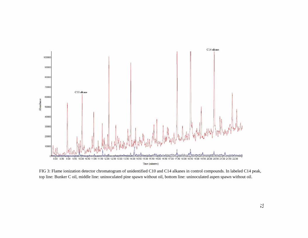
72
FIG 3: Flame ionization detector chromatogram of unidentified C10 and C14 alkanes in control compounds. In labeled C14 peak,
top line: Bunker C oil, middle line: uninoculated pine spawn without oil, bottom line: uninoculated aspen spawn without oil.

73
Literature Cited
Adams J, Beam M, Christy O, Todd J. 2007. Fisherville eco-machine pilot final report.
John Todd Ecological Design.
Aggelis G, Iconomou D, Christou M, Bokas D, Kotzailias S, Christou G, Tsagou V,
Papanikolaou S. 2003. Phenolic removal in a model olive oil mill wastewater using
Pleurotus ostreatus in bioreactor cultures and biological evaluation of the process.
Water Research 37(16):3897-3904.
Ahearn DG, Crow SA. 1986. Fungi and hydrocarbons in the marine environment. In: Moss
ST, ed. The biology of marine fungi. Cambridge: Cambridge University Press. p.
11-18.
Ander P, Eriksson K-E. 1976. The importance of phenol oxidase activity in lignin
degradation by the white-rot fungus Sporotrichum pulverulentum. Archives of
Microbiology 109(1):1-8.
Andersson BE, Henrysson T. 1996. Accumulation and degradation of dead-end
metabolites during treatment of soil contaminated with polycyclic aromatic
hydrocarbons with five strains of white-rot fungi. Applied Microbiology and
Biotechnology 46(5):647-652.
Arora D. 1986. Mushrooms demystified. Berkeley: Ten Speed Press.
Asgher M, Bhatti HN, Ashraf M, Legge RL. 2008. Recent developments in biodegradation
of industrial pollutants by white rot fungi and their enzyme system. Biodegradation
19(6):771-783.
Aske N, Kallevik H, Johnsen EE, Sjöblom J. 2002. Asphaltene aggregation from crude oils
and model systems studied by high-pressure NIR spectroscopy. Energy & Fuels
16(5):1287-1295.
Aust SD. 1995. Mechanisms of degradation by white rot fungi. Environmental Health
Perspectives 103(5):Medium: X; Size: pp. 59-61.
Baldrian P. 2006. Fungal laccases: occurrence and properties. FEMS Microbiology
Reviews 30(2):215-242.
Bamforth SM, Singleton I. 2005. Bioremediation of polycyclic aromatic hydrocarbons:
current knowledge and future directions. Journal of Chemical Technology &
Biotechnology 80(7):723-736.
Bao W, Fukushima Y, Jensen Jr KA, Moen MA, Hammel KE. 1994. Oxidative
degradation of non-phenolic lignin during lipid peroxidation by fungal manganese
peroxidase. FEBS Letters 354(3):297-300.
Barr DP, Aust SD. 1994. Pollutant degradation by white rot fungi. Reviews of
Environmental Contamination and Toxicology 138:49.
Bartha R. 1986. Biotechnology of petroleum pollutant biodegradation. Microbial Ecology
12(1):155-172.

74
Bauman JW, Liu J, Klassen CD. 1993. Production of metallothionein and heat-shock
proteins in response to metals. Toxicological Sciences 21(1):15-22.
Bezalel L, Hadar Y, Cerniglia CE. 1996a. Mineralization of polycyclic aromatic
hydrocarbons by the white rot fungus Pleurotus ostreatus. Applied and
Environmental Microbiology 62(1):292-5.
Bezalel L, Hadar Y, Cerniglia CE. 1997. Enzymatic mechanisms involved in phenanthrene
degradation by the white rot fungus Pleurotus ostreatus. Applied and
Environmental Microbiology 63(7):2495-501.
Bezalel L, Hadar Y, Fu PP, Freeman JP, Cerniglia CE. 1996b. Initial oxidation products in
the metabolism of pyrene, anthracene, fluorene, and dibenzothiophene by the white
rot fungus Pleurotus ostreatus. Applied and Environmental Microbiology
62(7):2554-9.
Bezalel L, Hadar Y, Fu PP, Freeman JP, Cerniglia CE. 1996c. Metabolism of phenanthrene
by the white rot fungus Pleurotus ostreatus. Applied and Environmental
Microbiology 62(7):2547-53.
Bhatt M, Cajthaml T, Šašek V. 2002. Mycoremediation of PAH-contaminated soil. Folia
Microbiologica 47(3):255-258.
Boddy L. 1992. Development and function of fungal communities in decomposing wood.
Mycology Series 9:749-782.
Bogan BW, Lamar RT. 1995. One-electron oxidation in the degradation of creosote
polycyclic aromatic hydrocarbons by Phanerochaete chrysosporium. Applied and
Environmental Microbiology 61(7):2631-5.
Bogan BW, Lamar RT. 1996. Polycyclic aromatic hydrocarbon-degrading capabilities of
Phanerochaete laevis HHB-1625 and its extracellular ligninolytic enzymes. Applied
and Environmental Microbiology 62(5):1597-603.
Bogan BW, Lamar RT, Hammel KE. 1996a. Fluorene oxidation in vivo by Phanerochaete
chrysosporium and in vitro during manganese peroxidase-dependent lipid
peroxidation. Applied and Environmental Microbiology 62(5):1788-92.
Bogan BW, Schoenike B, Lamar RT, Cullen D. 1996b. Expression of lip genes during
growth in soil and oxidation of anthracene by Phanerochaete chrysosporium.
Applied and Environmental Microbiology 62(10):3697-703.
Bogan BW, Schoenike B, Lamar RT, Cullen D. 1996c. Manganese peroxidase mRNA and
enzyme activity levels during bioremediation of polycyclic aromatic hydrocarbon-
contaminated soil with Phanerochaete chrysosporium. Applied and Environmental
Microbiology 62(7):2381-6.
Bourbonnais R, Paice MG. 1990. Oxidation of non-phenolic substrates: an expanded role
for laccase in lignin biodegradation. FEBS Letters 267(1):99-102.
Buettner GR. 1993. The pecking order of free radicals and antioxidants: lipid peroxidation,
alpha-tocopherol, and ascorbate. Archives of Biochemistry and Biophysics
300(2):535-43.
Bumpus JA. 1989. Biodegradation of polycyclic hydrocarbons by Phanerochaete
chrysosporium. Applied and Environmental Microbiology 55(1):154-158.

75
Buswell JA. 2001. Fungal biodegradation of chlorinated monoaromatics and BTEX
compounds. In: Gadd GM, ed. Fungi in Bioremediation. Cambridge: Cambridge
University Press. p. 113-135.
Cajthaml T, Erbanová P, Kollmann A, Novotný Č, Šašek V, Mougin C. 2008. Degradation
of PAHs by ligninolytic enzymes of Irpex lacteus. Folia Microbiologica 53(4):289-
294.
Camarero , arkar , Ruiz-Due as , Mart nez Ma , Mart nez T. 1999. Description of
a versatile peroxidase involved in the natural degradation of lignin that has both
manganese peroxidase and lignin peroxidase substrate interaction sites. Journal of
Biological Chemistry 274(15):10324-10330.
Cerniglia C, Yang S. 1984. Stereoselective metabolism of anthracene and phenanthrene by
the fungus Cunninghamella elegans. Applied and Environmental Microbiology
47(1):119-124.
Cerniglia CE. 1992. Biodegradation of polycyclic aromatic hydrocarbons. Biodegradation
3(2):351-368.
Cerniglia CE. 1997. Fungal metabolism of polycyclic aromatic hydrocarbons: past, present
and future applications in bioremediation. Journal of Industrial Microbiology and
Biotechnology 19(5-6):324-33.
Cerniglia CE, Sutherland JB. 2001. Bioremediation of polycyclic aromatic hydrocarbons
by ligninolytic and non-ligninolytic fungi. In: Gadd GM, ed. Fungi in
Bioremediation. Cambridge: Cambridge University Press. p. 136-187.
Cerniglia CE, Sutherland JB. 2006. Relative roles of bacteria and fungi in polycyclic
aromatic hydrocarbon biodegradation and bioremediation of contaminated soils. In:
Gadd GM, ed. Fungi in biogeochemical cycles. Cambridge: Cambridge University
Press. p. 182-211.
Chigu NL, Hirosue S, Nakamura C, Teramoto H, Ichinose H, Wariishi H. 2010.
Cytochrome P450 monooxygenases involved in anthracene metabolism by the
white-rot basidiomycete Phanerochaete chrysosporium. Applied Microbiology and
Biotechnology 87(5):1907-1916.
Collins PJ, Dobson ADW. 1996. Oxidation of fluorene and phenanthrene by Mn(II)
dependent peroxidase activity in whole cultures of Trametes (coriolus) versicolor.
Biotechnology Letters 18(7):801-804.
Collins PJ, Kotterman M, Field JA, Dobson A. 1996. Oxidation of anthracene and
benzo[a]pyrene by laccases from Trametes versicolor. Applied and Environmental
Microbiology 62(12):4563-7.
Dhawale SW, Dhawale SS, Dean-Ross D. 1992. Degradation of phenanthrene by
Phanerochaete chrysosporium occurs under ligninolytic as well as nonligninolytic
conditions. Applied and Environmental Microbiology 58(9):3000-3006.
Doddapaneni H, Yadav JS. 2005. Microarray-based global differential expression profiling
of P450 monooxygenases and regulatory proteins for signal transduction pathways
in the white rot fungus Phanerochaete chrysosporium. Molecular Genetics and
Genomics 274(5):454-466.

76
Elisashvili V, Kachlishvili E, Tsiklauri N, Metreveli E, Khardziani T, Agathos SN. 2008.
Lignocellulose-degrading enzyme production by white-rot basidiomycetes isolated
from the forests of Georgia. World Journal of Microbiology and Biotechnology
25(2):331-339.
Eshghi H, Alishahi Z, Zokaei M, Daroodi A, Tabasi E. 2011. Decolorization of methylene
blue by new fungus: Trichaptum biforme and decolorization of three synthetic dyes
by Trametes hirsuta and Trametes gibbosa. European Journal of Chemistry
2(4):463-468.
Evans CS. 1985. Laccase activity in lignin degradation by Coriolus versicolor: in vivo and
in vitro studies. FEMS Microbiology Letters 27(3):339-343.
Evans CS, Hedger JN. 2001. Degradation of plant cell wall polymers. In: Gadd GM, ed.
Fungi in Bioremediation. Cambridge: Cambridge University Press. p. 1-26.
Faison BD, Kirk TK. 1983. Relationship between lignin degradation and production of
reduced oxygen species by Phanerochaete chrysosporium. Applied and
Environmental Microbiology 46(5):1140-1145.
Fernando T, Aust SD. 1994. Biodegradation of toxic chemicals by white rot fungi. In:
Chaudhry GR, ed. Biological Degradation and Bioremediation of Toxic Chemicals.
London: Chapman & Hall. p. 386-402.
Field JA, de Jong E, Feijoo Costa G, de Bont JA. 1992. Biodegradation of polycyclic
aromatic hydrocarbons by new isolates of white rot fungi. Applied and
Environmental Microbiology 58(7):2219-26.
Floudas D, Binder M, Riley R, Barry K, Blanchette RA, Henrissat B, Martínez AT, Ortillar
R, Spatafora JW, Yadav JS et al. . 2012. Diversity and evolution of the wood decay
enzymatic apparatus in saprotrophic Agaricomycotina. Science (in press).
Freitag M, Morrell JJ. 1992. Decolorization of the polymeric dye Poly R-478 by wood-
inhabiting fungi. Canadian Journal of Microbiology 38(8):811-822.
Gadd GM. 2001. Preface. In: Gadd GM, ed. Fungi in Bioremedation. Cambridge:
Cambridge University Press. p. xi-xiii.
Garcia-Sandoval R, Wang Z, Binder M, Hibbett DS. 2011. Molecular phylogenetics of the
Gloeophyllales and relative ages of clades of Agaricomycotina producing a brown
rot. Mycologia 103(3):510-524.
Gierer J, Opara AE. 1973. Studies of the enzymatic degradation of lignin. Acta Chemika
Scandinavica 27:2909-2922.
Gilbertson RL, Ryvarden L. 1986a. North American Polypores. Oslo: Fungiflora.
Gilbertson RL, Ryvarden L. 1986b. North American Polypores. Oslo: Fungiflora.
Ginns J, Lefebvre M. 1993. Lignicolous corticoid fungi (Basidiomycota) of North
America: systematics, distribution, and ecology. American Phytopathological
Society.
Gramss G, Kirsche B, Voigt K, Gunther T, Fritsche W. 1999. Conversion rates of five
polycyclic aromatic hydrocarbons in liquid cultures of fifty-eight fungi and the
concomitant production of oxidative enzymes. Mycological Research 103(8):1009-
1018.
77
Haemmerli SD, Leisola MS, Sanglard D, Fiechter A. 1986. Oxidation of benzo(a)pyrene
by extracellular ligninases of Phanerochaete chrysosporium: veratryl alcohol and
stability of ligninase. Journal of Biological Chemistry 261(15):6900-3.
Hammel KE. 1995. Mechanisms for polycyclic aromatic hydrocarbon degradation by
ligninolytic fungi. Environmental Health Perspectives 103:41-3.
Hammel KE. 1997. Fungal Degradation of Lignin. In: Cadisch G, Giller KE, eds. Driven
by Nature: Plant Litter Quality and Decomposition. Wallingford: CABI.
Hammel KE, Gai WZ, Green B, Moen MA. 1992. Oxidative degradation of phenanthrene
by the ligninolytic fungus Phanerochaete chrysosporium. Applied and
Environmental Microbiology 58(6):1832-1838.
Hammel KE, Kalyanaraman B, Kirk TK. 1986. Oxidation of polycyclic aromatic
hydrocarbons and dibenzo[p]-dioxins by Phanerochaete chrysosporium ligninase.
Journal of Biological Chemistry 261(36):16948-16952.
Harvey PJ, Thurston CF. 2001. The biochemistry of ligninolytic fungi. In: Gadd GM, ed.
Fungi in Bioremediation. Cambridge: Cambridge University Press. p. 27-51.
Head IM. 1998. Bioremediation: towards a credible technology. Microbiology 144(3):599-
608.
Hess. No. 6 Fuel Oil; MSDS No. 9907. Woodbridge, NJ.
Hibbett DS, Donoghue MJ. 2001. Analysis of character correlations among wood decay
mechanisms, mating systems, and substrate ranges in Homobasidiomycetes.
Systematic Biology 50(2):215-242.
Hirano T, Honda Y, Watanabe T, Kuwahara M. 2000. Degradation of bisphenol A by the
lignin-degrading enzyme, manganese peroxidase, produced by the white-rot
basidiomycete, Pleurotus ostreatus. Bioscience, Biotechnology, and Biochemistry
64(9):1958-1962.
Horvath RS. 1972. Microbial co-metabolism and the degradation of organic compounds in
nature.
in der Wiesche C, Martens R, Zadrazil F. 1996. 2-step degradation of pyrene by white-rot
fungi and soil-microorganisms. Applied Microbiology and Biotechnology 46(5-
6):653-659.
Jobson A, Cook FD, Westlake DWS. 1972. Microbial utilization of crude oil. Applied
Microbiology 23(6):1082-1089.
Johannes C, Majcherczyk A. 2000. Natural mediators in the oxidation of polycyclic
aromatic hydrocarbons by laccase mediator systems. Applied and Environmental
Microbiology 66(2):524-528.
Kanaly R, Hur H. 2006. Growth of Phanerochaete chrysosporium on diesel fuel
hydrocarbons at neutral pH. Chemosphere 63(2):202-211.
Kim HY, Song HG. 2003. Transformation and mineralization of 2, 4, 6-trinitrotoluene by
the white rot fungus Irpex lacteus. Applied Microbiology and Biotechnology
61(2):150-156.
Kirk PM, Ainsworth GC. 2008. Ainsworth & Bisby's dictionary of the fungi. Cabi
Publishing.

78
Komagata K, Nakase T, Katsuya N. 1964. Assimilation of hydrocarbons by yeasts. The
Journal of General and Applied Microbiology 10:313-321.
Kotterman MJJ, Heessels E, Jong Ed, Field JA. 1994. The physiology of anthracene
biodegradation by the white-rot fungus Bjerkandera sp. strain BOS55.
Lamar RT, White RB, Ashley KC. 2002. Evaluation of white-rot fungi for the remediation
of creosote-contaminated soil. Remediation Journal 12(4):97-106.
Lang E, Nerud F, Novotná E, Zadrazil F, Martens R. 1996. Production of ligninolytic
exoenzymes and 14C-pyrene mineralization by Pleurotus sp. in lignocellulose
substrate. Folia Microbiologica 41(6):489-493.
Leahy JG, Colwell RR. 1990. Microbial degradation of hydrocarbons in the environment.
Microbiological Reviews 54(3):305-315.
Lee H, Choi YS, Kim MJ, Huh NY, Kim GH, Lim YW, Kang SM, Cho ST, Kim JJ. 2010.
Degrading ability of oligocyclic aromates by Phanerochaete sordida selected via
screening of white rot fungi. Folia Microbiologica 55(5):447-53.
Levin L, Viale A, Forchiassin A. 2003. Degradation of organic pollutants by the white rot
basidiomycete Trametes trogii. International Biodeterioration & Biodegradation
52(1):1-5.
Li H, Durbin R. 2010. Fast and accurate long-read alignment with Burrows–Wheeler
transform. Bioinformatics 26(5):589-595.
Markovetz AJ, Cazin JR, Allen JE. 1968. Assimilation of alkanes and alkenes by fungi.
Applied Microbiology 16(3):487-489.
Martinez AT. 2002. Molecular biology and structure-function of lignin-degrading heme
peroxidases. Enzyme and Microbial Technology 30(4):425-444.
Moen MA, Hammel KE. 1994. Lipid peroxidation by the manganese peroxidase of
Phanerochaete chrysosporium is the basis for phenanthrene oxidation by the intact
fungus. Applied and Environmental Microbiology 60(6):1956-1961.
Moore D, Robson GD, Trinci APJ. 2011. 21st Century Guidebook to Fungi. New York:
Cambridge University Press.
Morgan P, Lewis ST, Watkinson RJ. 1991. Comparison of abilities of white-rot fungi to
mineralize selected xenobiotic compounds. Applied Microbiology and
Biotechnology 34(5):693-696.
Morgenstern I, Klopman S, Hibbett DS. 2008. Molecular evolution and diversity of lignin
degrading heme peroxidases in the Agaricomycetes. Journal of Molecular
Evolution 66(3):243-257.
Mougin C, Jolivalt C, Briozzo P, Madzak C. 2003. Fungal laccases: from structure-activity
studies to environmental applications. Environmental Chemistry Letters 1(2):145-
148.
Niku-Paavola ML, Karhunen E, Salola P, Raunio V. 1988. Ligninolytic enzymes of the
white-rot fungus Phlebia radiata. Biochemical Journal 254(3):877.
Novotny C, Cajthaml T, Svobodova K, Susla M, Sasek V. 2009. Irpex lacteus, a white-rot
fungus with biotechnological potential--review. Folia Microbiologica 54(5):375-90.

79
Novotny C, Erbanova P, Cajthaml T, Rothschild N, Dosoretz C, Sasek V. 2000. Irpex
lacteus, a white rot fungus applicable to water and soil bioremediation. Applied
Microbiology and Biotechnology 54(6):850-3.
Novotny C, Erbanova P, Sasek V, Kubatova A, Cajthaml T, Lang E, Krahl J, Zadrazil F.
1999. Extracellular oxidative enzyme production and PAH removal in soil by
exploratory mycelium of white rot fungi. Biodegradation 10(3):159-68.
Ogbo E, Okhuoya J. 2008. Biodegradation of aliphatic, aromatic, resinic and asphaltic
fractions of crude oil contaminated soils by Pleurotus tuber-regium Fr. Singer - a
white rot fungus. African Journal of Biotechnology 7(23):7.
Okino LK, Machado KMG, Fabris C, Bononi VLR. 2000. Ligninolytic activity of tropical
rainforest basidiomycetes. World Journal of Microbiology and Biotechnology
16(8):889-893.
Ollila P, Soukup J, Nowack B, Bernat E, Brammer D, Hultstrom E, Tsang J. 2008.
Fisherville Mill – a case study – cost effective remediation through collaboration.
Proceedings of the Annual International Conference on Soils, Sediments, Water
and Energy. Berkeley Electronic Press. p. 108-151.
Palmieri G, Giardina P, Bianco C, Fontanella B, Sannia G. 2000. Copper induction of
laccase isoenzymes in the ligninolytic fungus Pleurotus ostreatus. Applied and
Environmental Microbiology 66(3):920-924.
Parkhomchuk D, Borodina T, Amstislavskiy V, Banaru M, Hallen L, Krobitsch S, Lehrach
H, Soldatov A. 2009. Transcriptome analysis by strand-specific sequencing of
complementary DNA. Nucleic Acids Research 37(18):e123-e123.
Pérez-Boada M, Ruiz-Dueñas FJ, Pogni R, Basosi R, Choinowski T, Martínez MJ, Piontek
K, Martínez AT. 2005. Versatile peroxidase oxidation of high redox potential
aromatic compounds: site-directed mutagenesis, spectroscopic and crystallographic
investigation of three long-range electron transfer pathways. Journal of Molecular
Biology 354(2):385-402.
Pickard MA, Roman R, Tinoco R, Vazquez-Duhalt R. 1999. Polycyclic aromatic
hydrocarbon metabolism by white rot fungi and oxidation by Coriolopsis gallica
UAMH 8260 laccase. Applied and Environmental Microbiology 65(9):3805-3809.
Pouli M, Agathos SN. 2011. Bioremedation of PAH-contaminated sties: from pathways to
bioreactors. In: Koukkou A, ed. Microbial Bioremediation of Non-metals. Norfolk,
UK: Caister University Press. p. 119-147.
Pozdnyakova NN, Dubrovskaya EV, Makarov OE, Nikitina VE, Turovskaya OV. 2011a.
Production of ligninolytic enzymes by white-rot fungi during bioremediation of oil-
contaminated soil. In: Shukla G, Varma A, eds. Soil Enzymology. Berlin: Springer-
Verlag. p. 15.
Pozdnyakova NN, Nikiforova SV, Makarov OE, Turkovskaya OV. 2011b. Effect of
polycyclic aromatic hydrocarbons on laccase production by white rot fungus
Pleurotus ostreatus D1. Applied Biochemistry and Microbiology 47(5):543-548.

80
Pozdnyakova NN, Nikiforova SV, Turkovskaya OV. 2010. Influence of PAHs on
ligninolytic enzymes of the fungus Pleurotus ostreatus D1. Central European
Journal of Biology 5(1):83-94.
Pozdnyakova NN, Nikitina VE, Turovskaya OV. 2008. Bioremediation of oil-polluted soil
with an association including the fungus Pleurotus ostreatus and soil microflora.
Applied Biochemistry and Microbiology 44(1):60-65.
Pozdnyakova NN, Rodakiewicz-Nowak J, Turkovskaya OV. 2004. Catalytic properties of
yellow laccase from Pleurotus ostreatus D1. Journal of Molecular Catalysis
30(1):19-24.
Reddy CA, Mathew Z. 2001. Bioremediation potential of white rot fungi. In: Gadd GM,
ed. Fungi in Bioremediation. Cambridge: Cambridge University Press. p. 52-78.
Rehm H, Reiff I. 1981. Mechanisms and occurrence of microbial oxidation of long-chain
alkanes. Reactors and Reactions. Springer Berlin / Heidelberg. p. 175-215.
Rogalski J, Lundell TK, Leonowicz A, Hatakka AI. 1991. Influence of aromatic
compounds and lignin on production of ligninolytic enzymes by Phlebia radiata.
Phytochemistry 30(9):2869-2872.
Rothschild N, Novotn C, asek V, Dosoretz CG. 2002. Ligninolytic enzymes of the
fungus Irpex lacteus (Polyporus tulipiferae): isolation and characterization of lignin
peroxidase. Enzyme and Microbial Technology 31(5):627-633.
Sack U, Heinze TM, Deck J, Cerniglia CE, Martens R, Zadrazil F, Fritsche W. 1997.
Comparison of phenanthrene and pyrene degradation by different wood-decaying
fungi. Applied and Environmental Microbiology 63(10):3919-25.
Salas C, Lobos S, Larraín J, Salas L, Cullen D, Vicuna R. 1995. Properties of laccase
isoenzymes produced by the basidiomycete Ceriporiopsis subvermispora.
Biotechnology and Applied Biochemistry 21(3):323-333.
Saloheimo M, Niku-Paavola ML, Knowles JKC. 1991. Isolation and structural analysis of
the laccase gene from the ligninegrading fungus Phlebia radiata. Journal of
General Microbiology 137(7):1537.
Sambrook J, Russell DW. 2001. Molecular cloning: a laboratory manual. CSHL press.
Schultz A, Jonas U, Hammer E, Schauer F. 2001. Dehalogenation of chlorinated
hydroxybiphenyls by fungal laccase. Applied and Environmental Microbiology
67(9):4377-4381.
Shin K, Kim YH, Lim J. 2005. Purification and characterization of manganese peroxidase
of the white-rot fungus Irpex lacteus. Journal of Microbiology Seoul 43(6):503.
Sims R. 1990. Soil remediation techniques at uncontrolled hazardous waste sites: a critical
review. Journal of the Air & Waste Management Association 40(5):704-732.
Singer ME, Finnerty WR. 1984. Microbial metabolism of straight-chain and branched
alkanes. In: Atlas RM, ed. Petroleum Microbiology. New York: Macmillan
Publishing. p. 1-59.
Steffen KT, Hatakka A, Hofrichter M. 2003. Degradation of benzo[a]pyrene by the litter-
decomposing basidiomycete Stropharia coronilla: role of manganese peroxidase.
Applied and Environmental Microbiology 69(7):3957-3964.

81
Sutherland JB, Selby AL, Freeman JP, Evans FE, Cerniglia CE. 1991. Metabolism of
phenanthrene by Phanerochaete chrysosporium. Applied and Environmental
Microbiology 57(11):3310-3316.
Svobodová K, Majcherczyk A, Novotn C, Kües U. 2008. Implication of mycelium-
associated laccase from Irpex lacteus in the decolorization of synthetic dyes.
Bioresource Technology 99(3):463-471.
Syed K, Doddapaneni H, Subramanian V, Lam YW, Yadav JS. 2010. Genome-to-function
characterization of novel fungal P450 monooxygenases oxidizing polycyclic
aromatic hydrocarbons (PAHs). Biochemical and Biophysical Research
Communications 399(4):492-497.
ten Have R, Teunissen. 2001. Oxidative mechanisms involved in lignin degradation by
white-rot fungi. Chemical Reviews 101:18.
Teramoto H. 2004. Fungal cytochrome P450s catalyzing hydroxylation of substituted
toluenes to form their hydroxymethyl derivatives. FEMS Microbiology Letters
234(2):255-260.
Thurston CF. 1994. The structure and function of fungal laccases. Microbiology
140(1):19-26.
Tien M, Kirk TK. 1984. Lignin-degrading enzyme from Phanerochaete chrysosporium:
purification, characterization, and catalytic properties of a unique H2O2-requiring
oxygenase. Proceedings of the National Academy of Sciences 81(8):2280-2284.
Torres-Duarte C, Roman R, Tinoco R, Vazquez-Duhalt R. 2009. Halogenated pesticide
transformation by a laccase–mediator system. Chemosphere 77(5):687-692.
Tyman J. 1996. Synthetic and Natural Phenols. Amsterdam: Elsevier.
Van Aken B, Hofrichter M, Scheibner K, Hatakka AI, Naveau H, Agathos SN. 1999.
Transformation and mineralization of 2, 4, 6-trinitrotoluene (TNT) by manganese
peroxidase from the white-rot basidiomycete Phlebia radiata. Biodegradation
10(2):83-91.
Vares T, Kalsi M, Hatakka A. 1995. Lignin peroxidases, manganese peroxidases, and other
ligninolytic enzymes produced by Phlebia radiata during solid-state fermentation
of wheat straw. Applied and Environmental Microbiology 61(10):3515-3520.
Walker JD, Petrakis L, Colwell RR. 1976. Comparison of the biodegradability of crude
and fuel oils. Canadian Journal of Microbiology 22(4):598-602.
Wariishi H, Valli K, Gold MH. 1989. Oxidative cleavage of a phenolic diarylpropane
lignin model dimer by manganese peroxidase from Phanerochaete chrysosporium.
Biochemistry 28(14):6017-6023.
Wariishi H, Valli K, Gold MH. 1991. In vitro depolymerization of lignin by manganese
peroxidase of Phanerochaete chrysosporium. Biochemical and Biophysical
Research Communications 176(1):269-275.
Wessels JGH. 1996. Fungal hydrophobins: proteins that function at an interface. Trends in
Plant Science 1(1):9-15.

82
Whitelaw ML, McGuire J, Picard D, Gustafsson JA, Poellinger L. 1995. Heat shock
protein hsp90 regulates dioxin receptor function in vivo. Proceedings of the
National Academy of Sciences 92(10):4437-4441.
Wolter M, Zadrazil F, Martens R, Bahadir M. 1997. Degradation of eight highly
condensed polycyclic aromatic hydrocarbons by Pleurotus sp. Florida in solid
wheat straw substrate. Applied Microbiology and Biotechnology 48:7.
Xu . 1996. Oxidation of phenols, anilines, and benzenethiols by fungal laccases:
correlation between activity and redox potentials as well as halide inhibition.
Biochemistry 35(23):7608-7614.
Yadav JS, Reddy CA. 1993. Degradation of benzene, toluene, ethylbenzene, and xylenes
(BTEX) by the lignin-degrading basidiomycete Phanerochaete chrysosporium.
Applied and Environmental Microbiology 59(3):756-762.
Zille A, Gornacka B, Rehorek A, Cavaco-Paulo A. 2005. Degradation of azo dyes by
Trametes villosa laccase over long periods of oxidative conditions. Applied and
Environmental Microbiology 71(11):6711-6718.