
AAPC Regional Conference Springfield, MA Regional Conference Springfield, MA Presented by: David...
50
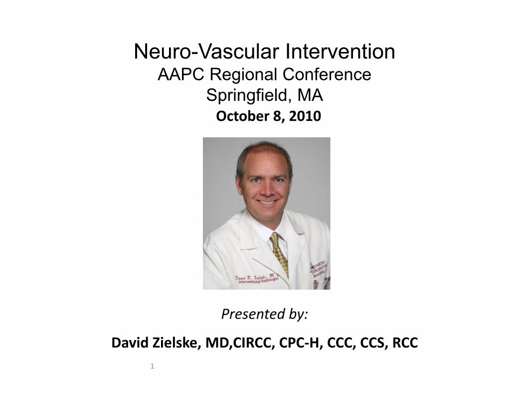
1 Neuro-Vascular Intervention AAPC Regional Conference Springfield, MA Presented by: David Zielske, MD,CIRCC, CPC‐H, CCC, CCS, RCC October 8, 2010
-
Upload
nguyenduong -
Category
Documents
-
view
215 -
download
0
Transcript of AAPC Regional Conference Springfield, MA Regional Conference Springfield, MA Presented by: David...
1
Neuro-Vascular InterventionAAPC Regional Conference
Springfield, MA
Presented by:
David Zielske, MD,CIRCC, CPC‐H, CCC, CCS, RCC
October 8, 2010

2
General Recommendations for Physician Dictations
• State the history, medical necessity, reasons for repeat diagnostic study after prior catheter angiography/CTA/MRA
• State the vascular access site(s)• State the vessels catheterized, describing the catheter tip
location, and any variant anatomy• State the vessels injected, the areas imaged (for medical
necessity), interpretation of findings, specific documentation of AVM, aneurysm, percentage stenosis, exact anatomic location of the lesions, spasm vs atherosclerotic stenoses. Thrombus removed or dissolved. CNS vs Extracranial work
• State the interventions and adjunctive procedures performed. Also any complications or additional treatments provided. Follow-up angiography after infusion or embolotherapy.
• State the specific devices and specialty supplies used during the procedure
3
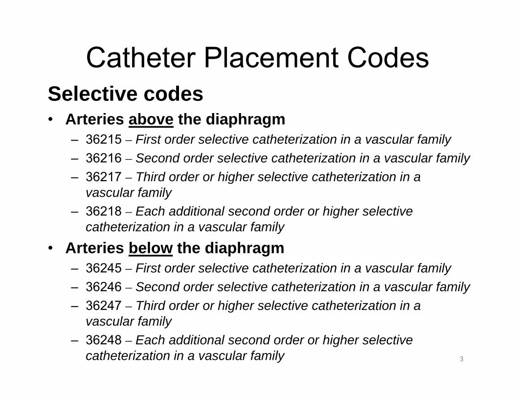
Selective codes• Arteries above the diaphragm
– 36215 – First order selective catheterization in a vascular family– 36216 – Second order selective catheterization in a vascular family– 36217 – Third order or higher selective catheterization in a
vascular family– 36218 – Each additional second order or higher selective
catheterization in a vascular family
• Arteries below the diaphragm– 36245 – First order selective catheterization in a vascular family– 36246 – Second order selective catheterization in a vascular family– 36247 – Third order or higher selective catheterization in a
vascular family– 36248 – Each additional second order or higher selective
catheterization in a vascular family
Catheter Placement Codes

4
Coding Rules
• Concept of Vascular Families (separate trunks off the aorta or access vessel and all its branches)
• Do code to where the tip of the catheter is.• Do code 36200 (aorta catheter placement) over other non‐selective codes (36140, 36120).
• Do code each of the four separate vascular systems separately: arterial, venous, portal and pulmonary.
• Do code each vascular family separately, using modifiers to distinguish the different vessels.
5
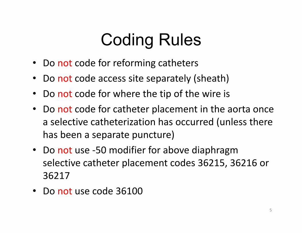
• Do not code for reforming catheters• Do not code access site separately (sheath)• Do not code for where the tip of the wire is• Do not code for catheter placement in the aorta once a selective catheterization has occurred (unless there has been a separate puncture)
• Do not use ‐50 modifier for above diaphragm selective catheter placement codes 36215, 36216 or 36217
• Do not use code 36100
Coding Rules

6
Procedures That Include Some Catheter Placements
• 37215 - Cervical carotid artery stent with distal protection• 37216 - Cervical carotid artery stent without distal protection• 0075T - Extracranial vertebral or intrathoracic carotid stent• 0076T - Extracranial vertebral or intrathoracic stent - each
additional vessel• 61623 - Endovascular temporary balloon occlusion• 61630 - Balloon angioplasty, intracranial• 61635 - Intravascular stent(s), intracranial• 61640 - Balloon dilation, intracranial vasospasm• 61641 - Balloon dilation, each additional vessel - same
vascular family• 61642 - Balloon dilation, each additional vessel - different
vascular family

7
3D Reconstructions• 76376 – 3-Dimensional reconstructions of CT,
MR, ultrasound including catheter based angiography not requiring an independent workstation
• 76377 – 3-Dimensional reconstructions of CT, MR, ultrasound including catheter based angiography which does require image post-processing on an independent workstation
• Need treating physician to order 3-D reconstructions
8
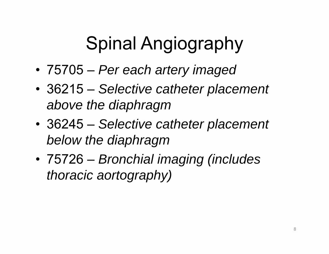
Spinal Angiography• 75705 – Per each artery imaged• 36215 – Selective catheter placement
above the diaphragm• 36245 – Selective catheter placement
below the diaphragm• 75726 – Bronchial imaging (includes
thoracic aortography)

9
Spinal Angiography/Embolization Case 1: Unexplained subarachnoid hemorrhage: Selective bilateral bronchial (separate origins) off the aorta), selective injection of the ascending branch to the cervical cord off the right bronchial, selective injection and imaging of bilateral L‐1 thru L3 lumbar arteries with additional selection and imaging of the artery of Adamkowicz off the right L‐2 lumbar artery. AVM is supplied by this artery and is embolized after placement of a microcatheter into a third order branch. Two follow‐up angiograms are necessary to complete the study.36215‐593621636247 36245‐5950 x 236245‐59
7572675726‐597570575705‐59 x 675774
61624759847589875898‐59

10
Spinal Angiography/Embolization Case 2:
Patient with L‐2 osseous metastasis with back pain.A sheath is placed in the left common femoral artery. Aselective catheter is advanced into both the right and leftlumbar arteries at the L‐1, L‐2 and L‐3 levels withdiagnostic angiography performed. The L1 and L‐2 leftsided lumbar arteries supplied a hypervascular L‐2vertebral body metastasis. No AV shunting was seen andthere appeared to be no supply to the cord. The otherfour vessels appeared normal with the Artery ofAdamkowicz arising from L‐1 on the right. A catheter isadvanced into a second order branch of both the L‐1 andL‐2 vessels for selective embolization. Embolization isperformed with a chemoembolic mixture until there isstasis of flow. Follow‐up angiography from each vesselshows complete occlusion of flow to the tumor.

11
Embolization Case 2 Codes:
75705 – L-1 right spinal angiography, S&I75705-59 x 5 – L-1 left, bilateral L-2 and L-3 spinal
angiography, S&I36245-59 x 4 – 1st order selective catheter placements36246 – 2nd order selective catheter placement36246-59 – 2nd order selective catheter placement37204 – non-neuro embolization of vertebral body75894 – embolization, S&I75898 – follow-up angiography after embolization

12
Cervicocerebral• Innominate artery = Brachiocephalic artery• Variant anatomy includes bovine arch, common origin of
the right brachiocephalic and the left common carotid, left vertebral originating directly off the arch, aberrant right subclavian and any combination of these variants
• The common and internal carotid arteries as well as the non-selective external carotid arteries are all included as a part of the cervical carotid angiography S&I codes (75676, 75680)
• Arch injection includes the transverse aorta, the proximal subclavian, vertebral and carotid arteries (not the carotid bifurcations or complete cervical vertebrals or complete extremities)

13
Cervicocerebral Case 3:Arch injection with cervicocerbral arch, bilateral carotid cervical, bilateral carotid cerebral and bilateral vertebral imaging
36200 – Catheter placement aorta75650 – Arch aortogram S&I75671 – Bilateral cerebral carotids, S&I75680 – Bilateral cervical carotids S&I75685 – Vertebral S&I75685-59 – Vertebral S&I

14
36215-59 – 1st order selective above diaphragm36216-59 – 2nd order selective above diaphragm36216 – 2nd order selective above diaphragm75650 – Cervicocerebral arch, S&I75680 – Bilateral cervical carotid angiogram, S&I75671 – Bilateral cerebral carotid angiogram, S&I75685 – Vertebral angiogram, S&I
Cervicocerebral Case 4:Arch, followed by selective bilateral carotid cervical and carotid cerebral imaging with selective left vertebral imaging. Normal anatomy.

15
Cervicocerebral Case 5:Selective bilateral carotid cervical and carotid cerebral imaging with selective right vertebral catheter placement and imaging, bovine arch.
36218 – Ea addtl selective above diaphragm36218 – Ea addtl selective above diaphragm36217 – 3rd order selective above diaphragm75680 – Bilateral cervical carotid angiogram, S&I75671 – Bilateral cerebral carotid angiogram, S&I75685 – Vertebral angiogram, S&I

16
36215-59 – 1st order selective above diaphragm36216-59 – 2nd order selective above diaphragm36218 – Ea addtl 2nd or higher selective above diaphragm36217 – 3rd order selective above diaphragm36218 x 2 – Ea addtl 2nd or higher selective above diaphragm75650 – Cervicocerebral arch, S&I75671 – Bilateral cerebral carotid angiogram, S&I75662 – Bilateral selective external carotid angiogram, S&I75685 – Vertebral angiogram, S&I75685-59 – Vertebral angiogram, S&I
Cervicocerebral Case 6:Arch followed by selective bilateral internal carotid, bilateral external carotid and bilateral vertebral artery catheter placement with cerebral imaging (aneurysm work-up, left vertebral arises directly from the arch)
17
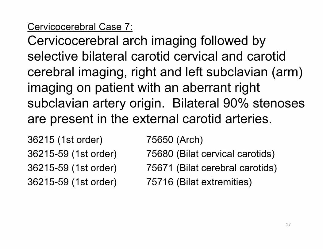
Cervicocerebral Case 7:Cervicocerebral arch imaging followed by selective bilateral carotid cervical and carotid cerebral imaging, right and left subclavian (arm) imaging on patient with an aberrant right subclavian artery origin. Bilateral 90% stenoses are present in the external carotid arteries.
75650 (Arch) 75680 (Bilat cervical carotids)75671 (Bilat cerebral carotids)75716 (Bilat extremities)
36215 (1st order) 36215-59 (1st order) 36215-59 (1st order)36215-59 (1st order)
18
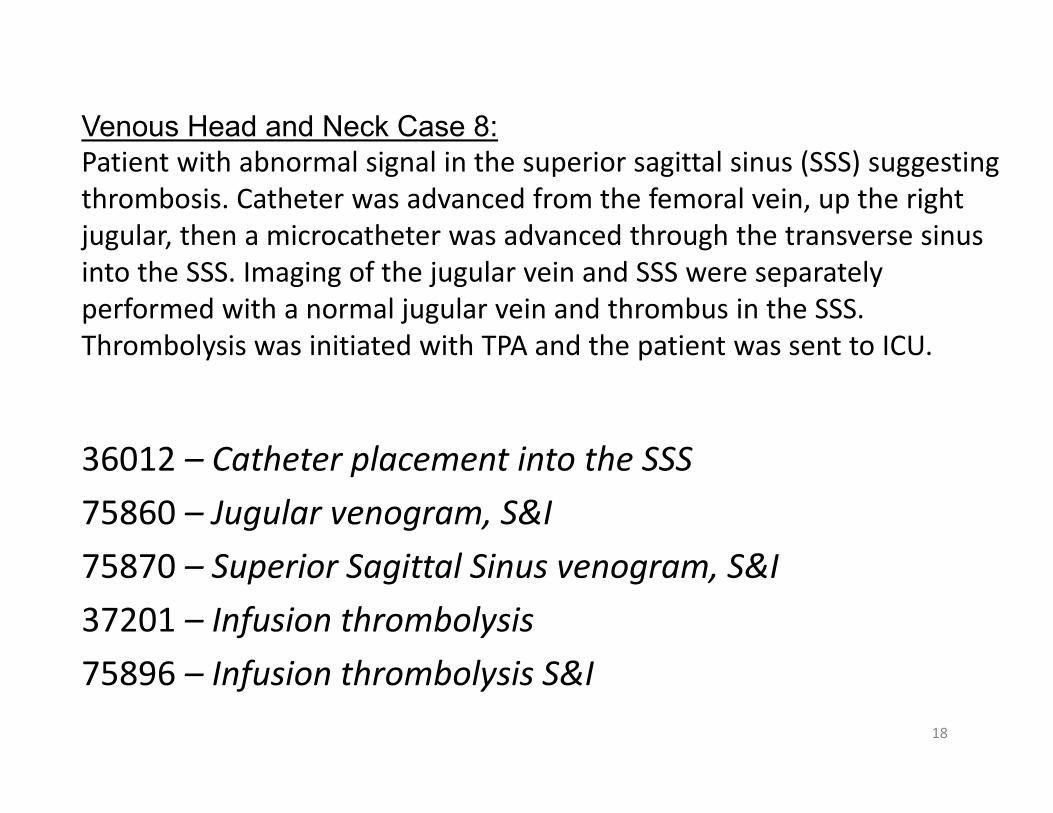
Venous Head and Neck Case 8:Patient with abnormal signal in the superior sagittal sinus (SSS) suggesting thrombosis. Catheter was advanced from the femoral vein, up the right jugular, then a microcatheter was advanced through the transverse sinus into the SSS. Imaging of the jugular vein and SSS were separately performed with a normal jugular vein and thrombus in the SSS. Thrombolysis was initiated with TPA and the patient was sent to ICU.
36012 – Catheter placement into the SSS75860 – Jugular venogram, S&I75870 – Superior Sagittal Sinus venogram, S&I37201 – Infusion thrombolysis75896 – Infusion thrombolysis S&I

19
Venous Head and Neck Case 9:
30yo post MVA with suspected carotid cavernous fistula and weeping exophthalmos. Via bilateral femoral vein punctures, catheters are advanced up the jugular veins into the cavernous sinus bilaterally. A catheter is placed via the femoral artery into both internal carotid arteries. Arterial imaging shows the fistula on the right and good cerebral filling without thrombus or occlusion and bilateral venous imaging showing retrograde filling of the opthalmic veins and rapid washout via the right side. The right side of the cavernous sinus is packed with coils to obliterate the fistula. Three follow‐up images are necessary. These show safe coil placement and no residual flow in the fistula.

20
Venous Head and Neck Case 9 Codes:
36012-50 – Bilateral venous catheter placement36216 -59 – Left ICA catheter placement36217 – Right ICA catheter placement75671 – Bilateral cerebral angiography75860, 75860-59 – Bilateral jugular venography61624 – Intracranial embolization75894 – Embolization S&I75898,75898-59 x 2 – Follow-up embolotherapy
per injection for cerebral therapy

Brachiocephalic AngioplastyCarotid and Vertebral artery angioplasty without stent placement are non‐covered services for Medicare patients. (CMS states the carotid artery is not a peripheral artery so do not use 75962, implies not to use 35475). Discuss this with your payer.
Brachiocephalic refers to the vessels arising from the cervicocerebral arch, including the upper extremities. The code 35475 applies to the right brachiocephalic artery, the right and left subclavian, axillary, brachial, radial and ulnar arteries of the upper extremities.
21
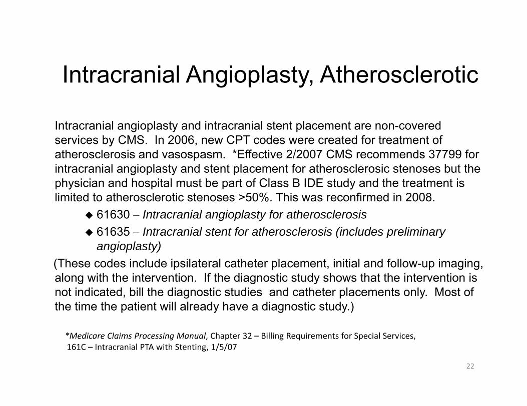
Intracranial angioplasty and intracranial stent placement are non-covered services by CMS. In 2006, new CPT codes were created for treatment of atherosclerosis and vasospasm. *Effective 2/2007 CMS recommends 37799 for intracranial angioplasty and stent placement for atherosclerosic stenoses but the physician and hospital must be part of Class B IDE study and the treatment is limited to atherosclerotic stenoses >50%. This was reconfirmed in 2008.
61630 – Intracranial angioplasty for atherosclerosis 61635 – Intracranial stent for atherosclerosis (includes preliminary
angioplasty)(These codes include ipsilateral catheter placement, initial and follow-up imaging, along with the intervention. If the diagnostic study shows that the intervention is not indicated, bill the diagnostic studies and catheter placements only. Most of the time the patient will already have a diagnostic study.)
Intracranial Angioplasty, Atherosclerotic
*Medicare Claims Processing Manual, Chapter 32 – Billing Requirements for Special Services, 161C – Intracranial PTA with Stenting, 1/5/07
22

• 61640 – Intracranial balloon angioplasty for vasospasm, initial vessel• 61641 – Intracranial balloon angioplasty for vasospasm, each
additional vessel in the same vascular family• 61642 – Intracranial balloon angioplasty for vasospasm, each
additional vessel in a different vascular family
(These codes include catheter placement, intra-procedural imaging, roadmapping, vessel measurements, and guidance, along with the intervention and follow-up imaging. If a diagnostic study is needed the day of the intervention, it is separately billable even if performed on the same date of service. Due to the rapidly changing clinical status in these patients it is common to have to perform repeat diagnostic studies. These codes also are non-covered by Medicare at this time).For Medicare, consider coding the catheter placements and imaging procedures along with the non-covered code with -52 modifier attached (SIR website, 9/23/2007). This should be discussed with your MAC.
Intracranial Angioplasty
23

Angioplasty Case 10:
48 year old patient with TIAs. Via right femoral approach aninitial evaluation of the cerebral vasculature was performedwith arch, bilateral cervical and cerebral angiographyperformed by selective common carotid catheter placements.This shows normal arch and left carotid cervical and cerebralvessels. The right M1 segment of the MCA shows a 90%atherosclerotic stenosis. This was treated initially with balloondilation, showing suboptimal results, requiring stentplacement. A Wingspan stent apparatus was initiallydeployed at 1.5mm in size but required subsequent furtherballoon dilation to 2.0mm. Stenosis in the right anteriorcerebral artery was separately treated with the Gatewayballoon for angioplasty alone without the need for stentplacement. Follow-up angiography at 10 minutes showed nothrombus and patent vessels. Catheters were removed.
24

Angioplasty Case 10 Codes:
36215-59 - 1st order above diaphragm catheter placement, left common carotid
75650 – Cervicocerebral arch, S&I75676 – Unilateral carotid cervical, S&I75665 – Unilateral carotid cerebral, S&I61630-59 – Balloon angioplasty, intracranial, initial vessel
(use 37799 for Medicare patients if criteria met and Class B IDE study participation)
61635 – Intracranial stent placement (use 37799 for Medicare patients if criteria met and Class B IDE study participation)
25
Angioplasty Case 11:
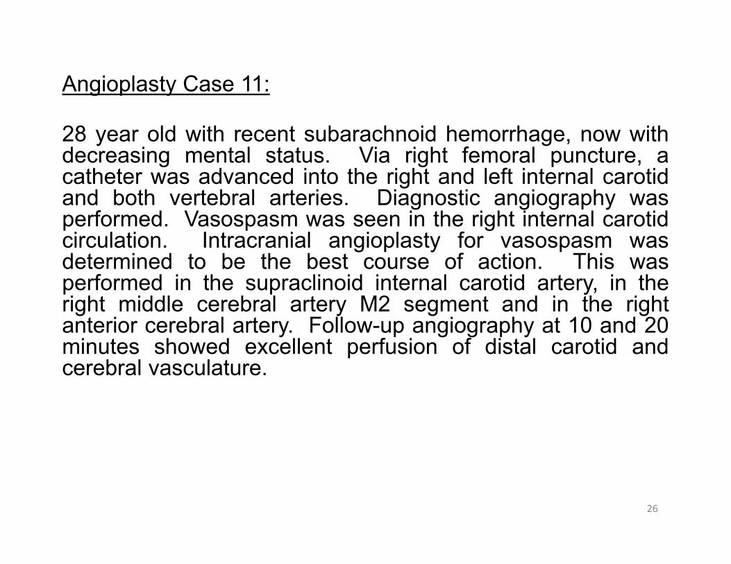
28 year old with recent subarachnoid hemorrhage, now withdecreasing mental status. Via right femoral puncture, acatheter was advanced into the right and left internal carotidand both vertebral arteries. Diagnostic angiography wasperformed. Vasospasm was seen in the right internal carotidcirculation. Intracranial angioplasty for vasospasm wasdetermined to be the best course of action. This wasperformed in the supraclinoid internal carotid artery, in theright middle cerebral artery M2 segment and in the rightanterior cerebral artery. Follow-up angiography at 10 and 20minutes showed excellent perfusion of distal carotid andcerebral vasculature.
26

Angioplasty Case 11 Codes:
36217 – 3rd order selective catheter placement - R vertebral36216-59 – 2nd order selective catheter placement –L-carotid36216-59 – 2nd order selective catheter placement – L- vertebral75685 – Vertebral angiography, S&I75685-59 – Vertebral angiography, S&I75671 – Bilateral carotid cerebral angiography, S&I61640 – Balloon dilation of intracranial vasospasm, initial vessel61641 – Balloon dilation of intracranial vasospasm, each additional vessel
in the same vascular family 61641 – Balloon dilation of intracranial vasospasm, each additional vessel
in the same vascular family
61640 and 61641 are non-covered services for Medicare at this time.
27

Carotid Stent Placement• 37215 – Carotid cervical stent placement with distal embolic
protection• 37216 – Carotid cervical stent placement without distal
embolic protection• 37215 & 37216 include:
– Ipsilateral selective catheterization – Ipsilateral carotid cervical and cerebral artery S&I – All other related S&I during stent placement procedure– All road-mapping, guiding shots and follow-up images– All angioplasties within the region of stent deployment– 37215 remains an inpatient C-status indicator procedure
(1/2010)• Medicare expects you to abandon the case if EPD not
possible• Code 75962 not appropriate as the carotid artery is not a
peripheral artery28

29
Common Carotid and Vertebral Stent Placement
– 0075T – Percutaneous placement extracranial vertebralor common carotid stent, initial vessel
– Includes radiological S&I, imaging and catheterplacement
– 0076T – Percutaneous placement of vertebral or common carotid stent, each additional vessel
– Includes radiological S&I, imaging and catheter placement
– This is an add-on code to 0075T

30
Stent Placement Case 12:
Patient with Doppler stenoses of the left carotid and leftvertebral arteries. Via femoral approach, arch examfollowed by selective catheter placements with injectionof contrast, imaging and findings via the right and leftcommon carotid arteries and left vertebral arteries withimaging of the head and neck is performed. Arch, rightcervical and cerebral arteries and basilar arteries arenormal. The left proximal internal carotid and leftvertebral origin are 90% stenosed. Using distal embolicprotection, stents were placed in both vessels. Follow-upimaging is normal.

31
Stent Placement Case 12 Codes:
37215 – Cervical carotid stent placement0075T – Vertebral artery stent placement36216 – Right common carotid cath placement75650 – Cervicocerebral arch S&I75676 – Right cervical carotid S&I75665 – Right cerebral carotid S&I
*This is an inpatient only procedure

Thrombolytic Infusion with Thrombectomy Case 13:43 year old patient presents with two hour history of left hemispheric stroke.Initial CT scan shows no intracranial bleed. Via a right femoral approach,arch, bilateral selective common carotid catheter placement with imaging ofthe cervical and cerebral vessels was performed. This demonstrated normalcarotid bifurcations and normal left cerebral vessels. Right cerebralangiogram showed thrombus and occlusion of the M-1 and M2 segments ofthe MCA with some clot seen in two branches. Intracranial thrombolysis wasinitiated for 20 minutes (after balloon maceration) with follow-up angiographyshowing some improvement, however MERCI device was necessary toremove significant clot in the M2 segment and the two branches off the M1segment. Follow-up showed further improvement. Thrombolysis wascontinued for another 20 minutes with 8mg TPA. The M1 and M2 segments ofthe middle cerebral artery along with two branches were selected with themicrocatheter during the exam while imaging was used for guidance in thesevessels. Symptoms were significantly improved as was intracranial flow onfollow-up imaging. The sheath was removed and hemostasis obtained.Cardiology was consulted to evaluate for PFO. 22mm PFO was found onTEE and 1 week later the PFO was closed with a PFO occluder device.
32

Thrombolytic Infusion with Thrombectomy Case 13:
36217 – catheter placement in the M2 segment of right carotid36218 x 2 – catheter placement into two branches of the M1 segment36215‐59 – catheter placement into the left common carotid 37201‐59 – intracranial thrombolysis in the right brain37184 – MERCI retrieval thrombectomy in the M2 segment right carotid37185 – MERCI retrieval thrombectomy in two separate M1 segment branches75650 – Cervicocerebral arch S&I75671 – Bilateral cerebral angiography S&I75680 – Bilateral carotid cervical angiography S&I75896 – thrombolysis S&I75898 – follow‐up angiography after thrombolysis S&I75898‐59 – additional follow‐up angiography after thrombolysis S&I
The PFO closure would be reported with code 93580 at the later session with Cardiology.
33

Non-Thrombolytic Infusion Therapy Papaverine, Verapamil, Vasopressin 37202, 75896 per separate vascular distribution (some payers allow only once per session)
37202 requires ‐59 modifier due to CCI edits with most procedures
Add selective catheter placement codes Add diagnostic imaging performed Follow up angiography – 75898 (‐59 for each additional) Do not use 37202 for injection of drugs (such as nitroglycerin, heparin or priscoline) into vessel
Do not use 37202 for infusion of chemotherapy into liver. Do not use 37202 for infusion of Fenoldopam with Benephit catheter (LCD Medical Necessity issue)
Use codes for balloon dilation of vasospasm if applicable (61640/41/42)
34

Non-Thrombolytic Infusion Case 14:
23 year old patient initially presented with a rupturedaneurysm and intracranial bleed treated with surgicalclipping. He now has decreased level of consciousnesssuggesting vasospasm. Arch angiography demonstratesbovine configuration. No FMD or proximal vesseldisease. Bilateral selective internal carotid and bilateralvertebral catheter placement with imaging demonstratedsevere narrowing of the carotid cerebral vesselsbilaterally. The vertebral cervical and cerebral arterieswere normal. Selective Verapamil infusion was started for30 minutes in each carotid vessel due to this severevasospasm. Follow-up angiography shows improvedflow in the intracranial vessels bilaterally.
35

Non-Thrombolytic Infusion Case 14 Codes:
3621736218 x 236216‐5975650756717568575685‐59
37202‐5975896‐59 75898‐59
36

37
Embolization• Peripheral
– 37204, 75894 (use all-inclusive code 37210 for fibroid embolization)
– Per surgical site– Add selective catheter placement codes– Add diagnostic imaging performed– Follow up angiography – 75898 (use 75898
only once per surgical site for completion study)

38
Embolization
• Head and Neck – Non-central nervous system– 61626, 75894– Per surgical site– Add selective catheter placement codes– Add diagnostic imaging performed– Follow up angiography – 75898 (use 75898
only once per surgical site for completion study)
39
Embolization• Head and Neck – Central nervous system
(brain/spinal cord)−61624, 75894−Per surgical site−Add selective catheter placement codes−Add diagnostic imaging performed−Follow up angiography – 75898 (use 75898 as
often as deemed necessary to safely complete the procedure in head and neck, may need to use multiple times in the same vessel for complex embolizations, e.g., aneurysms)

Embolization• Head and Neck – Central nervous system
(brain/spinal cord)−61624, 75894−Wide mouthed aneurysms may require use of a
specialized stent to prevent reflux of coils out of the aneurysm and into the intracerebral vessels
−These stents are the Neuroform and Enterprise−They are considered part of the embolization if
performed at the same setting as the coil deployment. If done a couple weeks earlier, use code 61635 (but NOT for Medicare) to describe the procedure.

41
• Carotid Test Occlusion – 61623– Includes (may not be billed separately)
Selective catheterization of vessel to be occludedMonitoringBalloon inflation
– Does not include (may be billed separately)Selective catheterization and angiography of other arteriesDiagnostic angiography of the test vessel if initial
diagnostic study performed immediately prior to occlusion
Embolization

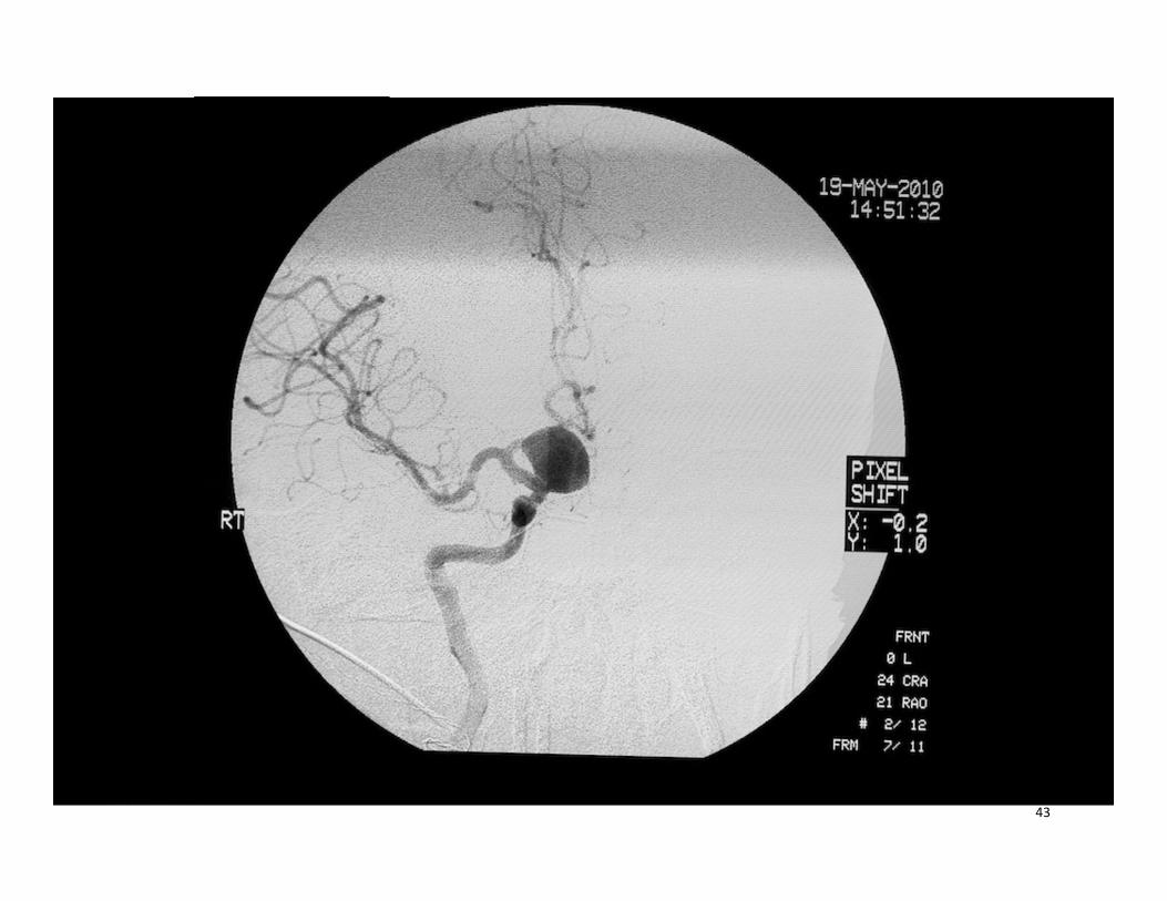
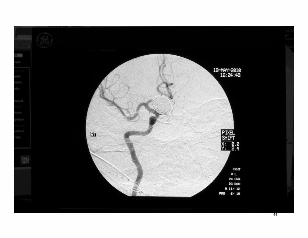
Intracranial Aneurysm Case 15:
39yo post subarachnoid hemorrhage with suspectedaneurysm. A catheter is placed via the femoral arteryinto both internal carotid and both vertebral arteries.Bilateral cervical and cerebral angiograms wereperformed demonstrating a wide necked 4mm aneurysmin the supraclinoid right ICA. A base guide sheath wasplaced in the right proximal ICA, followed by placementof an Enterprise stent apparatus. This was deployedacross the aneurysm, followed by placement ofnumerous helical platinum coils. Follow-up angiographywas necessary and was performed after each of thefollowing 14 coils that were placed. Coil positioning, flowdynamics and distal vasculature were evaluated prior toand after each coil detachment. Complete occlusion ofthe aneurysm was obtained.
42
43
44

Intracranial Aneurysm 15 Codes:
36217 – Catheter placement right ICA aneurysm36216-59 – Catheter placement left ICA36216-59 – Catheter placement left vertebral36218 – Catheter placement right vertebral75671 – Bilateral cerebral angiography S&I75685, 75685-59 – Bilateral vertebral angiography S&I 61624 – Intracranial embolization (includes stent)75894 – Embolization S&I75898,75898-59 x 13 – Follow-up embolotherapy per
injection for cerebral therapy
45

Head and Neck Embolization Case 16:70yo patient with nose bleed. Via transfemoralpuncture, a catheter was placed in the aortic arch forimaging, followed by bilateral common carotid cervicalimaging. The arch vessels were normal and there wasno significant carotid disease. The external carotidarteries were selected and imaged bilaterally showingtortuous vessels but no stenoses. Super-selectivecatheter placement through both internal maxillaryarteries into the sphenopalatine arteries was performedwith imaging of the sphenopalatine arteries. Thesestudies showed hypervascularity on the right and nointracranial collateralization. Embolization with 300-500micron particles was performed. Follow-upangiography on each side showed decreased flow tothe nasal region. The catheter and sheath wereremoved and the patient discharged home six hourslater.
46

Head and Neck Embolization Case 16 Codes:
36017 – Right sphenopalatine catheter placement36217-59 – Left sphenopalatine catheter placement75650 – Cervicocerebral arch angiography S&I75680 – Bilateral cervical carotid angiography S&I75662 – Bilateral external carotid angiography S&I75774 x 2 – Bilateral sphenopalatine artery angiography S&I61626 – Head and neck embolization75894 – Embolization S&I75898 – Follow-up embolotherapy per surgical site
61626 is an outpatient procedure
47
Embolization Case 17:
70yo patient with large falx meningioma. Via transfemoralpuncture, a catheter was placed in the aortic arch forimaging, followed by bilateral common carotid cervicalimaging. The arch vessels were normal and there was nosignificant carotid disease. The external carotid arterieswere selected and imaged bilaterally showing tortuousvessels but no stenoses. Super-selective catheterplacement through both internal maxillary arteries into themiddle meningeal arteries was performed followed bydiagnostic imaging of both the anterior and posteriordivisions bilaterally after selective catheterization of eachbranch. These studies showed hypervascularity of thetumor from both anterior division vessels. There was nocollateralization to non-target vessels. Embolization with150-200 micron particles was performed bilaterally. Follow-up angiography on each side showed decreased flow to thetumor. The catheter and sheath were removed.
48

Embolization Case 17 Codes:
36217 – Right middle meningeal anterior division cath36217-59 – Left middle meningeal anterior division cath36218 x 2 – Right and left middle meningeal posterior division75650 – Cervicocerebral arch angiography S&I75680 – Bilateral cervical carotid angiography S&I75662 – Bilateral external carotid angiography S&I75774 x 4 – Bilateral anterior and posterior branch middle
meningeal artery diagnostic angiography S&I61624 – Intracranial tumor embolization75894 – Embolization S&I75898 x 2 – Follow-up embolotherapy as medically necessary
for intracranial embolization
61624 is an inpatient procedure. This tumor is intracranial although the approach is via the external carotid artery.
49

www.zhealthpublishing.com
Copyright © 2010 ZHealth Publishing
David Zielske, MD, CIRCC, CPC‐H, CCC, CCS, RCC
CPT © 2009 American Medical Association