A STUDY ON CLINICAL PROFILE OF TUBERCULOSIS IN HIV...
109
A STUDY ON CLINICAL PROFILE OF TUBERCULOSIS IN HIV CHILDREN Dissertation submitted to THE TAMILNADU DR. M.G.R. MEDICAL UNIVERSITY in fulfillment of the regulations for the award of M.D DEGREE IN PAEDIATRIC MEDICINE BRANCH VII GOVERNMENT MOHAN KUMARAMANGALAM MEDICAL COLLEGE, SALEM. MARCH 2010
Transcript of A STUDY ON CLINICAL PROFILE OF TUBERCULOSIS IN HIV...

A STUDY ON CLINICAL PROFILE OF
TUBERCULOSIS IN HIV CHILDREN
Dissertation submitted to
THE TAMILNADU DR. M.G.R. MEDICAL UNIVERSITY
in fulfillment of the regulations
for the award of
M.D DEGREE IN PAEDIATRIC MEDICINE
BRANCH VII
GOVERNMENT MOHAN KUMARAMANGALAM
MEDICAL COLLEGE, SALEM.
MARCH 2010

CERTIFICATE
Certified that this dissertation entitled “A STUDY ON
CLINICAL PROFILE OF TUBERCULOSIS IN HIV
CHILDREN” is a bonafide work done by Dr.M.NIRMALA post
graduate student of Paediatric Medicine, Government Mohan
Kumaramangalam Medical College, Salem-636030, during the
Academic year 2008-2010.
Prof.Dr.R.SIVAGAMASUNDARI,M.D.,DCH.,
HOD of Paediatric Medicine, DEAN
Govt. Mohan Kumaramangalam Govt. Mohan Kumaramangalam
Medical College, Salem. Medical College, Salem.

DECLARATION
I declare that this dissertation entitled “A STUDY ON
CLINICAL PROFILE OF TUBERCULOSIS IN HIV
CHILDREN” done by me at Government Mohan Kumaramangalam
Medical College Hospital, Salem under the guidance and supervision
of my department chiefs Prof.R.SIVAGAMASUNDARI,
Prof.M.RATHINASAMY. It is submitted in part of fulfillment of the
award of the degree of MD (Paediatrics) for the March 2010
examination to be held under The TamilNadu Dr. MGR Medical
University, Chennai. This has not been submitted previously by me for
the award of any degree or diploma from any other university.
(Dr.M. NIRMALA)
ACKOWLEDGEMENT
I gratefully acknowledge and sincerely thank our beloved Former
Dean, Dr.P.SHANMUGAM, M.S., M.Ch., Govt. Mohan
Kumaramangalam Medical College and Hospital, for kindly giving me the
permission for conducting this study.
I am also thankful to Dr. K.V.KANAGASABAI, M.D., Present
Dean, Govt.Mohan Kumaramangalam Medical College and Hospital, for
his whole hearted co-operation and support for the completion of this
dissertation.
I am grateful to Prof.Dr.R.SIVAGAMASUNDARI, M.D., DCH.,
Prof. and Head of the Department of Pediatric Medicine, Govt. Mohan
Kumaramangalam Medical College and Hospital, for permitting me to do
the study and for her encouragement.
I am sincerely grateful to Associate Professor
Dr.M.RATHINASAMY, M.D., DCH., for his guidance and help in
conducting this study.
I extend my sincere thanks to Registrar Dr. T.S.SUNDARARAJAN,
M.D., D.C.H., for his valuable suggestions and guidance.
I am extremely thankful to all the Assistant Professors of Pediatrics
for their valuable guidance and encouragement.
I express my sincere thanks to Dr.L.ARUNACHALAM,M.D.,(S.T.D.)
and all medical officers, ART centre, for their help in conducting the study.
I express my sincere thanks to Dr.K.ARUNAGIRI, J.D., District
T.B. Centre for his valuable help in conducting the study.
I am also thankful to my colleagues for their full cooperation in this
study and my sincere thanks to all the patients who cooperated for this
study.

CONTENTS
SL.NO. TITLE PAGE NO.
1. INTRODUCTION 1
2. AIM OF THE STUDY 2
3. REVIEW OF LITERATURE 3
4. BACKGROUND OF THE STUDY 50
5. MATERIALS AND METHODS 51
6. OBSERVATION & RESULTS 54
7. DISCUSSION 69
8. CONCLUSION 73
BIBLIOGRAPHY
ANNEXURES
a. PROFORMA
b. MASTER CHART
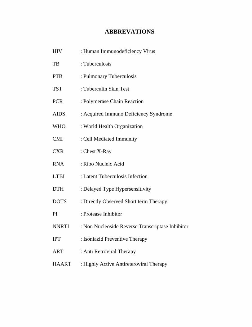
ABBREVATIONS
HIV : Human Immunodeficiency Virus
TB : Tuberculosis
PTB : Pulmonary Tuberculosis
TST : Tuberculin Skin Test
PCR : Polymerase Chain Reaction
AIDS : Acquired Immuno Deficiency Syndrome
WHO : World Health Organization
CMI : Cell Mediated Immunity
CXR : Chest X-Ray
RNA : Ribo Nucleic Acid
LTBI : Latent Tuberculosis Infection
DTH : Delayed Type Hypersensitivity
DOTS : Directly Observed Short term Therapy
PI : Protease Inhibitor
NNRTI : Non Nucleoside Reverse Transcriptase Inhibitor
IPT : Isoniazid Preventive Therapy
ART : Anti Retroviral Therapy
HAART : Highly Active Antireteroviral Therapy

INTRODUCTION
HIV is driving TB epidemic in many countries, especially in
sub-saharan Africa and increasingly, in Asia and South America. TB in
populations with high HIV prevalence is a leading cause of morbidity
and mortality.
Children who are HIV infected have a higher risk of progression
after primary infection. Children born to HIV positive parents who are
not infected with TB themselves, are also at higher risk of acquiring
TB because of exposure. The source of transmission of TB to a child is
usually an adult with sputum-smear positive PTB. Cases of TB in
children represent between 10% to 20% of all TB cases.
Extrapulmonary TB manifestations are Lymphadenopathy,
Pleural effusion, Miliary TB, TB meningitis, TB Abdomen,
Disseminated TB, Potts Spine.
2
AIM OF THE STUDY
1. To evaluate the clinical, bacteriological and radiological pattern
of TB in HIV seropositive children in correlation with CD4
count.
2. To find out the trends of TB infection in HIV seropositive
children in GMKMCH, Salem.
3. To find out the prevalence of TB among HIV seropositive
children in our hospital.

3
REVIEW OF LITERATURE
TUBERCULOSIS: An overview
Tuberculosis has been present in humans since antiquity, as the
origin of the disease are in the first demonstration of cattle (which also
gave human viral pores). Skeletal remains show prehistoric humans
(4000BC) had TB1 and tubercular decay has been found in the spines
of Egyptian mummies from 3000-2400BC2. There were references to
TB in India around 2000 BC and indications of lung scarring identical
to that of modern day TB sufferers in preserved bodies (such as
mummies) suggests that TB was present in the Americans from about
2000 BC. Phthisis is a Greek term for consumption. Around 460 BC,
Hippocrates identified phthisis as the most widespread disease of the
times which was almost always fatal3.
Due to the variety of the symptoms, TB was not identified as a
single disease until the 1820’s and was not named tuberculosis until
1839 by JL Schonlein4. The bacillus causing tuberculosis,
Mycobacterium tuberculosis, was identified and described on March
24, 1882 by Robert Koch. He received the Nobel Prize in physiology
(or) medicine in 1905 for his discovery5.

4
The other names for tuberculosis are:
TB (short for tuberculosis and also for Tubercle Bacillus).
Consumption (TB seemed to consume people from within with
its symptoms of bloody cough, fever, pallor and long relentless
wasting).
Wasting disease.
King’s evil.
White Plague (TB sufferers appear markedly pale)
Phthisis (Greek for consumption) and phthisis pulmonalis.
Miliary TB (X-ray lesions look like millet seeds) 6.
Koch’s disease named after Robert Koch who discovered the
tuberculosis bacilli7.
PATHOGENESIS:
While only 10% of TB infection progresses to TB disease8, if
untreated the death rate is 51%9.The primary complex of tuberculosis
includes local infection at the portal of entry and the regional lymph
nodes. TB infection begins when the MTB bacilli reach the pulmonary
alveoli, infecting alveolar macrophages 8, 10, where the mycobacteria
replicate. The primary site of infection in the lungs is called the Ghon

5
focus, which is the combination of a parenchymal pulmonary lesion
and a corresponding lymph node site.
The tubercle bacilli are carried to most tissues of the body
through the blood and lymphatic vessels to the more distant tissues and
organs where TB disease could potentially develop: lung apices,
peripheral lymph nodes, kidneys, brain and bone 8,11 .
Tuberculosis is classified as one of the granulomatous inflammatory
conditions. Macrophages, T lymphocytes, B lymphocytes and fibroblasts
are among the cells that aggregate to form a granuloma, with lymphocytes
surrounding infected macrophages. The granuloma functions not only to
prevent dissemination of mycobacteria, but also provides a local
environment for communication of cells of the immune system. Within
the granuloma, T lymphocytes (CD4 +) secrete a cytokine such as
interferon gamma, which activates macrophages to destroy the bacteria
with which they are infected12, making them better able to fight infection.
T lymphocytes (CD8 +) can also directly kill infected cells by secreting
perforin and granulysin13.
Importantly, bacteria are not eliminated within the granuloma, but
can become dormant resulting in a latent infection8. Latent infection can
be diagnosed by tuberculin skin test, which yields a delayed
hypersensitivity type response to purified protein derivatives of M.

6
tuberculosis in an infected person.Another feature of the granulomas of
human tuberculosis is the development of cell death, also called necrosis,
in the centre of tubercles. To the naked eye this has the texture of soft
white cheese and was termed caseous necrosis14.
If TB bacteria gain entry to the blood stream from an area of
tissue damage they spread through the body and set up myriad foci of
infection, all appearing as tiny white tubercles in the tissues. This is
called as miliary tuberculosis and has a high case fatality.
PROGRESSION:
In children in whom TB bacilli overcome the immune system
defenses and begin to multiply, there is progression from TB infection
to TB disease. This may occur soon after infection (primary TB
disease-1 to 5%) or many years after infection (post primary TB,
secondary TB, reactivation TB disease of dormant bacilli-5 to 9%).
About five percent of infected persons will develop TB in first two
years and another five percent will develop disease later in life. In
another words about 10% of infected persons with normal immune
systems will develop TB disease in their lifetime8.

7
Some medical conditions increase the risk of progression to TB
disease. In HIV infected children the rate of tuberculosis disease is 30
times higher than in non HIV infected children. Other conditions such
as prolonged corticosteroid therapy, immunosuppressive therapy,
leukemia, Hodgkins disease, end stage renal disease, recent TB
infection, (within two years, or history of inadequately treated
TB),chest Xray suggestive of previous TB (fibrotic lesion and
nodules),chronic malabsorption syndrome or low body weight (10%or
more below the ideal).
Primary complex includes the parenchymal pulmonary focus
and the regional lymph nodes15. About 70% of pulmonary foci are
subpleural and localized pleurisy is common. The hallmark of primary
tuberculosis is the relatively large size of regional lymphadenitis
compared with the relatively small size of initial lung focus
Children may have lobar pneumonia without impressive hilar
lymphadenopathy. If primary infection is destructive , liquefaction of
the lung parenchyma can lead to the formation of a thin walled primary
tuberculosis cavity. Rarely, bullous tuberculous lesions can occur in
lungs and lead to pneumothorax if they rupture15 Erosion of a
parenchymal focus of tuberculosis into a blood or lymphatic vessel
may result in dissemination of the bacilli and a miliary pattern.

8
No clinical disease
Positive tuberculosis
(usual outcome:90%of cases)
PRIMARY
COMPLEX
Disseminated disease
Lymphadenopathy (usually cervical)
Meningitis
Pericarditis
Miliary disease
TB Spine
Hypersensitivity reactions (eg) Erythema
nodosum,
Phlyctenular conjunctivitis
Dactylitis.
Primary and pleural complication (eg)TB
pneumonia, Hyper inflation and Collapse
Pleural effusion Consolidation.

9
More than 50% of infants and children with radiologically
moderate to severe pulmonary TB have no physical findings and are
discovered only by contact tracing .Non productive cough and dyspnea
are the most common symptoms. Systemic complaints such as fever,
night sweats, anorexia and decreased activity occur less often .
Older children and adolescents with reactivation tuberculosis are
more likely to experience fever, anorexia, malaise, weight loss, night
sweats, productive cough, hemoptysis and chest pain than children
with primary pulmonary TB.

10
POST-PRIMARY TB
PULMONARY TUBERCULOSIS
(eg) Cavities
Upper lobe infiltrates
Fibrosis
Progressive pneumonia
Endobronchial
EXTRAPULMONARY TB
COMMON LESS COMMON
Pleural effusion Empyema
Lymphadenopathy
(usually cervical) Kidney
Central nervous system
(Meningitis,tuberculoma) Adrenal gland
Pericarditis (Effusion/pericarditis) Skin (lupus vulgaris,
tuberculids, miliary)
Gastrointestinal
(ileocaecal, peritoneal)

11
DIAGNOSIS:
A complete evaluation of TB includes a history of contact with
adult TB, a physical examination, clinical features, a tuberculin skin
test, a chest x-ray, serological test and microbiological smears and
cultures. The interpretation of the tuberculin skin test depends upon the
persons risk factors for infection and progression to TB disease, such
as exposure to other cases of TB or immunosupression16 .Tuberculin
tests have the disadvantage in that they may produce false negatives,
especially when the patient is co-morbid with malnutrition, sarcoidosis,
Hodgkins lymphoma or most notably active tuberculosis disease8. New
TB tests are being developed that offer the hope of cheap ,fast and
more accurate TB testing. These include PCR detection of bacterial
DNA ,and assays to detect the release of interferon gamma in response
to mycobacterial proteins such as ESAT-617.These are not affected by
immunization or environment mycobacteria, so generate fewer false
positive results18. The development of a rapid and inexpensive
diagnostic test would be particularly valuable in the developing
world19.

12
HIV-AN INTRODUCTION

13
Ever since the first description of AIDS in 1981,researchers have
identified two types of HIV. HIV-1 Subtype C is the predominant type
worldwide. HIV-2 occurs mostly in West Africa and occasional
infections have occurred in Africa, Europe, Asia, and Latin America.
Both types cause AIDS and the routes of transmission are the same.
EPIDEMIOLOGY
The WHO estimated that more than 39 million persons
worldwide were living with HIV infection at the end of 2008,
including 2.2 million children < 15 years of age15. Sub-saharan Africa
accounts for the fastest growing epidemic, with almost 90% of worlds
total population of HIV infected children. India and Thailand dominate
the epidemic in Southeast Asia.
Virtually all HIV infections in children < 12 years of age are the
result of vertical transmission from an HIV infected mother. A
vanishing minority of children are infected by contaminated blood
products and or clotting factors. The risk of transmission without
Breast Feeding is 20-25%, with Breast Feeding for 6 months is 25-30%
and the risk increases to 30-35% with prolonged Breast feeding for
18-24 months.15

14
The risk of transmission can be reduced to 1% by:
a) ARV prophylaxis to women during pregnancy/labour and to the
infant in first week.
b) Elective caesarean section – prior to onset of labour and rupture
of membranes
c) Complete avoidance of Breast feeding.
The clinical consequences of HIV infection are due to the ability
of this virus to disarm the host immune system, a process that occurs
by virtue of the fact that the primary target for the virus is the helper
induced subset of lymphocytes. This lymphocyte subset, defined by its
surface expression of the CD4 molecule, acts as the pivotal
orchestrator of a myriad of immune functions. HIV infection can
therefore be considered a disease of the immune system, characterized
by the progressive loss of CD4 + lymphocytes, with ultimately total
consequences for the infected host8.
Despite the immunosuppression induced by HIV, a number of
specific immunologic defences against the virus are generated in
infected individuals and may contribute to the long asymptomatic
phase following infection by keeping the virus atleast partially
contained. The hallmark of HIV infection is progressive depletion of
the CD4 helper inducer subset of lymphocytes. Because of the central

15
role of these cells in immunologic functioning, the clinical disease
manifestation of immunosuppression and susceptibility to
opportunistic infections and neoplasms are not surprising. The
immunologic deficit associated with HIV infection are wide spread and
involve numerous interdependent effector arms of the immune system,
including both cellular and humoral elements.
Of all the opportunistic infections in persons infected with HIV
virus, tuberculosis remains the commonest20. HIV probably increases
the susceptibility to primary infection with M-tuberculosis, as well as
to reactivation of TB infection due to depressed CMI21,22. HIV
increases the risk of progression of M-tuberculosis infection to TB
disease. This risk increases with increasing immunosuppression. HIV
increases not only the risk but also the rate of progression of recent or
latent M-tuberculosis infection to disease.
WHO staging system for HIV infection and related disease in
children younger than 13 years23,24.
Two Classifications
i) WHO Classification – Symptomatic HIV divided into four
stages based on the severity of the symptoms.
ii) CDC Classification – Clinical classification and immunological
classification based on CD4.

16
Revised WHO Clinical Staging of HIV/AIDS for Infants and
Children with Confirmed HIV infection (> 18 months of age – HIV
Antibody Test Positive, < 18 months of age – Virologic Test
Positive)23
CLINICAL STAGE 1 (AYSMPTOMATIC)
Asymptomatic
Persistent generalized lymphadenopathy
CLINICAL STAGE 2 (MILD)
Unexplained persistent hepatosplenomegaly
Papular pruritic eruptions
Extensive wart virus infection
Extensive molluscum contagiosum
Fungal nail infections
Recurrent oral ulcerations
Unexplained persistent parotid enlargement
Linear gingival erythema
Herpes zoster
Recurrent or chronic upper respiratory tract infections (otitis media,
otorrhoea, sinusitis, tonsillitis)

17
CLINICAL STAGE 3 (ADVANCED)
Unexplained moderated malnutrition not adequately responding to
standard therapy
Unexplained persistent diarrhea (14 days or more)
Unexplained persistent fever (above 37.5°C intermittent or
constant, for longer than one month)
Persistent oral candidiasis (after first 6-8 weeks of life)
Oral hairy leukoplakia
Acute necrotizing ulcerative gingivitis/periodontitis
Lymph node TB
Pulmonary TB
Severe recurrent bacterial pneumonia
Symptomatic lymphoid interstitial pneumonitis
Chronic HIV – associated lung disease including bronchiectasis
Unexplained anemia (<8g/dl), neutropenia (<0.5x10⁹/L3 ) or chronic
thrombocytopenia (<50 x 109/L3 )
CLINICAL STAGE 4 (SEVERE)
Unexplained severe wasting, stunting or severe malnutrition not
responding to standard therapy
Pneumocystis pneumonia

18
Recurrent severe bacterial infections (e.g. empyema, pyomyositis,
bone or joint infection, meningitis, but excluding pneumonia)
Chronic herpes simplex infection; (orolabial or cutaneous of more
than one month’s duration or visceral at any site)
Extra pulmonary TB
Kaposi’s sarcoma
Oesophageal candidiasis (or candidiasis of trachea, bronchi or
lungs)
Central nervous system toxoplasmosis (after one month of life)
HIV encephalopathy
Cytomegalovirus infection: retinitis or CMV infection affecting
another organ, with onset at age over 1 month.
Extra pulmonary cryptococcosis (including meningitis)
Disseminated endemic mycosis (extrapulmonary histoplasmosis,
coccidiomycosis
Chronic cryptosporidiosis
Chronic isosporiasis
Disseminated non-tuberculous mycobacteria infection
Cerebral or B cell non-Hodgkin lymphoma
Progressive multifocal leukoencephalopathy
Symptomatic HIV – associated nephropathy or HIV – associated
cardiomyopathy

19
IMMUNOLOGICAL CLASSIFICATION BASED ON CD423
Immunologic
Definitions
< 12 months 1-5 years 6-12 years
µL % µL % µL %
No evidence of suppression
≥1500 ≥25 ≥1000 ≥25 ≥500 ≥25
Evidence of moderate suppression
750-1499 15-24 500-999 15-24 200-499 15-24
Severe suppression <750 <15 <500 <15 <200 <15
WHO case definitions for AIDS in children where HIV testing
facilities are not available.
MAJOR SIGNS:
Weight loss > 10% of body weight or failure to thrive
Chronic diarrhea (> one month)
Prolonged fever (> one month)
Severe or repeated pneumonia
MINOR SIGNS:
Generalised lymphadenopathy
Oro – Pharyngeal candidiasis

20
Repeated common infections (otitis, pharyngitis etc.,)
Generalised dermatitis
Confirmed maternal HIV infection.
Pediatric AIDS is suspected in an infant or child presenting with
atleast two major signs associated with atleast two minor signs in the
absence of known cases of immunosuppression.
These definitions detect the presence of symptomatic AIDS and
not HIV infection itself and are confounded by malnutrition, diarrheal
diseases and tuberculosis.
PRACTICAL POINT:
The term AIDS is used for epidemiological surveillance, not for
clinical case. For patients with TB, persistent cough for more than one
month should not be considered as a minor sign.
Picture showing estimated HIV & TB coinfection in India25
TB 20 Million
HIV & TB
2 Million
HIV 5.1 Million

21
Features Stage of HIV Infection
Early Late
Clinical
Presentation
Often resembles
post primary TB
Often resembles primary
TB; Extra pulmonary TB
common
Sputum smear
result Often positive Often negative
CXR Often shows
cavities
Atypical often infiltrates,
Lower lobe lesions or
intrathoracic lymphnodes
EPIDEMIOLOGY:
About a third of the HIV positive population worldwide is co-
infected with M-tuberculosis. This accounts to about 14 million people
worldwide. Globally, 9% of all tuberculosis cases in adults are
attributable to HIV26. Studies from sub-saharan Africa have recorded
HIV seroprevalance rates of 50 - 70% in patients with tuberculosis. In
Asia, where the HIV epidemic is still at early stage, the rate of HIV
infection in TB patient has been lower.
A HIV positive person infected with M-tuberculosis has a 50-
60% lifetime risk of developing TB disease as compared to an HIV
negative person who has only a 10% risk.

22
Using estimates of prevalence of TB and HIV in various regions,
It has been observed that by mid-1994 there were 5.6 million persons
co-infected with HIV and TB worldwide, more than 1.15 million of
these live in South-east Asia (including India)27. The proportion of
HIV-attributable tuberculosis death will increase from 4.6% to 14.2%.
In India the rates of HIV-tuberculosis co-infection is steadily
increasing. In Pune the prevalence of HIV infection among
tuberculosis patients increased from 3.2% in 1991 to 20.1%28. Most
available data on HIV and TB is from adults. Disease in children
follows progressions of primary infection. It is estimated that
worldwide there will be over 56,000 cases of HIV attributable
tuberculosis annually in children by 200629. A study from Mumbai
showed a prevalence of HIV of 18% among children with CNS
tuberculosis or miliary tuberculosis; the prevalence HIV seropositivity
among children with chronic diarrhea in same study was 24%30.
TUBERCULOSIS IN HIV INFECTION:
HIV has had as substantial effect on the incidence clinical
manifestations, treatment and outcome of TB. Globally, HIV and TB

23
are the 2 leading infectious causes of death.People who are infected
with HIV are at increased risk of contracting TB.
Co-infection with these pathogens can be particularly
devastating, especially in the developing world, where the burden of
disease is high and access to effective therapy in low. Among infection
associated with HIV, TB is unique in that it may be transmitted to
immunocompetent persons via the respiratory route, and is easily
treatable once identified, may occur in early stage HIV- disease and is
preventable with drug therapy. However, multidrug resistance
tuberculosis is a potentially serious problem, even though its incidence
has declined because of the use of directly observed therapy and other
improved practices31.
TB is a major opportunistic infection in HIV-infected patients
often representing their AIDS defining illness and the first indication
of immunodeficiency. Epidemiology, clinical manifestations and
management of TB are altered in HIV-infected patients.

24
IMPACT OF HIV ON TB
HIV is the most significant risk factor for progression from sub-
clinical infection with mycobacterium tuberculosis to active TB.22
However, when a person is dually infected with HIV and M-
tuberculosis, the risk of developing TB significantly increases from
10% in a lifetime to 5-15% per year31.
HIV infected children are at markedly increased risk for primary
or reactivation tuberculosis and for second episodes of tuberculosis
from exogenous reinfection32.
The lifetime risk for progression to active TB among HIV-
negative persons likely infected with M-tuberculosis is estimated at
10%. In contrast, among HIV infected children the risk for progression
to active disease is approximately 10% per year, and the immediate
risk for progressive primary disease after recent infection with M-
tuberculosis approaches 40%33.
This interaction between TB and HIV accounts for much of the
recent global resurgance in TB of late, however HIV infection does not

25
increase the infectiousness of persons with active TB. The introduction
of potent combination Anti-retroviral therapy in developing world,
which has dramatically decreased the risk for opportunities infections
and death among HIV infected children, has also decreased the risk for
developing active TB and the risk for death among HIV infected
persons who develop active TB.
Children born to HIV infected mother but who are not infected,
are also at higher risk of acquiring tuberculosis because of the
increased risk of exposure to tuberculosis from their parents. It is
estimated that tuberculosis rate in the first four years of life among
children born to HIV infected mothers is 10 times higher than in non
HIV infected and 30 times higher in HIV infected children as
compared to other children.

26
PATHOGENESIS:
Tuberculosis can develop through progression of recently
acquired infection (primary disease), reactivation of latent infection, or
exogenous reinfection.
Infection with M-tuberculosis can occur when a child is exposed
to an infectious case of TB particles (<5 cm in size) containing the
tubercle bacilli. If the bacilli reach the pulmonary alveoli, they may be
ingested by alveoli macrophages the first line of defence against
M-tuberculosis. Surviving tubercle bacilli multiply within the
macrophage and eventually undergo hematogenous spread to other
areas of the body.
After ingestion, alveoli macrophages present the mycobacterial
antigens to CD4 + T cells. This results in the release of interferon
gamma (IFN γ) which in turn activities macrophages to control the
mycobacterial infection. However the activated macrophages also
release interleukin – 1 which enhances HIV replication. Mycobacteria
also enhance HIV replication by inducing nuclear factor kappa-B, the
cellular factors that bends the promote region of HIV34,35.

27
Susceptibility to TB is related to the pattern of cytokines
produced by T lymphocytes. CD4 + lymphocytes which produce
interferon-γ are central to antimycobacterial immune defenses and fatal
mycobacterial disease develops in children who lack the interferon γ
receptor. In contrast to CD4+lymphocytes, CD8 + lymphocytes, which
produce interleukin -4 and interleukin -10 do not contribute to anti
mycobacterial immunity. When peripheral blood lymphocytes from
HIV infected patients with tuberculosis are exposed to M-tuberculosis
in vitro, they produce less interferon–γ but similar amounts of
interleukin-4 and interleukin-10, as compared with lymphocytes from
HIV- negative patients with tuberculosis. These findings suggest that
the reduced CD4 response in HIV-infected patients contributes to their
susceptibility to TB.
The hallmark of HIV infection is progressive deterioration and
depletion of CD4cells, coupled with defects in macrophage and
monocyte function. There is evidence that the immune response in
patients with TB might enhance HIV viral replication and accelerate
the natural progression of HIV infection 36.

28
TUBERCULOSIS AND THE COURSE OF HIV INFECTION:
Exposure of alveolar macrophages and lymphocytes from HIV
infected patients to M.Tuberculosis in vitro upregulates retroviral
replication34,35. TB has been associated with a 5 to 160 fold increase in
HIV viral replication, which may decrease after successful TB
treatment.
Pleural fluid from patients with tuberculosis increases HIV
replication in activated lymphocytes and in HIV-infected patients with
pulmonary tuberculosis, the concentrations of retroviral RNA in
bronchoalveolar lavage fluid are highest in areas of tuberculous
involvement37.M.Tuberculosis probably increases HIV replication by
inducing macrophages to produce tumor necrosis factor α, interleukin-
1 and interleukin-6 38,39.
CLINICAL MANIFESTATIONS OF TB IN HIV:
Unlike other opportunistic infections, TB can occur in persons
with early –stage HIV infection (CD4 count >300/cubic mm). Clinical
and radiological manifestations of TB are similar to those seen in non
HIV infected persons (37,40).

29
Children are less likely to present with extrapulmonary disease
at this stage of HIV infection. Because of the increased virulence in
immunocompetent hosts of M.Tuberculosis compared with other
opportunistic infections, tuberculosis can occur early in the course of
HIV infection. In several studies of HIV infected children with
pulmonary tuberculosis, the median CD4 T-cell count was >300
cells/cubic mm.
Studies have shown that 88-92% of HIV infected persons with
TB had only pulmonary involvement, whereas 0.6to3%had both
pulmonary and extrapulmonary and 8to12% had only extrapulmonary
involvement37. Of those with extrapulmonary involvement 85% had
lymphadenopathy (mainly cervical) 37.
Miliary tuberculosis was uncommon and found in only 1.7% of
HIV children with TB37. Though many HIV infected patients have
typical clinical and radiological manifestations of tuberculosis, atypical
presentations do occur frequently, especially in those with low CD4
counts. Thus cavitatory upper lobe tuberculosis is more common with
those with CD4 counts >200/cubic mm, whereas hilar/mediastinal

30
adenopathy and diffuse pulmonary infiltrates (without cavitation) are
more common in those with CD4 counts <200/cubic mm.
Typical symptoms include fever weight loss, cough of several
weeks duration and failure of pulmonary signs and symptoms to
subside despite adequate antibiotic therapy. Chest radiographs
demonstrate the presence of lobar infiltrates with or without hilar
adenopathy and diffuse infiltrates.In children with advanced HIV
disease, TB may present atypical and common extrapulmonary
manifestations were lymphadenopathy, pleural effusion, TB abdomen,
TB meningitis and miliary tuberculosis42.
The clinical feature of TB meningitis in children who are
seropositive for HIV are not siginificantly different from those in
seronegative children. However ventriculomegaly, gyral enhancement
and cortical atrophy on CT scan are more common in HIV seropositive
children43. Also,mortality and the incidence of severe neurological
sequelae are more common in HIV seropositive children. Co-existing
HIV encephalopathy and diminished immune function may account for
poorer prognosis43.
31
RADIOGRAPHIC FINDINGS:
The chest radiograph is the cornerstone of diagnosis for
pulmonary tuberculosis. Upper lobe infiltrates and cavities are the
typical findings in reactivation tuberculosis, whereas intrathoracic
lymphadenopathy and lower lobe disease are seen in primary
tuberculosis. In HIV-infected children with CD4 T cell counts >200/
cubic mm the radiographic pattern tends to be one of the reactivation
disease with upper lobe infiltrates with or without cavities.
In HIV-infected persons with a greater degree of
immunosuppression (e.g. CD4 T-cell count < 200 cells/mm3)
hilar/mediastinal lymphadenopathy and lower lobe infiltrates are
common 41. As chest radiographs may appear normal in 7-14% of
cases, a high index of suspicion must be maintained in evaluating an
HIV-infected patient with symptoms suggestive of TB. The finding of
low density lymph nodes with peripheral enhancement on a contrast-
enhanced chest computed tomography (CT) scan is highly predictive of
tuberculosis. Cavitation is unusual at advanced stage of
immunosuppression, and infiltrates more often are diffuse or
interstitial.

32
DIAGNOSIS
In children the majority of tuberculosis cases are diagnosed
clinically without microbiological confirmation. The diagnosis is based
mainly on clinical suspicion and radiological manifestation. In children
with TB and HIV, special consideration is required in assessing results
of tuberculin skin test and Acid-fast bacillus smear and culture.
TUBERCULIN SKIN TEST
HIV infection causes depression of cell mediated immunity,
which can reduce the sensitivity and reliability of the tuberculin skin
test. The TST is often negative, especially in those with more advanced
HIV disease. Only one-third to less than half of children co-infected
with TB and HIV have a positive tuberculin test (42,44,45) . Because of
the higher risk of tuberculosis in children living in households with
HIV-infected adults and because of diminished immune responses in
children with HIV infection, the American Academy of Pediatrics
recommends using induration ≥5mm as the criterion for diagnosis of
tuberculous infection46. These criteria have not been adequately
evaluated in developing countries, especially in those where BCG
vaccination is a part of routine childhood immunization.

33
Although a positive result increases the likelihood of TB, a
negative result does not exclude the diagnosis. Therefore diagnosis
evaluation for TB should be undertaken in all children with clinical
feature compatible with TB, regardless of the results of TB.
There are recent reports of restoration of delayed
hypersensitivity on skin testing, including tuberculin skin testing in
HIV-infected patients who begin highly active antiretroviral therapy.
This reaction reflects restoration of the anti M-tuberculosis cell
mediated immune response, a phenomenon that usually occurs within
the first month of therapy.
Polymerase chain reaction and gene probes are approved for
rapid identification of M.tuberculosis in sputum smears that are
positive for acid-fast bacilli. These rapid tests are more sensitive than
traditional staining methods but are not as sensitive as culture. Smears
from extrapulmonary sites (e.g., bone marrow, lymph nodes) are often
negative for acid-fast bacilli.

34
BACTERIOLOGICAL EXAMINATION
All patients suspected of having pulmonary tuberculosis should
have 3 sputum specimens obtained on 3 consecutive days, and these
specimens should be examined for AFB and cultured for mycobacteria.
Contrary to what one would expect, smears and cultures are more often
negative in HIV-infected adult patients with tuberculosis47. Similar
findings are reported among children with co-infection(42,44).
In general the rate of smear positivity correlates with the extent
of radiographic disease. For example, patients with cavitary lesions due
to active tuberculosis will almost always have positive smears, whereas
a negative smear in a patient with minimal disease on chest radiograph
would not be unusual, and would not rule out active TB. However, in
HIV infected patients positive smears may be seen with relatively little
radiograph involvement. Despite this, it is recommended that
aggressive attempts be made to obtain a positive culture to differentiate
between M.tuberculosis and other mycobacteria, as well as to
determine the antimicrobial susceptibility.
Smears from needle aspiration and Ziehl-Neelson staining are
often negative in HIV patients who have tuberculous lymphadenitis48.

35
Hence biopsy with histopathological examination and culture of lymph
node tissue are recommended to establish the diagnosis.
Diagnostic tests for TB can be broadly divided into
1. Demonstration/isolation of Mycobacterium tuberculosis
2. Demonstration of host response to exposure to M.tuberculosis
(A) DEMONSTRATION OF M.TUBERCULOSIS OR ITS
COMPONENTS
1. Ziehl-Neelson Staining
2. Special stains (Flurochrome stain, Auramine-O-stain etc.,)
3. Culture of mycobacterium tuberculosis
- LJ medium
- BACTEC radiometric assay
4. Polymerase chain reaction
(B) DEMONSTRATION OF HOST RESPONSE TO
EXPOSURE TO M.TUBERCULOSIS
1. Serodiagnosis
2. Tuberculin skin testing

36
Newer diagnostic modalities including molecular methods of
diagnosis of tuberculosis may yield better results than routine smear
and culture in HIV infected population, but these methods need further
evaluation. Moreover, these methods for diagnosis are seldom
available in regions where the two infections most frequently co-exist.
DIAGNOSIS OF LATENT TUBERCULOSIS INFECTION
Screening for latent tuberculous infection is an essential step in
controlling the spread of tuberculosis. Screening for LTBI is
recommended in persons at risk for recent infection and in those with
increased risk of progression to active disease once infected, including
HIV-infected persons.
The TST is currently the only method available for identifying
LTBI. Routine annual TST is recommended in HIV infected
individuals. A reaction of ≥5mm induration is considered positive for
HIV infected patients and those with other forms of severe
immunosuppression, person who are close contacts of infectious cases,
and persons with abnormal radiographs consistent with tuberculosis.
Use of 5mm cutoff is supported by a prospective study in the United
States demonstrating that the risk of tuberculosis was significantly

37
higher in HIV-infected persons with TST≥5mm of induration than in
those who have a reaction of <5mm 46.
Testing with tuberculin purified protein derivative is dependant
on the presence of an intact cell mediated immune response. In the
setting of HIV infection, reduced CMI can lead to decreased delayed-
type hypersensitivity responsiveness, resulting in false-negative skin
test. The prevalence of positive TST≥5mm was shown to decrease with
decreasing CD4 T cell counts.
Application of multiple skin antigens (e.g. candida, mumps,
tetanus toxoid etc.,) referred to as anergy testing, has been used to
access cell-mediated immune function and to distinguish true negative
from false negative tuberculin skin test results. In 1991, the CDC
recommended that anergy testing be performed in conjunction with
TST in HIV-infected persons based on the premise that anergic HIV-
infected individual at high risk for tuberculosis infection would benefit
from treatment with INH. In 1997, the CDC revised its
recommendations and no longer recommends anergy testing while
screening for M.tuberculosis infection in HIV-infected persons. The
revised recommendation is based on the following points. First, there

38
are no standardized guideline for performing anergy skin testing. The
appropriate number of control antigens to administer or the appropriate
cut off for interpreting a test as positive in nor known. Second, the
response to skin testing with control antigens as well with tuberculin
can vary over time. Several studies have demonstrated that HIV-1
seropositive individuals can regain DTH responsiveness with time 49.
The only factor associated with regaining DTH responsiveness was the
CD4-T cell count, the higher the CD4 count, the more likely the
individual regains DTH responsiveness.
In some with tuberculous infection, DTH responsiveness may
decrease with time. A second TST, applied weeks to month after the
first, can boost the DTH response resulting in a positive skin test
reaction. Such responses are considered true evidence of tuberculous
infection.
NATURAL HISTORY
Although the immune response to M. tuberculosis is important
in controlling disease, immune activation may also be associated with
increased HIV viral load and accelerated progression of HIV infection.
HIV-infected patients with tuberculosis do not survive as long as HIV

39
infected controls without tuberculosis, even after controlling for
baseline CD4 T-cell count. When tuberculin positive HIV infected
patients were given INH therapy, they were less likely to develop
AIDS and less likely to die. Thus, it is likely that tuberculosis acts to
accelerate the clinical course of HIV infection.
Although increased viral replication is thought to play a role, the
mechanisms by which tuberculosis accelerates progression of HIV
disease are not known with certainty. High levels of TNF-α which are
known to increase HIV replication in T-cell clones, have been
demonstrated in both HIV-1 seropositive and seronegative tuberculosis
cases.
Moreover, investigators have shown that M. tuberculosis or
purified protein derivative can also increase viral replication in infected
T lymphocytes and monocytes 36.
TREATMENT
Standard treatment regimens:
Anti-TB therapy is equally effective in HIV negative and HIV
positive patients. The weight of the evidence to date indicates that the
rate of TB relapse after short-course (6-month rifampicin based)
40
therapy is similar for HIV-positive and HIV negative patients, although
this remains somewhat controversial. In general the same treatment
regimen may be used regardless of HIV status 49. The American
Thoracic Society TB treatment guidelines (currently under revision)
will likely include a recommendation to extend the duration of therapy
to 9 months (regardless of HIV status) in persons who have both
cavitary pulmonary disease on initial presentation and positive-sputum
cultures after 2 months of treatment. These changes are based on
results of a recent TB-treatment study conducted by CDC, in which
HIV negative adults with pulmonary disease who met these criteria had
a relapse rate of >20% far higher than clinically acceptable relapse rate
of <5% 49.
PULMONARY TUBERCULOSIS
For drug susceptible pulmonary TB standard 6 month therapy
with isoniazid (INH), rifampicin (RIF) Pyrazinamide (PZA) and
Ethambutol (EMB) daily for intitial two months followed by INH and
RIF daily or twice weekly, for atleast four additional months is
recommended. Conversion from sputum positive to sputum negative
with this regimen is similar in HIV seropositive and seronegative
patients with tuberculosis. However it is not known if relapse rates are

41
higher in HIV infected patients. Hence some experts recommend
extending the treatment to 9 months in those who show slow response.
Slow response is defined as sputum positivity or persistence of signs
and symptoms of disease after 2 months of therapy. DOTS is
recommended wherever possible 49.
EXTRAPULMONARY TUBERCULOSIS
The drug regimen and duration of treatment used for pulmonary
tuberculosis are generally adequate to treat most forms of
extrapulmonary tuberculosis. However, for certain forms of
tuberculosis such as meningitis and bone and joint tuberculosis, a 9
month regimen of rifamycin containing regimen is recommended49.
DRUG RESISTANT TUBERCULOSIS
The risk of drug resistant tuberculosis is higher among those co-
infected with HIV50. The reasons for this is not clear, but this might
reflect the fact that a higher proportion of tuberculosis disease in HIV
infected individuals follows recently acquired infection.
In recent years there had been an increasing number of reports of
rifampicin monoresistance in HIV co-infected patients. The reasons for

42
this is not fully understood. Possible reasons include (1) increased rates
of bacterial replication in an environment of suppressed CMI. (2)
selective drug malabsorption and (3) inadequate tissue penetration of
the drug.
For isoniazid-resistant tuberculosis, a regimen containing
Rifamycin, Pyrazinamide and Ethambutol may be used for the full
duration of treatment 6-9 months. Intermittent therapy may be used
after daily therapy for the initial 8 weeks.
For tuberculosis resistant only to rifampicin a 9 month regime
consisting of INH, EMB, PZA and streptomycin for the initial two
months, followed by INH, PZA and streptomycin, for the next 7
months is recommended.
In multidrug resistant tuberculosis resistant to INH and RIF,
aggressive treatment with a regime that contains an aminoglycoside or
capreomycin and a fluroquinolone is recommended. The duration of
therapy must be at least 24 months after culture conversion.

43
PHARMACOKINETIC INTERACTION
A central aspect of TB treatment in HIV infected patients is the
pharmacokinetic interactions between rifamycins and PI and
NNRTI’S. Although these interactions do not preclude the concomitant
use of potent antireteroviral therapy and anti TB therapy clinicians
must be aware of these interactions and adjust dosages accordingly.
Although large studies of the effectiveness of rifabutin based regimens
in co-infected patients concomitantly receiving antireteroviral therapy
are underway, the results will not be available for 1 to 2 years.
Rifampicin induces the enzyme CPY450 that increases the
metabolism of protease inhibitors resulting in lower serum levels of
these drugs 49. Since PI resistant mutants of HIV may emerge if
optimal levels of the drug are not maintained during therapy
concomitant therapy with rifampicin and PI’s is not recommended.
On the other hand the protease inhibitor ritonavir inhibits
CPY450, which results in increased concentration of rifabutin and
resultant toxicity. Since current evidence indicates that the anti-
tuberculosis activity of rifabutin is equal to that of rifampicin, it is not
recommended that this drug be used instead of rifampicin in the

44
treatment of TB in patients receiving PI’s, the PI ritonavir should not
however, be used in treatment regimens containing rifabutin.
DIRECTLY OBSERVED THERAPY
DOT is becoming the standard of care of TB (51, 52). A large body
of evidence has conclusively proven that a large proportion of patients
do not take medications regularly as prescribed and that it is not
possible to predict which patients will not adhere to treatment. Surprise
home visits as per studies from Tuberculosis Research Centre, Chennai
showed a much greater degree of non-adherence than pill counts or
urine tests 53.
The efficacy of short-course intermittent treatment has been
conclusively demonstrated in controlled clinical trials in India and
elsewhere 54. Short course treatment has been documented to be
effective for extrapulmonary TB. Since the doubling time of
M.tuberculosis is 18-24 hours, compared with 12-20 minutes for most
bacteria intermittent therapy given twice or thrice weekly is as
effective as daily therapy 55. Among HIV infected children, directly
observed therapy has been associated with improved survival. Because

45
of these benefits, DOT therapy is recommended by the American
Thoracic Society, the CDC and WHO 51.
WHO recommended Treatment regimen
Category of
treatment Type of patients
Regimen
Initial phase Continuation
phase
Category I
New sputum smear positive
PTB, New sputum negative
PTB with extensive
parenchymal involvement.
New cases of severe forms of
extrapulmonary TB
2H3R3Z3E3 4H3R3
Category II
Sputum smear-positive relapse,
Treatment failure Treatment
after default
2H3R3Z3E3S3
/
1H3R3Z3E3
5H3R3E3
Category
III
New smear negative and
extrapulmonary, not seriously
ill
2H3R3Z3 4H3R3
Sputum examined after 2 months for patients for Category I and
after 3 months for patients in Category II 26. All treatment thrice
weekly. Category I and Category II extended one month if smear

46
positive at end of initial intensive phase. H-Isoniazid, R-Rifampicin, Z-
Pyrazinamide, E-Ethambutol, S-Streptomycin.
HIV AND PARADOXICAL REACTION TO TREATMENT
Patients who receive anti-tuberculosis treatment along with anti-
retroviral therapy may manifest a paradoxical worsening of symptoms,
which are attributable to a recovery of tuberculin hypersensitivity as a
result of therapy56. Such patients manifest with hectic fever,
lymphadenopathy, worsening of chest radiographic findings (miliary
infiltrates and pleural effusion) and worsening of original tuberculosis
lesions. Paradoxical worsening is thought to represent an improvement
of the host’s immune response to mycobacterial antigens during the
course of treatment leading to more intense inflammation at sites of TB
disease.
The course of paradoxical worsening can be brief or prolonged
with multiple exacerbations and recurrences. Discontinuation or
changes in tuberculosis or antiretroviral therapy is rarely required in
most situations. A short course of steroids to suppress the immune
response may ameliorate some of the signs and symptoms, such as
lymphadenopathy.

47
TREATMENT OF LATENT TB INFECTION
The risk of progressing to active TB is high among HIV
seropositive persons infected with M.tuberculosis. Therefore all HIV-
infected children with evidence of latent M.tuberculosis should receive
treatment as the WHO recommends treating latent TB infection.
Despite strong evidence supporting the rationale of INH preventive
therapy in latent TB infection, implementation of IPT has not been
widespread in countries with high TB burden. This is due to recurring
concerns over drug resistance, short course of IPT efficacy and
difficulties in ruling out active TB in populations with high HIV
prevalence with limited diagnostic tools available.

48
WHO recommendations for the treatment of HIV and TB co-
infection with reference to CD4 cell count
CD4 cell count Recommended regimen Comments
<200 mm³ Start TB treatment. Start ART as
soon as TB treatment is tolerated
(2 weeks to 2 months). EFV
containing regimens
Recommended
ART
200 to 350 mm³ Start TB treatment. Start one of
the below regimens after
initiation phase. EFV containing
regimens or NVP regimens in
case of rifampicin free
continuation phase TB treatment
regimen
Consider ART
>350 mm³ Start TB treatment Defer ART
CD4 count not
available
Start TB treatment Consider ART
BCG VACCINATION
There are a few case reports of disseminated BCG infection in
individuals with HIV. However several studies in developing countries
have documented that adverse reactions following BCG vaccination in
HIV infants are no higher than in non-infected infants 57. Therefore

49
BCG vaccination is recommended for infants of HIV infected mothers
in countries where it is a part of routine immunization schedule,
provided the children do not have evidence of advanced
immunodeficiency. However the efficacy of BCG in HIV infected
children is not known. One small case control study showed no
efficacy in HIV infected children compared to 59% efficacy in
uninfected children of HIV seropositive mothers 58.

50
BACKGROUND OF THE STUDY
Tuberculosis is a life threatening, transmissible and pandemic
disease, especially among HIV infected patients. In developing
countries like India, where HIV infection is becoming prevalent and
where TB infection has long been endemic, the incidence is increasing.
HIV infected children are at increased risk of developing
tuberculosis due to depressed cell mediated immunity.
This study is aimed to identify the prevalence and the clinical
profile of TB in HIV seropositive children in our region, which will
help in early diagnosis and management.

51
MATERIALS AND METHODS
Nature of the study: Descriptive study
Study population : 100 children screened positive for HIV at VCTC
in ART centre, and diagnosed to have TB infection as per RNTCP
guidelines at GMKMCH Salem.
Period and place of study: This study was conducted over a period of
12 months from May 2008- April 2009 at GMKMCH Salem.
Inclusion criteria:
Children who are seropositive in the age group of 18 months to
12 years registered in the ART centre.
Children with stigmata for TB like fever, cough with
expectoration lasting for more than three weeks, loss of appetite
and loss of weight.
Children who are suffering from Extra pulmonary TB infection
like TB pericarditis, TB Meningitis, TB Abdomen, Isolated TB
Potts spine, Disseminated TB.

52
EXCLUSION CRITERIA:
Children < 18 months were not taken into consideration, as the
facility for making diagnosis of HIV by PCR was not available in our
centre.
PROFORMA
A special proforma was designed to record the following
information.
Demo graphic data
History at presentation
Clinical findings
Nutritional status
Developmental history
Parental and sibling status
Mode of transmission
Socio economic history
Stage of the disease
Immunisation history
Informed consent was obtained from the parent/ guardian for
registering the required data. The seropositive children were subjected
to the following further investigation that include,

53
Blood hemoglobin
Total count
Differential count
Erythrocyte sedimentation ratio
Liver function test
Sputum for AFB/ Resting gastic juice analysis
Mantoux test
Chest x-ray PA view/ AP view
CD4 count / CD4 %
FNAC
USG Abdomen
C T scan brain
CSF
Others (x-ray spine) etc.
STUDY LIMITATIONS
The confirmation of HIV infection could not be done by
Western Blot analysis. The sputum positivity for tuberculosis infection
in HIV infected children is low. Hence for confirmation of
Tuberculosis infection in these seropositive individuals, sputum culture
must be done which could not be done in our study.

54
OBSERVATION & RESULTS
The study population was derived from the persons attending
outpatient department at GMKMCH, Salem. Out of 512 confirmed
cases of HIV infection, 100 were found to be suffering from
Tuberculosis, they formed the study population.
The prevalence of tuberculosis in HIV infected children was
19.53%.
512
100
Total No. of HIVInfected ChildrenNo. of patients with HIV& TB

55
SEXWISE DISTRIBUTION
Out of the 100 children with HIV and TB infection 62 were
males and 38 were females. The ratio was 1.63:1.
62
38
MaleFemale

56
AGEWISE DISTRIBUTION
The age-wise distribution of cases were as follows
Age group Male Female
1 ½ to 5 Years 19 11
6-12 Years 43 27
Total 62 38
19
43
11
27
05
101520253035404550
1 ½ to 5 Years 6-12 Years
Age Group
No. o
f Pat
ient
s
MaleFemale

57
MODE OF TRANSMISSION
Mother to child transmission accounted for the highest route of
transmission (97%).
In two children the actual mode of transmission could not be
identified (2%)
One child was most likely affected due to Blood transfusion
given during acute illness (1%)
97
1 2
0
20
40
60
80
100
No.
of P
atie
nts
Mother to Child BloodTransmission
Unknown
Mode of Transmission
58
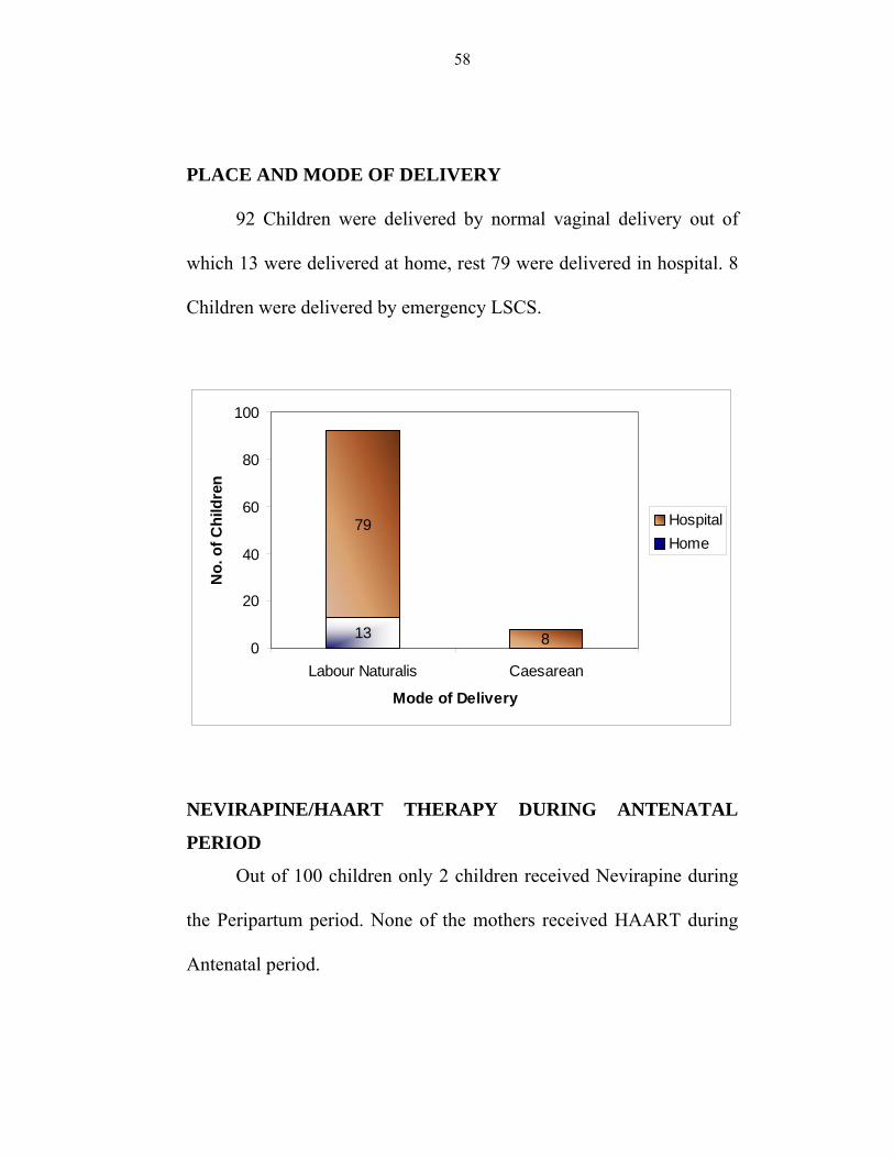
PLACE AND MODE OF DELIVERY
92 Children were delivered by normal vaginal delivery out of
which 13 were delivered at home, rest 79 were delivered in hospital. 8
Children were delivered by emergency LSCS.
13
79
80
20
40
60
80
100
Labour Naturalis Caesarean
Mode of Delivery
No.
of C
hild
ren
HospitalHome
NEVIRAPINE/HAART THERAPY DURING ANTENATAL
PERIOD
Out of 100 children only 2 children received Nevirapine during
the Peripartum period. None of the mothers received HAART during
Antenatal period.

59
FEEDING
82 Children received breast milk for the initial 4 months. 9
children received cow’s milk and 8 children received both cow’s milk
and breast milk. Only one child was completely replaced with milk
substitute.
82
9 81
0
10
20
30
40
50
60
70
80
90
No.
of C
hild
ren
Breast Milk Cow's Milk Mixed Replacement
Feeding
DEVELOPMENTAL HISTORY
98 Children presented with normal developmental history only 2
children had delayed motor milestones.

60
NUTRITIONAL STATUS
The nutritional status of the children was classified as per IAP
classification of malnutrition as follows.
Nutritional Status* Weight for age (% of expected) No. of Children
Normal > 80 15
Grade I PEM 71-80 17
Grade II PEM 61-70 27
Grade III PEM 51-60 26
Grade IV PEM < 50 15
1517
27 26
15
0
5
10
15
20
25
30
Normal Grade I PEM Grade II PEM Grade III PEM Grade IVPEM
Nutritional Status
No.
of C
hild
ren
61
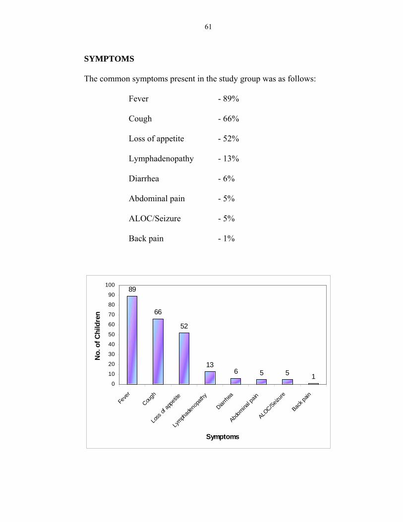
SYMPTOMS
The common symptoms present in the study group was as follows:
Fever - 89%
Cough - 66%
Loss of appetite - 52%
Lymphadenopathy - 13%
Diarrhea - 6%
Abdominal pain - 5%
ALOC/Seizure - 5%
Back pain - 1%
89
66
52
136 5 5 1
0
10
20
30
40
50
60
70
80
90
100
Feve
r
Cough
Loss
of ap
petite
Lymph
aden
opath
y
Diarrhe
a
Abdom
inal p
ain
ALOC/Seiz
ure
Back p
ain
Symptoms
No.
of C
hild
ren

62
SYSTEM EXAMINATION
The clinical presentations was Lymphadenopathy mostly
cervical (11%) and axillary in 1%. In respiratory system signs of
pneumonia was present in 60%, 2 children had pleural effusion, 2 had
bronchiectasis, 2 children had consolidation/collapse. Thus respiratory
system was involved in 66% of children.
Gastrointestinal system (9%) 5 children presented with
hepatomegaly, 5 children presented with hepatosplenomegaly and 4
children had ascites.
Oral lesion (24%) 14 children had Oral candidiasis, 4 children
had Apthous ulcers, 3 children had Angular stomatitis and 3 children
had Herpes simplex.
Skin Lesions (15%) 6 children had Herpes zoster, 4 children had
Molluscum contagiosum, 2 children had scabies, 2 had extensive
impetigo and 1 child had rash.
Central Nervous System (7%) Meningitis are present in 7
children out of which 1 child had Cranial nerve involvement.
Spinal involvement was present in 1 child (Pott’s spine).

63
SYSTEM INVOLVEMENT PRESENTATION
Respiratory System (66%)
Pneumonia – 60% Pleural Effusion – 2% Bronchiectasis – 2% Consolidation/Collapse -2%
Gastrointestinal system (9%)
Hepatomegaly – 5% Hepatosplenomegaly – 5% Ascites – 4%
Lymphadenopathy (12%)
Cervical – 11% Axillary – 1%
Oral lesion (24%)
Oral candidiasis – 14% Apthous ulcers – 4% Angular stomatitis – 3% Herpes simplex – 3%
Skin Lesions (15%)
Herpes zoster-6% Molluscum contagiosum-4% Scabies-2% Extensive impetigo-2% Rash-1%
Central Nervous System (7%)
Meningitis -7% Cranial nerve involvement -1%
Spinal involvement (1%) Pott’s spine – 1%

64
EXTRAPULMONARY MANIFESTATIONS:
26 Children had extrapulmonary system involvement:
Lymphadenopathy - 13
TB Meningitis - 6
TB abdomen - 4
Disseminated TB - 2
TB Spine - 1
SPUTUM POSITIVITY
Sputum for AFB Number
Sputum Positive 9
Sputum Negative 91
CORRELATION BETWEEN SPUTUM POSITIVITY AND
CD4 COUNT
CD4 count Sputum Positive Sputum Negative
0-100 1 13
101-200 - 7
201-300 1 11
>300 7 60
Total 9 91
65
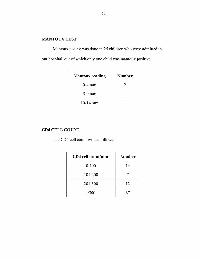
MANTOUX TEST
Mantoux testing was done in 25 children who were admitted in
our hospital, out of which only one child was mantoux positive.
Mantoux reading Number
0-4 mm 2
5-9 mm -
10-14 mm 1
CD4 CELL COUNT
The CD4 cell count was as follows:
CD4 cell count/mm3 Number
0-100 14
101-200 7
201-300 12
>300 67

66
WHO CLINICAL STAGING
WHO STAGE NUMBER
Stage III 87
Stage IV 13
TOTAL 100
The CD4 count correlation with WHO clinical staging was as
follows:
CD4 COUNT WHO STAGE
STAGE III STAGE IV
0-100 9 5
101-200 6 2
201-300 12 2
>300 59 5
Total 86 14

67
CHEST X-RAY FINDINGS
CHEST X-RAY FINDINGS NUMBER
Lobar/segmental infiltrates 41
Diffuse infiltrates 25
Miliary mottling 5
Pleural effusion 4
Hilar adenopathy 4
Bronchiectasis 3
Normal 18
Total 100
41
25
5 4 4 3
18
0
5
10
15
20
25
30
35
40
45
Loba
r/seg
men
tal
infil
trate
s
Diff
use
infil
trate
s
Mili
ary
mot
tling
Ple
ural
effu
sion
Hila
rad
enop
athy
Bro
nchi
ecta
sis
Nor
mal
Chest X-Ray Findings
No.
of C
hild
ren
68
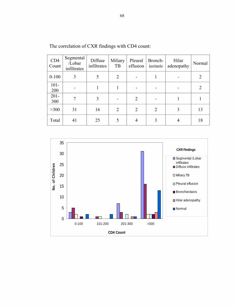
The correlation of CXR findings with CD4 count:
CD4 Count
Segmental /Lobar
infiltrates
Diffuse infiltrates
Miliary TB
Pleural effusion
Bronch-iectasis
Hilar adenopathy Normal
0-100 3 5 2 - 1 - 2
101-200 - 1 1 - - - 2
201-300 7 3 - 2 - 1 1
>300 31 16 2 2 2 3 13
Total 41 25 5 4 3 4 18
0
5
10
15
20
25
30
35
0-100 101-200 201-300 >300
CD4 Count
No.
of C
hild
ren
Segmental /LobarinfiltratesDiffuse infiltrates
Miliary TB
Pleural effusion
Bronchiectasis
Hilar adenopathy
Normal
CXR Findings

69
DISCUSSION
Among the 512 patients infected with HIV infection, 100
children were having TB co-infection, with a prevalence of 19.53%.
This corresponds to other studies like Merchant et al 59 where 29.5%
were co-infected. Lodha et al 60 (59.1%) and Dhurat et al 61 (67.5%)
have also recorded the increased prevalence of HIV-TB co-infection.
Out of 100 seropositive children with TB co-infection, 62%
were males and 38% were females. This indicates that the male:female
ratio is 1.6:1. The prevalence of HIB-TB co-infection is 70% in the age
group of 6-12 years. This correlates well with the observations of
Shahab, Afzal et al 62 where the male:female ratio was 2.3:1 and
prevalence of coinfection was 72.4% in the age group of 1-9 years.
History of contact with TB was present in 38% as against 47%
in the results of Shahab, Afzal et al 62 . 73% of the children in our
study population were taken care by their parents (father 55% and
mother 18%) rest 27% were orphaned and taken care by their grand
parents.
97 Children acquired infection by perinatal transmission, one
child has infected following blood transfusion. In two children the
70
actual mode of transmission could not be identified. Verghese VP,
Cherian et al63 study reported that perinatal transmission was the
predominant root of transmission (87%) followed by blood transfusion
(10%) and the mode of transmission could not be ascertained in rest
3%. Blood and blood products remain an important source of infection
in 10-30% of total cases in developing countries60.
The children in stage IV were severely malnourished and
belonged to Grade III & Grade IV PEM. All children in our study
population were on regular Cotrimoxazole prophylaxis.
Analysis of the clinical manifestations of TB in seropositive
children
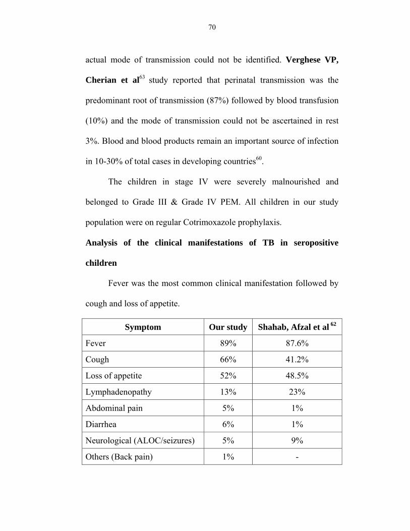
Fever was the most common clinical manifestation followed by
cough and loss of appetite.
Symptom Our study Shahab, Afzal et al 62
Fever 89% 87.6%
Cough 66% 41.2%
Loss of appetite 52% 48.5%
Lymphadenopathy 13% 23%
Abdominal pain 5% 1%
Diarrhea 6% 1%
Neurological (ALOC/seizures) 5% 9%
Others (Back pain) 1% -

71
Systemic examination revealed respiratory system findings and
Lymphadenopathy were present in 66% and 12% respectively. About
9% had hepatomegaly/hepatosplenomegaly with ascites in 4% of the
children. 24% of children presented with oral lesions(mostly oral
candidiasis 14%) and skin lesions was present in 15%.
General/system examination Our study Shahab, Afzal et al 62
Respiratory system findings 66% 36.5%
Lymphadenopathy 13% 28%
Hepatomegaly 5% -
Hepatosplenomegaly 4% 11.5%
Skin lesions 15% 3%
Oral candidiasis 14% 30%
CNS involvement 7% 5%
Extrapulmonary TB was seen in 26% of children in our study
which includes Lymphadenopathy 13%, TB Meningitis 6%, TB
abdomen 4%, Disseminated TB 2% and TB spine 1%. This finding
corresponds to the earlier observations of Dhurat et al61 and
Merchant et al59.
The sputum positivity in our study shows that only 9% of the
children are sputum positive. Sputum culture for M.Tuberculosis
72
remains the gold standard for the diagnosis of Pulmonary TB. In
resource poor countries the diagnosis is heavily dependent on the
sputum AFB smear. HIV infected children have reported to have a
lower yield on AFB smears as observed in Mukadi et al42, Garay JE
et al44. Verghese VP et al63, reported only 4% smear positive cases in
HIV children.
In our study Mantoux was done in 25 children who were
admitted in our hospital and only one child was Mantoux positive with
more than 10 mm induration. This correlates well with study by
Mukadi et al42 which shows an increased incidence of anergy in HIV
infected and AIDS patients.
In our study CD4 cell count less than 300 was observed in 33
children. In these children the predominant X-ray lesions were Hilar
adenopathy, lower lobe infiltrations, diffuse infiltrates and miliary
mottling. Upper lobe infiltrates was common with higher CD4 count
mean 350. These finding correlates well with Verghese VP et al63 and
Shahab, Afzal et al 62.
The most important finding of this study is the impact of HIV
related immunosuppression among children with TB. 8 children with
CD4 % of less than 10% died during 6 months of therapy. This finding
correlates well with the study of Mukadi et al42

73
CONCLUSION
Tuberculosis is a common opportunistic infection among HIV
seropositive individuals.
Males are commonly affected than females.
Tuberculosis occurs early in HIV infection even before CD4 count
falls to very low levels.
Fever, cough, loss of appetite and loss of weight are the most
common symptoms observed in HIV-TB co-infection.
Extrapulmonary TB is present in 30% of HIV seropositive children.
Sputum negativity was more commonly seen in TB of HIV
seropositive individuals.
Mantoux anergy was observed with lower CD4 counts.
This study documents the importance of HIV infection as an
independent risk factor for the development of TB in children and
also demonstrate that HIV related immunosuppression as critical
risk factor for mortality in this population.
With the conventional sputum positivity and Tuberculin test not
providing an adequate diagnostic help, familiarity with clinico
radiological spectrum of TB and HIV co-infection will help in early
diagnosis and improve survival among HIV seropositive children.

Sl. N
o.
Age
(yrs
.)
Sex
(M/F
)
Wei
ght (
kg.)
Mod
e of
Tr
ansm
issi
on
AN
C
POD
/MO
D
Feed
ing
Con
tact
His
tory
Soci
o-Ec
onom
ic
Stat
us
Clin
ical
Fea
ture
s
Syst
em
Exam
inat
ion
Sput
um fo
r AFB
MTX
CXR
CD
4 C
ount
FNA
C
USG
Abd
omen
CT
Scan
, CSF
, O
ther
s
WH
O S
tage
ATT
CA
T
Rx
Out
com
e
Ctx
1 12 M 17 MTC - I/LN BF - ↓ 1,2 IV - - UZ 577 (17%) - - - III P I C
2 9 F 16 MTC - I/CS BF - ↓ 1,6 VII - - LZ 89 (10%) - - CS IV EP I C
3 7 M 17 MTC - I/LN BF + ↓ 1,2,3 IV - - LZ 374 (14%) - - - III P I C
4 4 1/2 F 14.5 MTC - I/LN BF - ↓ 1,2 IV - - UZ 693 (19%) - - - III P I T
5 10 M 11 MTC - I/LN BF + ↓ 4 I - - LZ 283 (9%) + - - III EP II DE
6 12 M 25 MTC - I/LN BF + ↓ 1,3,6 VI - - LZ 28 (5%) - - CSF+ IV EP I D
7 7 F 17 MTC - I/LN BF - ↓ 1,2 IV - - LZ 777 (11%) - - - III P I C
8 6 M 15 MTC - I/LN BF + ↓ 1,2,3 IV - - M 98 (4%) - - - III P I C
9 4 F 14.5 MTC NVP I/LN CM - ↓2,3, 5,6 IV, V - + PE 1070
(16%) - HSM, PE, A - III EP I C
10 11 F 20 MTC - I/LN BF - ↓ 1,2,5 V - - B 671 (11%) - HEP,
A, N - IV EP I C
11 9 M 18 MTC - I/LN Mix - ↓ 1,2,6 IV, V - - DI 81 (4%) - HEP,
A
CT Chest,
BIV EP I C
12 3 1/2 M 10 MTC - I/LN Mix + ↓ 1,2 IV - - DI 889 (13%) - HEP - III P I C
13 5 F 11 MTC - I/LN BF - ↓ 1,2 IV, V - - B 282 (10%) - HSM - III P I C
MASTER CHART

14 11 1/2 M 13 MTC - I/LN BF - ↓ 1,2,6 II, V - - LZ 680 (15%) - - CT
Norm III P I C
15 11 M 19.6 MTC - H/LN BF - ↓ 1,2 V - - DI 82 (8%) - - - III P I C
16 12 M 20 MTC - I/LN BF - ↓ 1,2,3 IV - - LZ 356 (26%) - - - III P I C
17 9 M 17 MTC - I/LN BF - ↓ 1,2,3 IV - - LZ 188 (16%) - - - III P I C
18 8 M 18.6 MTC - I/LN Mix - ↓ 4 I, IV - - UZ 604 (15%) + - - III P I, II T
19 11 M 20 MTC - H/LN BF - ↓ 1,2,5 II, IV - - LZ 252 (12%) - Norm - III P I C
20 9 M 18 MTC - I/LN BF - ↓ 1,2,3 IV - - LZ, MZ
1024 (12%) - - - III P I C
21 10 M 18 MTC - I/LN BF - ↓ 1,2,3 IV - - LZ 89 (3%) - - - III P I C
22 4 M 10 MTC - I/LN BF + ↓ 1,2,3 IV - - LZ 206 (11%) - - - III P I D
23 4 F 15 MTC - I/LN BF - ↓ 1,2 IV - - LZ 773 (15%) - - - III P I C
24 9 F 18 MTC - I/LN BF - ↓ 1,2,3 IV - - LZ 656 (11%) - - - III P I C
25 11 F 20 MTC - I/LN BF + ↓ 1,2,3 IV - - M 805 (25%) - - - III P, EP II C
26 12 F 29 MTC - I/LN BF + ↓ 1,2,3 IV - - LZ 269 (12%) - - - III P I T
27 6 M 12 MTC - I/LN BF + ↓1,2, 3,4 I, II - - - 294
(13%) + - - III EP I C
28 11 M 24 MTC - I/LN BF - ↓ 1,3 II, V - - - 16 (3%) - HEP CT + IV EP I D
29 9 M 18 MTC - H/LN BF + ↓1,2, 3,4 I, II - - - 1019
(37%) + - - III EP I C
30 3 F 10 MTC - I/LN BF - ↓ 1,2,3 II, IV + - LZ 853 (17%) - HSM - III P I C
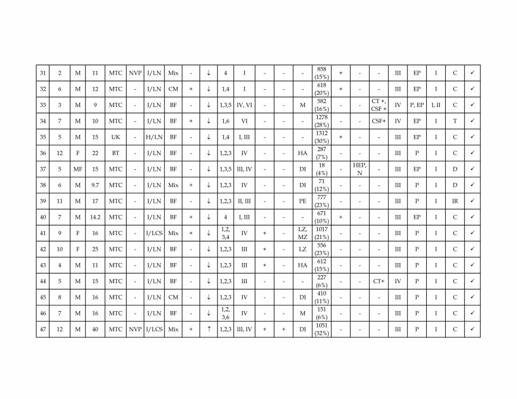
31 2 M 11 MTC NVP I/LN Mix - ↓ 4 I - - - 858 (15%) + - - III EP I C
32 6 M 12 MTC - I/LN CM + ↓ 1,4 I - - - 618 (20%) + - - III EP I C
33 3 M 9 MTC - I/LN BF - ↓ 1,3,5 IV, VI - - M 582 (16%) - - CT +,
CSF + IV P, EP I, II C
34 7 M 10 MTC - I/LN BF + ↓ 1,6 VI - - - 1278 (28%) - - CSF+ IV EP I T
35 5 M 15 UK - H/LN BF - ↓ 1,4 I, III - - - 1312 (30%) + - - III EP I C
36 12 F 22 BT - I/LN BF - ↓ 1,2,3 IV - - HA 287 (7%) - - - III P I C
37 5 MF 15 MTC - I/LN BF - ↓ 1,3,5 III, IV - - DI 18 (4%) - HEP,
N - III EP I D
38 6 M 9.7 MTC - I/LN Mix + ↓ 1,2,3 IV - - DI 71 (12%) - - - III P I D
39 11 M 17 MTC - I/LN BF - ↓ 1,2,3 II, III - - PE 777 (23%) - - - III P I IR
40 7 M 14.2 MTC - I/LN BF + ↓ 4 I, III - - - 671 (10%) + - - III EP I C
41 9 F 16 MTC - I/LCS Mix + ↓1,2, 3,4 IV + - LZ,
MZ1017
(21%) - - - III P I C
42 10 F 25 MTC - I/LN BF - ↓ 1,2,3 III + - LZ 556 (23%) - - - III P I C
43 4 M 11 MTC - I/LN BF - ↓ 1,2,3 III + - HA 612 (15%) - - - III P I C
44 5 M 15 MTC - I/LN BF - ↓ 1,2,3 III - - - 227 (6%) - - CT+ IV P I C
45 8 M 16 MTC - I/LN CM - ↓ 1,2,3 IV - - DI 410 (11%) - - - III P I C
46 7 M 16 MTC - I/LN BF - ↓1,2, 3,6 IV - - M 151
(6%) - - - III P I C
47 12 M 40 MTC NVP I/LCS Mix + ↑ 1,2,3 III, IV + + DI 1051 (32%) - - - III P I C

48 8 F 10 MTC - H/LN BF + ↓ 1,2,3 II, III - + LZ 260 (8%) - - - IV P I C
49 5 M 11.2 MTC - I/LN CM - ↓ 1,2,3 IV + - LZ 505 (8%) - - - III P I C
50 4 M 11.5 MTC - H/LN BF + ↓ 1,2,3 IV - - UZ 575 (25%) - - - III P I C
51 10 F 19.5 MTC - H/LN BF - ↓ 1,2,3 IV - - LZ 425 (23%) - - - III P I C
52 5 F 13 MTC - I/LN CM - ↓ 1,2,3 IV - - UZ 538 (15%) - - - III P I C
53 11 M 24 MTC - I/LN BF - ↓ 1,2,3 IV - - LZ 237 (16%) - - - III P I C
54 8 M 15 MTC - I/LN BF - ↓ 1,2,3 IV + - LZ 205 (21%) - - - III P I C
55 10 F 20 MTC - I/LN BF - ↓ 1,2,3 IV - - - 692 (29%) - - - III P I T
56 5 M 15 MTC - I/LN BF + ↓ 1,2,3 III - - DI 212 (7%) - - - III P I C
57 7 M 16 MTC - I/LN BF + ↓ 1,2,3 IV - - LZ 609 (18%) - - - III P I C
58 10 F 14.8 MTC - I/LN BF + ↓ 1,2,3 IV + - UZ 918 (25%) - - - III P I C
59 10 F 14 MTC - I/LN BF + ↓ 1,2,3 II, III, IV - - DI 106
(10%) - - - III P I C
60 5 M 14 MTC - I/LN Mix - ↓ 1,2,3 III, IV - - UZ 893 (28%) - - - III P I C
61 3 M 10 MTC - I/LN BF - ↓ 1,2,3 III, IV - - PE 945 (12%) - - - III P I C
62 6 M 11 MTC - H/LN BF + ↓ 4 I - - - 576 (17%) + HEP,
A, N - III EP I C
63 12 F 28 MTC - I/LCS CM + ↓ 1,2,3 III, IV + - DI 416 (15%) - - - III P I IR
64 7 M 19 MTC - I/LN BF + ↓ 3,5 IV, V - - DI 106 (4%) - - - III EP I C

65 5 M 10.5 MTC - I/LCS CM + ↓ 1,2,3 III, IV - - DI 624 (18%) - - - III P I IR
66 12 M 19 MTC - I/LN BF - ↓ 1,2,3 II, IV - - UZ 1299 (20%) - - - III P I C
67 10 M 23 MTC - I/LN Mix + ↓ 1,2,3 II, IV - - DI 135 (6%) - - - III P I T
68 11 F 18 MTC - H/LN BF + ↓ 1,3,6 VI - - - 117 (4%) - N CSF+ IV EP I D
69 4 F 9 MTC - I/LN BF - ↓ 1,2,3 III, IV - - HA 1524 (32%) - - - III P I C
70 7 M 18 MTC - I/LCS BF - ↓ 1,2,3 IV - - MZ 378 (19%) - - - III P I C
71 5 M 13.6 MTC - I/LN CM - ↓ 1,2,3 III, IV - - LZ 1183 (19%) - - - III P I C
72 11 F 12.5 MTC - I/LN BF + ↓ 1,2,3 III, IV - - DI 512 (14%) - - - III P I C
73 5 M 15 MTC - I/LN BF + ↓ 1,2,3 II, III - - - 382 (16%) - - - III P I C
74 8 F 14 MTC - I/LN BF + ↓ 1,2,3 IV + - DI 266 (16%) - - - III P I C
75 3 M 10 MTC - H/LN BF + ↓ 1,2,3 IV - - DI 1163 (34%) - - - III P I C
76 3 F 8 MTC - I/LN BF - ↓1,2, 3,6 IV - - UZ 905
(19%) - - - III P I C
77 10 F 21 MTC - H/LN BF + ↓ 1,2,3 IV - - DI 698 (29%) - - - III P I C
78 8 F 13 MTC - I/LCS BF + ↓ 1,2,3 I, III - - DI 386 (20%) - - - III P I C
79 6 F 16 MTC - I/LN BF - ↓ 4 IV - - - 600 (13%) + - - III EP I C
80 5 M 13 MTC - I/LN BF - ↓ 1,2,3 III, V - - DI 449 (17%) - - - III P I C
81 11 M 20 UK - I/LN BF - ↓ 2,3 IV - - - 10 (2%) - HEP,
N - IV EP I D

82 10 M 16 MTC - I/LN BF - ↓ 1,2,3 IV - - HA 81 (6%) - - - III P I T
83 5 F 15 MTC - I/LN BF - ↓ 1,2,3 IV - - DI 539 (12%) - - - III P I C
84 8 M 17 MTC - I/LN CM - ↓ 3,4 I - - - 554 (13%) + - - III EP I C
85 6 M 16 MTC - I/LN BF - ↓ 1,2,3 IV - - HA 495 (23%) - - - III P I C
86 10 M 26 MTC - I/LN BF - ↓ 1,3,4 I - - - 743 (16%) + - - III P I C
87 8 F 16 MTC - I/LN BF + ↓ 1,2,3 IV - - DI 574 (17%) - - - III P I C
88 9 F 18 MTC - I/LN BF + ↓ 1,2,3 IV - - DI 595 (29%) - - - III P I C
89 11 F 26 MTC - I/LN BF + ↓1,2, 3,4 I - - - 342
(21%) + - - III EP I C
90 5 M 14 MTC - I/LN BF + ↓ 1,2 IV - - B 615 (19%) - - - III P I IR
91 11 M 17 MTC - I/LN BF - ↓ 1,2 IV - - DI 777 (29%) - - - III P I C
92 3 F 9 MTC - I/LN BF - ↓ 1,2 IV - - LZ 576 (17%) - - - III P I C
93 7 M 38 MTC - I/LN BF - ↓ 1 IV - - TE 791 (32%) - - - III P I C
94 5 M 15 MTC - I/LCS BF - ↓ 1,2 IV - - DI 372 (18%) - - - III P I C
95 2 M 8 MTC - I/LN BF - ↓ 1,2 IV - - DI 890 (28%) - - - III P I T
96 9 M 18 MTC - I/LN BF - ↓ 1,2 IV - - HA 911 (25%) - - - III P I C
97 10 F 16 MTC - I/LN BF - ↓ 1,2 IV - - UZ 909 (20%) - - - III P I C
98 11 M 15 MTC - I/LN BF - ↓ 1,2,3 III + - M 31 (8%) - - - III P I D

99 11 F 12 MTC - I/LN BF + ↓ 1,2,3 IV - - MZ 84 (10%) - - - III P I C
100 9 F 15 MTC - I/LN BF + ↓ 1,2,3 III, VI - - - 180 (11%) - - CSF+ IV EP I C
Abbreviations to Master Chart:
MTC - Mother to Child System Examination USG Abdomen
BT - Blood Transmission I - Lymph Nodes HEP - Hepatomegaly
UK - Unknown II - Skin HSM - Hepatosplenomegaly
NVP - Nevirapine III- Oral Cavity N - Nodes
POD - Place of Delivery IV - Respiratory System Norm. - Normal
MOD - Mode of Delivery V - Abdomen A - Ascites
I/LN - Institution / Labour Naturalis VI - CNS, Others ATT
I/LCS - Institution / Lower Caesarean Section CXR P - Pulmonary
H/LN - Home / Labour Naturalis UZ - Upper Zone EP - Extra Pulmonary
BF - Breast Feed MZ - Middle Zone Rx Outcome
CM - Cow's Milk LZ - Lower Zone C - Cured
Mix - Mixed M - Miliary D - Death
Clinical Features PE - Pleural Effusion T - Transfer
1 - Fever HA - Hilar Adenopathy DF - Default
2 - Cough B - Bronchiectasis IR - Irregular
3 - Loss of appetite DI - Diffuse Infiltrates Ctx - Cotrimoxazole Prophylaxis
4 - Lymphadenopathy CS- Caries Spine
5 - Abdominal Pain FNAC

6 - Seizures, Back Pain, etc. + : Granulomatous changes

AR
T
-
-

-

-
-
-
-
-
-
-

-
-
-
-
-
-
-
-
-
-

-
-
-
-
-
-
-
-
-
-

-
-
-
-
-
-
-
-
-


BIBILIOGRAPHY
1. Hershkovitz I, Donoghue HD, Minnikin DE, Besra GS, Lee OY-
C, et al. (15 October 2008). "Detection and Molecular
Characterization of 9000-Year-Old Mycobacterium tuberculosis
from a Neolithic Settlement in the Eastern Mediterranean.". PLoS
ONE 3 (10): e3426. doi:10.1371/journal.pone.0003426.
2. Zink A, Sola C, Reischl U, Grabner W, Rastogi N, Wolf H,
Nerlich A (2003). "Characterization of Mycobacterium
tuberculosis complex DNAs from Egyptian mummies by
spoligotyping". J Clin Microbiol 41 (1): 359–67.
doi:10.1128/JCM.41.1.359-367.2003. PMID 12517873
3. Hippocrates. Aphorisms. Accessed 7 October 2006.
4. Zur Pathogenie der Impetigines. Auszug aus einer brieflichen
Mitteilung an den Herausgeber. [Müller’s] Archiv für Anatomie,
Physiologie und wissenschaftliche Medicin. 1839, page 82.
5. Nobel Foundation. The Nobel Prize in Physiology or Medicine
1905. Accessed 7 October 2006.
6. Tuberculosis Encyclopedia Britannica, 11th ed. Disseminated
tuberculosis NIH Medical Encyclopedia. Accessed 9 October
2006.
7. Bhansali SK. "Abdominal tuberculosis. Experiences with 300
cases". Am. J. Gastroenterol. 67 (4): 324–37. PMID 879148
8. Kumar, Vinay; Abbas, Abul K.; Fausto, Nelson; & Mitchell,
Richard N. (2007). Robbins Basic Pathology (8th ed.). Saunders
Elsevier. pp. 516-522 ISBN 978-1-4160-2973-1
9. Onyebujoh, Phillip and Rook, Graham A. W. World Health
Organization Disease Watch: Focus: Tuberculosis. December
2004. Accessed 7 October 2006.
10. Houben E, Nguyen L, Pieters J (2006). "Interaction of pathogenic
mycobacteria with the host immune system". Curr Opin
Microbiol 9 (1): 76–85. doi:10.1016/j.mib.2005.12.014. PMID
16406837
11. Herrmann J, Lagrange P (2005). "Dendritic cells and
Mycobacterium tuberculosis: which is the Trojan horse?". Pathol
Biol (Paris) 53 (1): 35–40. PMID 15620608.
12. Kaufmann S (2002). "Protection against tuberculosis: cytokines, T
cells, and macrophages". Ann Rheum Dis 61 Suppl 2: ii54–8.
PMID 12379623
13. Houben E, Nguyen L, Pieters J (2006). "Interaction of pathogenic
mycobacteria with the host immune system". Curr Opin

Microbiol 9 (1): 76–85. doi:10.1016/j.mib.2005.12.014. PMID
16406837
14. Grosset J (2003). "Mycobacterium tuberculosis in the
extracellular compartment: an underestimated adversary".
Antimicrob Agents Chemother 47 (3): 833–6.
doi:10.1128/AAC.47.3.833-836.2003. PMID 12604509
15. Nelson Text book of Pediatrics 18th edition volume-I
16. Centers for Disease Control and Prevention (CDC), Division of
Tuberculosis Elimination. Core Curriculum on Tuberculosis:
What the Clinician Should Know. 4th edition (2000). Updated
August 2003.
17. Nahid P, Pai M, Hopewell P (2006). "Advances in the diagnosis
and treatment of tuberculosis". Proc Am Thorac Soc 3 (1): 103–
10. doi:10.1513/pats.200511-119JH. PMID 16493157
18. Pai M, Zwerling A, Menzies D (June 2008). "Systematic Review:
T-Cell-Based Assays for the Diagnosis of Latent Tuberculosis
Infection: An Update". Ann. Intern. Med. 149 (3): 1–9. PMID
18593687
19. Reddy JR, Kwang J, Lechtenberg KF, Khan NC, Prasad RB,
Chengappa MM (January 2002). "An immunochromatographic
serological assay for the diagnosis of Mycobacterium

tuberculosis". Comp. Immunol. Microbiol. Infect. Dis. 25 (1): 21–
7. doi:10.1016/S0147-9571(01)00016-9. PMID 11831744.
20. HIV and TB: A guide for counsellors, New Delhi, Ministry of
Health and Family welfare, NACO and Government of India.
21. Abrams EJ. Opportunistic Infections and other clinical
manifestations of HIV disease in children. Pediatri Cli North Am
2000; 47:79-108
22. Mumbai Districts AIDS Control Society, Training manual for
Doctors, New Delhi, NACO.
23. 2006 Report the committee on Infectious Diseases, 27th edition
ELK Grove village, American Academy of Pediatrics 2006, PP
380-381; P 382
24. HIV infection codes and official guidelines for coding and
reporting ICD-9-CM, MMWR Recomm. Rep 1994; 43(RR-12);
1-19
25. NACO 2003-available from: www.nacoonline.org
guidelines/HIV-TB guidelines.
26. Corbett. Arch Intern Med 2003;163:1009-21. (s)
27. Raviglione MC, sinder DE, Global epidemiology of tuberculosis.
Morbidity and mortality of a worldwide epidemic. JAMA 1995;
273:220-6

28. Paranjape RS, Tripathy SP, Menon PA etal. Increasing trend of
HIV seroprevalence among pulmonary tuberculosis patients in
Pune. Indian Journal Med Res 1997:106:207-11.
29. Merchant RS, Shroff RC. HIV seroprevalence in disseminated
tuberculosis and chronic diarrhea. Indian Pediatr;35:883-7
30. Husson RN. Tuberculosis. In: Pizzo PA Wilfert CM (eds)
Pediatric AIDS. 3rd edition. 1998;139-56
31. Misra SN, Sengupta D. AIDS in India:Southeast Asian J Trop
Med Public Health 1998;29:37-6
32. Sharma SK, Mohan A. Co-infection of HIV and TB Indian
perspective. Indian J Tuberc 2004;51:5-16
33. Bornschlegel K, Thomas P. Tuberculosis in children born to HIV
infected human in New York City. XI International Conference
on AIDS, Vancouvr, Canada. 1996.
34. Lederman MM, Georges DL, Kusner DJ. M.tuberculosis and its
purified protein derivative activate expression of HIV. J Acquire
immune Defic Syndr 1994; 7:727-33.
35. Zhang Y, Nakata K. M tuberculosis enhances HIV-1 replication
by transcriptional activation at the long terminal repeat. J Clin
Invest 1995;95:2324-31

36. Whalen C, Copathogenicity of tuberculosis and HIV disease. Clin
Infec. Dis (In Press)
37. Harries AD. TB and HIV infection in developing countries.
Lancet 1990; 335:387-90
38. Folks TM, Kinter A, Fauci AS. Cytokine-induced expression of
HIV-1 in a chronically infected promonocyte cell line. Science;
238:800-2
39. Osoborn L, Kunkel S. TNF α and interleukin 1 stimulate the HIV
enhancer by activation of nuclear factor kB. Proc Natl Acad Sci
USA; 86:2336-40
40. Harries AD, Kangombe C et al. The scourge of HIV-related TB a
cohort study in a district hospital in Malawi. Ann Trop Med
Parasitol 1997;91:771-6
41. Perlman DC, Nelson ET et al. Variation of chest radiographic
patterns in pulmonary tuberculosis by degree of HIV-related
immuno suppression. Clin Infect Dis 1997;25:242-6
42. Mukadi YD, Coulibaly IM et al. Impact of HIV infection on the
development clinical presentation and outcome of tuberculosis
among children in Abidjan, Cote d’Ivoire. AIDS 1997;11:1151-8
43. Topley JM, Bamber S. TB meningitis and co-infection with HIV.
Ann Trop Pediatr 1998;18:261-6

44. Garay JE. Clinical presentation of pulmonary tuberculosis in
under 10s and differences in AIDS-relatedcases: a cohort study of
115 patients. Trop Doctor 1997;27:139-42
45. Jeena PM, Mitha T, Bamber S, Wesly A, Coutsoudis A. Effects of
the HIV on TB in children. Tuberc Lung Dis 1996;77:437-43
46. American Academy of Pediatrics, Committee on Pediatric AIDS.
Evaluation and medical treatment of HIV-exposed infant.
Pediatrics 1997;99:909-17
47. Johnson JL, Okwera et al. Impact of HIV-1 infection on the initial
bacteriologic and radiographic manifestations of pulmonary TB in
Uganda. Int J Tuberc Lung Dis 1998;2:397-404
48. Bekedam HJ, Kamenya A et al. Tuberculous lymphadenitis a
diagnostic problem in areas of high prevalence of HIV and TB.
Trans Roy Soc Trop Med HYg 1997;91:294-7
49. Centers for Disease Control and Prevention. Prevention and
treatment of TB among patients infected HIV. MMWR
1998;47(RR-20):1-58
50. Moore M, McCray E, Castro KG. Trends in drug-resistant TB in
United States, 1993-1996. JAMA 1997;278:833-7
51. CDC Initial therapy for tuberculosis in the era of multidrug
resistance. Recommendations of the advisory council for the

elimination of TB MMWR North Mortal wkly Rep 1993;42 (RR-
7):1-8
52. Bayer R, Wilkinson D. Directly observed therapy for TB: history
of an idea Lancet 1995;345:1545-1548
53. Fox W. Self administration of medications. A review of published
work and a study of problems. Bull int union Tuberc; 37:307-331
54. Chaisson RE, Holt EA et al. Six month supervised intermittent TB
therapy in Haitian patients with and without HIV infection. Am J
Respir Crit Care Med 1996;154:1034-8
55. Mitchison DA, Dickinson JM. Laboratory aspects of intermittent
drug therapy. Postgrad Med J; 47:737-741
56. Narita M, Ashkin D. Paradoxical worsening of TB following anti-
retroviral therapy in patients with AIDS. Am J Respir Crit Care
Med 1998;158:157-61
57. O’Brien KL, Ruff AJ, Lous MA et al. Bacillus Calmette-Guerin
complications in children born to HIV-1 infected women with a
review of the literature. Pediatrics 1995;95:414-8
58. Bhat G, Diwan V, Masona J. HIV, BCG and TB in children. J
Trop. Pediatr. 1993;39:219-23
59. Merchant RH, Oswal JS, et al, clinical profile of HIV infection.
Indian Pediatr 2001;38:239-246.

60. Lodha R, Singhal T, Jain Y, et al. Pediatric HIV infection in a
tertiary care centre in North India; early impression. Indian
Pediatr 2000;37:982-986.
61. Dhurat R, Manglani M, Sharma M, et al. Clinical spectrum of
HIV infection. Indian Pediatr 2000;37:831-836
62. Shahab T, Zoha MS, Afzal K et al. Prevalence of HIV in children
with TB. Indian Pediatr 2004;41:595-599.
63. Cherian T, Verghese PV. TB and HIV infection. Indian J Pediatr
2000;67:S47-S52.

PROFORMA
TB MANIFESTATIONS IN PEDIATRIC HIV
Name :
Age / Sex:
Address:
Parent/Guardians Name & Address:
Date of Sero positivity :
Place of Sero positivity :
Mode of Transmission :
a) Mother to child :
b) Blood transfusion:
c) Other modes (sexual, injection etc.,)
d) Unknown
Mother ANC History : HAART / NVP / NIL
Place of Delivery : Institution / Home
Mode of Delivery :
a) Labour Naturalis:
b) Caesarean section: Elective /
Emergency
c) Vacuum
d) Assisted forceps:

Infant feeding Duration :
a) Exclusive Breast feeding
b) Artificial feeding
c) Cow’s milk
d) Mixed
Developmental Milestones : Normal / Abnormal
FAMILY HISTORY
Members Age / Sex
HIV Status CD4
On ART Y/N
TB status Y/N
On ATT Y/N
Father
Mother
Brothers
Sisters
Others
Socio Economic H of Parent/Guardian:
Education Status:
Illiterate
Primary School
Higher Secondary
College
Employed : Yes/No
Occupation :
Per capita income :

Clinical Manifestations :
Fever :
Cough :
Breathlessness :
Loss of appetite :
Abdominal Pain :
Diarrhea :
Lymphadenopathy:
ALOC/ Seizures/mental deterioration:
Osteoarthritis :
Others
General Examination Height: Weight: MAC: BMI:
Built: (well) (moderate) (poor) (Febrile)
(pallor) ( cyanosis) (Tachypnoeic)
(pedal edema) (Jaundice) (others) Lymph nodes: Y/N (Region) (Nos:)
Cervical / Axillary / Others
(Tender/non tender) (Matted/Discrete)
Skin: (Normal) (PPE) (IBA) (Scabies) (Herpes Zoster) (Seb.derm) (Tinea) (xerosis) (others)
Oral Cavity (Normal) (Oral candidiasis) (OHL) (Hyper pigmentation) (Aphthous ulcer) (Herpes simplex) (Angular stomatitis) (others)
RS: (normal) (breath sounds) (added sounds) (others)
CVS: (Normal) (Abnormal)
Abdomen: (Normal) (Tenderness) (Organomegaly)
Genitals: Normal / Abnormal
CNS: (Normal) (Consciousness) (Neck stiffness) (plantar reflex) (DTR)
(Memory) (Motor weakness) (Sensory signs) (visual disturbance)

Immunisation H : BCG / OPV
Investigation :
Hb% :
TC :
DC :
ESR :
Sugar :
Urea :
Creatinine :
LFT :
1.
Sputum for AFB 2.
3.
Mantoux test :
CXR :
CD 4 Count / CD 4 % :
FNAC :
USG Abdomen :
CT Scan Brain :
CSF :
Others :
Δ : HIV C−
TB WHO Stage I / II / III / IV

TREATMENT HISTORY:
ATT Pulmonary Sputum +ve/-ve Extra pulmonary
I Category II III
• Date of Treatment Started Completed • Adherence
Regular / Irregular
• Treatment outcome
• Cure
• Treatment completed
• Treatment Failure
• Default
• Died
• Transfer out
Side Effects
Re infection
Relapse
Recurrence
Cotrimoxazole prophylaxis: Y / N Regular / Irregular
ART
Regimen : ZLN / ZLE / SLE / SLN
Date of treatment started:
Adherence: Good / Poor / occasionally missed
Reasons for missing:
Side Effects
IRIS:
![Sobotta...V. pulmonalis sinistra Pulmo sinister Oesophagus N. laryngeus recurrens N. vagus [X] Plexus pulmonalis A. pulmonalis dextra Pulmo dexter Bronchus principalis dexter Ductus](https://static.fdocuments.net/doc/165x107/5e5dc0c9b4bb8d4ca0665540/sobotta-v-pulmonalis-sinistra-pulmo-sinister-oesophagus-n-laryngeus-recurrens.jpg)