A Simple Hydrothermal Synthesis of Luminescent Carbon...
21
A Simple Hydrothermal Synthesis of Luminescent Carbon Quantum Dots from Different Molecular Precursors A dissertation Submitted for the partial fulfilment for the Degree of Master of Science in Chemistry Submitted by Ms. Sonali Paikaray & Ms. Priyanka Moharana Roll no: 411CY2027 Roll no: 411CY2024 Under the supervision of Dr. Sasmita Mohapatra Department of chemistry National Institute of Technology (NIT) Rourkela, Odisha, India. Pin: 769008
Transcript of A Simple Hydrothermal Synthesis of Luminescent Carbon...
A Simple Hydrothermal Synthesis of
Luminescent Carbon Quantum Dots from
Different Molecular Precursors
A dissertation
Submitted for the partial fulfilment for the
Degree of
Master of Science in Chemistry
Submitted by
Ms. Sonali Paikaray & Ms. Priyanka Moharana
Roll no: 411CY2027 Roll no: 411CY2024
Under the supervision of
Dr. Sasmita Mohapatra
Department of chemistry
National Institute of Technology (NIT)
Rourkela, Odisha, India.
Pin: 769008
Acknowledgement
We owe our cordial gratitude to my respected teacher and supervisor Dr. Sasmita
Mohapatra, Assistant Professor, Department of Chemistry, National Institute of Technology,
Rourkela, whose splendid guidance, authentic supervision, assiduous cooperation, moral
support and constant encouragement enabled me to make out our research problem in the
present form.
It is our great pleasure to acknowledge to Prof. B.G. Mishra, Head of the Chemistry
Department, National Institute of Technology, Rourkela for providing us the necessary
facilities for making this research work a success.
We are highly indebted to all our teachers of this department for their kind help and
expert suggestions. We express our profound gratitude to Ms. Swagatika Sahu &Mr. Smruti
Ranjan Rout for their ceaseless encouragement, immense help and hearty encouragement
during our project work.
We wish to thank all of friends for making our stay in this institute a memorable
experience.
Finally, we are honestly grateful to our family members for their endless love
unending support & blessing& GOD who has always been a source of our strength,
inspiration and our achievements.

Dr. Sasmita Mohapatra
Assistant Professor,
Department of Chemist
National Institute of Technology
Rourkela
Odisha-769008
CERTIFICATE
This is to certify that the dissertation entitled, “A Simple Hydrothermal Synthesis of
Luminescent Carbon Quantum Dots from Different Molecular Precursors” submitted by
Ms. SonaliPaikaray& Ms. Priyanka Moharana for the award of Master of Science in
Chemistry during the period of August 2011- May2013 in the Department of Chemistry,
National Institute of Technology, Rourkela, is a record of authentic work carried out by them
under my supervision. To the best of my knowledge, the matter embodied in this dissertation
has not been previously submitted for any degree in this/any other institute.
Date: Dr. Sasmita Mohapatra

Content
Introduction 1
A brief review of previous work 1-6
Objective of the present work 6
Experimental 6-7
Results and discussion 7-13
Conclusion 14
References 14-16

ABSTRACT
In this work,highly photoluminescent C-dots have been synthesised from different
precursors such as sucrose, ascorbic acid, citric acid and their combinations under similar
conditions to that of recently reported synthesis of C-dots from orange juice.The synthesized
carbon nanoparticles have been characterized by XRD, FTIR, UV, and fluorescence
measurements. The fluorescence quantum yields of carbon dots synthesized from different
precursors have been compared to verify the suitability of C-dots for different applications.
We have also thoroughly investigated the effect of excitation wavelength, pH, and electrolyte
concentration on luminescent properties.
Keywords: Fluorescent, C-dot, Quantum Yield, Hydrothermal Synthesis
1. Introduction
Photoluminescent materials have drawn increasing attention due to their promising
and diverse applications ranging from optoelectronics to biomedical fields, such as cellular
imaging, biosensing,and drug delivery.1-4
The most widely used fluorescent materials are
organic dyes and/or CdSe/ZnS quantum dots (QDs). However the photobleaching, quenching
of dye molecules, toxicity of QDs limit their applications in biomedical field.5,6
Most recently,
fluorescent carbon dots (C-dots) a new class of carbon-based nanomaterials serve as a
suitable alternative of these traditional fluorescent materials due to their excellent
luminescence properties, including large two-photon excitation cross-sections, lack of
blinking, high water solubility, low cytotoxicity, and excellent biocompatibility.7-12
During the past few years, much progress has been achieved inthe synthesis,
properties and applications of C-dots, as recently reviewed by Baker et al. and Li et al.13,
14Several top down approaches such as laser ablation,
8 electrochemical synthesis,
12arc
discharge15
have been proposed to produce fluorescent C-dots.These methods however
involve a non- selective exfoliation process, and therefore may require toxic reagents and
special equipment. On the other hand, bottom up approaches like carbonization of glucose,
sucrose, glycol, ascorbic acid, citric acids etc. have achieved significant attention for the
production of fluorescent C-dots.16-20
However most of these synthesis methods need several
steps and strong acids and post treatment with surface passivating agents8 are essential in
order to improve their water solubility and luminescence property. Urgently one-step, facile
approach with economicchemistry is required to produce self-passivatedphotoluminescent C-
dots.Among them, syntheses of amino-functionalized fluorescent carbon nanoparticles by
hydrothermal carbonization of chitosan at high temperature,21
microwave assisted
hydrothermal carbonization of sucrose,22
and carbonization of glucose in reverse micelle23
are
few scanty examples for the fabrication of fluorescent carbon nanoparticles without any
surface passivation. Inparticular, it is possible to control the size, shape, and
physicalproperties of the carbon nanoparticles by careful selection ofcarbon source and
surface modifier.24
Howeversubstantial production of C-dots with tailored composition,
structure, morphology andsize by a simple and cheap method is still challenging.
2. Review on various Synthetic methods

2
Synthetic approaches for C-dots can generally be classified into two main groups:
top-down and bottom-up methods. Top-down approaches, where larger carbon structures are
broken off to smaller c-dots include arc discharge, electrochemical oxidation and laser
ablation technique. Bottom-up methods include combustion/thermal/hydrothermal, supported
synthesis, microwave/ultrasonic, solution chemistry methods during which C-dots are formed
from molecular precursors.
2.1.Top-down approaches
2.1.1 Arc-discharge Methods
Ironically, C-dots were discovered by Xu et al. while purifying single-walled carbon
nanotubes (SWCNTs) derived from arc-discharge soot.15
They fractionated these
carbonaceous materials into a number of components which showed size-dependent
fluorescent properties. They oxidized thearc soot with 3.3 M HNO3 to introduce carboxyl
functionalgroups,which improve the hydrophilicity of the material, and then the sediment was
extracted with NaOH solution(pH 8.4) to produce a stable black suspension. The suspension
was separated by gel electrophoresis into SWCNTs, shorttubular carbons, and fluorescentC-
dots. The absence of characteristic C-H out of plane bending modes of
polyaromatichydrocarbons (PAHs) in FTIR spectrum indicated that the origin of thePL was
not derived from PAH sources.
2.1.2 Electrochemical Oxidation
For the first time Zhou et al. demonstrated the electrochemical synthesis of C-
dots.2They grew multiwall carbonnanotubes (MWCNTs) formed from scrolled
graphenelayerson carbon paper by chemical vapour deposition. They used these nanotubes as
the working electrode in an electrochemical cellconsisting of a Pt wire counter electrode and
a Ag/AgClO4reference electrode with degassed acetonitrile solution containing
0.1Mtetrabutylammonium perchlorate (TBA+ClO4
-)as the electrolyte.The C-dots produced
were spherical and exhibited λex -dependent PL. No C-dots were formedeither when carbon
paper without any MWCNTs was used. Chi and co-worker also synthesisedC-dots from
electrochemical oxidation of graphite rod with a Pt mesh counter electrode, and
Ag/AgClreferenceelectrode assembly in phosphate buffer solution (pH 7.0).25

3
Fig.1. Electrochemical production of C-dots from a graphite rod.25
2.1.3 Laser-Ablation Methods
Sun et al. Produced C-dots using Laser ablation technique, by hot-pressing a mixture
of graphite powder and cement, followed by stepwise baking, curing, and annealing under an
argon flow at 900oC and 75 kPa.
8 For better fluorescence they passivated the surface of C-
dots using different polymeric agents such as diamine-terminated poly(ethylene
glycol),8poly(propionyl-ethylenimine-co-ethylenimine).
9 Then highly fluorescent pure C-dots
were separated by dialysis against water, followed by a centrifugation step.26
A slightly
modified technique using 13
C powder and more rigorous control was introduced to produce
C-dots with high quantum yield of 20%.10
Hu et al. introduced a single-step procedure where
a pulsed Nd:YAG laser was used to irradiate graphite or carbon black dispersed in diamine
hydrate, diethanolamine, or PEG200N for 2 h, which serve as surface passivating agent (Fig.
2).27
Fig.2. One step synthesis of C-dots in PEG200N solvent.27
2.2 Bottom-up Approaches
2.2.1 Combustion/Thermal/Hydrothermal Methods
An elegantly simple source of C-dots is the soot derived from the combustion of
unscented candles or natural gas burners.3,28
Mao et al. produced water soluble multicolour
fluorescent c-dots (< 2 nm) from the combustion soot of candlesthrough oxidative acid
treatment, which introduced OH and COOH groups to the C-dot surfaces.3They purified the
particles by using polyacrylamide gel electrophoresis(PAGE) fraction.

4
Surface passivated C-dots were produced using one-step thermal decomposition of
low-temperature-melting molecular precursors.4Careful selection of the carbon source and
surface modifier resulted better control over the geometry and physical properties of the C-
dots. Highly blue luminescent C-dots with PL QY of 31.6–40.6% were prepared by a one-
step pyrolytic route from ethylenediamine–tetraacetic acid salts. Also chemical oxidation of
carbohydrates (glycerol, glycol, glucose, sucrose, citric acid, etc.) was another important
approach to obtain C-dots.24,29
However, most of these synthesis methods need several steps
and strong acid, and further treatment with other compounds to improve the water
solubility and PL properties.Wu et al. exploited a high-yield synthesis of hydrophilic C-dots
by controlled carbonization of sucrose.30
They effectively separated green luminescent C-dots
and non-luminous C-dots, which on functionalization with PEG emitted blue fluorescence.
Other groups also exploited hydrothermal technique for produce C-dots with high quantum
yield taking different molecular precursor such as glucose, fructose sucrose, and ascorbicacid
etc.18,31,32
Recently,in our laboratory, large scale synthesis of highly photoluminescent C-dots
was achieved by hydrothermal treatment of cheap and readily available orange juice.33
Due to
high photostability and low toxicity these C-dots are demonstrated as excellent probes in
cellular imaging. Zhu et al. also produced bifunctional florescent C-dots by hydrothermal
treatment of soya milk, which exhibited good electrocatalytic activity towards oxygen
reduction.34
Fig.3. Illustration of formation of CDs from hydrothermal treatment of orange juice.33
2.2.2 Microwave/Ultrasonic Synthesis
Yang et al. reported a microwave pyrolysis approach to synthesize C-dots with
electrochemiluminescence properties by combining PEG200 and a saccharide (for example,
glucose, fructose) in water to form a transparent solution, followed by heating in a 500 W
microwave oven for 2–10 min.35
Size and PL properties of these C-dots are related to the

5
duration of the microwave heating. Kang et al. synthesised C-dots, those exhibited colourful
PL covering the entire visible-to-NIR spectral range from glucose or active carbon by using
an ultrasonic treatment method.17
Same group also synthesised water-soluble fluorescent C-
dots from active carbon by a one-step hydrogen peroxide-assisted ultrasonic treatment.36
These C-dots exhibited up-conversion fluorescent properties and emitted bright and colorful
photoluminescence covering the entire visible-to-near infrared spectral range. Recently
phocatalytic active fluorescent N-doped C-dots (NCDs) was synthesised via one-pot
ultrasonic synthesis, which was used in the photodegradation of methyl orange under visible
light.37
Fig.4.Schematic presentation for the formation process of the NCDs.37
2.2.3 Supported Synthetic Methods
Supported synthetic method has been adopted for the synthesis of mono-disperse
C-dots, where the supportserves to localize the growth of C-dots, by blocking nanoparticle
agglomeration during high-temperature treatment. Li et al. used surfactant-modified silica
spheres as supports for C-dot growth, the silica spheres were then removed by etching with
2M NaOH solution.38
The surface passivation and oxidation with 3M HNO3 was done to
produce hydrophilic photoluminescent C-dots. Giannelisand co-workers used an
appropriately ion-exchanged NaY zeolite as support for the synthesis of C-dots.39
Hydrophilic C-dots was synthesised by Zhu et al. using mesoporous silica (MS)
spheres as nanoreactors.40
MS spheres were impregnated with a mixed solution of complex
salts and citric acid, which on subsequent calcination and removal of supports generated
monodisperse, hydrophilic C-dots.

6
Fig.5. Synthesis of photoluminescentC-dots.40
3. Objective of the present work
A variety of carbon precursors used for the synthesis of C-dots can influence the size
shape and thus photoluminescent properties of C-dots. Therefore we aim to compare the
photoluminescent properties of C-dots synthesised from different precursors. For this, we
have used the individual constituents of orange juice, such as sucrose, ascorbic acid and citric
acid and their combinations as carbon precursors under similar conditions to that of reported
synthesis from orange juice.33
The synthesized carbon nanoparticles have been characterized
by XRD, FTIR, UV, and fluorescence measurements. The fluorescence quantum yields of
carbon dots synthesized from different precursors have been compared. We have also
thoroughly investigated the effect of excitation wavelength, pH, and electrolyte concentration
on luminescent properties.
4. Experimental
4.1.1. Materials and Methods
Sucrose, ascorbic acid, citric acid and ethanol were procured from Merck India and all
chemicals were used as supplied without further purification. Millipore water (18.2 MΩ cm)
was used throughout the experiment
C-dots were synthesized by hydrothermal treatment of different precursor such as
sucrose, citric acid, ascorbic acid separately and also their mixture. In a typical procedure, 5
wt% of carbon precursor was dissolved in water (30 ml) and ethanol (20 ml) mixture, and
then the mixture was transferred into an 80 ml Teflon-lined stainless-steel autoclave and was
heated at constant temperature of 120 oC for 150 min (1
oC/min). After the reaction, the
autoclave was cooled down naturally. The resulted solution was lyophilised to get the
fluorescent C-dots.
4.1.2. Characterization
Particle size and zeta potential of C-dots were measured after suitable dilution of its
solution at 25.0 ± 0.5 oC, by laser light scattering using a particle size analyzer (Nano ZS 90,

7
Malvern). The crystalline phase was investigated by an Expert Pro Phillips X-ray
diffractometer. The Fourier transform infrared spectroscopy (FTIR) spectra were measured by
a Thermo Nicolet Nexux FTIR model 870 spectrometer with the KBr pellet technique ranging
from 400 to 4000 cm -1
. Fluorescence spectroscopy was performed with a Horiba Fluoromax
4 spectrophotometer at different excitation energies ranging from 320 to 450 nm. UV-vis
absorption spectra were obtained using a Shimadzu 220V (E) UV-vis spectrophotometer.
4.1.3. Quantum yield Calculation
The quantum yield (Φ) of the C-dots was calculated using quinine sulfate as
reference.33
For calculation of quantum yield, five concentrations of each compound were
made, all of which had absorbance less than 0.1 nm at 340 nm. Quinine sulfate (literature Φ =
0.54) was dissolved in 0.1 M H2SO4 (refractive index (η) of 1.33) while the C-dots were
dissolved in water (η = 1.33). Their fluorescence spectra were recorded at same excitation of
340 nm. Then by comparing the integrated photoluminescence intensities (excited at 340 nm)
and the absorbency values (at 340 nm) of the C-dots with the references quinine sulfate
quantum yield of the carbon sample was determined.
The quantum yield was calculated using the below equation
Φx = ΦST (mx / mST) (η2
x /η2
ST )
Where Φ is the quantum yield, m is slope, η is the refractive index of the solvent, ST is the
standard and X is the sample.
5. Results and Discussion
In our experiment, a facile and one-step hydrothermal synthetic route was employed
for the fabrication of fluorescent C-dots. The C-dots were prepared from different carbon
precursor: sucrose (S), citric acid (C), ascorbic acid (A), sucrose-citric acid (SC), sucrose-
ascorbic acid (SA) and they all exhibit strong photoluminescence in visible region.
5.1.C-Dot formation Mechanism
The mechanism for synthesis of carbon dots from sucrose/ascorbic acid/citric acid
involves their carbonization. In fact, in the hydrothermal carbonisation of sucrose for the
formation of final material structure is complicated and a clear scheme has not yet been

8
reported. The plausible mechanism for the formation of carbons dots may be illustrated as
follows.
Fig.6.
Schematic presentation of formation mechanism of carbon dots
Sucrose when hydrothermally treated undergoes hydrolysis to form glucose and fructose.
Glucose subsequently isomerises to form fructose.41
The dehydration and decomposition of
fructose/glucose gives rise to different soluble products such as furfural compounds (for ex:
5-hydroxymethyl furfural, furfural, 5-methyl furfural etc), several organic acids such as
acetic, lactic, propionic, livulinic and formic acids, aldehydes and phenols. It is worth
mentioning that the hydronium ion formed from these acids act as a catalyst in subsequent
decomposition reaction stages. The polymerisation and condensation of these products gives
rise to soluble polymers. Aromatization and formation of aromatic clustures take place via
aldol condensation, cycloaddition and a hydroxymethyl mediated furan resin condensation.
When concentration of aromatic clusters reaches a critical supersaturation point a burst
nucleation takes place and carbon dots are formed.
5.2. Characterization
An XRD pattern and FTIR spectrum were used to characterize the C-dots. The XRD
profiles of C-dots from ascorbic acid (A) shows the [002] peak centred at 19.0 degree which
has been shifted to 19.6 degree in case of SA (Fig. 6a). The corresponding interlayer spacing
in graphite (0.34 nm) becomes larger in SA (0.45 nm) which shifts to further larger end in A
(0.46 nm). The increase in d value indicates increase in amorphous nature attributed to the
introduction of more oxygen containing groups.FTIR spectrum of the synthesised C-dots
shows the presence of C=C, C-O, COOH bonds which clearly indicates that the C-dots are
9
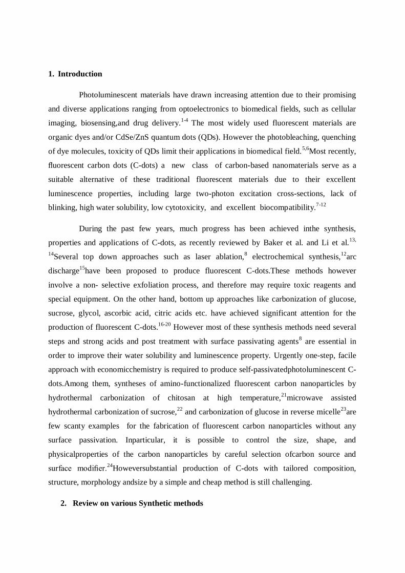
functionalized with hydroxyl, epoxy, carbonyl, and carboxylic acid groups (Fig. 6b). The
presence of these functional groups imparts excellent solubility in water without further
chemical modification.Also the presence of surface –COOH group is also evidenced of zeta
potential measurement. Measurement of zeta potential against pH shows that at low pH zeta
potential is positive which shifts to negative at higher pH which indicates the presence of –
COOH groups on the surface (Fig. 6c).
Fig.7. XRD patterns of A and SA (a), FTIR spectra of A, Sand AS (b), Zetapotential of A
with pH (c)
The as synthesised C-dots without any surface modification could easily disperse in
millipore water with an average diameter in between 16 nm to 40 nm. Among the different C-
dots the hydrodynamic diameter increases in the order SA < S < SC < A < C, which is in
agreement with red shift of emission spectra of the sample from AS to C. This change in
optical property with increase in particle size is consistent with the reported result.42
10
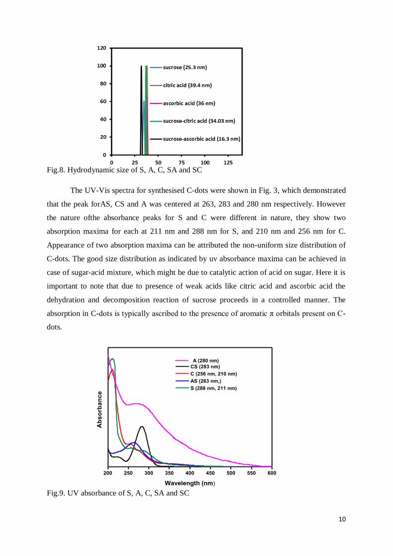
Fig.8. Hydrodynamic size of S, A, C, SA and SC
The UV-Vis spectra for synthesised C-dots were shown in Fig. 3, which demonstrated
that the peak forAS, CS and A was centered at 263, 283 and 280 nm respectively. However
the nature ofthe absorbance peaks for S and C were different in nature, they show two
absorption maxima for each at 211 nm and 288 nm for S, and 210 nm and 256 nm for C.
Appearance of two absorption maxima can be attributed the non-uniform size distribution of
C-dots. The good size distribution as indicated by uv absorbance maxima can be achieved in
case of sugar-acid mixture, which might be due to catalytic action of acid on sugar. Here it is
important to note that due to presence of weak acids like citric acid and ascorbic acid the
dehydration and decomposition reaction of sucrose proceeds in a controlled manner. The
absorption in C-dots is typically ascribed to the presence of aromatic π orbitals present on C-
dots.
Fig.9. UV absorbance of S, A, C, SA and SC
200 250 300 350 400 450 500 550 600
Ab
so
rba
nc
e
Wavelength (nm)
CS (283 nm)
C (256 nm, 210 nm)
AS (263 nm,)
S (288 nm, 211 nm)
A (280 nm)

11
At a fixed λex=330 nm, C-dots from different source show emission peak at 416, 421,
427, 432 and 438 nm for SA, S, SC, A and C respectively. This difference in position of
eission peak is attributed to the variation in size, as recently reported by Penget. al.43
This
result is also in consistence with hydrodynamic diameter. Furthermore, the unique
phenomemon of excitation dependent photoluminescence was observed in case of all
synthesised C-dots consistency with the PL property of CQDs reported by other groups (Fig.
3b). Apart from reasons like excitons of carbon,2 emissive traps,
44 aromatic conjugate
structures45
and free zig-zag sites,46
the excitation dependent PL behavior could arise because
of presence of “surface states”.22
Fig.10. Emission spectra of S (a), A (b), C (c), SC (d), and SA (e) recorded for progressively
longer excitation wavelength of 10 nm increments
Another interesting phenomenon here is the pH dependent PL behaviour, which is
shown by our all synthesised C-dots. The photoluminescent intensity of all C-dots gradually
decrease from acidic to basic medium, the trend is however not smooth. In case of SA and SC
the photoluminescent intensity is nearly constant in the neutral range (pH = 6-8). Due to
consistent PL within biological pH range, these C-dots may be suitable for biological
imaging.
12
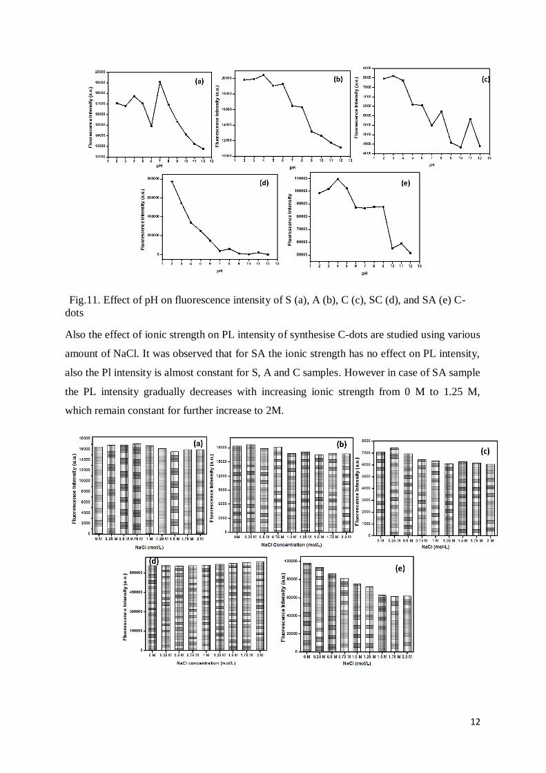
Fig.11. Effect of pH on fluorescence intensity of S (a), A (b), C (c), SC (d), and SA (e) C-
dots
Also the effect of ionic strength on PL intensity of synthesise C-dots are studied using various
amount of NaCl. It was observed that for SA the ionic strength has no effect on PL intensity,
also the Pl intensity is almost constant for S, A and C samples. However in case of SA sample
the PL intensity gradually decreases with increasing ionic strength from 0 M to 1.25 M,
which remain constant for further increase to 2M.

13
Fig.12. Effect of ionic strength on the fluorescence intensity of S (a), A (b), C (c), SC (d),
and SA (e) C-dots (ionic strengths were controlled by various concentrations of NaCl)
The PL quantum yields of the synthesised C-dots were calculated using quinine sulphate as
standard. The PL quantum yield of the sample S was as high as 13.5%, which is much higher
than other samples. Table 1 presents PL quantum yields of C-dots synthesized from different
precursors.
Fig.13. Fluorescence and Absorbance of S, A, C, SA, SC and Quinine Sulfate (QS)
Table-1: Comparison of HD size, λem, PL quantum yield (Φ)of C-dots synthesised from
different precursors
Sample ID composition Hydrodynamic
Size
λemission at
λex = 330 nm
Quantum Yield
(Φ)
S Sucrose 25.3 nm 421 nm 13.5 %
A Ascorbic acid 36.0 nm 432 nm 9.2 %
C Citric acid 39.4 nm 438 nm 6.9 %
SA Sucrose-Ascorbic acid 16.3 nm 416 nm 9.7 %
SC Sucrose-Citric acid 34.03 nm 427 nm 7.3 %
14
6. Conclusion
A simple economic synthesis for the preparation of highly photoluminescent C-dots
from different carbon precursor has been developed.The size distribution of the synthesised
C-dots was controlled by the addition of ascorbic and citric acid to sugar. By this approach
we were able to fabricate individual carbon quantum dots with different quantum yield, where
sucrose shows the highest value. The effect of pH and ionic strength on synthesised C-dots
has been investigated. Due to consistent PL within biological pH range for SA and SC
sample, they may be suitable for biological imaging.
References
1. S. J. Yu, M. W. Kang, H. C. Chang, K. M. Chen and Y. C. Yu, J. Am. Chem. Soc., 2005,
27, 17604.
2. J. Zhou, C. Booker, R. Li, X. Zhou, T. Sham, X. Sun and Z. Ding, J. Am. Chem. Soc., 2007,
129, 744.
3. H. P. Liu, T. Ye and C. D. Mao, Angew. Chem., Int. Ed., 2007, 46, 6473.
4. A. B.Bourlinos,A.Stassinopoulos, D.Anglos,R.Zboril,M.Karakassides, E. P. Giannelis,
Small, 2008, 4, 455.
5. A. P. Alivisatos, Science, 1996, 271, 933.
6. Y. W. Lin, W. L. Tseng, H. T. Chang, Adv. Mater., 2006, 18, 1381.
7. A. Safavi, F. Sedaghati, H. Shahbaazi, E. Farjami, RSC Adv., 2012, 2, 7367.
8. Y. P. Sun, B. Zhou, Y. Lin, W. Wang, K. A. S. Fernando, P. Pathak, M. J. Meziani, B. A.
Harruff, X. Wang, H. F. Wang, P. J. G. Luo, H. Yang, M. E. Kose, B. L. Chen, L. M. Veca,
S. Y. Xie, J. Am. Chem. Soc,. 2006, 128, 7756.
9. L. Cao, X. Wang, M. J. Meziani, F. S. Lu, H. F. Wang, P. J. G. Luo, Y. Lin, B. A. Harruff,
L. M. Veca, D. Murray, S. Y. Xie, Y. P. Sun, J. Am. Chem. Soc., 2007, 129, 11318.
10. S. T. Yang, X. Wang, H. Wang, F. Lu, P. G. Luo, L. Cao, M. J. Meziani, J. H. Liu, Y. Liu,
M. Chen, Y. Huang, Y. P. Sun, J. Phys. Chem. C, 2009,113, 18110.
11. S. T. Yang, L. Cao, P. G. Luo, F. S. Lu, X. Wang, H. F. Wang, M. J. Meziani, Y. F. Liu, G.
Qi, Y. P. Sun, J. Am. Chem. Soc., 2009, 131, 11308.
12. Q. L. Zhao, Z. L. Zhang, B. H. Huang, J. Peng, M. Zhang, D. W. Pang, Chem. Commun.,
2008, 5116.
13. S. N. Baker, G. A. Baker, Angew. Chem. Int. Ed., 2010, 49, 6726.
15
14. H. Li, Z. Kang, Y. Liu, S. T. Lee, J. Mater. Chem., 2012 (DOI: 10.1039/c2jm34690g).
15. X. Y.Xu, R. Ray, Y. L. Gu, H. J. Ploehn, L. Gearheart, K. Raker, W. A. Scrivens, J. Am.
Chem. Soc., 2004, 126, 12736.
16. F. Wang, Z. Xie, H. Zhang, C. Liu, Y. Zhang, Adv. Funct. Mater., 2011, 21, 1027.
17. H. T. Li, X. D. He, Y. Liu, H. Huang, S. Y. Lian, S. T. Lee, Z. H. Kang, Carbon, 2011,
49, 605.
18. B. Zhang, C. Liu, Y.un Liu, Eur. J. Inorg. Chem., 2010, 4411.
19. A. B. Bourlinos, A. Stassinopoulos, D. Anglos, R. Zboril, M. Karakassides, E. P.
Giannelis, Small, 2008, 4, 455.
20. D. Y. Pan, J. C. Zhang, Z. Li, C. Wu, X. M. Yan M. H. Wu, Chem. Commun., 2010, 46,
3681.
21. Y. Yang, J. Cui, M. Zheng, C. Hu, S. Tan, Y. Xiao, Q. Yang, Y. Liu,Chem. Commun.,
2012, 48, 380.
22. L. Tang, R.Ji, X.Cao, J.Lin, H. Jiang, X. Li, K.S.Teng,C. M. Luk, S. Zeng, J. Hao, S. P.
Lau, ACS Nano, 2012, 6, 5102.
23. W. Kwon, S. W. Rhee, Chem. Commun., 2012, 48, 5256.
24. F. Wang, M. Kreiter, B. He, S. Pang, C. Liu, Chem. Commun., 2010, 46, 3309.
25. L. Y. Zheng, Y. W. Chi, Y. Q. Dong, J. P. Lin, B. B. Wang, J. Am. Chem. Soc., 2009, 131,
4564.
26. Y. P. Sun, X. Wang, F. S. Lu, L. Cao, M. J. Meziani, P. J. G. Luo, L. R. Gu, L. M. Veca, J.
Phys. Chem. C,2008, 112, 18295.
27. S. L. Hu, K. Y. Niu, J. Sun, J. Yang, N. Q. Zhao, X. W. Du, J. Mater. Chem. 2009, 19, 484.
28. S. C. Ray, A. Saha, N. R. Jana, R. Sarkar, J. Phys. Chem. C, 2009,113, 18546.
29. M. J. Krysmann, A. Kelarakis, P. Dallas, E. P. Giannelis, J. Am. Chem. Soc., 2012,134, 747.
30. J. C. Zhang, W. Q. Shen, D. Y. Pan, Z. W. Zhang, Y. G. Fang, M. H. Wu, New J. Chem.,
2010, 34, 591.
31. X. He, H. Li, Y Liu, H. Huang, Z. Kang, S. T. Lee, Colloids Surf. B, 2011, 87, 326..
32. Z. C. Yang, X. Li, J. Wang, Carbon, 2011, 49, 5207.
33. S. Sahu, B. Behera, T. K. Maiti, S. Mohapatra, Chem. Commun., 2012, 48, 8835.
34. C. Zhu, J. Zhai, S. Dong, Chem. Commun.,2012, 48, 9367.
35. H. Zhu, X. L.Wang, Y. L.Li, Z. J. Wang, F.Yang, X. R. Yang, Chem.Commun., 2009, 5118.
36. H. T. Li, X. D. He, Y. Liu, H. Yu, Z. H. Kang, S. T. Lee, Mater.Res. Bull., 2011, 46, 147.
37. Z. Ma, H. Ming, H. Huang, Y. Liu, Z. Kang, New J. Chem., 2012, 36, 861.

16
38. R. L. Liu, D. Q. Wu, S. H. Liu, K. Koynov, W. Knoll, Q. Li, Angew. Chem. Int. Ed., 2009,
48, 4598.
39. A. B. Bourlinos, A.Stassinopoulos, D. Anglos, R. Zboril,V.Georgakilas, E. P. Giannelis,
Chem. Mater., 2008, 20, 4539.
40. J. Zong, Y. H. Zhu, X. L. Yang, J. H. Shen, C. Z. Li, Chem. Commun., 2011, 47, 764.
41. M. Sevilla, A.B. Fuertes, Carbon, 2009,47, 2281–2289
42. W. Kwon and S-W Rhee, Chem. Commun. 2012, 48, 5256.
43. J. Peng,W. Gao, B. K. Gupta, Z. Liu, R. Romero-Aburto, L. Ge, Li Song, L. B. Alemany,
X. Zhan, G. Gao, S. A. Vithayathil, B. A. Kaipparettu, A. A. Marti, T. Hayashi, J. Zhu and
Pulickel M. Ajayan, Nano Lett. 2012, 12, 844.
44. V. N. Mochalin and Y. Gogotsi. J. Am. Chem. Soc., 2009, 131, 4594.
45. S. Zhu, J. Zhang, C. Qiao, S. Tang, Y. Li, W. Yuan, B. Li, L. Tian, F. Liu, R. Hu, H. Gao,
H. Wei, H. Zhang, H. Sun and B. Yang, Chem. Commun. 2011, 48, 6858.
46. D. Y. Pan, J. C. Zhang, Z. Li and M. H. Wu, Adv. Mater., 2010, 22, 734.


![Electrochemical Synthesis of Luminescent MoS2 … · Electrochemical Synthesis of Luminescent MoS 2 ... [BMIM]Cl) from Sigma Aldrich, were used as received. In a typical experiment,](https://static.fdocuments.net/doc/165x107/5b02b6c87f8b9a65618fa48e/electrochemical-synthesis-of-luminescent-mos2-synthesis-of-luminescent-mos-2.jpg)