A role for the hepatobiliary system in IgE-mediated intestinal inflammation in the rat
9
A role for the hepatobiliary system in IgE-mediated intestinal inflammation in the rat A. M. COLLINS, S. LEACH, J. PAYNE A. MITCHELL, Y. DAI and G. D. F. JACKSON² School of Microbiology and Immunology, The University of New South Wales, Sydney, Australia Summary Background For many years the central focus of research into gastrointestinal hypersen- sitivity reactions has been the mast cell population of the intestinal lamina propria. Since bile is known to deliver immunological mediators to the gastrointestinal tract, the possibility arises that extra-intestinal populations of mast cells may also contribute to IgE-mediated intestinal damage. Objectives To characterize hepatic mast cells in the rat and to investigate the role of the hepatobiliary system in a model of IgE-mediated reactivity to dietary antigen. Methods Wistar rats were passively sensitized with monoclonal antidinitrophenyl (DNP) IgE antibodies, and were later challenged orogastrically with DNP-HSA. Additional animals were sensitized, then bile duct-cannulated prior to antigen challenge. At various time points, liver and intestinal samples were collected for histological examination, and bile was collected and assayed for histamine and TNFa. Results Hepatic mast cells display a mucosal mast cell-like phenotype, and are closely associated with the vessels of the portal triads. Orogastric antigen challenge led to a rapid and significant decline (P < 0.0001) in detectable mast cells as a result of anaphylactic degranulation. The median number of granulated mast cells associated with each portal triad in liver sections declined from six per portal triad to one per portal triad post-antigen challenge. After 15 min, biliary histamine concentrations rose above background levels (P < 0.01). TNFa was also detectable in the majority (4/6) of bile samples within 15 min of challenge. Histological examination of the gastrointestinal mucosa revealed disruption to the villous epithelium ranging from oedematous changes to gross destruction. Such damage was not seen in animals in which bile had been externally drained. Conclusion The data indicate that biliary products are major contributors to the gastro- intestinal damage arising from IgE-mediated hypersensitivity reactions in the rat, and such hypersensitivity reactions may involve a population of mast cells which reside in the liver. Keywords: hepatic mast cell, IgE, hypersensitivity, hepatobiliary, food allergy Clinical and Experimental Allergy, Vol. 29, pp. 262–270. Submitted 1 August 1997; revised 21 November 1997; accepted 3 March 1998. Introduction Food hypersensitivity and food allergy are terms used to describe a diverse group of clinical conditions ranging from acute, life-threatening anaphylaxis to chronic, debilitating conditions such as food-induced eczema, eosinophilic gastroenteritis, diarrhoeic-type irritable bowel disease and other food-induced gastroenteropathies [1]. Nausea, vomit- ing, abdominal pain and diarrhoea are symptoms associated with most of these conditions. Gastrointestinal manifesta- tions are also a common feature of systemic anaphylaxis in humans [2] and in animal models [3], while allergic asthma may sometimes be triggered by food antigens [4]. Although the underlying pathophysiological basis for these symp- toms remains unclear, Type 1 IgE-mediated immediate Clinical and Experimental Allergy, 1999, Volume 29, pages 262–270 262 q 1999 Blackwell Science Ltd Correspondence: A. M. Collins, School of Microbiology and Immunology, University of New South Wales, Sydney 2052, Australia. ²Deceased
Transcript of A role for the hepatobiliary system in IgE-mediated intestinal inflammation in the rat
A role for the hepatobiliary system in IgE-mediated intestinalinflammation in the rat
A. M. COLLINS, S. LEACH, J. PAYNE A. MITCHELL, Y. DAI and G. D. F.JACKSON†
School of Microbiology and Immunology, The University of New South Wales, Sydney, Australia
Summary
Background For many years the central focus of research into gastrointestinal hypersen-sitivity reactions has been the mast cell population of the intestinal lamina propria. Sincebile is known to deliver immunological mediators to the gastrointestinal tract, the possibilityarises that extra-intestinal populations of mast cells may also contribute to IgE-mediatedintestinal damage.Objectives To characterize hepatic mast cells in the rat and to investigate the role of thehepatobiliary system in a model of IgE-mediated reactivity to dietary antigen.Methods Wistar rats were passively sensitized with monoclonal antidinitrophenyl (DNP)IgE antibodies, and were later challenged orogastrically with DNP-HSA. Additionalanimals were sensitized, then bile duct-cannulated prior to antigen challenge. At varioustime points, liver and intestinal samples were collected for histological examination, andbile was collected and assayed for histamine and TNFa.Results Hepatic mast cells display a mucosal mast cell-like phenotype, and are closelyassociated with the vessels of the portal triads. Orogastric antigen challenge led to a rapidand significant decline (P< 0.0001) in detectable mast cells as a result of anaphylacticdegranulation. The median number of granulated mast cells associated with each portal triadin liver sections declined from six per portal triad to one per portal triad post-antigenchallenge. After 15 min, biliary histamine concentrations rose above background levels(P<0.01). TNFa was also detectable in the majority (4/6) of bile samples within 15 min ofchallenge. Histological examination of the gastrointestinal mucosa revealed disruption tothe villous epithelium ranging from oedematous changes to gross destruction. Such damagewas not seen in animals in which bile had been externally drained.Conclusion The data indicate that biliary products are major contributors to the gastro-intestinal damage arising from IgE-mediated hypersensitivity reactions in the rat, and suchhypersensitivity reactions may involve a population of mast cells which reside in the liver.
Keywords: hepatic mast cell, IgE, hypersensitivity, hepatobiliary, food allergy
Clinical and Experimental Allergy, Vol. 29, pp. 262–270. Submitted 1 August 1997;revised 21 November 1997; accepted 3 March 1998.
Introduction
Food hypersensitivity and food allergy are terms used todescribe a diverse group of clinical conditions ranging fromacute, life-threatening anaphylaxis to chronic, debilitatingconditions such as food-induced eczema, eosinophilic
gastroenteritis, diarrhoeic-type irritable bowel disease andother food-induced gastroenteropathies [1]. Nausea, vomit-ing, abdominal pain and diarrhoea are symptoms associatedwith most of these conditions. Gastrointestinal manifesta-tions are also a common feature of systemic anaphylaxis inhumans [2] and in animal models [3], while allergic asthmamay sometimes be triggered by food antigens [4]. Althoughthe underlying pathophysiological basis for these symp-toms remains unclear, Type 1 IgE-mediated immediate
Clinical and Experimental Allergy,1999, Volume 29, pages 262–270
262 q 1999 Blackwell Science Ltd
Correspondence: A. M. Collins, School of Microbiology and Immunology,University of New South Wales, Sydney 2052, Australia.†Deceased

hypersensitivity reactions are clearly associated with muchfood sensitivity [5].
The development of animal models has been crucial tothe advancement of our understanding of IgE-mediatedgastrointestinal hypersensitivity over the last 20 years,with published models focusing on the actions of mastcells from the gastrointestinal lamina propria and submu-cosa [3,6,7]. While these cells can certainly be accorded awatchkeeping role at mucosal surfaces, key features ofhypersensitivity reactions suggest that the emphasis onthese cells might be misplaced. For example, studies ofIgE-mediated antiparasite defences have shown that there isneither a change in the morphology nor in the numbers ofmucosal mast cells of the intestine in response to luminalantigen [6]. The failure ofin vitro and ex vivostudies todemonstrate key observations arising fromin vivo studieshas also been noted [8].
The gastrointestinal damage reported fromin vivomodelsshares many features with that seen in a model developed bythe authors to study the nature and function of biliary factorsproduced in response to challenge with bacterial lipopoly-saccharide (LPS) [9]. The LPS model developed from ourrealization that changes to the gastrointestinal epitheliumseen in a variety of clinical conditions point to the impor-tance of factors operating within the gastrointestinal lumen.Recent research has established that high concentrations ofcytokines such as TNFa can be delivered to the gut in thebile of rats [10] and humans [11]. Such cytokines have alsobeen shown to remain active in that environment, wherethey are able, for example, to regulate the expression ofMHC class II by the intestinal epithelium [12], and wherethey mediate tissue damage in our rat model of endotoxicshock [9]. Here evidence is presented suggesting that thehepatobiliary system plays an important role in IgE-mediated reactions to antigens originating from the gastro-intestinal tract lumen, and that two mast cell products,histamine and TNFa, are present in the bile of sensitizedand challenged animals.
Materials and methods
Animals and experimental design
Inbred SPF Wistar rats (8–10 weeks) were obtained fromCulas Ltd (Little Bay, NSW, Australia) and for the durationof the experiments were housed in the University of NewSouth Wales School of Microbiology and Immunologyanimal house. Conventional outbred Wistar rats were bredand maintained in the School of Microbiology and Immu-nology animal house. Murine IgE antidinitrophenyl (DNP)antibodies were raised as ascites using the H-1e-DNP cellline, and IgE antiovalbumin (OVA) antibodies were raisedas ascites using the OVA6B cell line. IgE antibody
concentrations were quantified by ELISA assay and by ratbasophil leukaemia cell degranulation assay as previouslydescribed [13]. Groups of rats were sensitized by i.v.injection with 3 mg mouse monoclonal anti-DNP IgE ormonoclonal anti-OVA IgE. Animals were returned to theircages with free access to food and water for 48 h, and weresubsequently denied further access to food for 24 h, afterwhich 1 mL of dinitrophenylated-human serum albumin(DNP32-HSA: Sigma Chemical Co., St Louis, MO, USA)in PBS (5 mg/mL) was given by gavage.
Prior to antigen challenge, some groups of rats weredeprived of intestinal bile by bile duct cannulation (BDC)and external drainage using the method of Lambert [14].Briefly, rats were anaesthetized with ether, laparotomieswere performed and cannuli (inner diameter 0.4 mm, outerdiameter 0.8 mm) were inserted into the bile ducts. Tubingwas secured in two places with suture cotton and passed outthrough the rats’ flanks. Bile was collected externally over a2-h period and then stored at – 208C until required. Controlanimals were either sham operated or left untreated. The ratswere observed for up to 2 h post-challenge before beinganaesthetized with ether and killed by cervical dislocation.Livers and intestines were removed, divided into portionsand immediately fixed in either Carnoy’s solution, for latermast cell staining, or in formaldehyde for H & E staining.
All surgical procedures were carried out under etheranaesthesia, and the experiments were approved by theUniversity of New South Wales Animal Care and EthicsCommittee.
Histology
Tissue sections which had been fixed in Carnoy’s solutionwere blocked in 0.5 mol/L HCl for 10 min, then stained intoluidine blue (0.5% w/v in 0.5 mol/L HCl) overnight.Additional sections were stained in Alcian blue (0.5% w/vin 0.7 mol/L HCl) overnight and then counter stained withsafranin (1% w/v in 0.125 mol/L HCl).
Toluidine blue-stained liver sections from four unsensi-tized controls and from four sensitized and challengedanimals were scanned by phase contrast microscopy tolocate portal triads. This technique allowed the portaltriads to be located independently of the presence of mastcells as stained mast cells can not be identified under phasecontrast. An eye-piece graticule was used to measure thelong and short diameters of each elliptical portal vein. Thesame portal triad was then viewed under normal illumina-tion and the surrounding mast cells counted and scored asmast cells per portal triad. Intestinal mast cells were countedas mast cells per villus crypt unit, where a villus crypt unit(VCU) was defined as one villus and the crypt immediatelybelow that villus, extending to the smooth muscle of theintestine [15]. All mast cell counts were conducted blind.
The hepatobiliary system and inflammation of the gut263
q 1999 Blackwell Science Ltd,Clinical and Experimental Allergy, 29, 262–270
Segments of duodenum, jejunum and ileum were col-lected from normal controls, sensitized and challengedanimals, and sensitized animals which were bile ductcannulated prior to antigen challenge. Tissue was col-lected from four animals from each group. From eachanimal, two jejunal segments were sectioned and stainedwith haematoxylin and eosin, and the full length of eachcentimetre long section was assessed for intestinaldamage. The extent of damage was judged on a gradedscale; 0 (no visible damage), 1 (slight oedema of villi), 2(Oedema with a few enterocytes breaking away from endsof villi), 3 (Clear oedema with almost all enterocytes lostfrom ends of villi). Four sections of intestine were viewedand graded per animal. The mean of the four grades wastaken as the overall grade of intestinal damage for thatanimal. All evaluations of tissue were conducted blind.
Western immunoblots
Biliary TNFa was investigated by Western blotting. SDS-PAGE gels were first run using a mini gel system (BioRad)and 12% separating gels. Proteins were then transferred ontonitrocellulose membranes using a mini trans-blot cell(BioRad). The membranes were then blocked with 1.0%(w/v) BSA in PBS and washed in PBS-0.05% Tween 20.Rabbit antimouse TNFa antiserum (Genzyme, Cambridge,MA, USA), which is known to cross-react with rat TNFa
[16], was added at a dilution of 1:100, and incubated for 1 hat room temperature. Membrane-bound antibodies werethen identified by further incubation with a 1:1000 dilutionof alkaline phosphatase-conjugated goat anti-rabbit immu-noglobulin (Southern Biotechnology Associates, Birming-ham, AL, USA), followed by incubation with the substratesnitroblue tetrazolium and 5-bromo-4-chloro-3-indolyl phos-phate (Sigma).
Measurement of histamine content by radioimmunoassay
A commercial histamine radioimmunoassay (Immunotech,Marseille, France) was used for the quantitative determina-tion of histamine levels in bile collected from five sensitizedand challenged animals and from five unsensitized controls.Twenty five microlitres of acylation solution and 25mL ofacylation buffer was added to 100mL of sample or standardsolution, and the resulting solution vortexed immediately.To each antibody-coated tube, 50mL of acylated sample orstandard and 500mL of iodinated tracer were added andincubated at 48C for 2 h. Test solutions were then aspiratedand discarded, and the tubes counted in a gamma counter(Packard Instrument Co., Meriden, CT, USA). Sampleconcentrations were finally calculated with reference to aseries of histamine standard solutions (0 –10 ng/mL).Where activity was too high to allow quantification
against the standard curve, samples were reanalysed afterappropriate dilution.
Statistical analysis
Data relating to mast cell numbers in the intestine wasanalysed using one wayANOVA, and where differencesbetween groups were shown, further analysis was performedusing the BonferoniiT-test. Western blot results wereanalysed byx2 test. All other data were analysed using theMann–WhitneyU-test. Significance was accepted at the0.05 level.
Results
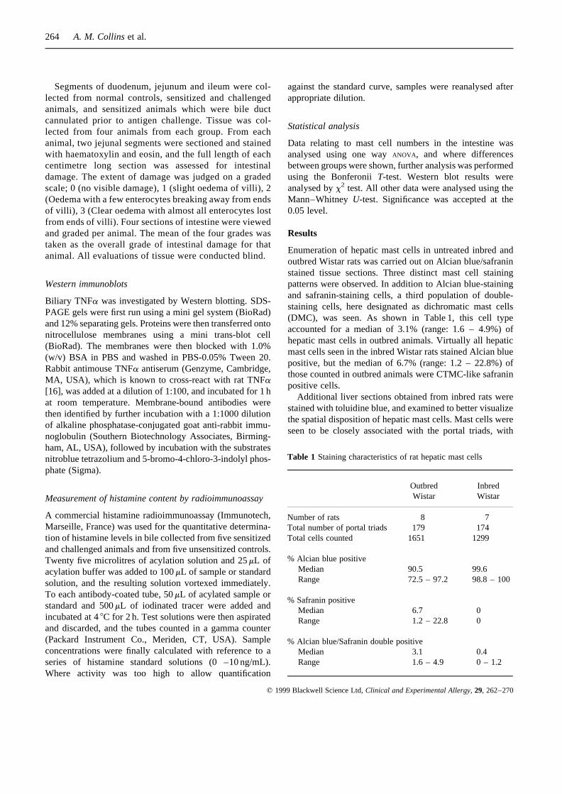
Enumeration of hepatic mast cells in untreated inbred andoutbred Wistar rats was carried out on Alcian blue/safraninstained tissue sections. Three distinct mast cell stainingpatterns were observed. In addition to Alcian blue-stainingand safranin-staining cells, a third population of double-staining cells, here designated as dichromatic mast cells(DMC), was seen. As shown in Table 1, this cell typeaccounted for a median of 3.1% (range: 1.6 – 4.9%) ofhepatic mast cells in outbred animals. Virtually all hepaticmast cells seen in the inbred Wistar rats stained Alcian bluepositive, but the median of 6.7% (range: 1.2 – 22.8%) ofthose counted in outbred animals were CTMC-like safraninpositive cells.
Additional liver sections obtained from inbred rats werestained with toluidine blue, and examined to better visualizethe spatial disposition of hepatic mast cells. Mast cells wereseen to be closely associated with the portal triads, with
264 A. M. Collinset al.
q 1999 Blackwell Science Ltd,Clinical and Experimental Allergy, 29, 262–270
Table 1 Staining characteristics of rat hepatic mast cells
Outbred InbredWistar Wistar
Number of rats 8 7Total number of portal triads 179 174Total cells counted 1651 1299
% Alcian blue positiveMedian 90.5 99.6Range 72.5 – 97.2 98.8 – 100
% Safranin positiveMedian 6.7 0Range 1.2 – 22.8 0
% Alcian blue/Safranin double positiveMedian 3.1 0.4Range 1.6 – 4.9 0 – 1.2

many mast cells clustered within the larger portal triads(Fig. 1). The number of mast cells per portal triad correlatedwell with the circumference of the portal vein (r2¼ 0.694)(Fig. 2a).
Liver sections obtained from IgE-sensitized and antigen-challenged animals showed a striking diminution in stain-able mast cells. Within 30 min of antigen challenge, thenumbers of identifiable mast cell declined dramatically(P<0.0001). As shown in Fig. 2(b), the median number ofmast cells declined from six per portal triad in unsensitizedcontrol animals (n¼ 4) to one per portal triad in sensitizedand challenged animals (n¼ 4). Two sections containing atleast 10 portal triads were examined per animal.
Mast cell numbers in the intestine were also investigatedand the results are presented in Fig. 3. Granulated mast cellnumbers declined in the duodenum and jejunum, within10 min of antigen challenge. Although the decline wasstatistically significant (P<0.05), the magnitude of thechange was relatively small compared with that seen inthe liver. While the mean number of mast cells per VCUseen in the duodenum of normal animals was 13.4, thisdeclined to 10.1 mast cells per VCU after antigen challenge.A significant decline in mast cell numbers was not seen inthe ileum until 30 min post-antigen challenge. Mast celldegranulation and the subsequent decline in visible mastcell numbers were unaffected by bile duct cannulation.
Sections of duodenum were collected and stained forhistological examination. The appearance of the villousepithelium from an unsensitized animal is shown inFig. 4(a). Sections from animals sensitized with anti-OVAIgE antibodies and challenged with DNP-HSA all showednormal, undamaged villous epithelium. A typical section isshown as Fig. 4(b). In contrast, sections obtained fromanimals sensitized with anti-DNP IgE and challenged withDNP-HSA revealed increasing intestinal damage over time.
The damage ranged from minor oedematous changes in thevillous epithelium over the first 15 min post-challenge, tototal obliteration of the villous architecture which could be
The hepatobiliary system and inflammation of the gut265
q 1999 Blackwell Science Ltd,Clinical and Experimental Allergy, 29, 262–270
Fig. 1. Toluidine blue stained section of normal liver from aninbred Wistar rat showing the typical distribution of mast cellsaround a portal triad.
75 (a)
Portal vein circumference (µm)
50
25
0M
ast
ce
lls p
er
po
rta
l tr
iad
10
00
0
10
00
10
0
10
60(b)
30
20
10
0
Ma
st
ce
lls p
er
po
rta
l tr
iad
Unsensitized Control
Sensitized and Challenged
Fig. 2. Hepatic mast cell counts. Sections of liver from inbred ratswere stained with toluidine blue. Portal triads were identified andthe circumference of the portal veins were measured. The numberof mast cells associated with each portal triad was then counted. (a)The relationship between portal vein circumference and mast cellnumbers. (b) Mast cell counts in control animals (n¼ 4) and inIgE-sensitized animals (n¼ 4) 30 min post antigen challenge. Solidbars show the median values for each group. The differencebetween the two groups was highly significant (P<0.0001).

seen after 30 min. Typical epithelial damage seen at the latertime point is shown in Fig. 4(c), which also shows dark-staining erythrocytes in areas of haemorrhage. To investi-gate a possible role for biliary factors in the changes seenwithin the gastrointestinal tract upon sensitization with IgEand challenge with antigen, animals were sensitized, thenbile duct cannulated prior to antigen challenge. No damageoccurred to the villi of bile duct cannulated animals(Fig. 4d).
Scores of intestinal damage seen in various animalsgroups are presented in Fig. 5. Intestinal damage was
particularly striking 30 min after antigen challenge ofsensitized animals. All sections from this timepointshowed intestinal damage scores of at least two, and somesections from all animals showed damage scores of three.These scores were significantly different to those seen innormal unchallenged controls (P< 0.05). Bile duct cannula-tion of sensitized and challenged animals appeared toabrogate the intestinal damage. No significant differencewas seen between these animals and normal controls.
Bile samples from five control animals and five sensitizedand challenged animals were analysed for histamine con-
266 A. M. Collinset al.
q 1999 Blackwell Science Ltd,Clinical and Experimental Allergy, 29, 262–270
15
10
5
0
Ma
st
ce
lls p
er
VC
U
(a)
* * *U
nch
all
en
ge
d
s/c
30
min
s/c
20
min
s/c
10
min
15
10
5
0M
ast
ce
lls p
er
VC
U
(b)
* *
Un
ch
all
en
ge
d
s/c
30
min
s/c
20
min
s/c
10
min
15
10
5
0
Ma
st
ce
lls p
er
VC
U
(c)
**
Un
ch
all
en
ge
d
s/c
30
min
s/c
20
min
s/c
10
min
Fig. 3. Intestinal mast cell counts. (a) Inbred rat duodenum, (b) jejunum and (c) ileum sections were stained with toluidine blue, and thenumber of mast cells per villus crypt unit (VCU) was determined. Small intestine was collected from normal and sensitized/challenged (s/c)rats at 10, 20 and 30 min post-antigen challenge. ** and * indicatep-values for comparisons with the normal group of less than 0.01 and0.001, respectively.
Fig. 4.H & E stained jejunum from (a) normal rat, (b) rat sensitized with monoclonala-OVA IgE then orally challenged with DNP-HSA,(c) rat sensitized with monoclonala-DNP IgE then orally challenged with DNP-HSA and (d) rat sensitized with monoclonala-DNP IgE,bile duct canulated, then orally challenged with DNP-HSA. Jejunum was collected 2 h post-antigen challenge in each case.

tent, and the results are shown as Fig. 6. Samples werecollected from sensitized animals 15 min after antigenchallenge, and showed a significant increase over thelevels seen in control rats (P<0.01).
Analysis of biliary proteins by SDS-PAGE showed arapid change in the protein profiles of challenged animals.Within 15 min of challenge, a range of new low molecularweight proteins appeared in bile (results not shown). Wes-tern blotting of gels showed one such protein to be TNFa.TNFa was detected in one of six unsensitized controlsamples and in none of six samples collected from sensi-tized animals challenged with HSA. However, a significant
upregulation of TNFa expression was seen in animalssensitized and challenged with DNP-HSA, where TNFa
was detected in four of six bile samples collected within 15min of antigen challenge (P<0.05). A representative blotshowing the time course for the appearance of TNFa is
The hepatobiliary system and inflammation of the gut267
q 1999 Blackwell Science Ltd,Clinical and Experimental Allergy, 29, 262–270
3
2
1
0
Inte
sti
na
l d
am
ag
e s
co
re
Un
ch
all
en
ge
d B
DC
s/c
30
min
BD
C
s/c
30
min
s/c
20
min
s/c
10
min
Un
ch
all
en
ge
d
Fig. 5. Intestinal damage scores. Inbred Wistar rats were sensitizedwith monoclonala-DNP IgE then orally challenged with DNP-HSA. Jejunal segments were collected 10, 20 and 30 min afterantigen challenge, with tissue from four rats collected at eachtimepoint. Segments were also collected 30 min after antigenchallenge of sensitized animals which had been bile duct cannu-lated (BDC) prior to antigen challenge. Additional segments werecollected from normal control animals and from BDC controls. H& E stained jejunal sections were scored for intestinal damage asfollows: 0 (undamaged normal jejunum), 1 (slight odema of villi),2 (clear odema with some loss of enterocytes from tip of villi), 3(complete loss of all enterocytes from tips of villi). The final scorefor each rat is given as the mean score from four sectionsexamined from each animal. Damage at 30 min was significantlydifferent to unchallenged and bile duct cannulated groups(P<0.05).
125
Unsensitized Control
Sensitized and Challenged
100
75
50
25
0
His
tam
ine
(n
g/m
L)
Fig. 6. Histamine concentrations in bile of five normal and fiveIgE-sensitized and antigen-challenged inbred rats. Bile was col-lected 15 min post-challenge. The difference between the twogroups was significant (P<0.01).
Fig. 7.Representative Western blot of TNFa in bile collected froman IgE-sensitized, antigen-challenged inbred rat. Bile was collectedfor 1 h post-antigen challenge. Lane 1 — MW markers; lane 2 —pre-challenge bile; lane 3 — 0–15 min; lane 4 — 15–30 min; lane5 — 30–45 min; lane 6 — 45–60 min; lane 7 — recombinant ratTNFa.

shown as Fig. 7. Biliary TNFa appeared as a band with amolecular weight of around 20 kDa, slightly higher than thatof the recombinant TNFa control.
Discussion
A simple model was developed to investigate whether thehepatobiliary system could be involved in IgE-mediatedreactions to antigens originating from the lumen of thegastrointestinal tract. Both the macroscopic and micro-scopic features seen were in accordance with previousreports of animal models of allergic reactions to dietaryantigens [3,17]. Sensitization of rats with monoclonal IgEanti-DNP antibodies, and later challenge with the appro-priate antigen, led to a rapid accumulation of fluid in the gutand inflammatory changes suggestive of tissue destruction.Histological examination of intestinal segments confirmedthe presence of gross destruction of the villous epithelia oftest animals.
To establish whether biliary factors could be involved inthe changes seen, IgE-sensitized animals were bile ductcannulated prior to antigen challenge. Strikingly, both themacroscopic and microscopic appearance of the intestineremained unchanged in these animals, and all animalsappeared well.
Large numbers of mast cells are resident in the liver andthey would seem a likely source of any inflammatorymediators released into bile in an IgE-dependent fashion.Although their presence in the liver has been recognized forsome time [18] little is known of the nature or function ofhepatic mast cells. The second aim of this investigation was,therefore, to better characterize rat hepatic mast cells, whichlike human hepatic mast cells [19] are found almost exclu-sively in association with portal triads.
Two apparently distinct subpopulations of mast cellshave long been recognized in the rat: mucosal mast cells(MMC) and connective tissue mast cells (CTMC). Thesesubpopulations have been most easily distinguished by thefact that MMC can be stained with Alcian blue, whileCTMC can be stained with safranin [20,21]. In this study,the livers of inbred Wistar rats were shown to be almostentirely populated by Alcian blue positive cells. A minorpopulation of dichromatic mast cells (DCM) was also seen.Cells with these same staining characteristics have beenreported previously from the dermis of Wistar rats [22].Enerback similarly reported that cells in the dermis ofSprague-Dawley rats that contained both blue and redgranules after staining with astra blue: safranin [20]. TheDMC population of the liver may represent a distinct andstable cell type, or may perhaps be a population of immaturecells.
Interestingly, in additional experiments conducted usingconventionally housed outbred animals, such DCM were
found at higher numbers. CTMC-like safranin positive cellsaccounted for almost 10% of the hepatic mast cells in theseanimals. These observations lend some credence to thenotion that hepatic mast cells may be capable of phenotypicdifferentiation, perhaps under the influence of the intestinalmicrobiota.
Hepatic mast cells belonging to the CTMC phenotype hasoften been assumed. The Alcian blue/safranin stainingproperties observed challenge this view. Additional studieswill be required to confirm the phenotype of the alcian blue-staining cells seen. However, a number of recent reports ofMMC-specific chymase activity in the liver support thisview [23–25].
Toluidine blue staining was used to monitor changes inhepatic mast cells after IgE-sensitization and antigenchallenge. The toluidine blue stain is specific for mastcell granules [26], and therefore a completely degranu-lated yet intact cell will not be seen in toluidine blue-stained tissue sections. Although mast cell degranulationmay often be incomplete or ‘piecemeal’, complete or‘anaphylactic’ degranulation has been reported in thehuman intestinal tract [27]. Similar anaphylactic degranu-lation has been inferred from the decline in granulatedintestinal mast cell numbers reported from animal modelsafter antigen challenge [28]. A similarly slight but sig-nificant reduction in mast cell numbers per villous cryptunit was also noted in stained sections of the intestine ofsensitized and antigen-challenged animals in this study. Incontrast, a dramatic decline in granulated hepatic mastcells was seen. Degranulating mast cells were also aconspicuous feature of histological sections of testanimal livers, while intestinal mast cells from these ratswere not seen to degranulate, as has previously beenreported [28].
To see whether mast cells are able to deliver inflamma-tory mediators to the gut via the hepatobiliary system, bilewas analysed for the presence of mast cell products. Mastcells release a range of preformed and newly formedmediators upon activation [29]. Histamine and TNFa aretwo such mediators which have been widely studied in thecontext of allergic disease, and serve as good indicators ofmast cell activation. Although TNFa is produced by manycell types, it is only known to be stored in mast cells [30]. Itsearly detection can therefore be taken as indirect evidence ofmast cell degranulation. Analysis of bile for these mediatorswas therefore included in this study.
Biliary histamine levels rise within 15 min of antigenchallenge of sensitized animals. TNFa was also identified inbile by Western blot, appearing as a band with an apparentmolecular weight of around 20 kDa. This is in line withprevious reports which have shown rat TNFa bands rangingfrom 17 kDa to 22 kDa [16]. While the authors present noevidence that TNFa is responsible for the intestinal damage
268 A. M. Collinset al.
q 1999 Blackwell Science Ltd,Clinical and Experimental Allergy, 29, 262–270

seen in the proposed model, the possibility can not bediscounted. Some cytokines, including TNFa, can retainmuch of their biological activity within the harsh environ-ment of the gastrointestinal tract [9,31], and the authorshave recently observed similar intestinal damage inanimals infused intraduodenally with TNFa (manuscriptin preparation).
The present model highlights the hepatobiliary system asa conduit for the delivery of mast cell products to the gastro-intestinal tract of the rat. Whether or not the humanhepatobiliary system is similarly involved in gastrointestinalhypersensitivity reactions awaits further investigation.
Acknowledgements
This study was supported, in part, by a grant from theNational Health and Medical Research Council.
References
1 Metcalfe DD. Food Allergy. In. Ogra PL, Mestecky J, LammME et al.eds Handbook of Mucosal Immunology. San Diego:Academic Press 1994 493–504.
2 Marquardt DL, Wasserman SI. Anaphylaxis. In Middleton E Jr,ed. Allergy. Principles and practice. St. Louis: Mosby, 1993:1525–37.
3 Pellon MI, Steil AA, Furio V, Sanchez CM. Study of theeffector mechanism involved in the production of haemorrha-gic necrosis of the small intestine in rat passive anaphylaxis. BrJ Pharmacol 1994; 112:1101–8.
4 James JM, Bernhisel BJ, Sampson HA. Respiratory reactionsprovoked by double-blind food challenges in children. Am JResp Crit Care Med 1994; 149:59–64.
5 Kemeny DM, Price JF, Richardson V, Richards D, Lessof MH.The IgE and IgG subclass antibody response to foods in babiesduring the first year of life and their relationship to feedingregimen and the development of food allergy. J Allergy ClinImmunol 1991; 87:920–9.
6 Ahmad A, Wang CH, Bell RG. A role for IgE in intestinalimmunity. Expression of rapid expulsion of Trichinella spiralisin rats transfused with IgE and thoracic duct lymphocytes. JImmunol 1991; 146:3563–70.
7 Crowe SE, Soda K, Stanisz AM, Perdue MH. Intestinalpermeability in allergic rats — nerve involvement inantigen-induced changes. Am J Physiol 1993; 264:G617–G623.
8 Scudamore CL, Pennington AM, Thornton E et al. Basalsecretion and anaphylactic release of rat mast cell protease-II(RMCP-II) from ex vivo perfused rat jejunum — translocationof RMCP-II into the gut lumen and its relation to mucosalhistology. Gut 1995; 37:235–41.
9 Jackson GDF, Dai Y. Biliary tumour necrosis factor alphamediates endotoxin-induced gastrointestinal damage. ClinImmunol Immunopathol 1995; 76:S41.
10 Jackson GDF, Dai Y, Fung C, Hansen PG. Tumour necrosis
factor alpha – a constitutive protein in bile. In: Mestecky J, ed.Advances in Mucosal Immunology. New York: Plenum Press1995: 1083–6.
11 Rosen HR, Winkle PJ, Kendall BJ, Diehl DL. Biliary inter-leukin-6 and tumor necrosis factor-alpha in patients under-going endoscopic retrograde cholangiopancreatography. DigDis Sci 1997; 42:1290–4.
12 Hamilton LJ, Dai Y, Jackson GDF. Bile regulates the expres-sion of MHC class II antigens on rat intestinal epithelium.Gastroenterology 1997; 113:1901–5.
13 Collins AM, Thelian D, Basil M. Antigen valency as adeterminant of the responsiveness of IgE-sensitised Rat Baso-phil Leukemia cells. Int Arch Allergy Immunol 1995;107:547–56.
14 Lambert R. (1965). Surgery of the Digestive System in the Rat.Springfield: Charles C. Thomas.
15 Befus AD, Bienenstock J. Immunologically mediated intestinalmastocytosis in Nippostrongylus brasiliensis-infected rats.Immunology 1979; 38:95–101.
16 Merrick BA, He CY, Craig WA et al. Two dimensional gelelectrophoresis of cellular and secreted proteins from ratalveolar macrophages after lipopolysaccharide treatment.Appl Theoret Electrop 1992; 2:177–87.
17 Curtis GH, Patrick MK, Catto SA, Gall DG. Intestinal anaphy-laxis in the rat. Effect of chronic antigen exposure. Gastro-enterology 1990; 98:1558–66.
18 Dimlich RV, Reilly FD, Meineke HA, McCuskey RS. Char-acterization of intensely fluorescent cells in the liver of the rat.i. Histochemistry and 48/80-induced degranulation. AnatRecord 1980; 198:475–84.
19 Farrell DJ, Hines JE, Walls AF et al. Intrahepatic mast cells inchronic liver diseases. Hepatology 1995; 22:1175–81.
20 Enerback L. Mast cells in rat gastrointestinal mucosa. 2. Dye-binding and metachromatic properties. Acta Pathol MicrobiolScan 1966; 66:303–12.
21 Befus D, Swieter M, Fujimaki H, Lee T. Investigations of ratmast cell heterogeneity. Int Arch Allergy Appl Immunol 1987;82:256–8.
22 Elsayed SO, Dyson M. Histochemical heterogeneity of mastcells in rat dermis. Biotech Histochem 1993; 68:326–32.
23 Rioux KP, Sharkey KA, Wallace JL, Swain MG. Hepaticmucosal mast cell hyperplasia in rats with secondary biliarycirrhosis. Hepatology 1996; 23:888–95.
24 Miller HR, Newlands GF, McKellar G et al. Hepaticrecruitment of mast cells occurs in rats but not mice infectedwith Schistosoma mansoni. Parasite Immunol 1994; 16:145–55.
25 Huntley JF, Mackellar A, Newlands GF, Irvine J, Miller HR.Mapping of the rat mast cell granule proteinases RMCPI and IIby enzyme-linked immunosorbent assay and paired immuno-fluorescence. Apmis 1990; 98:933–44.
26 Blumenkrantz N, Asboe HG. A selective stain for mast cells.Histochem J 1975; 7:277–82.
27 Dvorak AM, McLeod RS, Onderdonk A et al. Ultrastructuralevidence for piecemeal and anaphylactic degranulation ofhuman gut mucosal mast cells in vivo. Int Arch AllergyImmunol 1992; 99:74–83.
The hepatobiliary system and inflammation of the gut269
q 1999 Blackwell Science Ltd,Clinical and Experimental Allergy, 29, 262–270

28 D’Inca R, Hunt RH, Perdue MH. Mucosal damage duringintestinal anaphylaxis in the rat. Effect of betamethasone anddisodium cromoglycate. Dig Dis Sci 1992; 37:1704–8.
29 Schwartz LB. Mast cells – function and contents. Curr OpinImmunol 1994; 6:91–7.
30 Gordon JR, Galli SJ. Mast cells as a source of both preformedand immunologically inducible TNF-a/cachectin. Nature 1990;346:274–6.
31 Rollwagen FM, Baqar S. Oral cytokine administration. Immu-nol Today 1996; 17:548–50.
270 A. M. Collinset al.
q 1999 Blackwell Science Ltd,Clinical and Experimental Allergy, 29, 262–270