A Quick Reference for Gross Anatomy · ANATOMY HANDBOOK A Quick Reference for Gross Anatomy S....
136
ANATOMY HANDBOOK A Quick Reference for Gross Anatomy S. Christopher Bennett, Ph.D. Version 3.1 – August 30, 2003
Transcript of A Quick Reference for Gross Anatomy · ANATOMY HANDBOOK A Quick Reference for Gross Anatomy S....
ANATOMY HANDBOOK
A Quick Reference for Gross Anatomy
S. Christopher Bennett, Ph.D.
Version 3.1 – August 30, 2003

ii
i
TABLE OF CONTENTS
Introduction ............................................................... 1Terms of Orientation ....................................................... 2Terms of Movement .......................................................... 2Basic Principles ........................................................... 3
HEAD AND NECK
Osteology of the Head ...................................................... 5Joints of the Skull ........................................................ 6Muscles of the Posterior Triangle .......................................... 9Prevertebral Muscles ...................................................... 11Infrahyoid Muscles ........................................................ 13Suprahyoid Muscles ........................................................ 15Facial Muscles ............................................................ 17Muscles of Mastication .................................................... 19Muscles of the Tongue ..................................................... 21Extrinsic Muscles of the Eye .............................................. 23Cranial Nerves ............................................................ 24Functions of Cranial Nerves ............................................... 25Spinal Nerves of the Head and Neck ........................................ 27Arteries of the Head and Neck ............................................. 29Arteries and Veins of the Brain ........................................... 31Veins of the Head and Neck ................................................ 33Cross-section of the Neck ................................................. 34
BACK
Osteology of the Postcranial Axial Skeleton ............................... 35Joints of the Postcranial Axial Skeleton .................................. 36Intermediate Muscles of the Back .......................................... 39Erector Spinae Muscles ................................................... 41Transversospinalis Muscles ................................................ 43Muscles of the Suboccipital Region ........................................ 45
THORAX AND ABDOMEN
Muscles of the Thoracic Wall .............................................. 47Muscles of the Abdominal Wall ............................................. 49Muscles of the Pelvis ..................................................... 51Muscles of the Perineum ................................................... 53Nerves of the Thorax ...................................................... 55Nerves of the Abdomen ..................................................... 57Arteries of the Thorax and Abdomen ........................................ 59Unpaired Visceral Branches of the Abdominal Aorta ......................... 61Veins of the Thorax and Abdomen ........................................... 63
UPPER EXTREMITY
Osteology of the Pectoral Girdle and Upper Extremity ...................... 64Joints of the Upper Extremity ............................................. 65Muscles attaching Upper Extremity to Trunk - Anterior Side ................ 67Muscles attaching Upper Extremity to Trunk - Posterior Side ............... 69
ii
Muscles of the Shoulder Joint - Scapular Muscles .......................... 71Muscles of the Arm ........................................................ 73Superficial Extensors of the Forearm ...................................... 75Deep Extensors of the Forearm ............................................. 77Superficial Flexors of the Forearm ........................................ 79Deep Flexors of the Forearm ............................................... 81Other Muscles of the Forearm .............................................. 83Thenar and Hypothenar Muscles ............................................. 85Other Muscles of the Hand ................................................. 87Brachial Plexus ........................................................... 89Nerves of the Upper Extremity ............................................. 91Arteries of the Upper Extremity ........................................... 93Cutaneous Veins of the Upper Extremity .................................... 95Cross-section of the Arm .................................................. 96Cross-section of the Forearm .............................................. 97
LOWER EXTREMITY
Osteology of the Pelvic Girdle and Lower Extremity ........................ 98Joints of the Lower Extremity ............................................. 99Muscles of the Anterior Thigh ............................................ 101Muscles of the Medial Thigh .............................................. 102Muscles of the Posterior Thigh ........................................... 103Lateral Rotators of the Femur ............................................ 107Gluteal Muscles .......................................................... 109Muscles of the Anterior Crural Compartment - Dorsiflexors ................ 111Muscles of the Posterior Crural Compartment - Superficial Plantarflexors . 113Muscles of the Posterior Crural Compartment - Deep Plantarflexors ........ 115Muscles of the Lateral Crural Compartment ................................ 117Muscles of the Dorsum of the Foot ........................................ 119Muscles of the Sole of the Foot - Layers 1-4 ............................. 121Nerves of the Lower Extremity ............................................ 125Nerves and Arteries of the Gluteal Region ................................ 127Arteries of the Lower Extremity .......................................... 129Cutaneous Veins of the Lower Extremity ................................... 130Cross-section of the Thigh ............................................... 131Cross-section of the Leg ................................................. 132
1
INTRODUCTION
This handbook contains much of the basic information about the bones,muscles, nerves, arteries, and veins of the body that is needed in a typicalgross anatomy course. Such a course typically follows a regional approach, andso does the handbook. Within regions I have divided the material into smallpackets of information (a group of muscles; the pattern of arteries; etc.). Iorganize information visually, and so most packets consist of a drawing andsome accompanying textual material, usually arranged on facing pages.
I am often asked how one should study anatomy. I do not know how oneshould do it, I only know how I would study it. First I would divide up thematerial. In any particular region of the body the muscles can be divided intoa number of groups, each containing a small number of muscles. Again Iorganize information visually so I usually have a drawing or an outline. Ihave already divided up the material for you. Then to study a group of musclesI would sketch them and label or note the origins, insertions, actions, andinnervations. To study the pattern of arteries or nerves I would draw thepattern, name them, note the branching pattern, and where they go or what theydo. All the drawings, in this handbook are simple enough that you can drawthem. At first I might simply be copying my notes, but soon I would try to putthe things down on paper from memory. If I could not remember some things, Iwould check my notes or look them up in the book, and try again until I hadmemorized the material and could put it down on a blank sheet of paper. If Ican put it down on a blank piece of paper, I know I know it, and I know I canput it down on the test from memory.
After having learned the muscles and the patterns of nerves and vessels,one other thing that I find helpful is to look over illustrations in anatomytexts and atlases and make sure you can understand them.
2
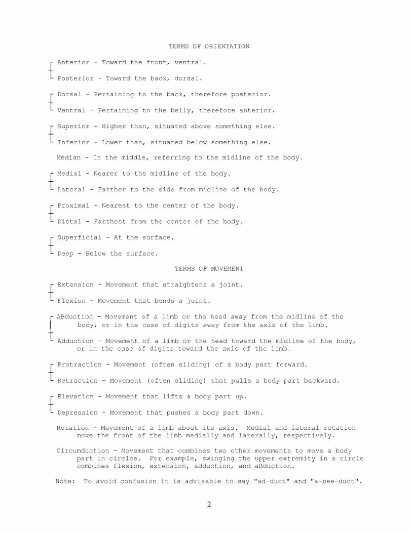
TERMS OF ORIENTATION
┌ Anterior - Toward the front, ventral.┼└ Posterior - Toward the back, dorsal.
┌ Dorsal - Pertaining to the back, therefore posterior.┼└ Ventral - Pertaining to the belly, therefore anterior.
┌ Superior - Higher than, situated above something else.┼└ Inferior - Lower than, situated below something else.
Median - In the middle, referring to the midline of the body.
┌ Medial - Nearer to the midline of the body.┼└ Lateral - Farther to the side from midline of the body.
┌ Proximal - Nearest to the center of the body.┼└ Distal - Farthest from the center of the body.
┌ Superficial - At the surface.┼└ Deep - Below the surface.
TERMS OF MOVEMENT
┌ Extension - Movement that straightens a joint.┼└ Flexion - Movement that bends a joint.
┌ ABduction - Movement of a limb or the head away from the midline of the│ body, or in the case of digits away from the axis of the limb.┼└ Adduction - Movement of a limb or the head toward the midline of the body, or in the case of digits toward the axis of the limb.
┌ Protraction - Movement (often sliding) of a body part forward.┼└ Retraction - Movement (often sliding) that pulls a body part backward.
┌ Elevation - Movement that lifts a body part up.┼└ Depression - Movement that pushes a body part down.
Rotation - Movement of a limb about its axis. Medial and lateral rotation move the front of the limb medially and laterally, respectively.
Circumduction - Movement that combines two other movements to move a body part in circles. For example, swinging the upper extremity in a circle combines flexion, extension, adduction, and aBduction.
Note: To avoid confusion it is advisable to say "ad-duct" and "a-bee-duct".
3
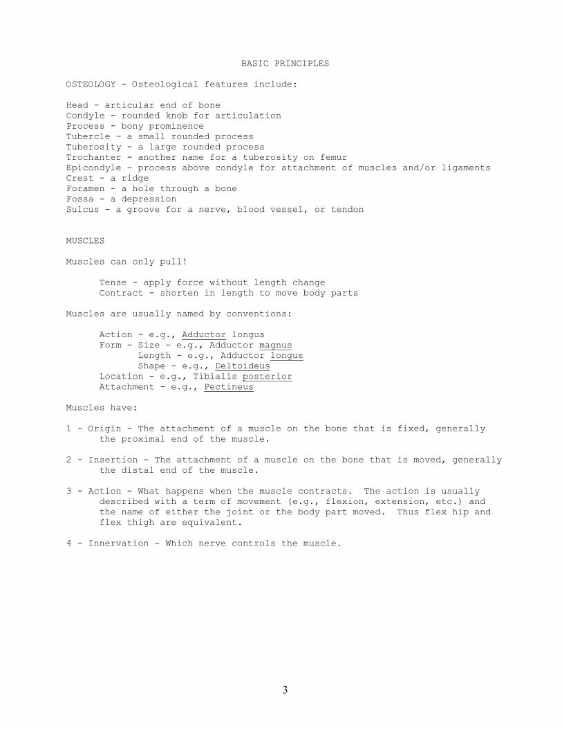
BASIC PRINCIPLES
OSTEOLOGY - Osteological features include:
Head - articular end of boneCondyle - rounded knob for articulationProcess - bony prominenceTubercle - a small rounded processTuberosity - a large rounded processTrochanter - another name for a tuberosity on femurEpicondyle - process above condyle for attachment of muscles and/or ligamentsCrest - a ridgeForamen - a hole through a boneFossa - a depressionSulcus - a groove for a nerve, blood vessel, or tendon
MUSCLES
Muscles can only pull!
Tense - apply force without length changeContract - shorten in length to move body parts
Muscles are usually named by conventions:
Action - e.g., Adductor longusForm - Size - e.g., Adductor magnus Length - e.g., Adductor longus Shape - e.g., DeltoideusLocation - e.g., Tibialis posteriorAttachment - e.g., Pectineus
Muscles have:
1 - Origin - The attachment of a muscle on the bone that is fixed, generallythe proximal end of the muscle.
2 - Insertion - The attachment of a muscle on the bone that is moved, generallythe distal end of the muscle.
3 - Action - What happens when the muscle contracts. The action is usuallydescribed with a term of movement (e.g., flexion, extension, etc.) andthe name of either the joint or the body part moved. Thus flex hip andflex thigh are equivalent.
4 - Innervation - Which nerve controls the muscle.
4
ARTERIES AND VEINS
Arteries carry blood from the heart out to the body and veins returnblood to the heart. The pattern of arteries and veins is described in thedirection that blood flows. Large arteries branch into smaller arteries.Small veins join to become, or empty into, larger veins. Both are subject totopographic name changes, in which the name of a vessel changes after it haspassed an anatomical landmark. Thus the External Iliac Artery becomes theFemoral Artery after passing under the Inguinal Ligament. Arteries and veinsgenerally run together and have the same names. Therefore, if one learns thepattern of arteries one knows most veins as well. However, cutaneous veins donot accompany arteries.
NERVES
Nerves carry impulses from and to the brain. Nerves can be sensory,carrying impulses from receptors to the brain; motor, carrying impulses fromthe brain to muscles; or mixed, both sensory and motor. Nerves generallybranch into smaller nerves, but small nerves can join to form a larger nerve.Nerves often run with arteries and veins, but the branching pattern of nervesusually differs from that of arteries and veins, and so the names of nervesusually differ from those of arteries and veins.
VARIATIONS
Variations on the pattern of structures presented in this handbook andmost anatomy texts are common. Veins are particularly prone to variation, butnerves, arteries, and muscles also are variable. This will not be a problem inthe lecture course because we will learn the "normal" pattern, but those incadaver labs should be aware that variations occur and not be concerned bythem.
ABBREVIATIONS
Terms of Orientation: Anterior Ant. Posterior Post. Superior Sup. Inferior Inf. Medial Med. Lateral Lat. Superficial Superf. External Ext. Internal Int.
SINGULAR PLURAL ————————————————————————Structures: Muscle M. Mm. Artery A. Aa. Vein V. Vv. Nerve N. Nn. Ligament Lig. Ligg.
5
OSTEOLOGY OF THE HEAD
SKULL - consists of two distinct parts: Braincase and Face
BRAINCASE - Bones: Frontal Parietals (2) Temporals (2) Occipital Sphenoid
Sutures: Sagittal - between parietals Coronal - between frontal and parietals Lambdoidal - between parietals and occipital
FACE - Bones: Zygomatics (2) Nasals (2) Lacrimals (2) Maxillae (2) Palatines (2) Sphenoid Ethmoid Vomer
FEATURES OF THE SKULL
Orbits - sockets for eyeballsOptic Canal - for Optic N. and Ophthalmic A.Superior orbital fissure - between wings of sphenoid for CN III, IV, VI and
ophthalmic division of Trigeminal N.Nasal aperature - external opening of nasal passagesNasal septum - separates nasal passagesInfraorbital foramen - for Infraorbital NAV to supply nasal regionZygomatic archExternal auditory meatus - opening of auditory canal
Greater and lesser wings of sphenoidSella turcica [=Turkish saddle] - depression for pituitary glandCribriform plate of ethmoid - for Olfactory N.
Hard palate - formed of palatine and maxillaePterygoid plates - origin of pterygoid Mm., wall of pharynxMandibular fossa - articulates with condyloid process of mandibleStyloid process of temporal - origin of Stylohyoid, Styloglossus, and
StylopharyngeusMastoid process of temporal - origin of Digastric, insertion of
Sternocleidomastoid and Splenius capitusOccipital condyles - articulate with atlas
Foramen ovale - for CN V2Foramen rotundum - for CN V3Foramen spinosum - for Middle meningeal A.Foramen lacerum - closed by cartilageCarotid canal - for Internal carotid A.Jugular foramen - for Internal jugular V. and CN IX-XIStylomastoid foramen - for CN VIIForamen magnum - for spinal cord
6
MANDIBLE
BodyRamusAngle - insertion of Masseter and Medial pterygoidCondyloid process - articulates with skullCoronoid process - insertion of TemporalisMandibular foramen - for Inferior alveolar A+NMental foramen - for Mental A+N to supply chinMylohyoid Line - insertion of MylohyoidMental Spines - origin of Geniohyoid
TEETH - on each side, top and bottom
2 Incisors1 Canine2 Premolars ("bicuspids")3 Molars - not preceded by deciduous teeth
HYOID BONE
——————————————————————————————————————————————————————————————————————————————
JOINTS OF THE SKULL
Movement of Temporo-mandibular joint
- rotation to elevate and depress mandible- lateral displacement- protraction and retraction
Movements of Atlanto-occipital joint
Skull-atlas - flexion and extension

7
NOTES
8
9
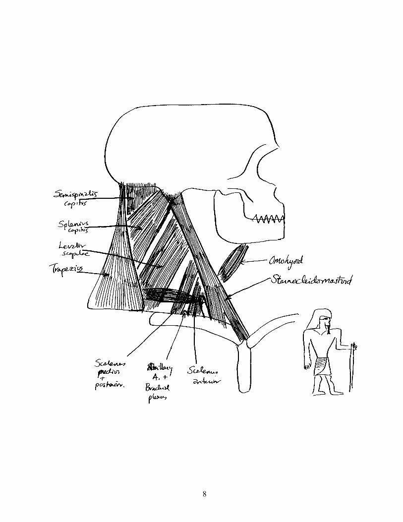
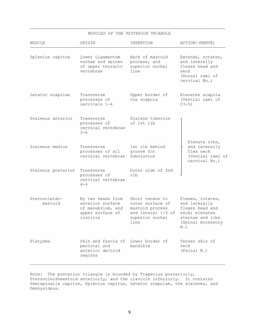
——————————————————————————————————————————————————————————————————————————————MUSCLES OF THE POSTERIOR TRIANGLE
MUSCLE ORIGIN INSERTION ACTION/(NERVE)——————————————————————————————————————————————————————————————————————————————
Splenius capitus Lower Ligamentum Back of mastoid Extends, rotates, nuchae and spines process, and and laterally of upper thoracic superior nuchal flexes head and vertebrae line neck
(Dorsal rami ofcervical Nn.)
Levator scapulae Transverse Upper border of Elevates scapula processes of the scapula (Ventral rami of cervicals 1-4 C3-5)
Scalenus anterior Transverse Scalene tubercle ┐ processes of of 1st rib │ cervical vertebrae │ 3-6 │ │ │ Elevate ribs,Scalenus medius Transverse 1st rib behind ├ and laterally processes of all groove for │ flex neck cervical vertebrae Subclavius │ (Ventral rami of │ cervical Nn.) │Scalenus posterior Transverse Outer side of 2nd │ processes of rib ┘ cervical vertebrae 4-6
Sternocleido- By two heads from Short tendon to Flexes, rotates, mastoid anterior surface outer surface of and laterally of manubrium, and mastoid process flexes head and upper surface of and lateral 1/3 of neck; elevates clavicle superior nuchal sternum and ribs line (Spinal Accessory N.)
Platysma Skin and fascia of Lower border of Tenses skin of pectoral and mandible neck anterior deltoid (Facial N.) regions
——————————————————————————————————————————————————————————————————————————————
Note: The posterior triangle is bounded by Trapezius posteriorly,Sternocleidomastoid anteriorly, and the clavicle inferiorly. It containsSemispinalis capitus, Splenius capitus, Levator scapulae, the scalenes, andOmohyoideus.
10
11
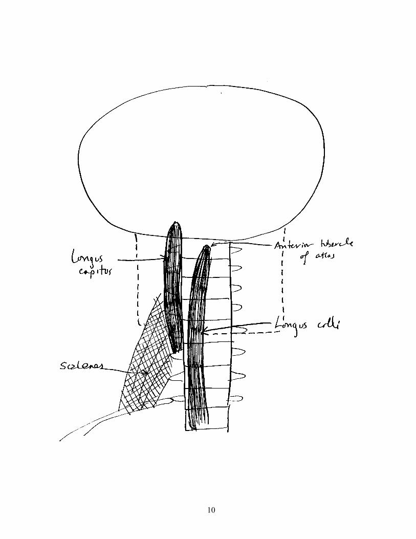
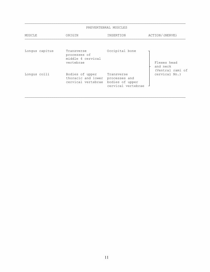
——————————————————————————————————————————————————————————————————————————————PREVERTEBRAL MUSCLES
MUSCLE ORIGIN INSERTION ACTION/(NERVE)——————————————————————————————————————————————————————————————————————————————
Longus capitus Transverse Occipital bone ┐ processes of │ middle 4 cervical │ vertebrae │ Flexes head ├ and neck │ (Ventral rami ofLongus colli Bodies of upper Transverse │ cervical Nn.) thoracic and lower processes and │ cervical vertebrae bodies of upper │ cervical vertebrae ┘
——————————————————————————————————————————————————————————————————————————————
12
13
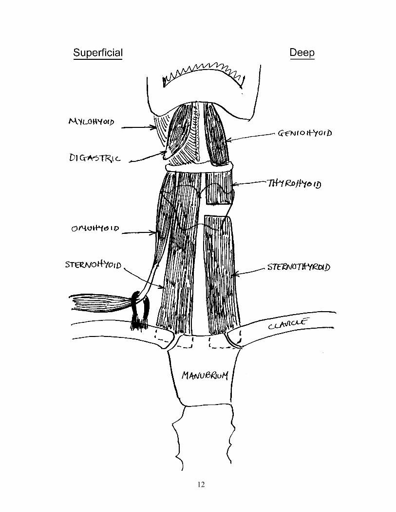
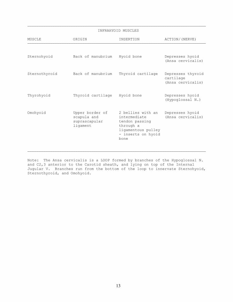
——————————————————————————————————————————————————————————————————————————————INFRAHYOID MUSCLES
MUSCLE ORIGIN INSERTION ACTION/(NERVE)——————————————————————————————————————————————————————————————————————————————
Sternohyoid Back of manubrium Hyoid bone Depresses hyoid (Ansa cervicalis)
Sternothyroid Back of manubrium Thyroid cartilage Depresses thyroid cartilage (Ansa cervicalis)
Thyrohyoid Thyroid cartilage Hyoid bone Depresses hyoid (Hypoglossal N.)
Omohyoid Upper border of 2 bellies with an Depresses hyoid scapula and intermediate (Ansa cervicalis) suprascapular tendon passing ligament through a ligamentous pulley - inserts on hyoid bone
——————————————————————————————————————————————————————————————————————————————
Note: The Ansa cervicalis is a LOOP formed by branches of the Hypoglossal N.and C2,3 anterior to the Carotid sheath, and lying on top of the InternalJugular V. Branches run from the bottom of the loop to innervate Sternohyoid,Sternothyroid, and Omohyoid.
14

15
——————————————————————————————————————————————————————————————————————————————SUPRAHYOID MUSCLES
MUSCLE ORIGIN INSERTION ACTION/(NERVE)——————————————————————————————————————————————————————————————————————————————
Stylohyoid Styloid process Passes around Elevate and Digastric and retract hyoid inserts on greater (Facial N.) horn of hyoid bone
Mylohyoid Mylohyoid line of Body of hyoid bone Elevate hyoid mandible (Inferior alveolar N.)
Geniohyoid Mental spines of Body of hyoid bone Protracts hyoid mandible below (Hypoglossal N.) Genioglossus
Digastric Posterior belly Passes by Elevates hyoid; from mastoid intermediate depresses and notch of temporal tendon through retracts mandible bone Stylohyoid; (Facial and anterior belly Inferior alveolar inserts on Nn.) inferior surface of mandible near symphysis
——————————————————————————————————————————————————————————————————————————————
Note: Mylohyoid forms the floor of the mouth and supports Geniohyoid, themuscles of the tongue, salivary glands, etc.
16
17
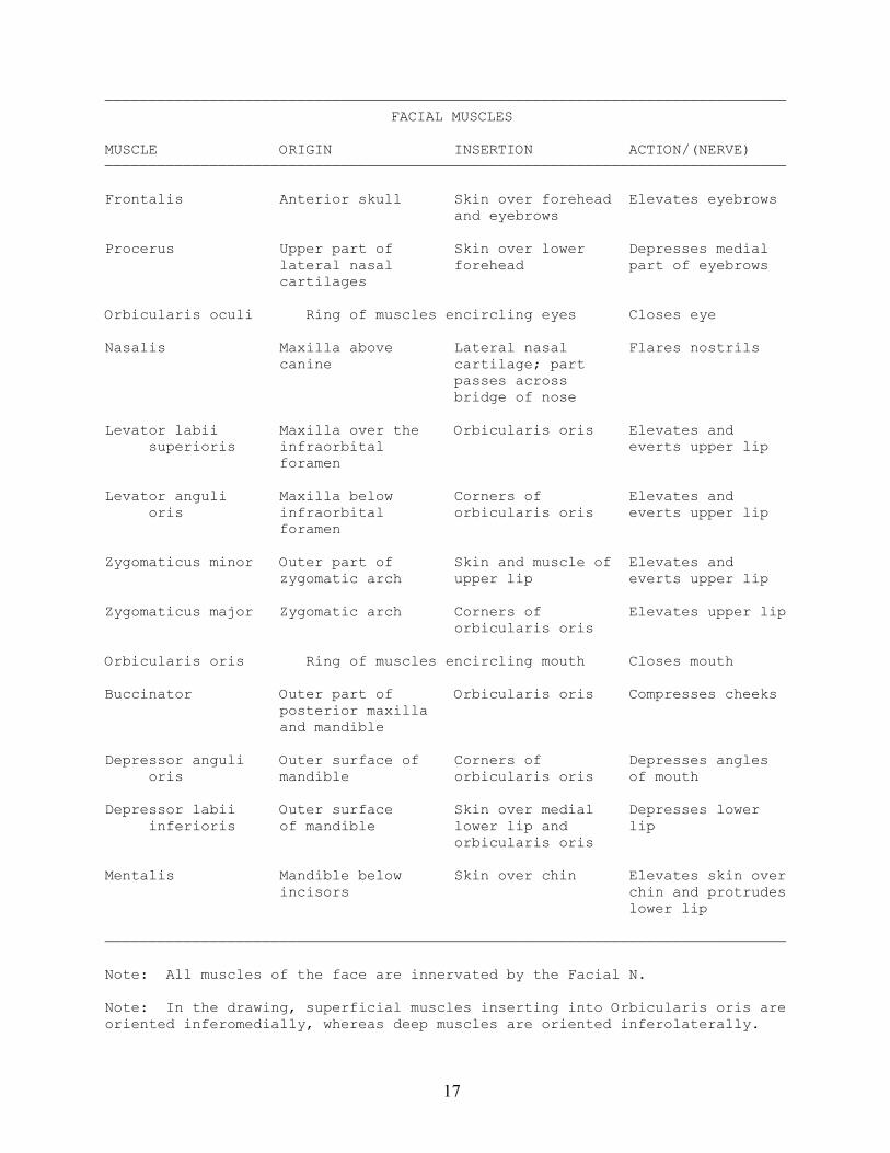
——————————————————————————————————————————————————————————————————————————————FACIAL MUSCLES
MUSCLE ORIGIN INSERTION ACTION/(NERVE)——————————————————————————————————————————————————————————————————————————————
Frontalis Anterior skull Skin over forehead Elevates eyebrows and eyebrows
Procerus Upper part of Skin over lower Depresses medial lateral nasal forehead part of eyebrows cartilages
Orbicularis oculi Ring of muscles encircling eyes Closes eye
Nasalis Maxilla above Lateral nasal Flares nostrils canine cartilage; part passes across bridge of nose
Levator labii Maxilla over the Orbicularis oris Elevates and superioris infraorbital everts upper lip foramen
Levator anguli Maxilla below Corners of Elevates and oris infraorbital orbicularis oris everts upper lip foramen
Zygomaticus minor Outer part of Skin and muscle of Elevates and zygomatic arch upper lip everts upper lip
Zygomaticus major Zygomatic arch Corners of Elevates upper lip orbicularis oris
Orbicularis oris Ring of muscles encircling mouth Closes mouth
Buccinator Outer part of Orbicularis oris Compresses cheeks posterior maxilla and mandible
Depressor anguli Outer surface of Corners of Depresses angles oris mandible orbicularis oris of mouth
Depressor labii Outer surface Skin over medial Depresses lower inferioris of mandible lower lip and lip orbicularis oris
Mentalis Mandible below Skin over chin Elevates skin over incisors chin and protrudes lower lip
——————————————————————————————————————————————————————————————————————————————
Note: All muscles of the face are innervated by the Facial N.
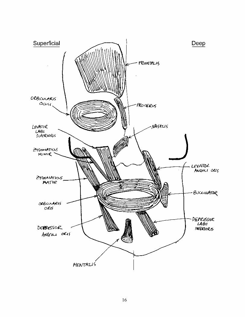
Note: In the drawing, superficial muscles inserting into Orbicularis oris areoriented inferomedially, whereas deep muscles are oriented inferolaterally.
18
19
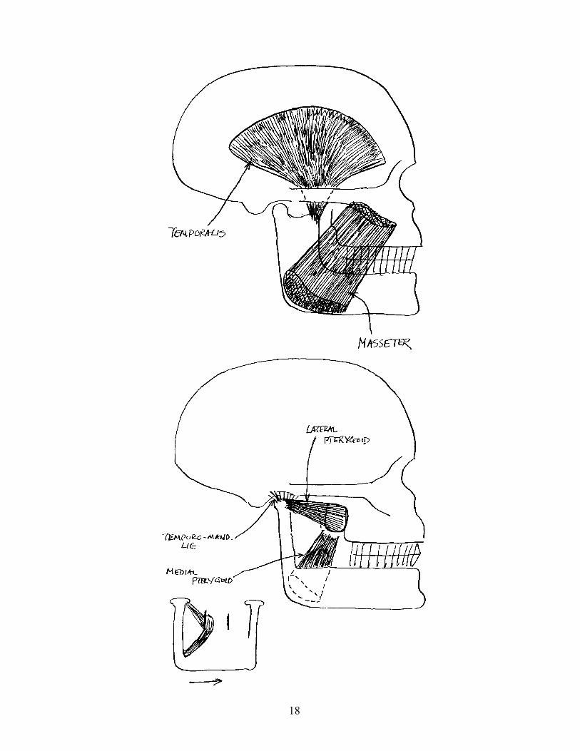
—————————————————————————————————————————————————————————————————————————————— MUSCLES OF MASTICATION
MUSCLE ORIGIN INSERTION ACTION/(NERVE)——————————————————————————————————————————————————————————————————————————————
Masseter Zygomatic arch Outer surface of Elevates and ramus of mandible protrudes mandible
(Trigeminal N.)
Temporalis Fan-shaped area Coronoid process Elevates and on side of skull of mandible retracts mandible
(Trigeminal N.)
Medial pterygoid Medial surface of Ramus of mandible Elevates and lateral pterygoid near angle laterally plate displaces mandible
(Trigeminal N.)
Lateral pterygoid Lateral surface of Neck of mandible Protrudes mandible lateral pterygoid near articulation (Trigeminal N.) plate
——————————————————————————————————————————————————————————————————————————————
Note: All muscles of mastication are innervated by branches of the TrigeminalN.
20
21
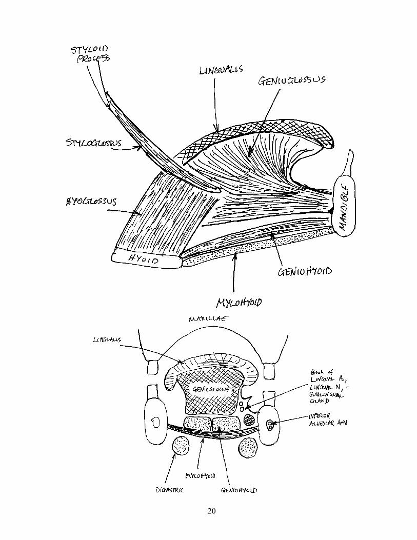
——————————————————————————————————————————————————————————————————————————————MUSCLES OF THE TONGUE
MUSCLE ORIGIN INSERTION ACTION/(NERVE)——————————————————————————————————————————————————————————————————————————————
Styloglossus Styloid process Into side of Retracts and tongue elevates tongue (Hypoglossal N.)
Hyoglossus Hyoid bone Into side of Retracts and tongue depress tongue; deflects tongue to side (Hypoglossal N.)
Genioglossus Mental spines of Fan out beneath Retracts, mandible body of tongue protrudes, and depresses tongue (Hypoglossal N.)
Lingualis Muscular body of tongue that Changes shape of lies on top of Genioglossus tongue (Hypoglossal N.)
——————————————————————————————————————————————————————————————————————————————
Note: All muscles of the tongue are innervated by the Hypoglossal N.
22
23
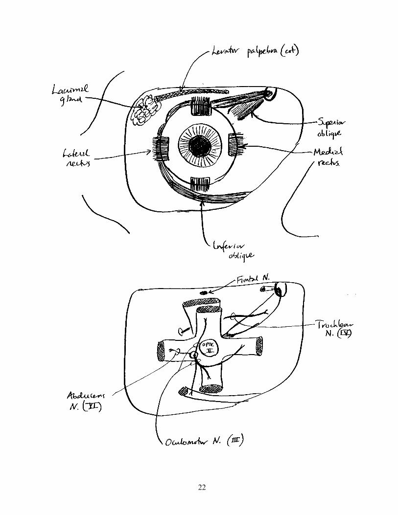
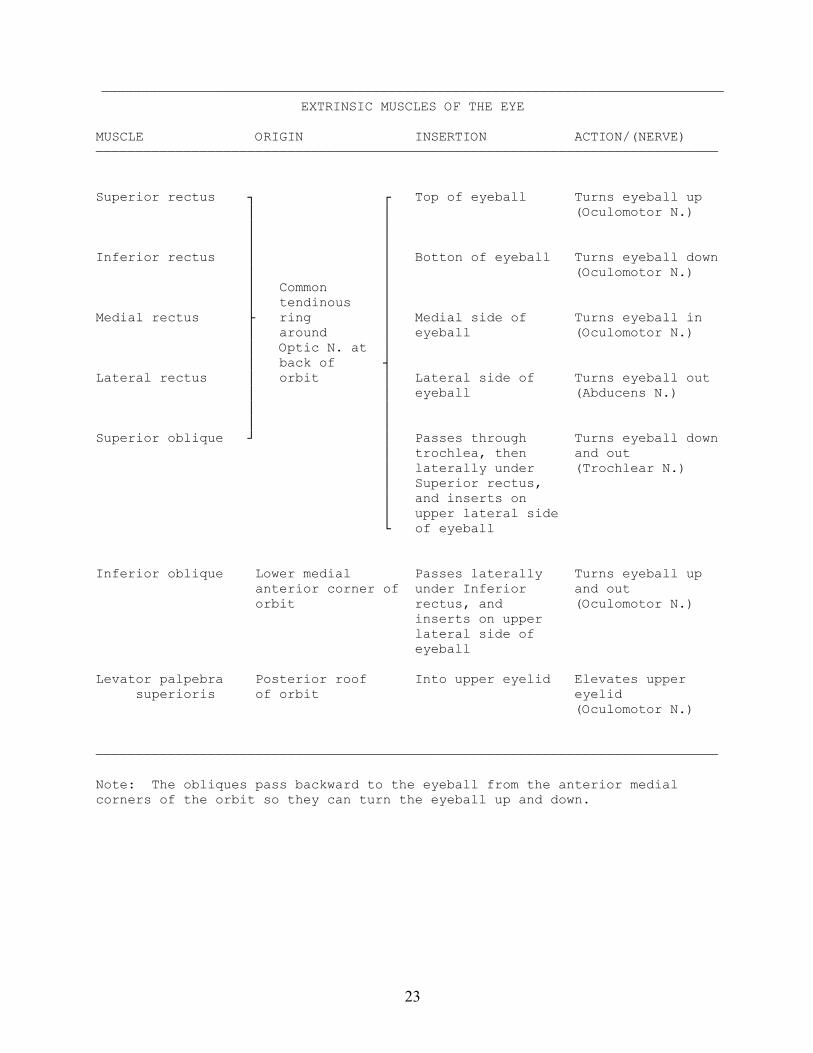
——————————————————————————————————————————————————————————————————————————————EXTRINSIC MUSCLES OF THE EYE
MUSCLE ORIGIN INSERTION ACTION/(NERVE)——————————————————————————————————————————————————————————————————————————————
Superior rectus ┐ ┌ Top of eyeball Turns eyeball up │ │ (Oculomotor N.) │ │ │ │Inferior rectus │ │ Botton of eyeball Turns eyeball down │ │ (Oculomotor N.) │ Common │ │ tendinous │Medial rectus ├ ring │ Medial side of Turns eyeball in │ around │ eyeball (Oculomotor N.) │ Optic N. at │ │ back of ┤Lateral rectus │ orbit │ Lateral side of Turns eyeball out │ │ eyeball (Abducens N.) │ │ │ │Superior oblique ┘ │ Passes through Turns eyeball down │ trochlea, then and out │ laterally under (Trochlear N.) │ Superior rectus, │ and inserts on │ upper lateral side └ of eyeball
Inferior oblique Lower medial Passes laterally Turns eyeball up anterior corner of under Inferior and out orbit rectus, and (Oculomotor N.) inserts on upper lateral side of eyeball
Levator palpebra Posterior roof Into upper eyelid Elevates upper superioris of orbit eyelid (Oculomotor N.)
——————————————————————————————————————————————————————————————————————————————
Note: The obliques pass backward to the eyeball from the anterior medialcorners of the orbit so they can turn the eyeball up and down.
24
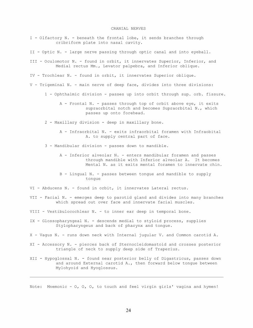
CRANIAL NERVES
I - Olfactory N. - beneath the frontal lobe, it sends branches throughcribriform plate into nasal cavity.
II - Optic N. - large nerve passing through optic canal and into eyeball.
III - Oculomotor N. - found in orbit, it innervates Superior, Inferior, andMedial rectus Mm., Levator palpebra, and Inferior oblique.
IV - Trochlear N. - found in orbit, it innervates Superior oblique.
V - Trigeminal N. - main nerve of deep face, divides into three divisions:
1 - Ophthalmic division - passes up into orbit through sup. orb. fissure.
A - Frontal N. - passes through top of orbit above eye, it exitssupraorbital notch and becomes Supraorbital N., whichpasses up onto forehead.
2 - Maxillary division - deep in maxillary bone.
A - Infraorbital N. - exits infraorbital foramen with InfraobitalA. to supply central part of face.
3 - Mandibular division - passes down to mandible.
A - Inferior alveolar N. - enters mandibular foramen and passesthrough mandible with Inferior alveolar A. It becomesMental N. as it exits mental foramen to innervate chin.
B - Lingual N. - passes between tongue and mandible to supplytongue
VI - Abducens N. - found in orbit, it innervates Lateral rectus.
VII - Facial N. - emerges deep to parotid gland and divides into many brancheswhich spread out over face and innervate facial muscles.
VIII - Vestibulocochlear N. - to inner ear deep in temporal bone.
IX - Glossopharyngeal N. - descends medial to styloid process, suppliesStylopharyngeus and back of pharynx and tongue.
X - Vagus N. - runs down neck with Internal jugular V. and Common carotid A.
XI - Accessory N. - pierces back of Sternocleidomastoid and crosses posteriortriangle of neck to supply deep side of Trapezius.
XII - Hypoglossal N. - found near posterior belly of Digastricus, passes downand around External carotid A., then forward below tongue betweenMylohyoid and Hyoglossus.
——————————————————————————————————————————————————————————————————————————————
Note: Mnemonic - O, O, O, to touch and feel virgin girls' vagina and hymen!

25
———————————————————————————————————————————————————————————————————————————————————————————————————FUNCTIONS OF CRANIAL NERVES
CN # NAME SPECIAL SENSE SENSORY MOTOR PARASYMPATHETIC———————————————————————————————————————————————————————————————————————————————————————————————————
I Olfactory Smell -- -- --
II Optic Sight -- -- --
III Oculomotor -- -- Most eye muscles Muscles of lens and iris
IV Trochlear -- -- Superior oblique --
V Trigeminal -- Face, mouth, Muscles of -- and 2/3 of mastication tongue
VI Abducens -- -- Lateral rectus --
VII Facial Taste 2/3 tongue -- Facial muscles Salivary and lacrimal glands
VIII Vestibulocochlear Hearing and -- -- -- equilibrium
IX Glossopharyngeal Taste 1/3 tongue 1/3 of tongue, Stylopharyngeus Parotid gland pharynx, middle ear
X Vagus -- Pharynx, larynx, Mm. of pharynx Viscera and viscera and larynx
XI Accessory -- -- Sternomastoid -- and trapezius
XII Hypoglossal -- -- Mm. of tongue --
———————————————————————————————————————————————————————————————————————————————————————————————————
Note: Mnemonic – Some say marry money, but my brother says big breasts matter most.
26
27
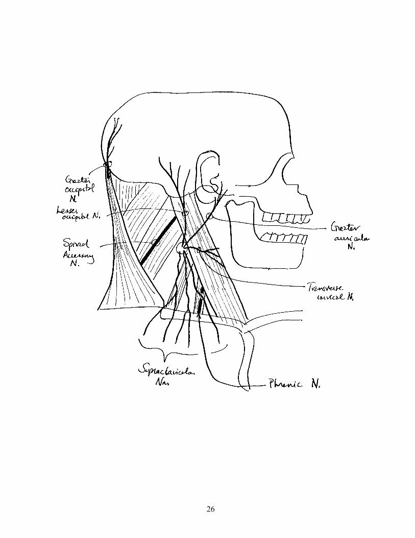
SPINAL NERVES OF THE HEAD AND NECK
I - Dorsal rami of Spinal Nn.
A - Suboccipital N. (C1) - found in suboccipital triangle with VertebralA. Innervates rectus capitus posterior muscles and Obliquuscapitus superior.
B - Greater occipital N. (C2) - pierces Semispinalis capitus nearmidline, then pierces Trapezius and runs up onto back ofskull.
II - Ventral rami of Spinal Nn. (=Cervical Plexus).
A - Lesser occipital N. (C2,3) - found in posterior triangle betweenLevator scapulae and Scalenus medius, it passes up onto skullbetween external ear and Greater occipital N.
B - Great auricular N. (C2,3) - found in posterior triangle betweenLevator scapulae and Scalenus medius, it passes up overSternocleidomastoid toward external ear.
C - Supraclavicular Nn. (C3,4) - found in posterior triangle betweenLevator scapulae and Scalenus medius, they passes down overclavicle.
D - Phrenic N. (C3-5) - descends neck on Scalenus anterior behindInternal jugular V., then passes into thorax behind first rib.In thorax it passes down to supply diaphragm.
III - Sympathetic trunk - deep to Carotid sheath lying on prevertebral muscles,it has a large cigar-shaped ganglion high in neck, a smallerganglion at base of neck, and passes down into thorax.
28
ARTERIES OF THE HEAD AND NECK
29
ARTERIES OF THE HEAD AND NECK
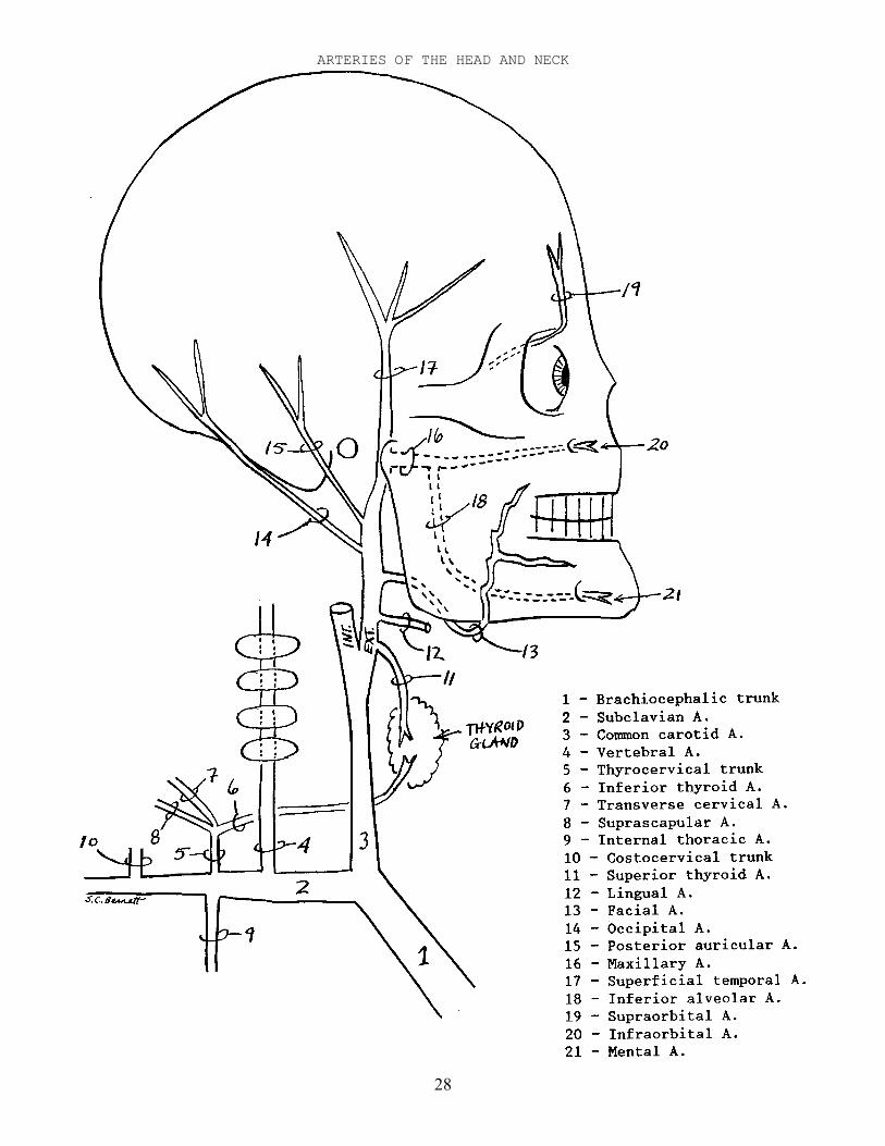
I - Common carotid A. - large branch off Brachiocephalic trunk (right) orAortic arch (left), passes up Carotid sheath with Internal jugularV., and divides into External and Internal carotid Aa.
A - External carotid A. - passes up side of neck and skull to temple.
1 - Superior thyroid A. - passes down to Thyroid gland.
2 - Lingual A. - passes anteriorly to tongue, then betweenHyoglossus and Genioglossus.
3 - Facial A. - passes through Submandibular gland, then up ontoface, and follows a sinuous path toward corner of mouth.
4 - Occipital A. - passes up posteriorly to back of skull, thenaccompanies Greater occipital N.
5 - Posterior auricular A. - passes up behind ear.
6 - Maxillary A. - large branch medial to mandible. It passesforward, sends a branch down to mandible, and continuesinto maxilla.
a - Inferior alveolar A. - branches off Maxillary A. near itsbeginning, and passes down into mandibularforamen, passes through mandible, exits mentalforamen, and becomes Mental A. to supply chin.
b - Infraorbital A. - terminal branch of Maxillary A., itexits Infraorbital foramen with Infraorbital N.
7 - Superficial temporal A. - terminal branch of External carotidA. It passes up onto scalp.
a - Transverse facial A. - small branch, passes forwardacross Masseter below zygomatic arch.
B - Internal carotid A. - passes up into skull to supply brain.
1 - Ophthalmic A. - passes through Optic canal into orbit, supplieseye, and sends a branch through supraorbital foramen tobecome Supraorbital A.
a - Supraorbital A. - passes onto forehead with Supraorbital N.
——————————————————————————————————————————————————————————————————————————————
Note: A mnemonic for the branches of the External carotid A. from inferior tosuperior is: Some like fast, others prefer much slower.
30
ARTERIES AND VEINS OF THE BRAIN
31
ARTERIES AND VEINS OF THE BRAIN
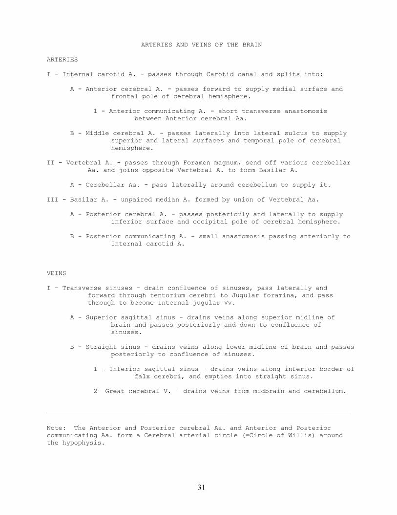
ARTERIES
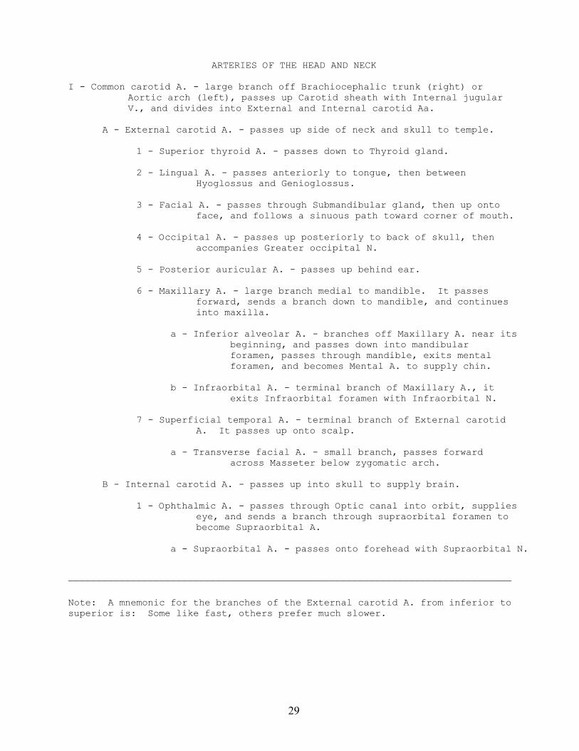
I - Internal carotid A. - passes through Carotid canal and splits into:
A - Anterior cerebral A. - passes forward to supply medial surface andfrontal pole of cerebral hemisphere.
1 - Anterior communicating A. - short transverse anastomosisbetween Anterior cerebral Aa.
B - Middle cerebral A. - passes laterally into lateral sulcus to supplysuperior and lateral surfaces and temporal pole of cerebralhemisphere.
II - Vertebral A. - passes through Foramen magnum, send off various cerebellarAa. and joins opposite Vertebral A. to form Basilar A.
A - Cerebellar Aa. - pass laterally around cerebellum to supply it.
III - Basilar A. - unpaired median A. formed by union of Vertebral Aa.
A - Posterior cerebral A. - passes posteriorly and laterally to supplyinferior surface and occipital pole of cerebral hemisphere.
B - Posterior communicating A. - small anastomosis passing anteriorly toInternal carotid A.
VEINS
I - Transverse sinuses - drain confluence of sinuses, pass laterally andforward through tentorium cerebri to Jugular foramina, and passthrough to become Internal jugular Vv.
A - Superior sagittal sinus - drains veins along superior midline ofbrain and passes posteriorly and down to confluence ofsinuses.
B - Straight sinus - drains veins along lower midline of brain and passesposteriorly to confluence of sinuses.
1 - Inferior sagittal sinus - drains veins along inferior border offalx cerebri, and empties into straight sinus.
2- Great cerebral V. - drains veins from midbrain and cerebellum.
——————————————————————————————————————————————————————————————————————————————
Note: The Anterior and Posterior cerebral Aa. and Anterior and Posteriorcommunicating Aa. form a Cerebral arterial circle (=Circle of Willis) aroundthe hypophysis.
32
VEINS OF THE HEAD AND NECK
33
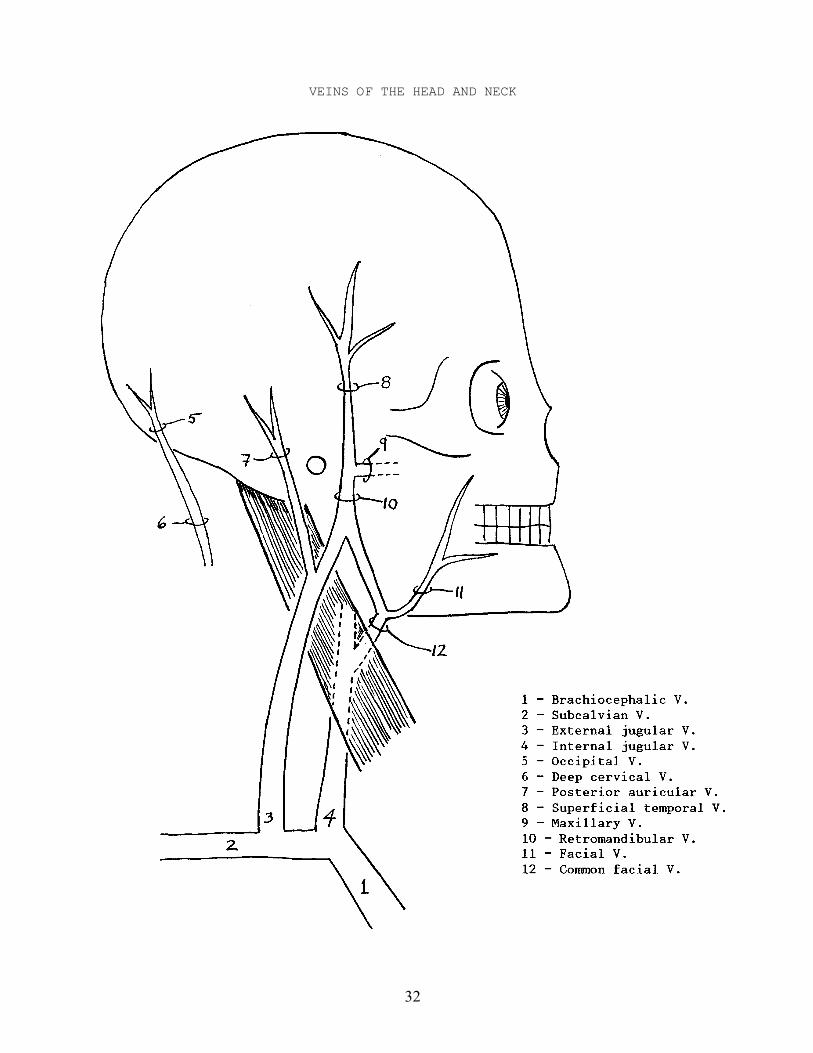
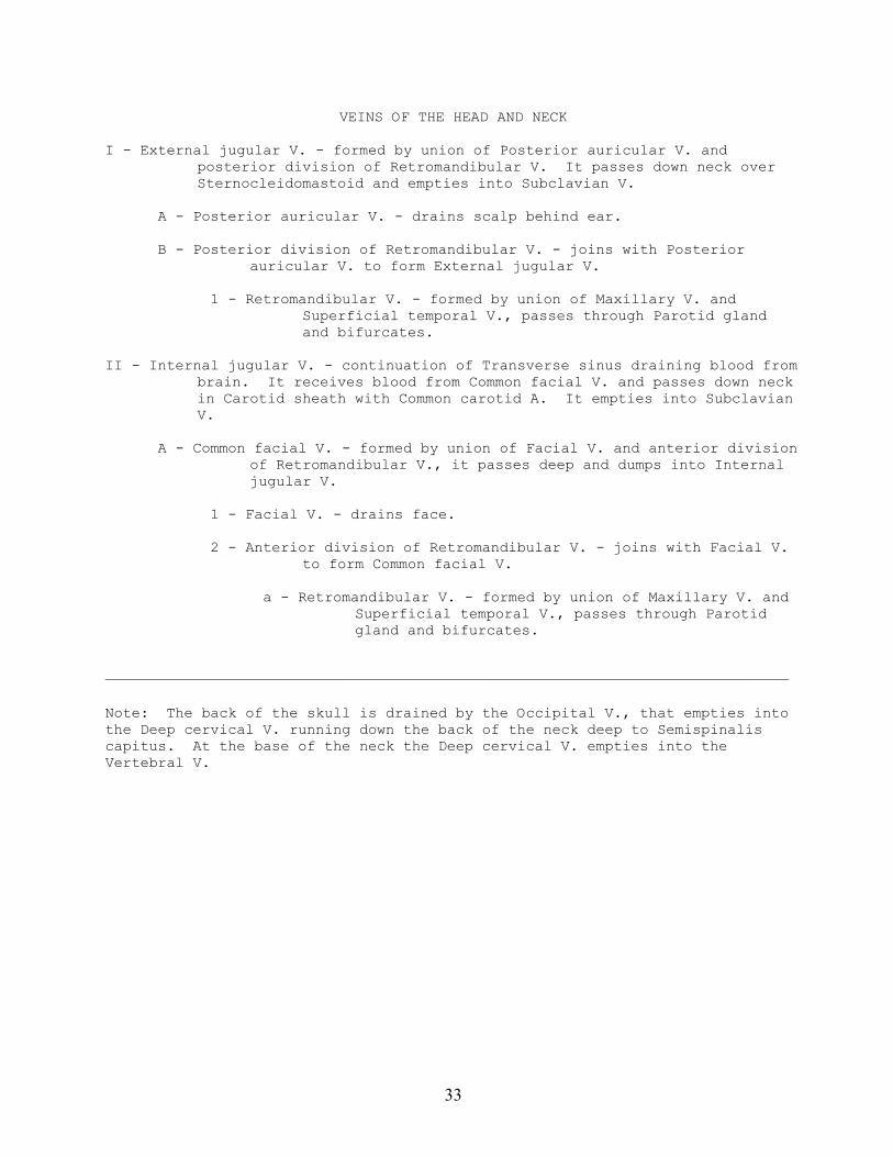
VEINS OF THE HEAD AND NECK
I - External jugular V. - formed by union of Posterior auricular V. andposterior division of Retromandibular V. It passes down neck overSternocleidomastoid and empties into Subclavian V.
A - Posterior auricular V. - drains scalp behind ear.
B - Posterior division of Retromandibular V. - joins with Posteriorauricular V. to form External jugular V.
1 - Retromandibular V. - formed by union of Maxillary V. andSuperficial temporal V., passes through Parotid glandand bifurcates.
II - Internal jugular V. - continuation of Transverse sinus draining blood frombrain. It receives blood from Common facial V. and passes down neckin Carotid sheath with Common carotid A. It empties into SubclavianV.
A - Common facial V. - formed by union of Facial V. and anterior divisionof Retromandibular V., it passes deep and dumps into Internaljugular V.
1 - Facial V. - drains face.
2 - Anterior division of Retromandibular V. - joins with Facial V.to form Common facial V.
a - Retromandibular V. - formed by union of Maxillary V. andSuperficial temporal V., passes through Parotidgland and bifurcates.
——————————————————————————————————————————————————————————————————————————————
Note: The back of the skull is drained by the Occipital V., that empties intothe Deep cervical V. running down the back of the neck deep to Semispinaliscapitus. At the base of the neck the Deep cervical V. empties into theVertebral V.
34
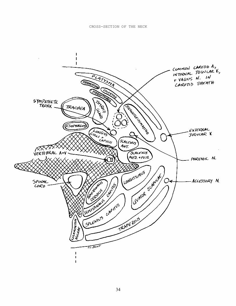
CROSS-SECTION OF THE NECK
35
OSTEOLOGY OF THE POSTCRANIAL AXIAL SKELETON
VERTEBRAL COLUMN - consists of 5 regions
7 cervical vertebrae12 thoracic vertebrae - bearing ribs5 lumbar vertebrae5 sacral vertebrae - fused to form Sacrum4 coccygeal vertebrae - fused to form Coccyx - said to resemble Cuckoo's beak
PARTS OF VERTEBRAE
Body or centrum - bears weight; articulates with adjacent vertebraeArch - encloses vertebral foramen, supports spinous and transverse processes
and superior and inferior articular surfacesVertebral foramen - for spinal cordSuperior and inferior articular processes and surfaces - articulate with
adjacent vertebraeSpinous process - for origin and insertion of musclesTransverse process - for origin and insertion of muscles; carry ribs in
thoracic regionIntervertebral disc - fibrous joint between vertebral centra
CERVICAL VERTEBRAE
All have transverse foramina (Foramina transversaria) for Vertebral A+V.C1 (=atlas) - articulates with occipital condylesC2 (=axis) - has odontoid process that permits atlas to rotate on axisC3-C5 have bifid spinous processes for Ligamentum nuchae
THORACIC VERTEBRAE
Costotransverse articulations - on transverse processes for tubercle of ribSuperior and inferior costal articulations - on centrum for head of rib
LUMBAR VERTEBRAE
More robust than thoracic vertebrae. Do not carry ribs, but with largetransverse process and mammillary processes for attachment of muscles.
SACRAL VERTEBRAE - Fused together to form sacrum
Body - fused bodies of centra of sacral vertebraeAlae - fused transverse processes of sacral vertebraeAuricular surface - articulates with pelvic boneMedian Crest - fused spinous processes of sacral vertebraeSacral Foramina - between transverse processes, for sacral spinal nerves
36
RIBS: True Ribs (pairs 1-7) False Ribs (pairs 8-12) Floating Ribs (pairs 11-12)
HeadSuperior and Inferior Articular Facets - articulate with vertebral centraNeckTubercle - articulates with transverse process of vertebraAngle - lateral margin of erector spinae musclesBodyChondral (=sternal) endCostal cartilages - cartilaginous joint between rib and sternum
STERNUM
ManubriumClavicular Notch - articulates with clavicle
BodyXiphoid Process
——————————————————————————————————————————————————————————————————————————————
JOINTS OF THE AXIAL SKELETON
Movements of Vertebral Column
Skull-atlas - flexion and extensionAtlas-axis - rotation, slight flexion and extensionAll others - flexion and extension in sagittal plane - lateral flexion (in coronal plane) - rotation
Movements of Ribs
- swing out, up, and forward like bucket handle to elevate sternum and increase volume of ribcage

37
NOTES
38
39
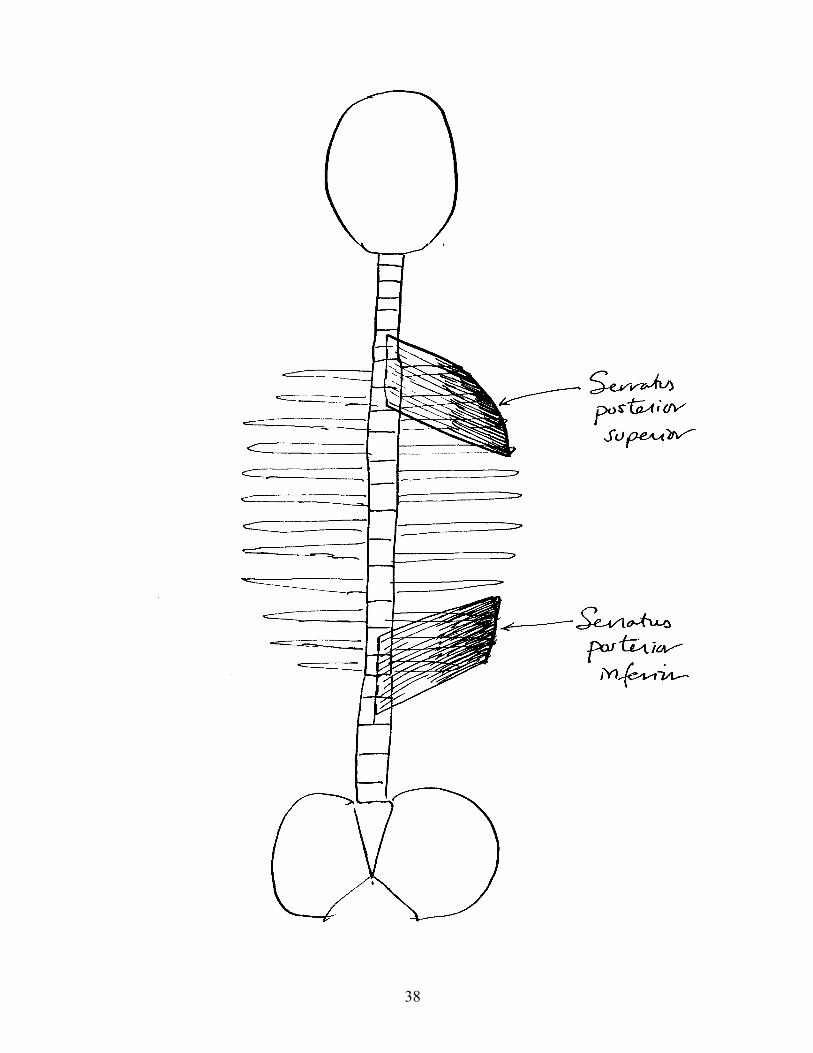
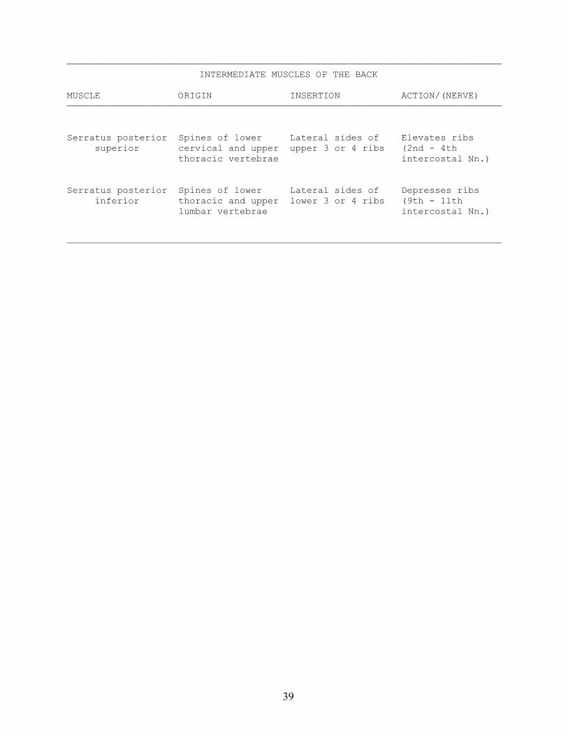
——————————————————————————————————————————————————————————————————————————————INTERMEDIATE MUSCLES OF THE BACK
MUSCLE ORIGIN INSERTION ACTION/(NERVE)——————————————————————————————————————————————————————————————————————————————
Serratus posterior Spines of lower Lateral sides of Elevates ribs superior cervical and upper upper 3 or 4 ribs (2nd - 4th thoracic vertebrae intercostal Nn.)
Serratus posterior Spines of lower Lateral sides of Depresses ribs inferior thoracic and upper lower 3 or 4 ribs (9th - 11th lumbar vertebrae intercostal Nn.)
——————————————————————————————————————————————————————————————————————————————
40
41
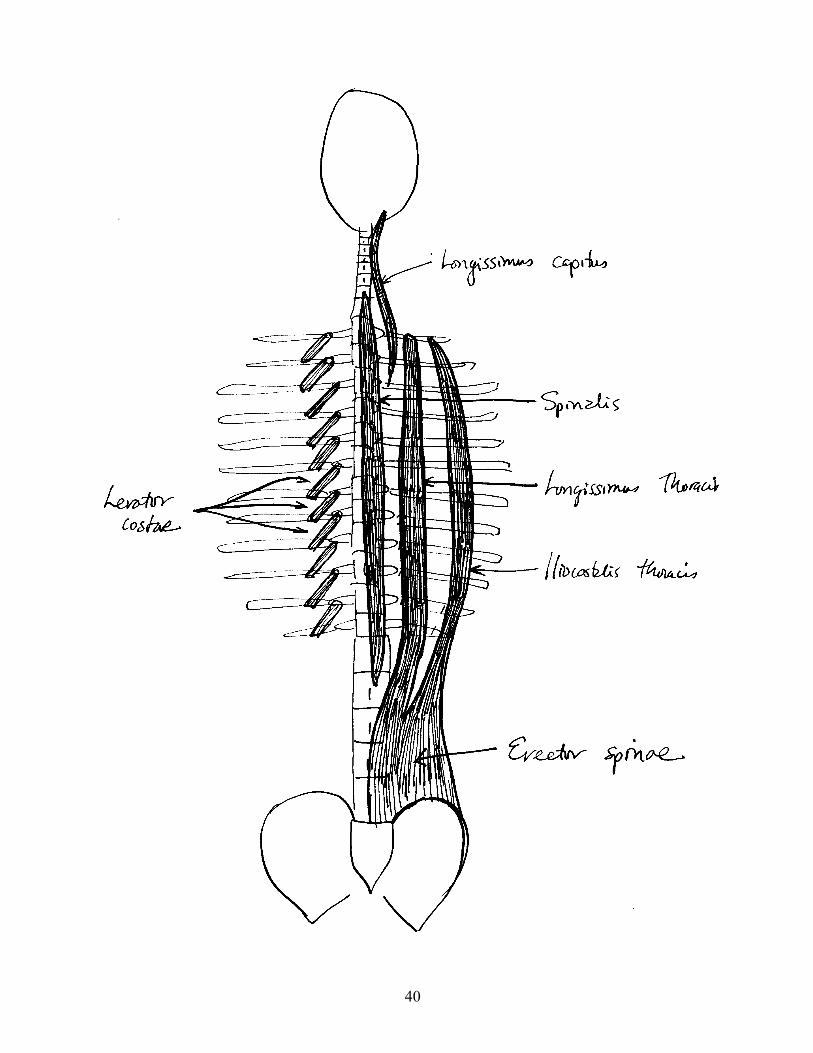
——————————————————————————————————————————————————————————————————————————————ERECTOR SPINAE MUSCLES
MUSCLE ORIGIN INSERTION ACTION/(NERVE)——————————————————————————————————————————————————————————————————————————————
Spinalis Spines of lower Spines of upper ┐ thoracic and upper thoracic vertebrae │ lumbar vertebrae │ │ │Longissimus With entire By slips - medial │ thoracis Erector spinae to transverse │ group from spines processes of all 12 │ of sacral and thoracic vertebrae; │ Extend, lumbar vertebrae and lateral to all │ rotate, and and iliac crest 12 ribs │ laterally flex ├ vertebral │ columnLongissimus Continuation of Tendon between │ (Dorsal rami capitus longissimus - from Splenius capitus │ of Spinal Nn.) transverse and Semispinalis │ processes of upper capitus, onto back │ thoracic and lower of mastoid process │ 4 cervical │ vertebrae │ │ │Iliocostalis With entire By slips to back │ Erector spinae of all 12 ribs ┘ group from spines of sacral and lumbar vertebrae and iliac crest
——————————————————————————————————————————————————————————————————————————————
Note: The three columns of muscle usually cannot be differentiated in thelumbar region and are best considered to be simply Erector spinae.
Note: The main function of these muscles is to maintain the curvatures of thespine and keep the trunk erect.
42
43
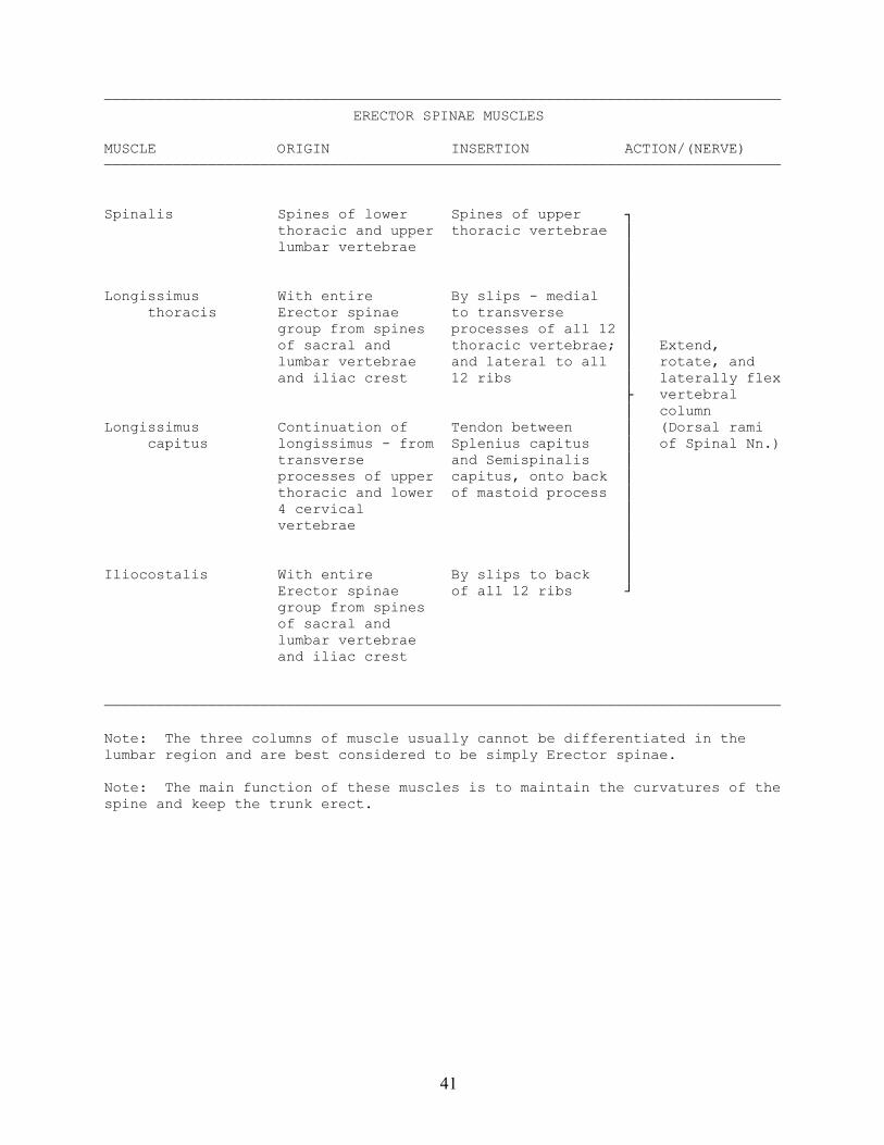
——————————————————————————————————————————————————————————————————————————————TRANSVERSOSPINALIS MUSCLES
MUSCLE ORIGIN INSERTION ACTION/(NERVE)——————————————————————————————————————————————————————————————————————————————
Semispinalis Transverse Between superior ┐ capitus processes of lower and inferior │ 4 cervical and nuchal lines on │upper 6 thoracic skull; just deep │vertebrae to Trapezius │ │ │Semispinalis Articular Spans 3 to 6 │ cervicis processes of lower vertebrae and │ cervical vertebrae inserts of spines │ Extend, and transverse of cervical │ rotate, and processes of upper vertebrae │ laterally flex thoracic vertebrae ├ vertebral │ column │ (Dorsal ramiMultifidus Transverse Spans 2 to 3 │ of Spinal Nn.) processes of vertebrae and │ vertebrae inserts on spines │ of vertebrae │ │ │Rotatores Transverse Spans from one │ processes of vertebra to next │ vertebrae above, inserts on │ spine of vertebra ┘
------------------------------------------------------------------------------
Levator costae Transverse Passes laterally Elevate ribs processes of last and down to back (Dorsal ram cervical to 11th of next rib Spinal Nn.) thoracic vertebrae between angle and tubercle
——————————————————————————————————————————————————————————————————————————————
Note: Levator costae is technically not a transversospinalis muscle, but wasderived from them.
Note: All transversospinalis muscles originate on a transverse process andinsert on a spinous process, hence the name. The transversospinalis musclesare best seen in different parts of the back: Semispinalis is best seen in thecervical region, Multifidus is best seen in the lumbar region, and Rotatoresare best seen in the thoracic region.
44
45
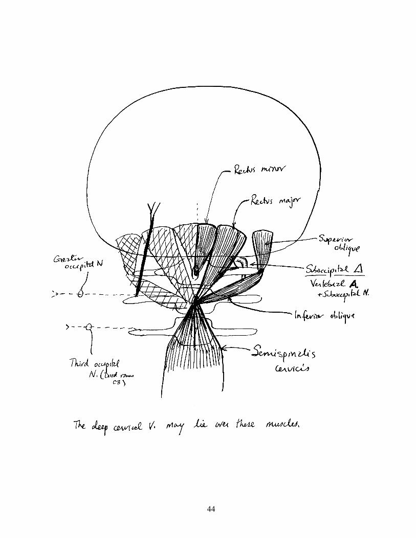
——————————————————————————————————————————————————————————————————————————————MUSCLES OF SUBOCCIPITAL REGION
MUSCLE ORIGIN INSERTION ACTION/(NERVE)——————————————————————————————————————————————————————————————————————————————
Obliquus capitis Transverse Occipital bone ┐ superior process of atlas │ │ │Obliquus capitis Spine of axis Transverse process │ inferior of atlas │ Extend and ├ rotate head │ (SuboccipitalRectus capitus Spine of axis Occipital bone deep │ N.) posterior to Semispinalis │ major │ │ │Rectus capitus Posterior tubercle Occipital bone deep │ posterior of atlas to Rectus major ┘ minor
——————————————————————————————————————————————————————————————————————————————
Note: There are similar short muscles (Rectus capitus anterior and Rectuscapitus lateralis) between the atlas and occipital bone on the anterior side.All of these muscles are primarily postural.
Note: The Greater occipital N. passes over these muscles and piercesSemispinalis capitus and trapezius. When Semispinalis capitus is reflected,the Greater occipital N. lies transversely on its deep surface. Also visibleare the 3rd occipital N. and the Deep cervical V.
46
47
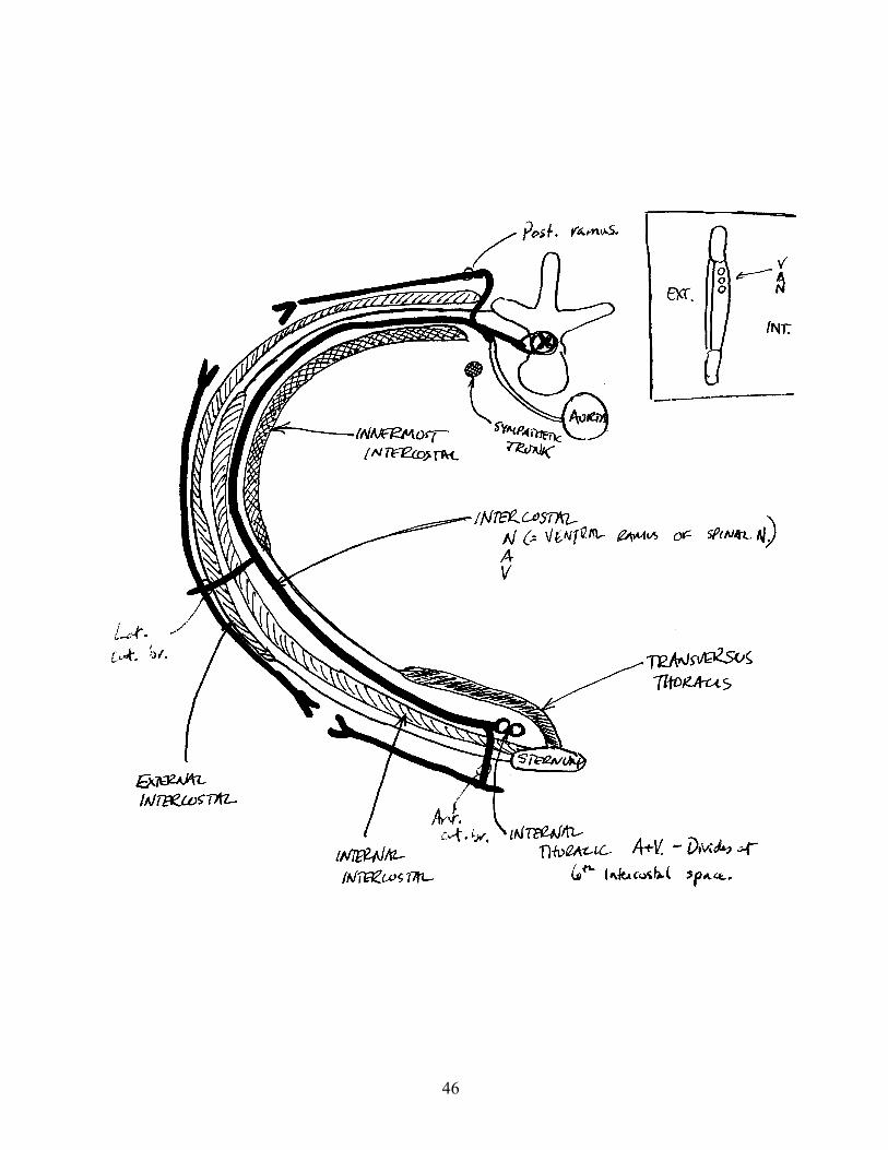
—————————————————————————————————————————————————————————————————————————————— MUSCLES OF THE THORACIC WALL
MUSCLE ORIGIN INSERTION ACTION/(NERVE)——————————————————————————————————————————————————————————————————————————————
External Lower border of Passes down and ┐ intercostals ribs medially to next │ lower rib │ │ │Internal Lower border of Passes down and │ intercostals ribs laterally to next │ lower rib │ Move ribcage │ during active│ ├ respirationInnermost Same origin and insertion as internal │ (Intercostal intercostals intercostals; variable layer lying │ Nn.) over Intercostal NAV, may be difficult │ to differentiate from Internal │ intercostals │ │ │Transversus Back of sternum Inner surface of │ thoracis costal cartilages ┘
——————————————————————————————————————————————————————————————————————————————
Note: The ribcage is also moved directly by Serratus posterior superior andinferior, Levator costae, and the scalenes. Other muscles can also move theribs during powerful respiration, e.g., Sternocleidomastoid, Rectus abdominus,etc.
48
49
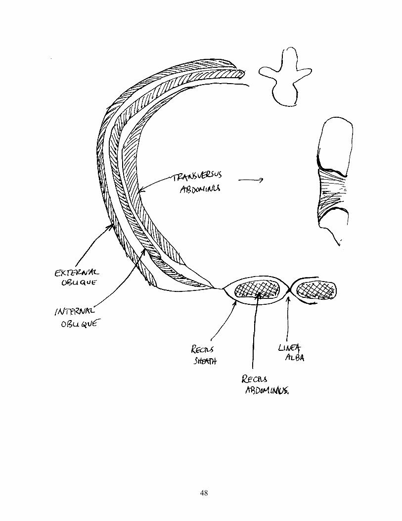
—————————————————————————————————————————————————————————————————————————————— MUSCLES OF THE ABDOMINAL WALL
MUSCLE ORIGIN INSERTION ACTION/(NERVE)——————————————————————————————————————————————————————————————————————————————
External oblique Outer surface of Passes down and ┐ lower 8 ribs medially to iliac │ crest and linea │ alba │ Flex and │ laterally flex │ lumbarInternal oblique Thoracolumbar Passes up and │ vertebrae; fascia, iliac medially to lower │ compress crest, and 3 ribs and linea ├ abdomen to inguinal ligament alba (splits │ expel contents; around Rectus │ obliques are abdominus) │ main rotators │ of lumbar spine │ (Ventral rami ofTransversus Deep on costal Aponeurosis of │ lower thoracic abdominus cartilages, iliac internal │ and first lumbar crest, and intercostal ┘ Nn.) inguinal ligament
Rectus abdominus Pubic symphysis Sternum and lower Flex lumbar spine, and pubic crest costal cartilages compress abdomen (Ventral rami of
lower thoracic Nn.)
——————————————————————————————————————————————————————————————————————————————
Note: A important function of the muscles of the abdominal wall is support theviscera against the pull of gravity.
50
MUSCLES OF THE PELVIS
51
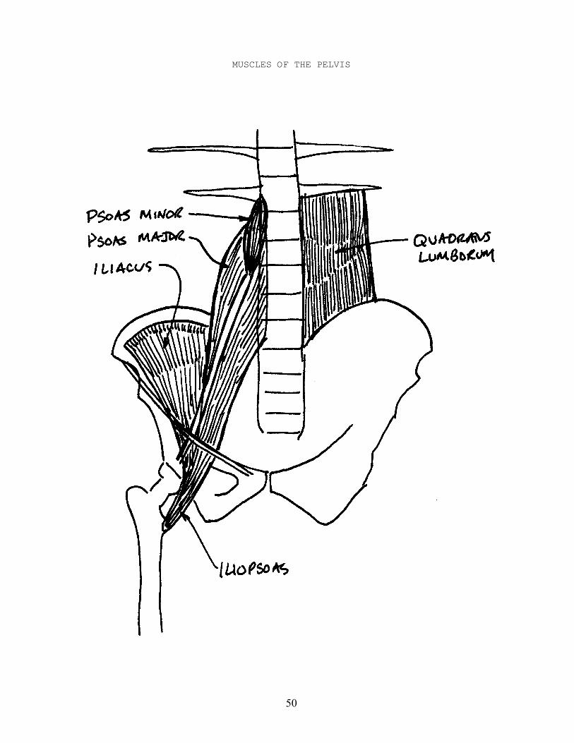
——————————————————————————————————————————————————————————————————————————————MUSCLES OF THE PELVIS
MUSCLE ORIGIN INSERTION ACTION/(NERVE)——————————————————————————————————————————————————————————————————————————————
Iliacus Iliac fossa Passes down to Flexes hip insert on lesser (Femoral N.) trochanter of femur with Psoas major
Psoas major Bodies of the Passes beneath Flexes hip lumbar vertebrae inguinal ligament (2nd and 3rd to insert on lumbar Nn.) lesser trochanter of femur with Iliacus
Psoas minor Bodies of the Iliopubic eminence Flexes lumbar last thoracic and near acetabulum vertebrae first lumbar (Ventral ramus of vertebrae L1)
Quadratus lumborum Iliac crest and Transverse Flexes lumbar transverse processes of vertebrae processes of lower upper lumbar laterally lumbar vertebrae vertebrae and (Ventral rami of last rib Subcostal N. and
L1-3 or 4)
——————————————————————————————————————————————————————————————————————————————
Note: Psoas minor is small, and is absent in 40% of individuals.
Note: Iliacus and Psoas major are discussed on p. 103 as Iliopsoas.
52
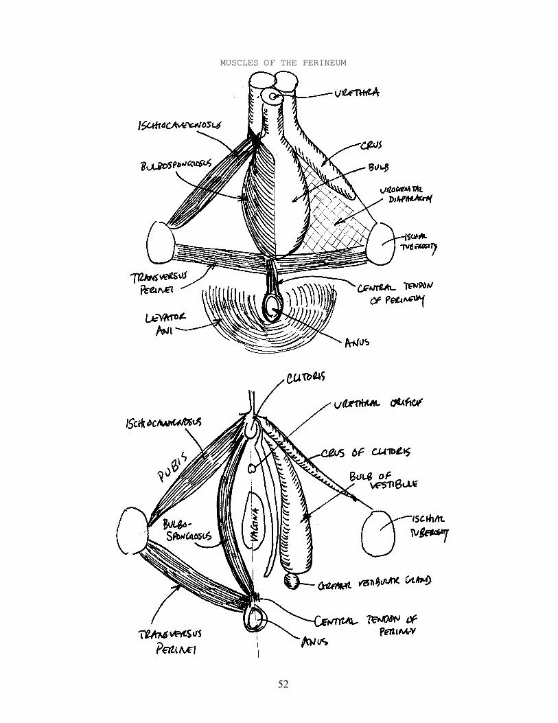
MUSCLES OF THE PERINEUM
53
——————————————————————————————————————————————————————————————————————————————MUSCLES OF THE PERINEUM - MALE
MUSCLE ORIGIN INSERTION ACTION/(NERVE)——————————————————————————————————————————————————————————————————————————————
Bulbospongiosus Central tendon of Pubic arch, Compresses bulb of perineum and urogenital corpus spongiosum median raphe with diaphragm, and during micturation opposite dorsum of penis and ejaculation; Bulbospongiosus compresses Dorsal V. of penis (Pudendal N.)
Ischiocavernosus Medial side of Pubic arch on Compresses crus of ischial tuberosity side of crus of penis to erect penis (Pudendal N.)
Transversus Medial side of Central tendon of Stabilizes central perinei ischial tuberosity perineum near anus tendon of perineum (Pudendal N.)
——————————————————————————————————————————————————————————————————————————————
——————————————————————————————————————————————————————————————————————————————MUSCLES OF THE PERINEUM - FEMALE
MUSCLE ORIGIN INSERTION ACTION/(NERVE)——————————————————————————————————————————————————————————————————————————————
Bulbospongiosus Central tendon of Pubic arch, and Sphincter of perineum root and dorsum vagina; compresses of clitoris Dorsal V. of clitoris (Pudendal N.)
Ischiocavernosus Medial side of Pubic arch on Compresses crus of ischial tuberosity side of crus of clitoris to erect clitoris (Pudendal N.)
Transversus Medial side of Central tendon of Stabilizes central perinei ischial tuberosity perineum tendon of perineum (Pudendal N.)
——————————————————————————————————————————————————————————————————————————————
Note: The central tendon of the perineum is a muscular/fibrous band extendinganteriorly on the midline from the anus to the urogenital diaphragm.

54
NOTES
55
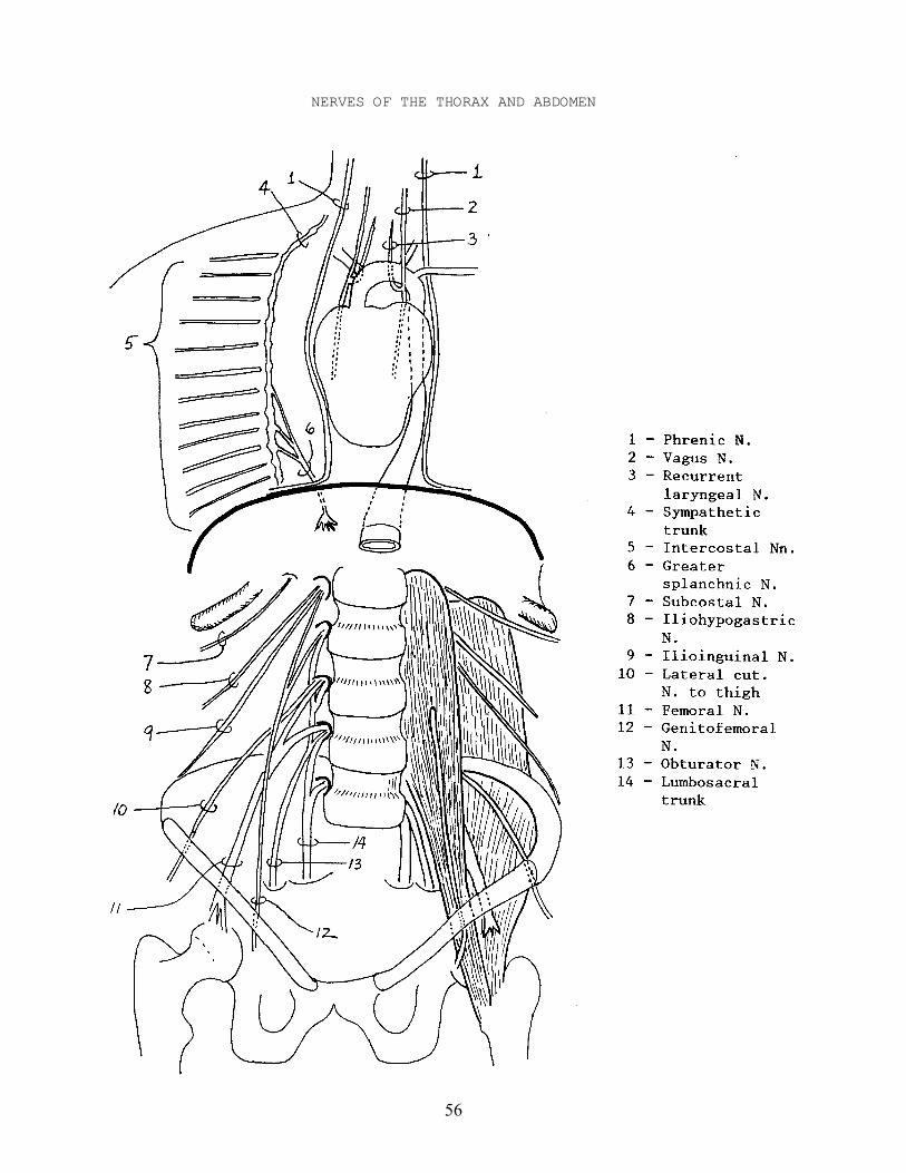
NERVES OF THE THORAX
I - Cranial Nerves
A - Vagus N. - passes down neck into mediastinum, in front of Aortic archand sends off Recurrent laryngeal N., then continues behindheart to form plexus around the esophagus.
1 - Recurrent laryngeal N. - passes behind Brachiocephalic trunk onright and Aortic arch on left, then up neck. Note theybegin at a different level on each side.
II - Cervical Plexus
A - Phrenic N. - passes down neck, on side of fibrous pericardium, infront of root of lung, pierces diaphragm and supplies itsventral surface.
III - Spinal Nerves
A - Intercostal Nn. - ventral rami of Spinal Nn. T1-T11. They runlaterally between ribs to supply thoracic wall.
IV - Sympathetic Trunk - continuation of trunk from neck, descends thorax oneither side of vertebral column. Has 11 ganglia in thorax whichgive it a lumpy appearance.
A - Greater splanchnic N. - main visceral branch of Sympathetic trunk.It passes anteriorly and inferiorly on side of vertebralcolumn, pierces the diaphragm, and ends in large Celiacganglion, which in turn sends branches to the abdominalviscera.
56
NERVES OF THE THORAX AND ABDOMEN
57
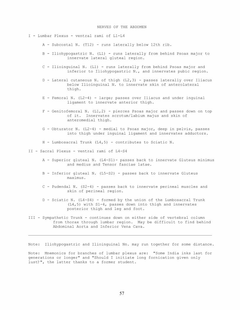
NERVES OF THE ABDOMEN
I - Lumbar Plexus - ventral rami of L1-L4
A - Subcostal N. (T12) - runs laterally below 12th rib.
B - Iliohypogastric N. (L1) - runs laterally from behind Psoas major toinnervate lateral gluteal region.
C - Ilioinguinal N. (L1) - runs laterally from behind Psoas major andinferior to Iliohypogastric N., and innervates pubic region.
D - Lateral cutaneous N. of thigh (L2,3) - passes laterally over Iliacusbelow Ilioinguinal N. to innervate skin of anterolateralthigh.
E - Femoral N. (L2-4) - large; passes over Iliacus and under inguinalligament to innervate anterior thigh.
F - Genitofemoral N. (L1,2) - pierces Psoas major and passes down on topof it. Innervates scrotum/labium majus and skin ofanteromedial thigh.
G - Obturator N. (L2-4) - medial to Psoas major, deep in pelvis, passesinto thigh under inguinal ligament and innervates adductors.
H - Lumbosacral Trunk (L4,5) - contributes to Sciatic N.
II - Sacral Plexus - ventral rami of L4-S4
A - Superior gluteal N. (L4-S1)- passes back to innervate Gluteus minimusand medius and Tensor fasciae latae.
B - Inferior gluteal N. (L5-S2) - passes back to innervate Gluteusmaximus.
C - Pudendal N. (S2-4) - passes back to innervate perineal muscles andskin of perineal region.
D - Sciatic N. (L4-S4) - formed by the union of the Lumbosacral Trunk(L4,5) with S1-4, passes down into thigh and innervatesposterior thigh and leg and foot.
III - Sympathetic Trunk - continues down on either side of vertebral columnfrom thorax through lumbar region. May be difficult to find behindAbdominal Aorta and Inferior Vena Cava.
——————————————————————————————————————————————————————————————————————————————
Note: Iliohypogastric and Ilioinguinal Nn. may run together for some distance.
Note: Mnemonics for branches of lumbar plexus are: "Some India inks last forgenerations or longer" and "Should I initiate long fornication given onlylust?", the latter thanks to a former student.
58
ARTERIES OF THE THORAX AND ABDOMEN

59
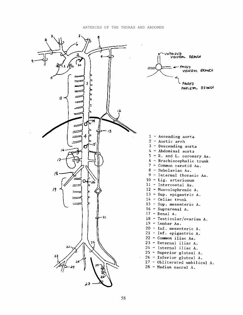
ARTERIES OF THE THORAX AND ABDOMEN
Ia - Ascending Aorta, Aortic Arch, and Descending Aorta in thorax.
A - Subclavian A.
1 - Internal thoracic A. - descends inside thoracic wall behindTransversus thoracis. Divides at 6th rib into:
a - Superior epigastric A. - pierces diaphragm and runs downRectus sheath, anastomose with Inferior epigastricA.
b - Musculophrenic A. - runs laterally on diaphragm.
B - Intercostal Aa. - pass laterally from Aorta between ribs.
Ib - Abdominal Aorta - continuation of Descending Aorta in abdomen.
A - CELIAC TRUNK - unpaired visceral branch supplying liver, spleen, andstomach. See p. 61 for branches.
B - SUPERIOR MESENTERIC A. - unpaired visceral branch supplying smallintestine and part of colon. See p. 61 for branches.
C - Suprarenal Aa. - small, pass to suprarenal glands.
D - Renal Aa. - large; pass laterally to kidneys.
E - Testicular/Ovarian Aa. - pass down over Psoas major to gonads.
F - Lumbar Aa. - pass laterally to abdominal wall.
G - INFERIOR MESENTERIC A. - unpaired visceral branch supplying part ofcolon and rectum. See p. 61 for branches.
H - Median sacral A. - passes down over sacrum between Common iliac Aa.
I - Common iliac Aa. - terminal branches, pass inferolaterally to supplulower extremilty.
1 - External iliac A. - passes down to thigh and becomes Femoral A.
a - Inferior epigastric A. - branches off just above inguinalligament, passes medially, then up rectus sheath.
2 - Internal iliac A. - passes down into pelvis.
a - Superior gluteal A. - passes back to gluteal regionbetween Lumbosacral trunk and 1st sacral ramus.
b - Inferior gluteal A. - passes back to gluteal regionbetween 1st and 3rd sacral rami.
c - Obliterated umbilical A. - fetal terminal branch, inadults a fibrous cord passing up abdominal wall toumbilicus.
60
UNPAIRED VISCERAL BRANCHES OF THE ABDOMINAL AORTA
61
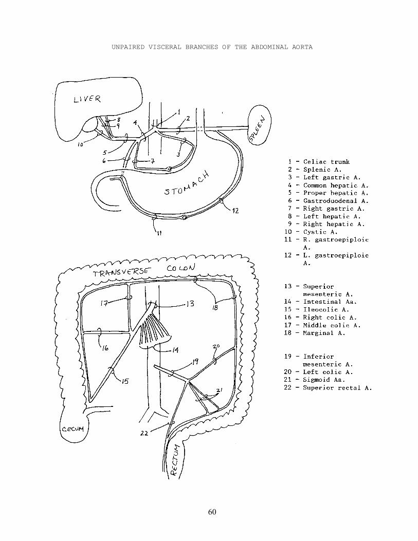
UNPAIRED VISCERAL BRANCHES OF THE ABDOMINAL AORTA
A - CELIAC TRUNK - unpaired visceral branch supplying liver, spleen, andstomach.
1 - Common hepatic A. - passes toward liver and gives off:
a - Gastroduodenal A. - passes to end of stomach and sends off:
i - Right Gastroepiploic A. - passes along greater curvatureof stomach and anastomoses with Leftgastroepiploic A.
b - Hepatic (Proper) A. - passes up to liver and splits into:
i - Right hepatic A. - enters Porta hepatis to supply liver.
ii - Left hepatic A - enters Porta hepatis to supply liver.
iii - Cystic A. - passes to gall bladder.
c - Right gastric A. - passes down to stomach and then along lessercurvature of stomach; anastomoses with Left gastric A.
2 - Splenic A. - passes left over to spleen; sends off:
a - Left gastroepiploic A. - passes along greater curvature ofstomach and anastomoses with Right gastroepiploic A.
3 - Left gastric A. - passes along lesser curvature of stomach andanastomoses with Right gastric A.
B - SUPERIOR MESENTERIC A. - unpaired visceral branch supplying small intestineand part of colon.
1 - Intestinal Aa. - 10-16 branches pass into mesentery to supply jejunumand ileum. Interconnected by anastomosing arcades.
a - Vasa recta - small straight vessels passing from ends ofanastomosing Intestinal Aa. to small intestine.
2 - Ileocolic A. - passes down to junction of ileum and colon.
3 - Right colic A. - passes right to ascending colon.
4 - Middle colic A. - passes up to transverse colon.
G - INFERIOR MESENTERIC A. - unpaired visceral branch supplying part of colonand rectum.
1 - Left colic A. - passes left to descending colon.
2 - Sigmoid Aa. - usually 2 or 3, pass down to sigmoid colon.
3 - Superior rectal A. - passes down to rectum.
62
VEINS OF THE THORAX AND ABDOMEN
63
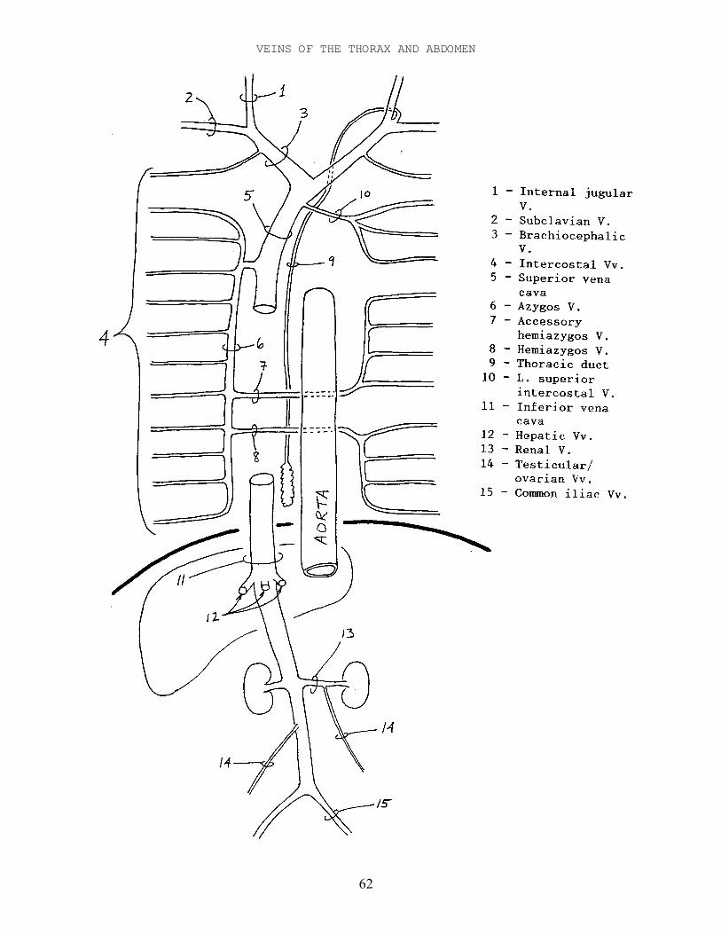
VEINS OF THE THORAX AND ABDOMEN
I - Superior Vena Cava - drains head, arms, and thorax and passes down toheart.
A - Brachiocephalic Vv. - drain blood from Subclavian V. and Jugular V.
1 - Left superior intercostal V. - union of 2nd and 3rd intercostalVv., drains into Left Brachiocephalic V.
B - Azygos V. - on right side of spinal column, it drains Rightintercostal Vv., Hemiazygos V. and Accessory hemiazygos V. Itpasses up and dumps into Superior Vena Cava from behind.
1 - Hemiazygos V. - drains lower Intercostal Vv. on left side.
2 - Accessory hemiazygos V. - drains upper Intercostal Vv. on leftside.
3 - Intercostal Vv. - on right side they drain into Azygos V.; onleft they drain into Hemiazygos and Accessory hemiazygosVv.
II - Inferior Vena Cava - drains abdomen and legs, and passes up to heart.
A - Hepatic Vv. - three or so short veins from back of liver.
III - Hepatic Portal V. - large vein entering Porta hepatis. It drains venousblood from organs to liver.
A - Splenic V. - drains blood from spleen.
B - Superior mesenteric V. - large; drains blood from Ascending andTransverse colon and small intestine.
C - Inferior mesenteric V. - drains blood from descending and sigmoidcolon.
D - Gastric V. - drains blood from stomach; may empty into Splenic orHepatic portal V.
——————————————————————————————————————————————————————————————————————————————
Note: The Left testicular/ovarian V. usually empties into the Left renal V.rather than the Inferior Vena Cava.
Note: The Thoracic Duct lies between the Descending Aorta and Azygos V. anddrains lymph and chyle from the Cisterna chyli up to dump into the union of theL. Internal jugular V. and the L. Subclavian V.
64
OSTEOLOGY OF THE PECTORAL GIRDLE AND UPPER EXTREMITY
CLAVICLE
Sternal Articular Surface - articulates with sternumAcromial Articular Surface - articulates with acromion processConoid Tubercle - attachment for conoid part of Coracoclavicular Ligament
SCAPULA
Supraspinous Fossa - origin of SupraspinatusInfraspinous Fossa - origin of Infraspinatus, Teres major, and Teres minorSubscapular Fossa - origin of SubscapularisSpine - insertion of TrapeziusVertebral border - insertion of Rhomboideus major and minor, and Serratus
anteriorCoracoid Process - origin of Coracobrachialis, and Biceps brachii short head;
insertion of Pectoralis minor, and attachment for trapezoid part ofCoracoclavicular Ligament
Acromion Process - roofs over shoulder joint and articulates with clavicleGlenoid fossa - articulates with humerusSupraglenoid Tubercle - origin of Biceps brachii long headInfraglenoid Tubercle - origin of Triceps brachii long head
HUMERUS
Head - articulates with scapulaAnatomical Neck - margin of articular surface of headSurgical Neck - where humerus often breaksGreater Tubercle - insertion of Supraspinatus, Infraspinatus, and Teres minorLesser Tubercle - insertion of SubscapularisIntertubercular Sulcus (=Bicipital Groove) - groove for tendon of Biceps
brachii long head; Latissimus dorsi, Teres major, and Pectoralismajor insert on its sides
Deltoid Tuberosity - insertion of DeltoideusRadial (=Spiral) Groove - for Radial N. between medial and lateral heads of
Triceps brachiiCapitulum - articulates with radiusTrochlea - articulates with ulnaMedial Epicondyle - origin of flexors of forearmLateral Epicondyle - origin of extensors of forearmOlecranon Fossa - receives olecranon process on extension of elbowCoronoid Fossa - receives coronoid process on flexion of elbow
ULNA
Olecranon Process - insertion of Triceps brachiiTrochlear Notch - articulates with trochlea of humerusCoronoid Process - forms inferior margin of trochlear notchRadial Notch - articulates with proximal end of radiusUlnar Tuberosity - insertion of BrachialisStyloid Process - attachment of ligaments
65
RADIUS
Head - articulates with humerus and ulnaFovea capitis - depression for capitulum of humerusRadial Tuberosity - insertion of Biceps brachiiStyloid Process - attachment of ligamentsUlnar Notch - articulates with distal end of ulnaLower Articular Facet - articulates with scaphoid and lunate
CARPUS
Scaphoid Mnemonics for these, Sally SomeLunate proximal thumbside to Lowers LoversTriquetrum digit V side, then Tom's TryPisiform distal thumbside to Pants PositionsTrapezium digit V side are: Then ThatTrapezoid Things TheyCapitate Can Can'tHamate Happen Handle
METACARPALS I-V AND PHALANGES
Digit I (=Pollex) has: Proximal and Distal PhalangesDigits II-V have: Proximal, Middle, and Distal Phalanges
——————————————————————————————————————————————————————————————————————————————
JOINTS OF THE UPPER EXTREMITY
Movements of scapula
- upward and downward rotation- elevation and depression- protraction and retraction
Movements of shoulder joint
- flexion and extension- aBduction and adduction- medial and lateral rotation
Movements of forearm
- flexion and extension- Pronation and supination
Movements of wrist and hand
- adduction and abduction at radio-carpal joint- extension at radio-carpal joint- flexion at intercarpal joint- flexion and extension of carpo-metacarpal joints- opposition of pollex by rotation- flexion and extension of metacarpo-phalangeal joints
66
67
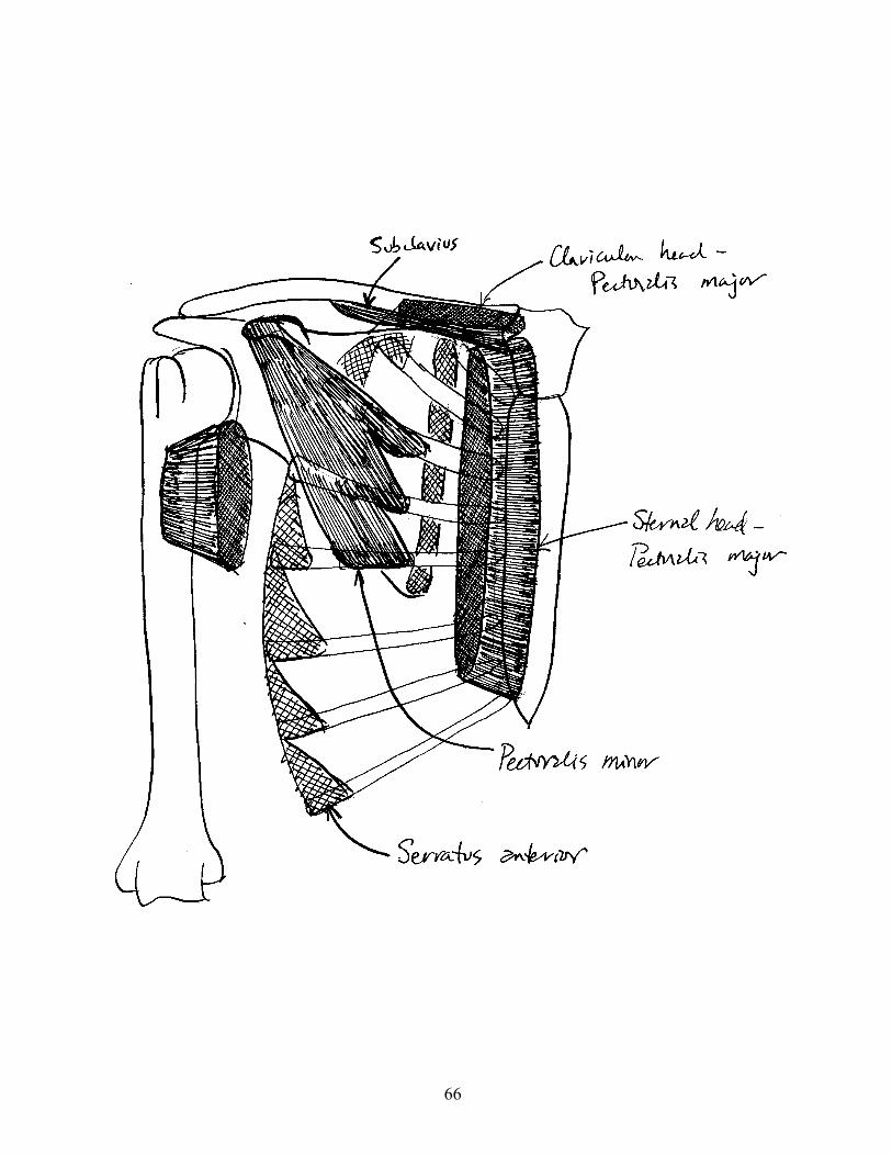
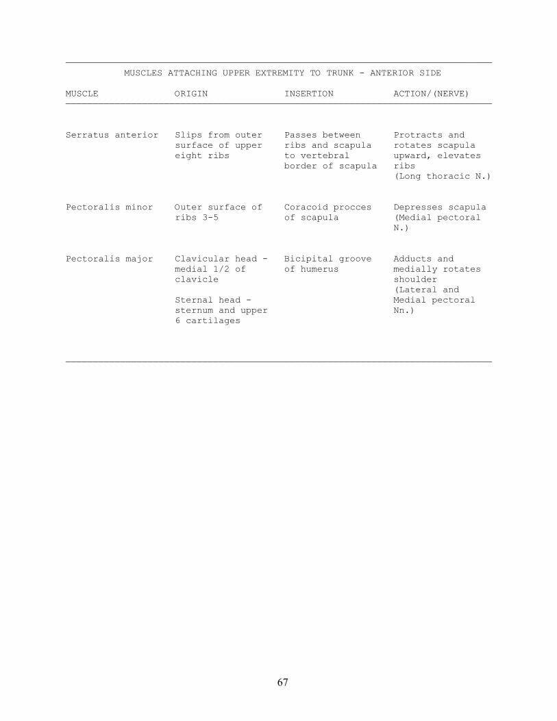
——————————————————————————————————————————————————————————————————————————————MUSCLES ATTACHING UPPER EXTREMITY TO TRUNK - ANTERIOR SIDE
MUSCLE ORIGIN INSERTION ACTION/(NERVE)——————————————————————————————————————————————————————————————————————————————
Serratus anterior Slips from outer Passes between Protracts and surface of upper ribs and scapula rotates scapula eight ribs to vertebral upward, elevates border of scapula ribs (Long thoracic N.)
Pectoralis minor Outer surface of Coracoid procces Depresses scapula ribs 3-5 of scapula (Medial pectoral N.)
Pectoralis major Clavicular head - Bicipital groove Adducts and medial 1/2 of of humerus medially rotates clavicle shoulder (Lateral and Sternal head - Medial pectoral sternum and upper Nn.) 6 cartilages
——————————————————————————————————————————————————————————————————————————————
68
69
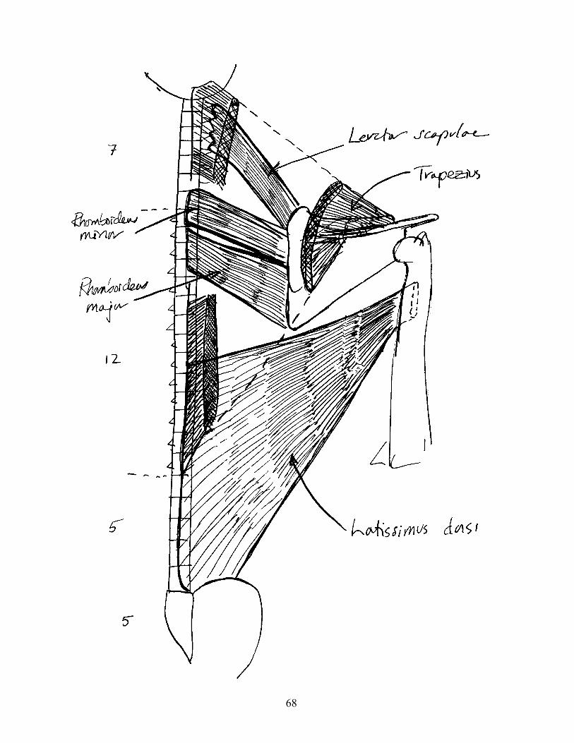
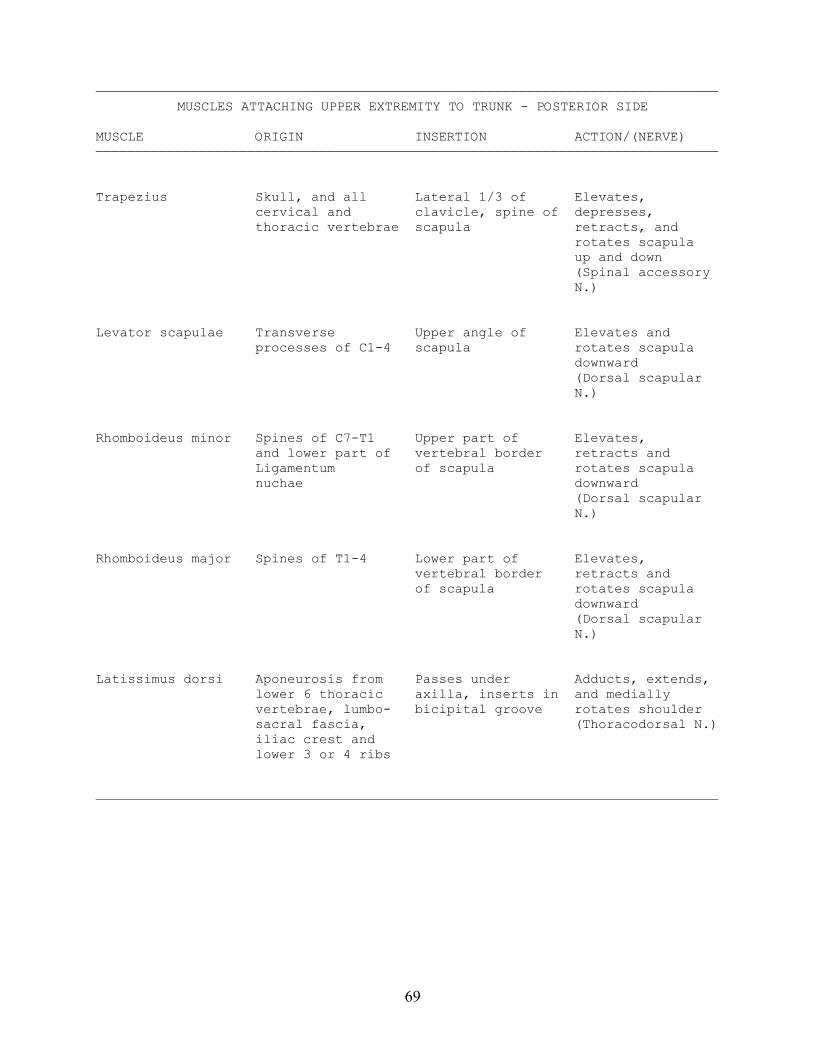
——————————————————————————————————————————————————————————————————————————————MUSCLES ATTACHING UPPER EXTREMITY TO TRUNK - POSTERIOR SIDE
MUSCLE ORIGIN INSERTION ACTION/(NERVE)——————————————————————————————————————————————————————————————————————————————
Trapezius Skull, and all Lateral 1/3 of Elevates, cervical and clavicle, spine of depresses, thoracic vertebrae scapula retracts, and rotates scapula up and down (Spinal accessory N.)
Levator scapulae Transverse Upper angle of Elevates and processes of C1-4 scapula rotates scapula downward (Dorsal scapular N.)
Rhomboideus minor Spines of C7-T1 Upper part of Elevates, and lower part of vertebral border retracts and Ligamentum of scapula rotates scapula nuchae downward (Dorsal scapular N.)
Rhomboideus major Spines of T1-4 Lower part of Elevates, vertebral border retracts and of scapula rotates scapula downward (Dorsal scapular N.)
Latissimus dorsi Aponeurosis from Passes under Adducts, extends, lower 6 thoracic axilla, inserts in and medially vertebrae, lumbo- bicipital groove rotates shoulder sacral fascia, (Thoracodorsal N.) iliac crest and lower 3 or 4 ribs
——————————————————————————————————————————————————————————————————————————————
70
71
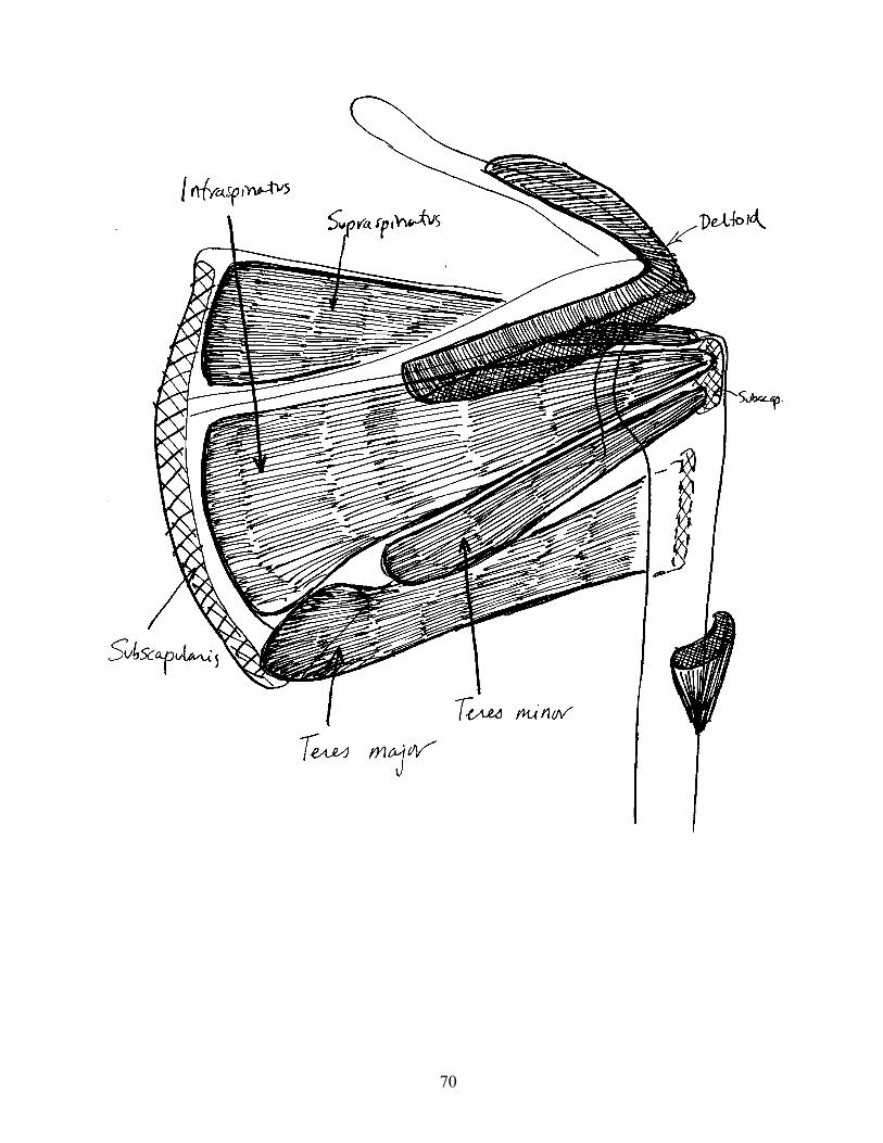
——————————————————————————————————————————————————————————————————————————————MUSCLES OF THE SHOULDER JOINT - SCAPULAR MUSCLES
MUSCLE ORIGIN INSERTION ACTION/(NERVE)——————————————————————————————————————————————————————————————————————————————
Subscapularis Subscapular fossa Passes in front of Medially rotates joint to lesser shoulder tubercle of (Upper subscapular humerus N.)
Supraspinatus Supraspinous fossa Passes above joint ABducts shoulder to greater (Suprascapular N.) tubercle of humerus
Infraspinatus Infraspinous fossa Passes behind Laterally rotates joint to greater shoulder tubercle of (Suprascapular N.) humerus
Teres minor Upper 2/3 of Passes behind Laterally rotates lateral border of joint to greater shoulder scapula tubercle of (Axillary N.) humerus
Teres major Inferior angle of Medial lip of Adducts and scapula bicipital groove medially rotates near Latissimus shoulder dorsi (Lower subscapular N.)
Deltoideus Lateral 1/3 of Deltoid tuberosity Flexes, aBducts, clavicle, upper of humerus and extends surface of shoulder acromion, spine of (Axillary N.) scapula
——————————————————————————————————————————————————————————————————————————————
Note: Subscapularis, Supraspinatus, Infraspinatus, and Teres minor togetherform the "Rotator Cuff", and share the common function of holding the head ofthe humerus in the glenoid fossa.
Note: Deltoideus cannot initiate aBduction of the shoulder, but Supraspinatuscan.
72
73
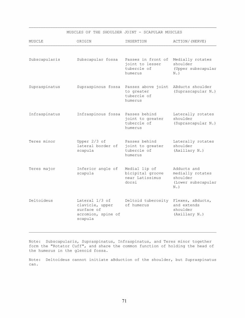
——————————————————————————————————————————————————————————————————————————————MUSCLES OF THE ARM
MUSCLE ORIGIN INSERTION ACTION/(NERVE)——————————————————————————————————————————————————————————————————————————————
Biceps brachii Short head - By tendon on back Flexes elbow and coracoid process of tuberosity of supinates forearm of scapula (w/ radius; tendon of (Musculocutaneous Coracobrachialis) long head runs in N.) bicipital groove Long head - supraglenoid tubercle of scapula
Coracobrachialis Coracoid process Middle of medial Flexes and adducts of scapula border of humerus shoulder (w/ Biceps (Musculocutaneous brachii - short) N.)
Brachialis Lower 2/3 of By tendon to Flexes elbow anterior humerus tuberosity of ulna (Musculocutaneous N.)
Triceps brachii Long head - Upper surface of Extends elbow infraglenoid olecranon (Radial N.) tubercle of scapula
Medial head - posterior surface of humerus below
spiral groove
Lateral head - posterior and lateral surface of humerus above
spiral groove
Anconeus Lateral epicondyle Lateral surface of Extends elbow of humerus olecranon and ulna (Radial N.)
——————————————————————————————————————————————————————————————————————————————
Note: Biceps brachii also inserts by way of the bicipital aponeurosis into thedeep fascia over the origin of the flexors of the wrist and hand. Theaponeurosis can be palpated easily in the living.
Note: Anconeus can be considered a detached part of Triceps brachii.
74
75
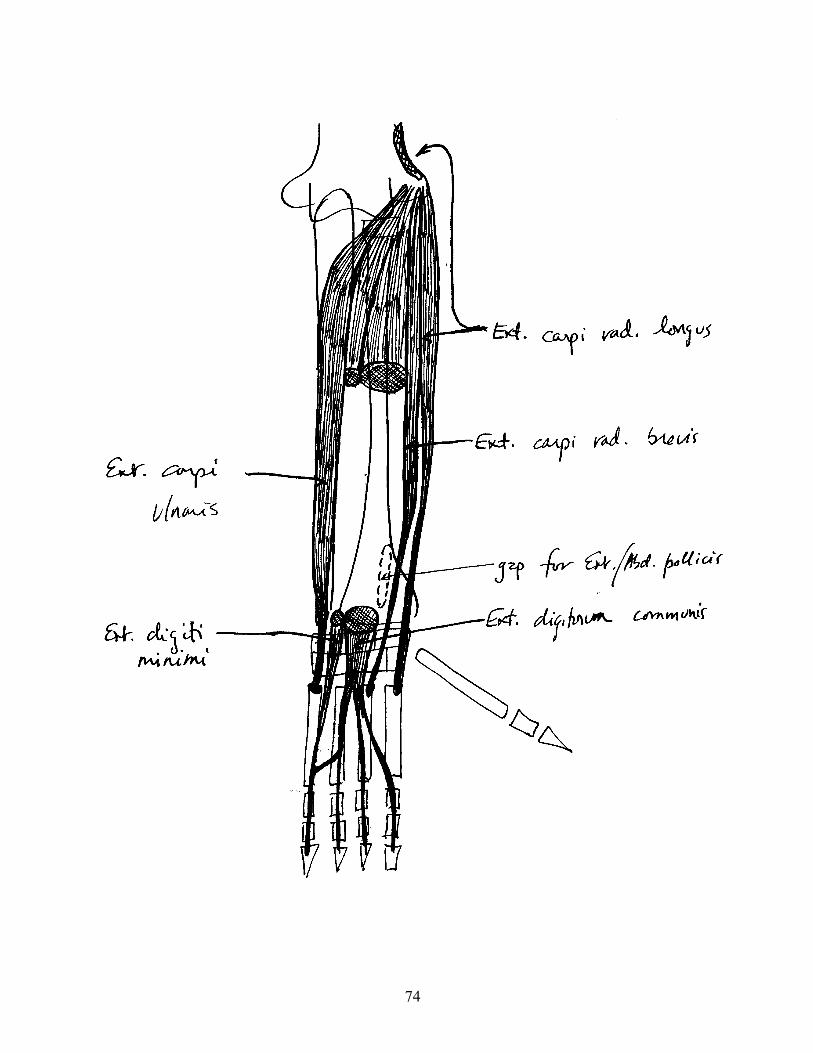
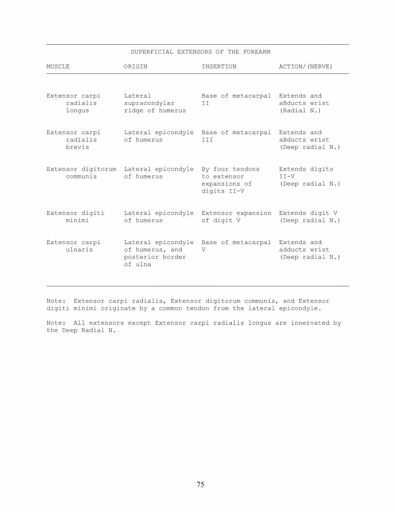
——————————————————————————————————————————————————————————————————————————————SUPERFICIAL EXTENSORS OF THE FOREARM
MUSCLE ORIGIN INSERTION ACTION/(NERVE)——————————————————————————————————————————————————————————————————————————————
Extensor carpi Lateral Base of metacarpal Extends and radialis supracondylar II aBducts wrist longus ridge of humerus (Radial N.)
Extensor carpi Lateral epicondyle Base of metacarpal Extends and radialis of humerus III aBducts wrist brevis (Deep radial N.)
Extensor digitorum Lateral epicondyle By four tendons Extends digits communis of humerus to extensor II-V expansions of (Deep radial N.) digits II-V
Extensor digiti Lateral epicondyle Extensor expansion Extends digit V minimi of humerus of digit V (Deep radial N.)
Extensor carpi Lateral epicondyle Base of metacarpal Extends and ulnaris of humerus, and V adducts wrist posterior border (Deep radial N.) of ulna
——————————————————————————————————————————————————————————————————————————————
Note: Extensor carpi radialis, Extensor digitorum communis, and Extensordigiti minimi originate by a common tendon from the lateral epicondyle.
Note: All extensors except Extensor carpi radialis longus are innervated bythe Deep Radial N.
76
77
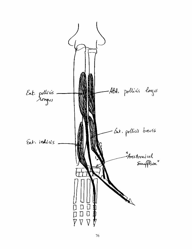
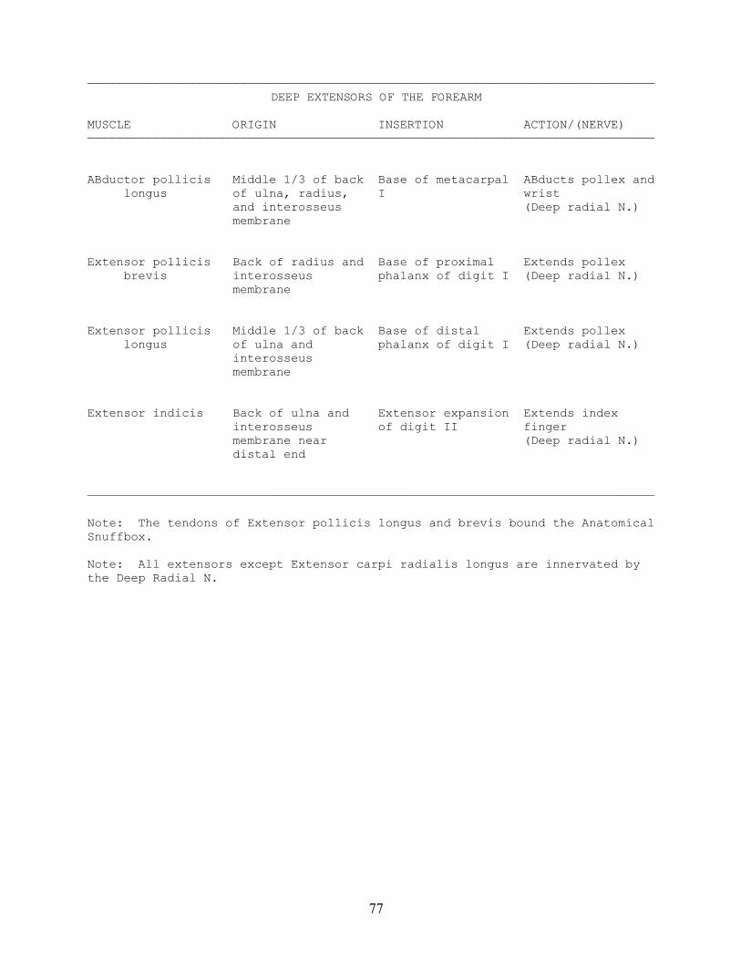
——————————————————————————————————————————————————————————————————————————————DEEP EXTENSORS OF THE FOREARM
MUSCLE ORIGIN INSERTION ACTION/(NERVE)——————————————————————————————————————————————————————————————————————————————
ABductor pollicis Middle 1/3 of back Base of metacarpal ABducts pollex and longus of ulna, radius, I wrist and interosseus (Deep radial N.) membrane
Extensor pollicis Back of radius and Base of proximal Extends pollex brevis interosseus phalanx of digit I (Deep radial N.) membrane
Extensor pollicis Middle 1/3 of back Base of distal Extends pollex longus of ulna and phalanx of digit I (Deep radial N.) interosseus membrane
Extensor indicis Back of ulna and Extensor expansion Extends index interosseus of digit II finger membrane near (Deep radial N.) distal end
——————————————————————————————————————————————————————————————————————————————
Note: The tendons of Extensor pollicis longus and brevis bound the AnatomicalSnuffbox.
Note: All extensors except Extensor carpi radialis longus are innervated bythe Deep Radial N.
78
79
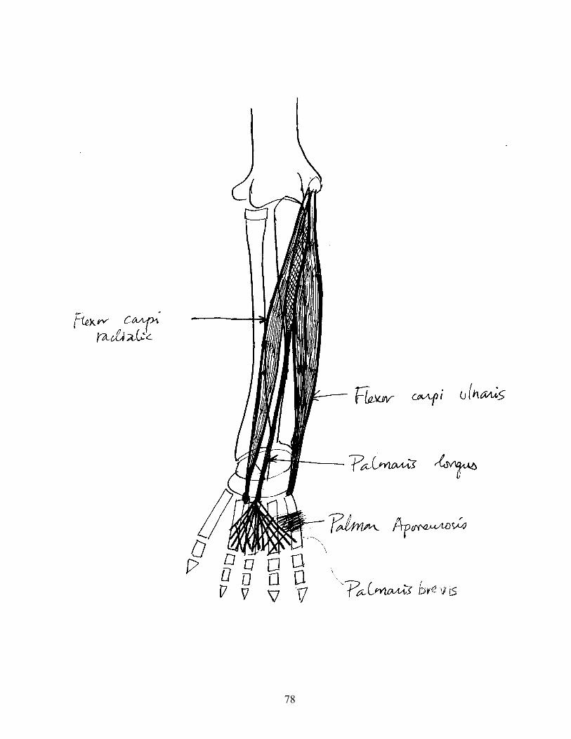
——————————————————————————————————————————————————————————————————————————————SUPERFICIAL FLEXORS OF THE FOREARM
MUSCLE ORIGIN INSERTION ACTION/(NERVE)——————————————————————————————————————————————————————————————————————————————
Flexor carpi Medial epicondyle Bases of Flexes wrist radialis of humerus metacarpals II-III (Median N.)
Palmaris longus Medial epicondyle Palmar aponeurosis Flexes wrist of humerus (Median N.)
Flexor carpi Medial epicondyle Pisiform, hamate, Flexes and ulnaris of humerus and base of adducts wrist metacarpal V (Ulnar N.)
——————————————————————————————————————————————————————————————————————————————
Note: Palmaris longus is absent in 10% of individuals. The trait isgenetically controlled, and absence of the muscle is dominant.
80
81
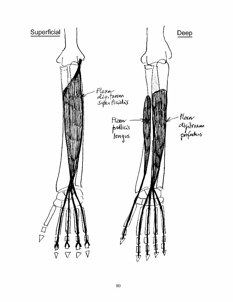
——————————————————————————————————————————————————————————————————————————————DEEP FLEXORS OF THE FOREARM
MUSCLE ORIGIN INSERTION ACTION/(NERVE)——————————————————————————————————————————————————————————————————————————————
Flexor digitorum Medial epicondyle By four tendons Flex digits II-V superficialis of humerus, to 2nd phalanges (Median N.) coronoid process of digits II-V of ulna, and (Tendons split oblique line of near insertion radius to allow Flexor digitorum profundus to pass through)
Flexor digitorum Middle 2/3 of By four tendons Flex digits II-V profundus ulna and to distal (Median and Ulnar interosseus phalanges of Nn.) membrane digits II-V
Flexor pollicis Middle 2/3 of Base of distal Flexes pollex longus radius and phalanx of (Median N.) interosseus thumb membrane
——————————————————————————————————————————————————————————————————————————————
82
83
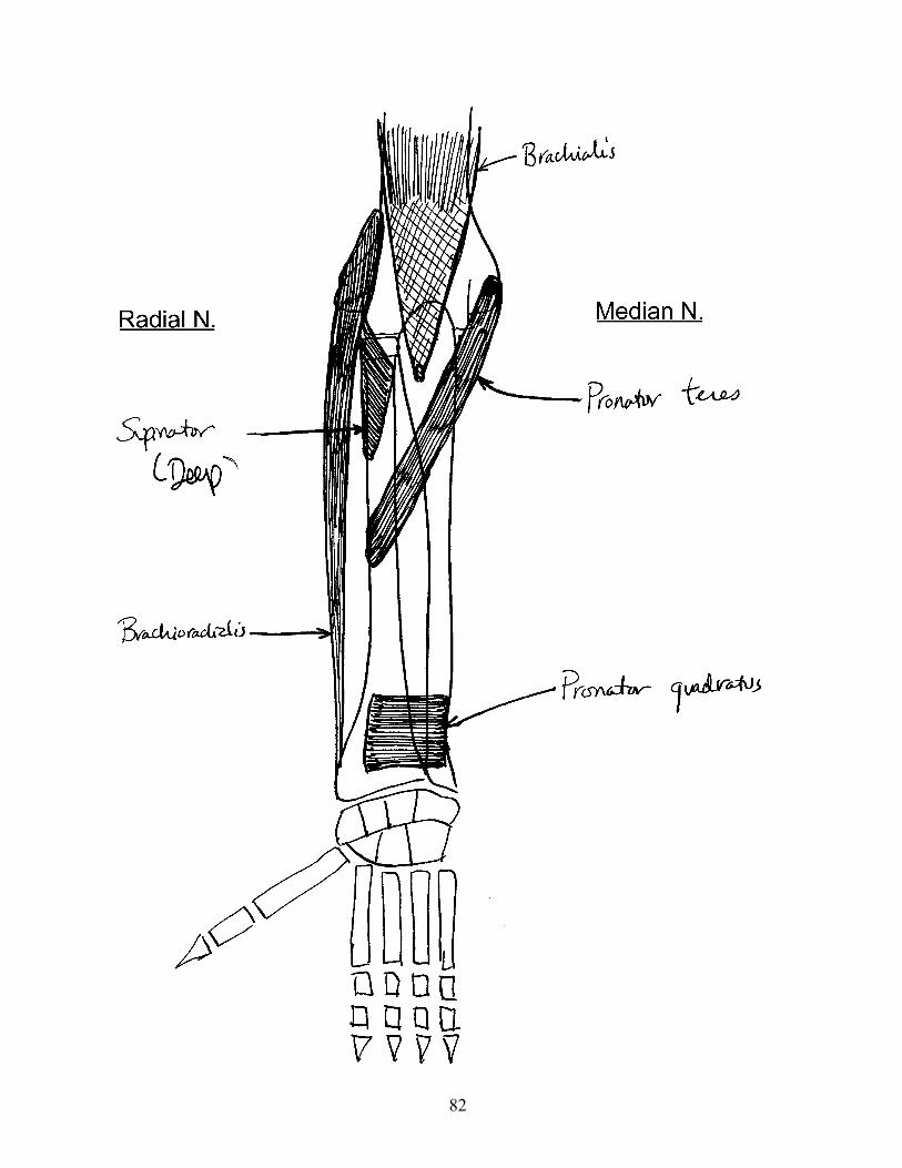
——————————————————————————————————————————————————————————————————————————————OTHER MUSCLES OF THE FOREARM
MUSCLE ORIGIN INSERTION ACTION/(NERVE)——————————————————————————————————————————————————————————————————————————————
Brachioradialis Lateral Styloid process of Flexes elbow supracondylar radius (Radial N.) ridge of humerus
Supinator By 2 heads, from Anterior and Supinates forearm lateral epicondyle lateral surface (Deep radial N.) of humerus and of radius supinator crest of
ulna
Pronator teres Medial epicondyle Middle of lateral Pronates forearm of humerus, and surface of radius (Median N.) coronoid process of ulna
Pronator quadratus Distal 1/4 of Distal 1/4 of Pronates forearm front of ulna lateral surface (Median N.) of radius
——————————————————————————————————————————————————————————————————————————————
Note: Brachioradialis can assist in supination of a pronated forearm, and usedto be called "supinator longus".
84
85
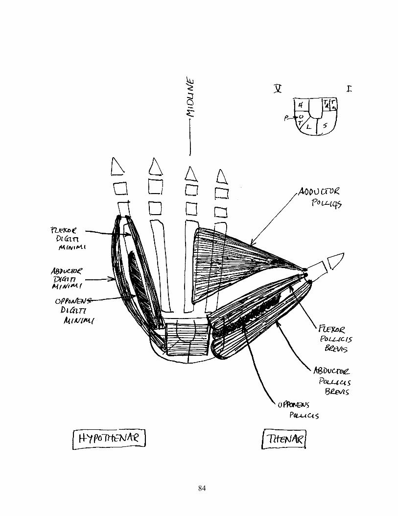
——————————————————————————————————————————————————————————————————————————————THENAR MUSCLES
MUSCLE ORIGIN INSERTION ACTION/(NERVE)——————————————————————————————————————————————————————————————————————————————
ABductor pollicis Flexor retinaculum Radial side of ABducts pollex brevis and adjacent base of 1st (Median N.) carpals phalanx of digit I
Opponens pollicis Flexor retinaculum Radial side of Flexes metacarpal and adjacent metacarpal I I; rotates carpals. Found metacarpal to deep to ABductor oppose pollicis brevis (Median N.)
Flexor pollicis Flexor retinaculum Base of 1st Flexes pollex brevis and adjacent phalanx of digit I (Median N.) carpals
——————————————————————————————————————————————————————————————————————————————
——————————————————————————————————————————————————————————————————————————————HYPOTHENAR MUSCLES
MUSCLE ORIGIN INSERTION ACTION/(NERVE)——————————————————————————————————————————————————————————————————————————————
ABductor digiti Pisiform and Ulnar side of base ABducts digit V minimi adjacent ligaments of 1st phalanx of (Ulnar N.) digit V
Opponens digiti Flexor retinaculum Ulnar side of Rotates metacarpal minimi and hook of hamate metacarpal V V (Ulnar N.)
Flexor digiti Flexor retinaculum Base of 1st Flexes digit V minimi and hook of hamate phalanx of digit V (Ulnar N.)
——————————————————————————————————————————————————————————————————————————————
86
87
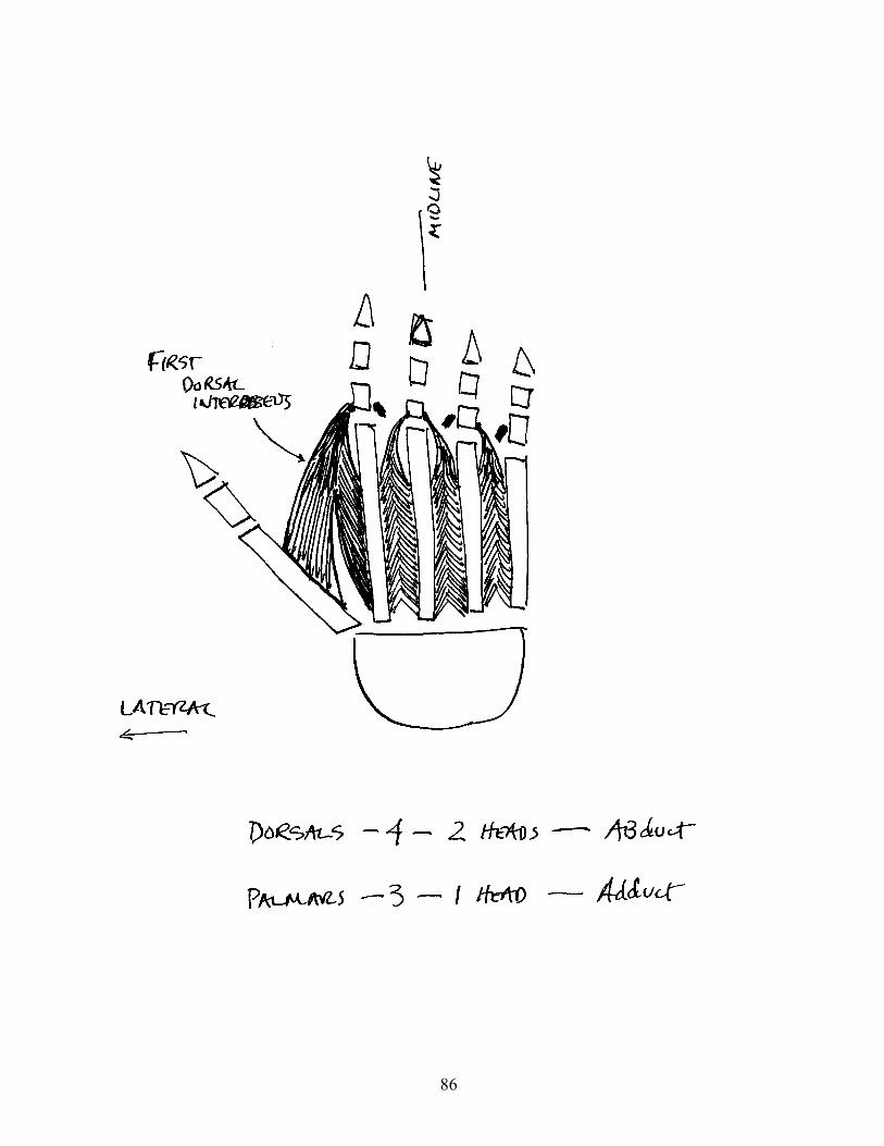
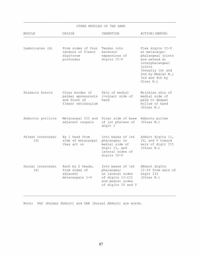
——————————————————————————————————————————————————————————————————————————————OTHER MUSCLES OF THE HAND
MUSCLE ORIGIN INSERTION ACTION/(NERVE)——————————————————————————————————————————————————————————————————————————————
Lumbricales (4) From sides of four Tendon into Flex digits II-V tendons of Flexor extensor at metacarpo- digitorum expansions of phalangeal joints profundus digits II-V and extend at interphalangeal joints (Usually 1st and
2nd by Median N.;3rd and 4th byUlnar N.)
Palmaris brevis Ulnar border of Skin of medial Wrinkles skin of palmar aponeurosis (=ulnar) side of medial side of and front of hand palm to deepen flexor retinaculum hollow of hand (Ulnar N.)
Adductor pollicis Metacarpal III and Ulnar side of base Adducts pollex adjacent carpals of 1st phalanx of (Ulnar N.) digit I
Palmar interossei By 1 head from Into bases of 1st Adduct digits II, (3) side of metacarpal phalanges; on IV, and V toward they act on medial side of axis of digit III digit II, and (Ulnar N.) lateral sides of digits IV-V
Dorsal interossei Each by 2 heads, Into bases of 1st ABduct digits (4) from sides of phalanges; II-IV from axis of adjacent on lateral sides digit III metacarpals I-V of digits II-III (Ulnar N.) and medial sides of digits IV and V
——————————————————————————————————————————————————————————————————————————————
Note: PAD (Palmar ADduct) and DAB (Dorsal ABduct) are words.
88
BRACHIAL PLEXUS
89
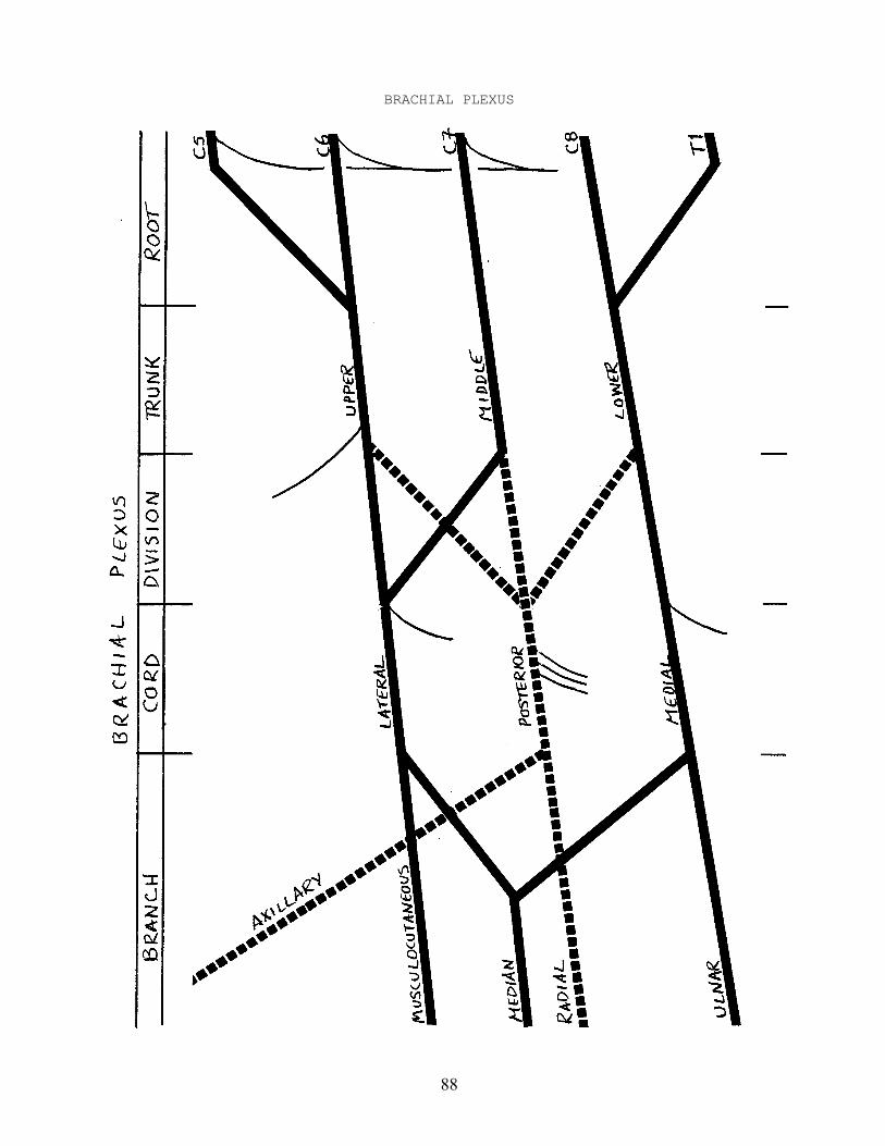
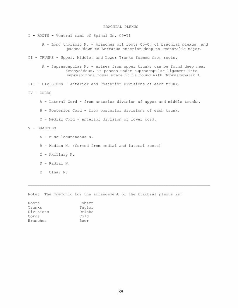
BRACHIAL PLEXUS
I - ROOTS - Ventral rami of Spinal Nn. C5-T1
A - Long thoracic N. - branches off roots C5-C7 of brachial plexus, andpasses down to Serratus anterior deep to Pectoralis major.
II - TRUNKS - Upper, Middle, and Lower Trunks formed from roots.
A - Suprascapular N. - arises from upper trunk; can be found deep nearOmohyoideus, it passes under suprascapular ligament intosupraspinous fossa where it is found with Suprascapular A.
III - DIVISIONS - Anterior and Posterior Divisions of each trunk.
IV - CORDS
A - Lateral Cord - from anterior division of upper and middle trunks.
B - Posterior Cord - from posterior divisions of each trunk.
C - Medial Cord - anterior division of lower cord.
V - BRANCHES
A - Musculocutaneous N.
B - Median N. (formed from medial and lateral roots)
C - Axillary N.
D - Radial N.
E - Ulnar N.
——————————————————————————————————————————————————————————————————————————————
Note: The mnemonic for the arrangement of the brachial plexus is:
Roots RobertTrunks TaylorDivisions DrinksCords ColdBranches Beer
90
NERVES OF THE UPPER EXTREMITY
91
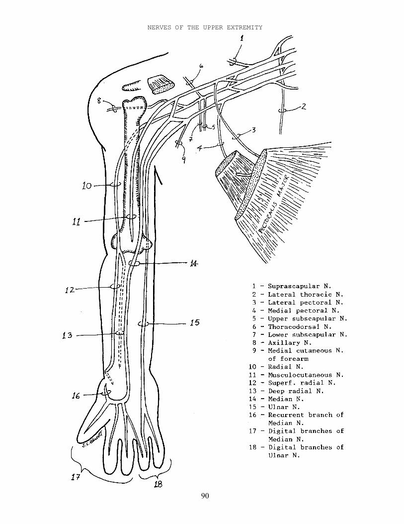
NERVES OF THE UPPER EXTREMITY
Ia - Lateral cord
A - Lateral pectoral N. - passes medial to Pectoralis minor and ends inPectoralis major.
B - Lateral root of Median N. - joins medial root to form Median N.
Ib - Musculocutaneous N. - continuation of Lateral Cord, piercesCoracobrachialis and descends arm between Biceps brachii andBrachialis. At elbow becomes Lateral cutaneous N. of forearm.
IIa - Medial cord
A - Medial pectoral N. - pierces Pectoralis minor and ends in Pectoralismajor.
B - Medial root of Median N. - joins lateral root to form Median N.
IIb - Ulnar N. - continuation of Medial Cord. Passes down arm with BrachialA., then behind medial epicondyle, and down forearm with Ulnar A.
A - Palmar digital branches of Ulnar N. - pass across palm to digits IV-V.
IIIa - Posterior cord - lies behind Axillary A.
A - Upper subscapular N. - arises in axilla, passes to Subscapularis.
B - Thoracodorsal N. - arises between Upper and Lower subscapular Nn. andpasses with Thoracodorsal A+V to Latissimus dorsi.
C - Lower subscapular N. - arises near Axillary N., passes to Teres maj.
D - Axillary N. - large branch, passes behind humerus and throughquadrangular space with Posterior humeral circumflex A+V.
IIIb - Radial N. - continuation of Posterior Cord. Passes behind humerus withProfunda brachii A., then anterior to lateral epicondyle, and splitsinto Superficial and Deep Radial Nn.
A - Superficial radial N. - passes down forearm deep to Brachioradialis.At wrist it passes onto dorsum of hand.
1 - [Dorsal] digital Nn. - small branches passing along digits.
B - Deep radial N. - Pierces Supinator and passes down extensor side offorearm deep to Extensor digitorum communis.
IV - Median N. - Descends arm with Brachial A., and passes down forearm deep toFlexor digitorum superficialis.
A - Palmar digital branches of Median N. - cross palm to digits I-IV.
B - Recurrent branch of median N. - passes laterally to thenar muscles.
Note: Long thoracic N. off roots C5-C7 and Suprascapular N. off Upper Trunkwere described on p. 89.
92
ARTERIES OF THE UPPER EXTREMITY
93
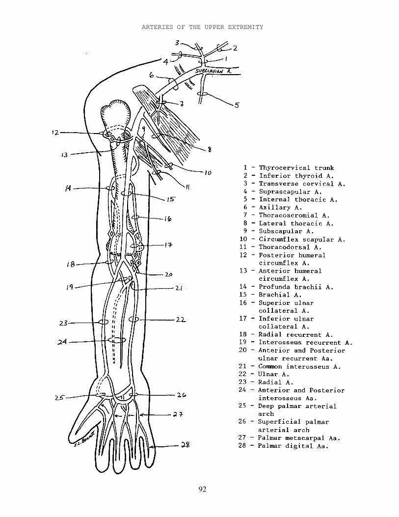
ARTERIES OF THE UPPER EXTREMITY - SHOULDER REGION
Ia - Subclavian A. - passes laterally from Brachiocephalic trunk or Aortic archto supply arm. At lateral border of 1st rib becomes Axillary A.
A - Vertebral A. - passes up through Foramina transversaria of cervicalvertebrae, through Suboccipital triangle, and into skull.
B - Thyrocervical trunk - short branch from top of Subclavian A.
1 - Inferior thyroid A. - passes up neck behind Carotid sheath toThyroid gland.
2 - Suprascapular A. - passes over Suprascapular Ligament and intosupraspinous fossa, a branch then passes laterally intoinfraspinous fossa.
3 - Transverse cervical A. - passes down to vertebral border ofscapula, and anastomoses with Suprascapular andCircumflex scapular Aa.
C - Internal thoracic A. - branches off opposite Thyrocervical trunk andpasses down into thorax.
D - Costocervical trunk - branches off back of Subclavian A. and sendsbranches up neck and down into thorax.
Ib - Axillary A. - continuation of Subclavian A. after first rib. Afterpassing inferior border of Teres major it becomes Brachial A.
A - Thoracoacromial A. - branches off Axillary A. medial to Pectoralisminor and branches on deep surface of Pectoralis major.
B - Lateral thoracic A. – arises deep to Pectoralis minor and passes downlateral border of Pectoralis minor to supply muscles andmammary gland.
C - Subscapular A. - short medial branch opposite Humeral circumflex Aa.It divides into Circumflex scapular A. and Thoracodorsal A.
1 - Circumflex scapular A. - passes backward into triangular space,enters infraspinous fossa and anastomoses withTransverse cervical A. and Suprascapular A.
2 - Thoracodorsal A. - passes down thoracic wall with ThoracodorsalN. to Latissimus dorsi.
D - Anterior humeral circumflex A. - small branch opposite SubscapularA., passes laterally around humerus.
E - Posterior humeral circumflex A. - large branch opposite SubscapularA., passes laterally behind humerus with Axillary N. throughquadrangular space.
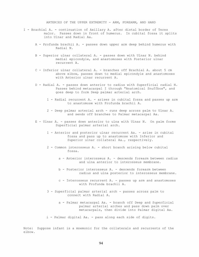
94
ARTERIES OF THE UPPER EXTREMITY - ARM, FOREARM, AND HAND
I - Brachial A. - continuation of Axillary A. after distal border of Teresmajor. Passes down in front of humerus. In cubital fossa it splitsinto Ulnar and Radial Aa.
A - Profunda brachii A. - passes down upper arm deep behind humerus withRadial N.
B - Superior ulnar collateral A. - passes down with Ulnar N. behindmedial epicondyle, and anastomoses with Posterior ulnarrecurrent A.
C - Inferior ulnar collateral A. - branches off Brachial A. about 5 cmabove elbow, passes down to medial epicondyle and anastomoseswith Anterior ulnar recurrent A.
D - Radial A. - passes down anterior to radius with Superficial radial N.Passes behind metacarpal I through "Anatomical Snuffbox", andgoes deep to form Deep palmar arterial arch.
1 - Radial recurrent A. - arises in cubital fossa and passes up armto anastomose with Profunda brachii A.
2 - Deep palmar arterial arch - runs deep across palm to Ulnar A.and sends off branches to Palmar metacarpal Aa.
E - Ulnar A. - passes down anterior to ulna with Ulnar N. On palm formsSuperficial palmar arterial arch.
1 - Anterior and posterior ulnar recurrent Aa. - arise in cubitalfossa and pass up to anastomose with Inferior andSuperior ulnar collateral Aa., respectively.
2 - Common interosseus A. - short branch arising below cubitalfossa.
a - Anterior interosseus A. - descends forearm between radiusand ulna anterior to interosseus membrane.
b - Posterior interosseus A. - descends forearm betweenradius and ulna posterior to interosseus membrane.
c - Interosseus recurrent A. - passes up arm and anastomoseswith Profunda brachii A.
3 - Superficial palmar arterial arch - passes across palm toconnect with Radial A.
a - Palmar metacarpal Aa. - branch off Deep and Superficialpalmar arterial arches and pass down palm overmetacarpals, then divide into Palmar digital Aa.
i - Palmar digital Aa. - pass along each side of digits.
Note: Suppose infant is a mnemonic for the collaterals and recurrents of theelbow.
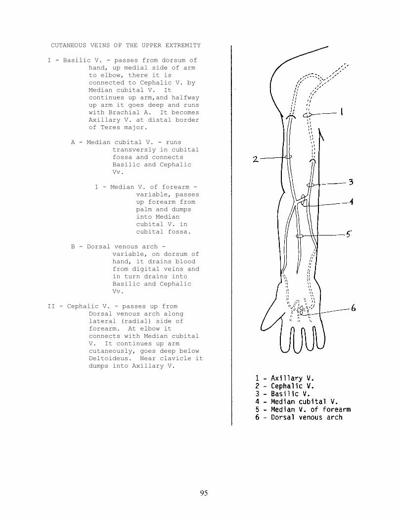
95
CUTANEOUS VEINS OF THE UPPER EXTREMITY
I - Basilic V. - passes from dorsum ofhand, up medial side of armto elbow, there it isconnected to Cephalic V. byMedian cubital V. Itcontinues up arm,and halfwayup arm it goes deep and runswith Brachial A. It becomesAxillary V. at distal borderof Teres major.
A - Median cubital V. - runstransversly in cubitalfossa and connectsBasilic and CephalicVv.
1 - Median V. of forearm -variable, passesup forearm frompalm and dumpsinto Mediancubital V. incubital fossa.
B - Dorsal venous arch -variable, on dorsum ofhand, it drains bloodfrom digital veins andin turn drains intoBasilic and CephalicVv.
II - Cephalic V. - passes up fromDorsal venous arch alonglateral (radial) side offorearm. At elbow itconnects with Median cubitalV. It continues up armcutaneously, goes deep belowDeltoideus. Near clavicle itdumps into Axillary V.
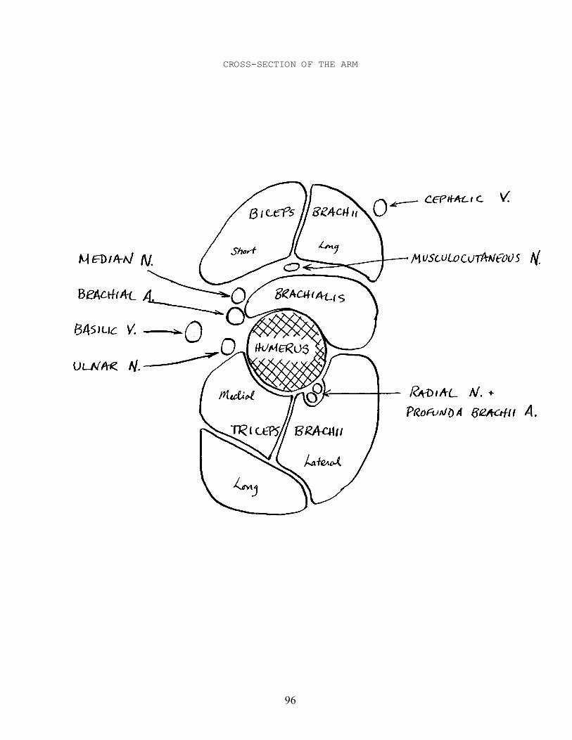
96
CROSS-SECTION OF THE ARM
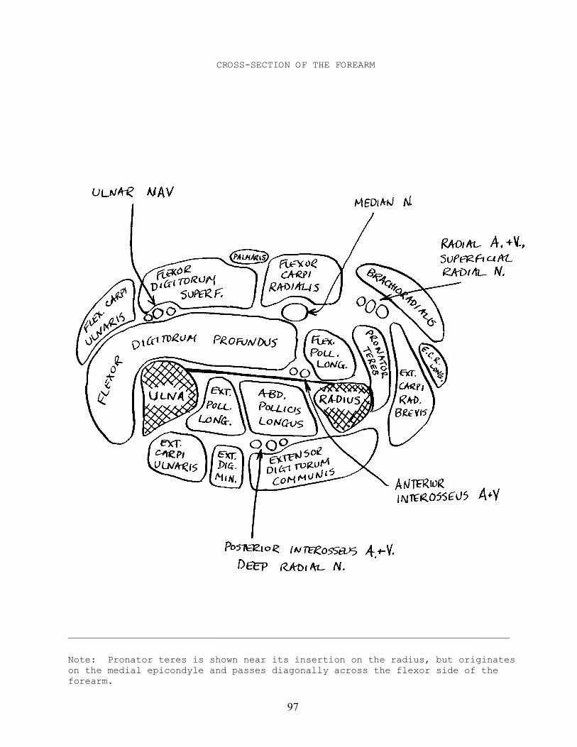
97
CROSS-SECTION OF THE FOREARM
——————————————————————————————————————————————————————————————————————————————
Note: Pronator teres is shown near its insertion on the radius, but originateson the medial epicondyle and passes diagonally across the flexor side of theforearm.
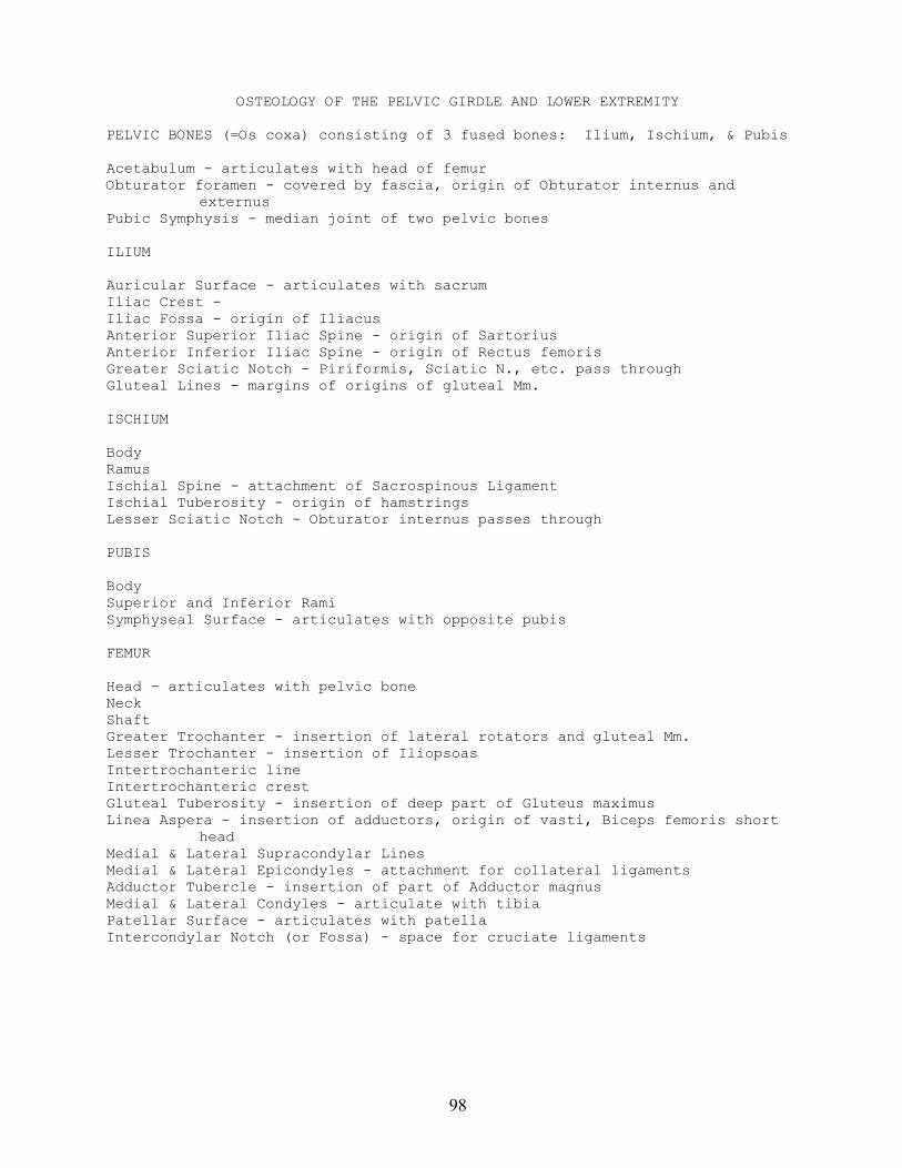
98
OSTEOLOGY OF THE PELVIC GIRDLE AND LOWER EXTREMITY
PELVIC BONES (=Os coxa) consisting of 3 fused bones: Ilium, Ischium, & Pubis
Acetabulum - articulates with head of femurObturator foramen - covered by fascia, origin of Obturator internus and
externusPubic Symphysis - median joint of two pelvic bones
ILIUM
Auricular Surface - articulates with sacrumIliac Crest -Iliac Fossa - origin of IliacusAnterior Superior Iliac Spine - origin of SartoriusAnterior Inferior Iliac Spine - origin of Rectus femorisGreater Sciatic Notch - Piriformis, Sciatic N., etc. pass throughGluteal Lines - margins of origins of gluteal Mm.
ISCHIUM
BodyRamusIschial Spine - attachment of Sacrospinous LigamentIschial Tuberosity - origin of hamstringsLesser Sciatic Notch - Obturator internus passes through
PUBIS
BodySuperior and Inferior RamiSymphyseal Surface - articulates with opposite pubis
FEMUR
Head - articulates with pelvic boneNeckShaftGreater Trochanter - insertion of lateral rotators and gluteal Mm.Lesser Trochanter - insertion of IliopsoasIntertrochanteric lineIntertrochanteric crestGluteal Tuberosity - insertion of deep part of Gluteus maximusLinea Aspera - insertion of adductors, origin of vasti, Biceps femoris short
headMedial & Lateral Supracondylar LinesMedial & Lateral Epicondyles - attachment for collateral ligamentsAdductor Tubercle - insertion of part of Adductor magnusMedial & Lateral Condyles - articulate with tibiaPatellar Surface - articulates with patellaIntercondylar Notch (or Fossa) - space for cruciate ligaments
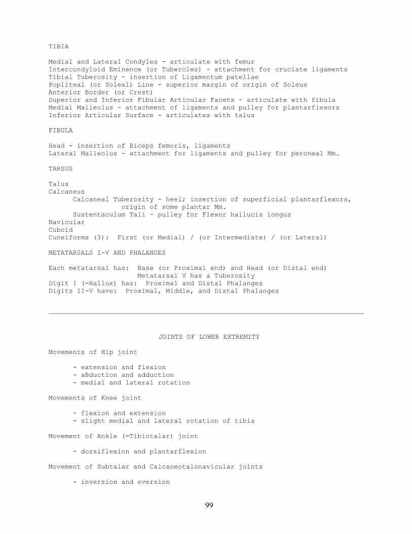
99
TIBIA
Medial and Lateral Condyles - articulate with femurIntercondyloid Eminence (or Tubercles) - attachment for cruciate ligamentsTibial Tuberosity - insertion of Ligamentum patellaePopliteal (or Soleal) Line - superior margin of origin of SoleusAnterior Border (or Crest)Superior and Inferior Fibular Articular Facets - articulate with fibulaMedial Malleolus - attachment of ligaments and pulley for plantarflexorsInferior Articular Surface - articulates with talus
FIBULA
Head - insertion of Biceps femoris, ligamentsLateral Malleolus - attachment for ligaments and pulley for peroneal Mm.
TARSUS
TalusCalcaneus
Calcaneal Tuberosity - heel; insertion of superficial plantarflexors,origin of some plantar Mm.
Sustentaculum Tali - pulley for Flexor hallucis longusNavicularCuboidCuneiforms (3): First (or Medial) / (or Intermediate) / (or Lateral)
METATARSALS I-V AND PHALANGES
Each metatarsal has: Base (or Proximal end) and Head (or Distal end) Metatarsal V has a TuberosityDigit I (=Hallux) has: Proximal and Distal PhalangesDigits II-V have: Proximal, Middle, and Distal Phalanges
——————————————————————————————————————————————————————————————————————————————
JOINTS OF LOWER EXTREMITY
Movements of Hip joint
- extension and flexion- aBduction and adduction- medial and lateral rotation
Movements of Knee joint
- flexion and extension- slight medial and lateral rotation of tibia
Movement of Ankle (=Tibiotalar) joint
- dorsiflexion and plantarflexion
Movement of Subtalar and Calcaneotalonavicular joints
- inversion and eversion
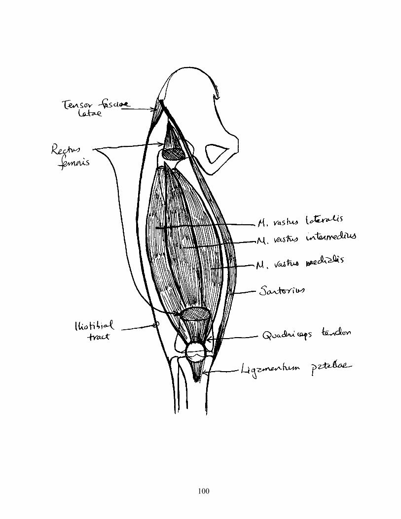
100
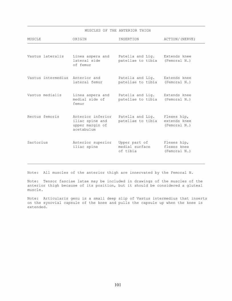
101
——————————————————————————————————————————————————————————————————————————————MUSCLES OF THE ANTERIOR THIGH
MUSCLE ORIGIN INSERTION ACTION/(NERVE)——————————————————————————————————————————————————————————————————————————————
Vastus lateralis Linea aspera and Patella and Lig. Extends knee lateral side patellae to tibia (Femoral N.) of femur
Vastus intermedius Anterior and Patella and Lig. Extends knee lateral femur patellae to tibia (Femoral N.)
Vastus medialis Linea aspera and Patella and Lig. Extends knee medial side of patellae to tibia (Femoral N.) femur
Rectus femoris Anterior inferior Patella and Lig. Flexes hip, iliac spine and patellae to tibia extends knee upper margin of (Femoral N.) acetabulum
Sartorius Anterior superior Upper part of Flexes hip, iliac spine medial surface flexes knee of tibia (Femoral N.)
——————————————————————————————————————————————————————————————————————————————
Note: All muscles of the anterior thigh are innervated by the Femoral N.
Note: Tensor fasciae latae may be included in drawings of the muscles of theanterior thigh because of its position, but it should be considered a glutealmuscle.
Note: Articularis genu is a small deep slip of Vastus intermedius that insertson the synovial capsule of the knee and pulls the capsule up when the knee isextended.
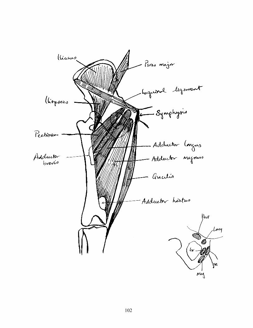
102
103
——————————————————————————————————————————————————————————————————————————————MUSCLES OF THE MEDIAL THIGH
MUSCLE ORIGIN INSERTION ACTION/(NERVE)——————————————————————————————————————————————————————————————————————————————
Iliopsoas Iliacus - iliac Lesser trochanter Flexes hip fossa of femur (Femoral N.)
Psoas major - (Upper 3 or 4 lumbar vertebrae lumbar Nn.)
Pectineus Superior ramus of Line from lesser Adducts, flexes, pubis trochanter to and laterally linea aspera rotates hip (Femoral N.)
Adductor brevis Anterior surface Linea aspera Adducts hip of inferior ramus (Obturator N.) of pubis
Adductor longus Anterior surface Linea aspera Adducts hip of body of pubis (Obturator N.)
Adductor magnus Inferior ramus of Linea aspera and Adducts hip pubis and ischial medial supra- (Obturator N.) tuberosity condylar line to adductor tubercle
Gracilis Body and inferior Upper part of Adducts hip; ramus of pubis medial surface flexes and near the symphysis of tibia medially rotates knee (Obturator N.)
——————————————————————————————————————————————————————————————————————————————
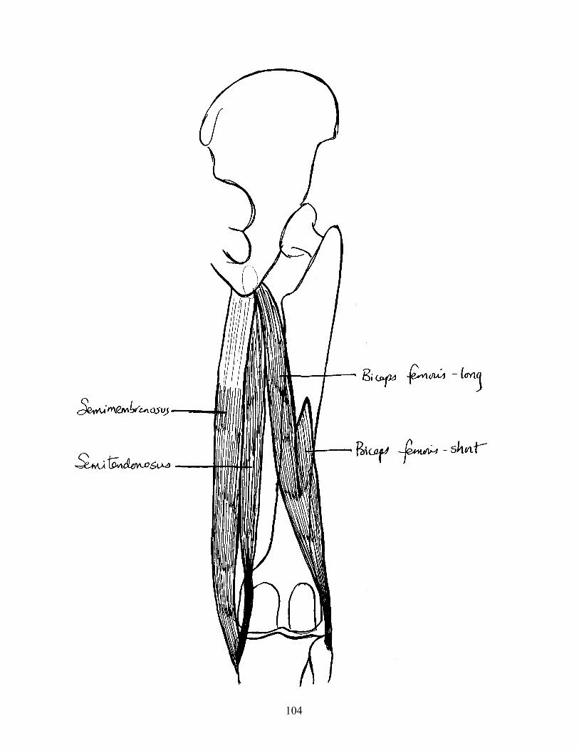
104
105
——————————————————————————————————————————————————————————————————————————————MUSCLES OF THE POSTERIOR THIGH
MUSCLE ORIGIN INSERTION ACTION/(NERVE)——————————————————————————————————————————————————————————————————————————————
Semimembranosus Ischial tuberosity Posterior surface Extends hip, of medial condyle flexes knee of tibia (Sciatic N.)
Semitendinosus Ischial tuberosity Upper part of Extends hip, medial surface flexes knee of tibia (Sciatic N.)
Biceps femoris Long head - Head of fibula Extends hip, ischial tuberosity flexes knee (Sciatic N.)
Short head - linea Flexes knee aspera and lateral (Sciatic N.) supracondylar line
——————————————————————————————————————————————————————————————————————————————
Note: All muscles of the posterior thigh are innervated by the tibial divisionof the Sciatic N., and some books state that Semimembranosus and Semitendinosusare innervated by the Tibial N.
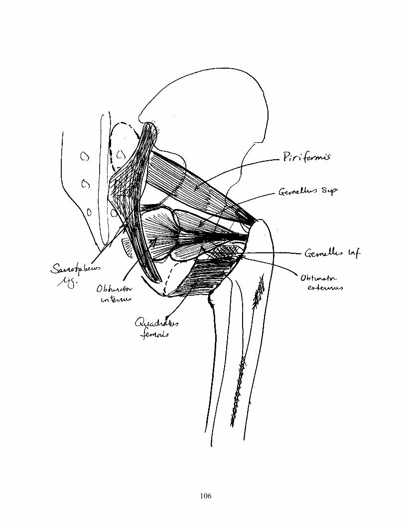
106
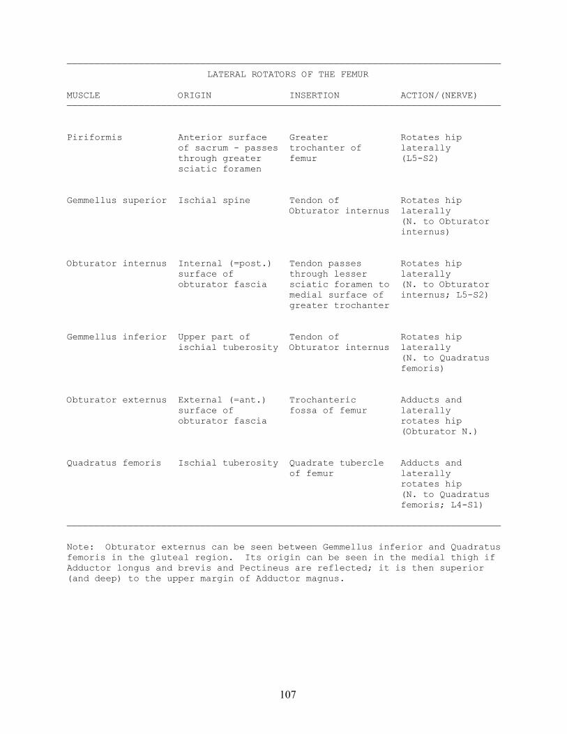
107
——————————————————————————————————————————————————————————————————————————————LATERAL ROTATORS OF THE FEMUR
MUSCLE ORIGIN INSERTION ACTION/(NERVE)——————————————————————————————————————————————————————————————————————————————
Piriformis Anterior surface Greater Rotates hip of sacrum - passes trochanter of laterally through greater femur (L5-S2) sciatic foramen
Gemmellus superior Ischial spine Tendon of Rotates hip Obturator internus laterally
(N. to Obturatorinternus)
Obturator internus Internal (=post.) Tendon passes Rotates hip surface of through lesser laterally obturator fascia sciatic foramen to (N. to Obturator medial surface of internus; L5-S2) greater trochanter
Gemmellus inferior Upper part of Tendon of Rotates hip ischial tuberosity Obturator internus laterally (N. to Quadratus
femoris)
Obturator externus External (=ant.) Trochanteric Adducts and surface of fossa of femur laterally obturator fascia rotates hip
(Obturator N.)
Quadratus femoris Ischial tuberosity Quadrate tubercle Adducts and of femur laterally rotates hip
(N. to Quadratusfemoris; L4-S1)
——————————————————————————————————————————————————————————————————————————————
Note: Obturator externus can be seen between Gemmellus inferior and Quadratusfemoris in the gluteal region. Its origin can be seen in the medial thigh ifAdductor longus and brevis and Pectineus are reflected; it is then superior(and deep) to the upper margin of Adductor magnus.
108
109
——————————————————————————————————————————————————————————————————————————————GLUTEAL MUSCLES
MUSCLE ORIGIN INSERTION ACTION/(NERVE)——————————————————————————————————————————————————————————————————————————————
Gluteus minimus Outer surface of Anterior border ABducts and ilium between of greater medially rotates anterior and trochanter with hip inferior gluteal Gluteus medius (Sup. gluteal N.) lines
Gluteus medius Upper part of the Lateral surface ABducts and outer surface of of greater medially rotates ilium trochanter hip (Sup. gluteal N.)
Gluteus maximus Posterior gluteal Posterior femur Extends, adducts, line of ilium and and iliotibial and laterally posterior surface tract rotates hip of sacrum and (Inf. gluteal N.) coccyx
Tensor fasciae Outer surface of Iliotibial tract ABducts and latae ilium behind medially rotates anterior superior hip iliac spine (Sup. gluteal N.)
——————————————————————————————————————————————————————————————————————————————
Note: The main function of Tensor fasciae latae and Gluteus minimus and mediusis to brace the hip during walking to keep the pelvis level when the oppositefoot is off the ground.

110
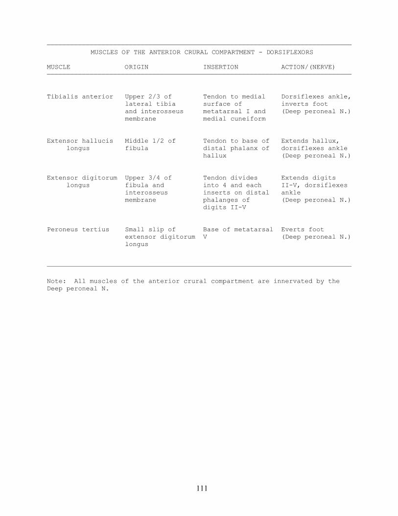
111
——————————————————————————————————————————————————————————————————————————————MUSCLES OF THE ANTERIOR CRURAL COMPARTMENT - DORSIFLEXORS
MUSCLE ORIGIN INSERTION ACTION/(NERVE)——————————————————————————————————————————————————————————————————————————————
Tibialis anterior Upper 2/3 of Tendon to medial Dorsiflexes ankle, lateral tibia surface of inverts foot and interosseus metatarsal I and (Deep peroneal N.) membrane medial cuneiform
Extensor hallucis Middle 1/2 of Tendon to base of Extends hallux, longus fibula distal phalanx of dorsiflexes ankle hallux (Deep peroneal N.)
Extensor digitorum Upper 3/4 of Tendon divides Extends digits longus fibula and into 4 and each II-V, dorsiflexes interosseus inserts on distal ankle membrane phalanges of (Deep peroneal N.) digits II-V
Peroneus tertius Small slip of Base of metatarsal Everts foot extensor digitorum V (Deep peroneal N.) longus
——————————————————————————————————————————————————————————————————————————————
Note: All muscles of the anterior crural compartment are innervated by theDeep peroneal N.
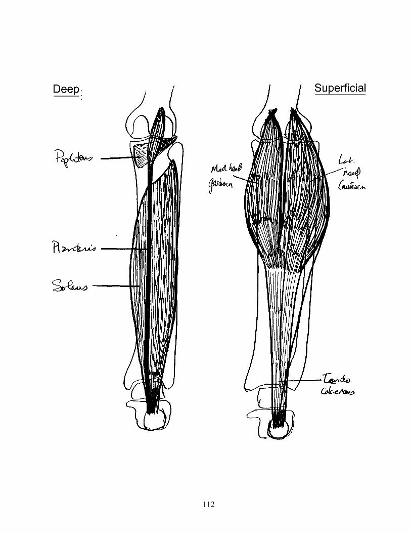
112
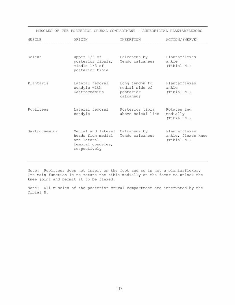
113
——————————————————————————————————————————————————————————————————————————————MUSCLES OF THE POSTERIOR CRURAL COMPARTMENT - SUPERFICIAL PLANTARFLEXORS
MUSCLE ORIGIN INSERTION ACTION/(NERVE)——————————————————————————————————————————————————————————————————————————————
Soleus Upper 1/3 of Calcaneus by Plantarflexes posterior fibula, Tendo calcaneus ankle middle 1/3 of (Tibial N.) posterior tibia
Plantaris Lateral femoral Long tendon to Plantarflexes condyle with medial side of ankle Gastrocnemius posterior (Tibial N.) calcaneus
Popliteus Lateral femoral Posterior tibia Rotates leg condyle above soleal line medially (Tibial N.)
Gastrocnemius Medial and lateral Calcaneus by Plantarflexes heads from medial Tendo calcaneus ankle, flexes knee and lateral (Tibial N.) femoral condyles, respectively
——————————————————————————————————————————————————————————————————————————————
Note: Popliteus does not insert on the foot and so is not a plantarflexor.Its main function is to rotate the tibia medially on the femur to unlock theknee joint and permit it to be flexed.
Note: All muscles of the posterior crural compartment are innervated by theTibial N.
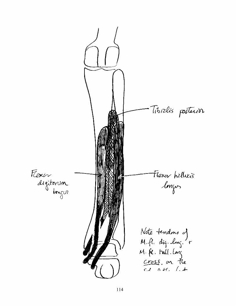
114
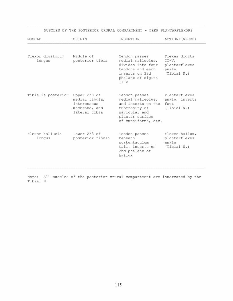
115
——————————————————————————————————————————————————————————————————————————————MUSCLES OF THE POSTERIOR CRURAL COMPARTMENT - DEEP PLANTARFLEXORS
MUSCLE ORIGIN INSERTION ACTION/(NERVE)——————————————————————————————————————————————————————————————————————————————
Flexor digitorum Middle of Tendon passes Flexes digits longus posterior tibia medial malleolus, II-V, divides into four plantarflexes tendons and each ankle inserts on 3rd (Tibial N.) phalanx of digits II-V
Tibialis posterior Upper 2/3 of Tendon passes Plantarflexes medial fibula, medial malleolus, ankle, inverts interosseus and inserts on the foot membrane, and tuberosity of (Tibial N.) lateral tibia navicular and plantar surface of cuneiforms, etc.
Flexor hallucis Lower 2/3 of Tendon passes Flexes hallux, longus posterior fibula beneath plantarflexes sustentaculum ankle tali, inserts on (Tibial N.) 2nd phalanx of hallux
——————————————————————————————————————————————————————————————————————————————
Note: All muscles of the posterior crural compartment are innervated by theTibial N.
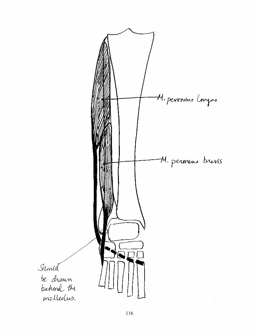
116
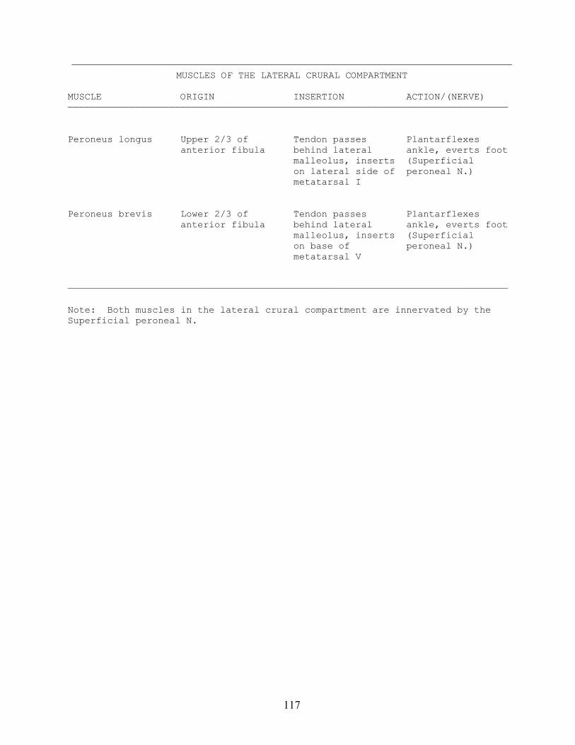
117
——————————————————————————————————————————————————————————————————————————————MUSCLES OF THE LATERAL CRURAL COMPARTMENT
MUSCLE ORIGIN INSERTION ACTION/(NERVE)——————————————————————————————————————————————————————————————————————————————
Peroneus longus Upper 2/3 of Tendon passes Plantarflexes anterior fibula behind lateral ankle, everts foot malleolus, inserts (Superficial on lateral side of peroneal N.) metatarsal I
Peroneus brevis Lower 2/3 of Tendon passes Plantarflexes anterior fibula behind lateral ankle, everts foot malleolus, inserts (Superficial on base of peroneal N.) metatarsal V
——————————————————————————————————————————————————————————————————————————————
Note: Both muscles in the lateral crural compartment are innervated by theSuperficial peroneal N.
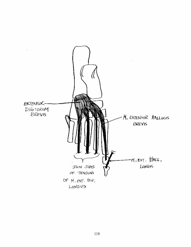
118
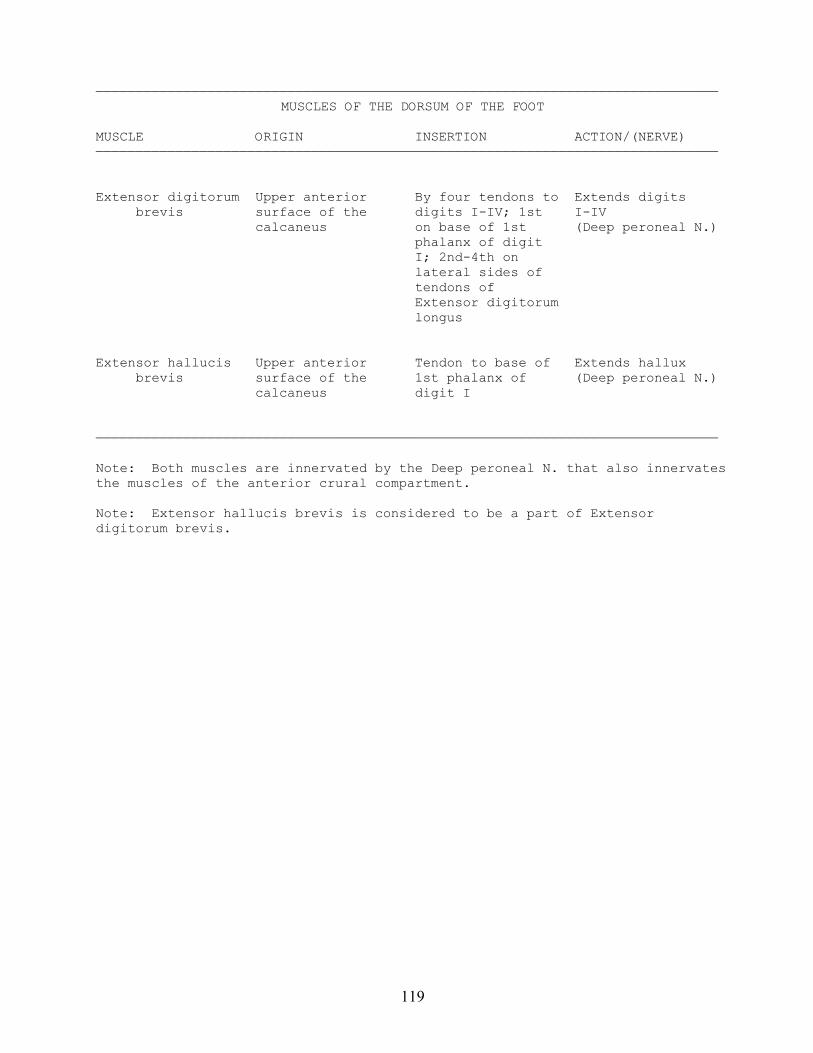
119
——————————————————————————————————————————————————————————————————————————————MUSCLES OF THE DORSUM OF THE FOOT
MUSCLE ORIGIN INSERTION ACTION/(NERVE)——————————————————————————————————————————————————————————————————————————————
Extensor digitorum Upper anterior By four tendons to Extends digits brevis surface of the digits I-IV; 1st I-IV calcaneus on base of 1st (Deep peroneal N.) phalanx of digit I; 2nd-4th on lateral sides of tendons of Extensor digitorum longus
Extensor hallucis Upper anterior Tendon to base of Extends hallux brevis surface of the 1st phalanx of (Deep peroneal N.) calcaneus digit I
——————————————————————————————————————————————————————————————————————————————
Note: Both muscles are innervated by the Deep peroneal N. that also innervatesthe muscles of the anterior crural compartment.
Note: Extensor hallucis brevis is considered to be a part of Extensordigitorum brevis.
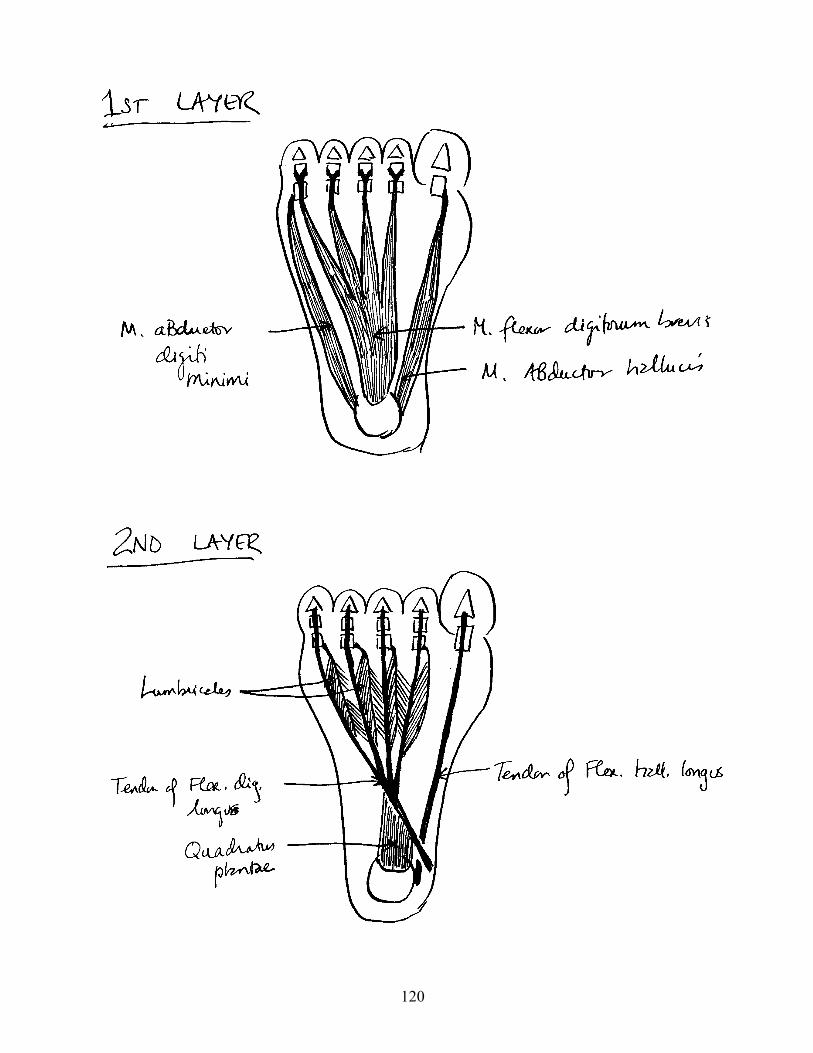
120
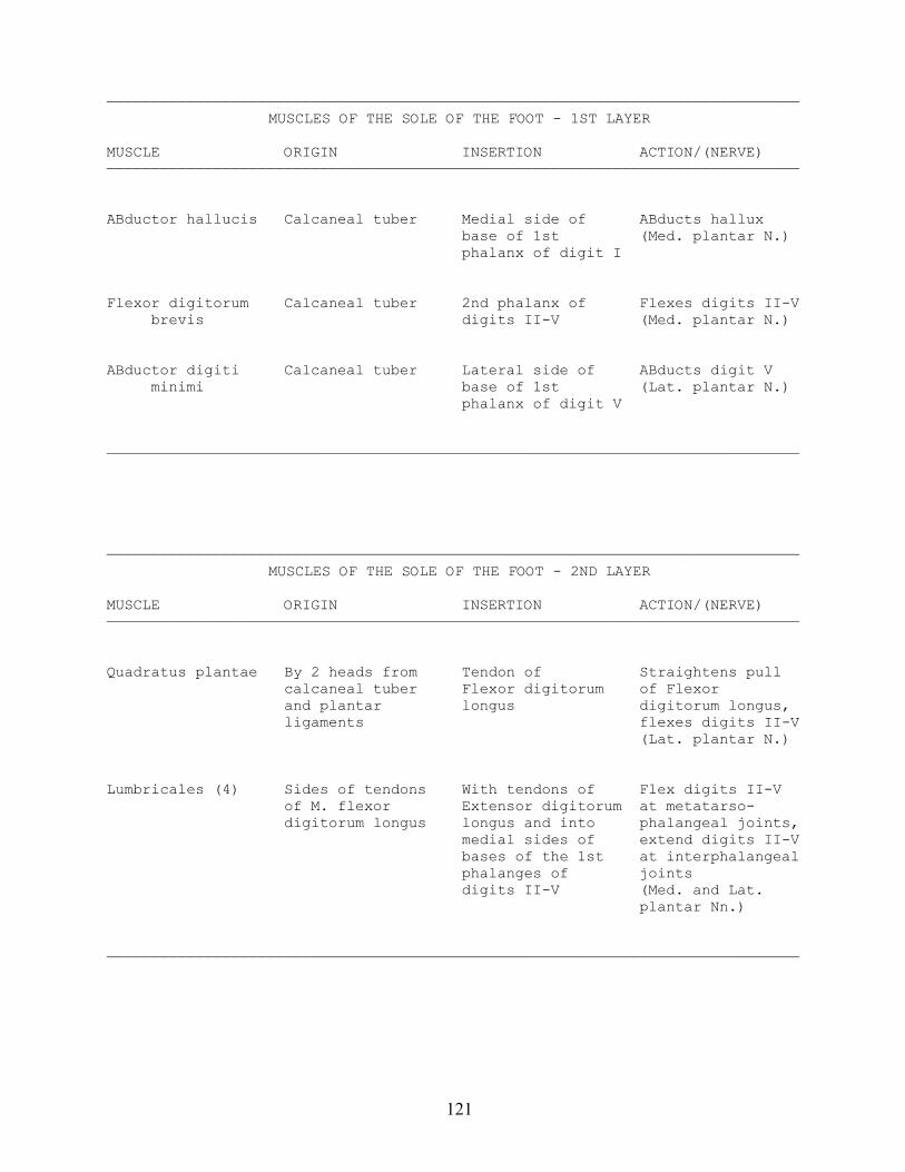
121
——————————————————————————————————————————————————————————————————————————————MUSCLES OF THE SOLE OF THE FOOT - 1ST LAYER
MUSCLE ORIGIN INSERTION ACTION/(NERVE)——————————————————————————————————————————————————————————————————————————————
ABductor hallucis Calcaneal tuber Medial side of ABducts hallux base of 1st (Med. plantar N.) phalanx of digit I
Flexor digitorum Calcaneal tuber 2nd phalanx of Flexes digits II-V brevis digits II-V (Med. plantar N.)
ABductor digiti Calcaneal tuber Lateral side of ABducts digit V minimi base of 1st (Lat. plantar N.) phalanx of digit V
——————————————————————————————————————————————————————————————————————————————
——————————————————————————————————————————————————————————————————————————————MUSCLES OF THE SOLE OF THE FOOT - 2ND LAYER
MUSCLE ORIGIN INSERTION ACTION/(NERVE)——————————————————————————————————————————————————————————————————————————————
Quadratus plantae By 2 heads from Tendon of Straightens pull calcaneal tuber Flexor digitorum of Flexor and plantar longus digitorum longus, ligaments flexes digits II-V (Lat. plantar N.)
Lumbricales (4) Sides of tendons With tendons of Flex digits II-V of M. flexor Extensor digitorum at metatarso- digitorum longus longus and into phalangeal joints, medial sides of extend digits II-V bases of the 1st at interphalangeal phalanges of joints digits II-V (Med. and Lat. plantar Nn.)
——————————————————————————————————————————————————————————————————————————————
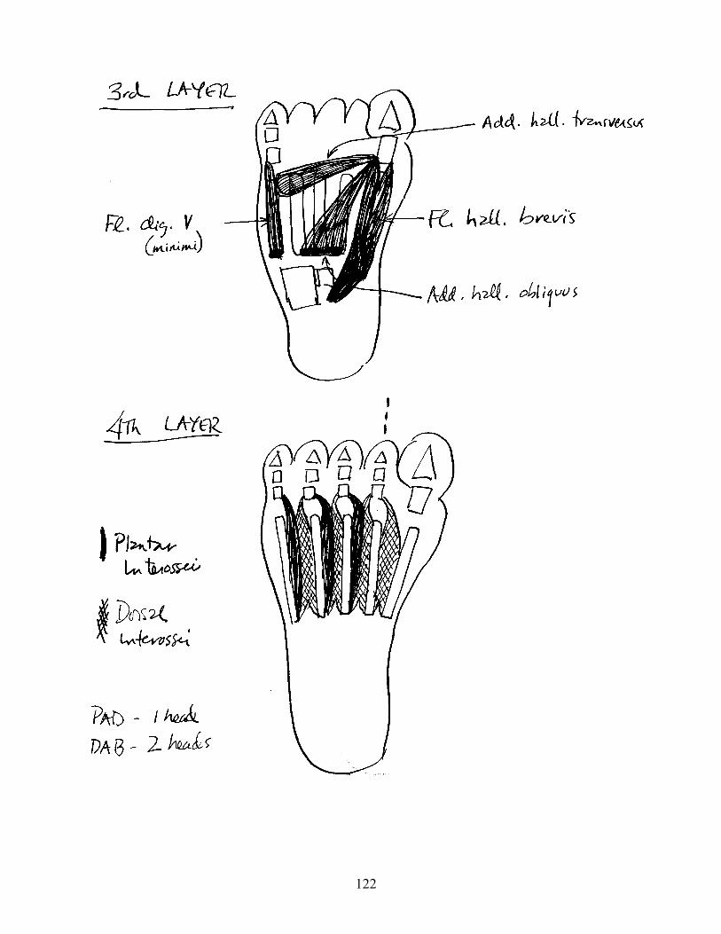
122
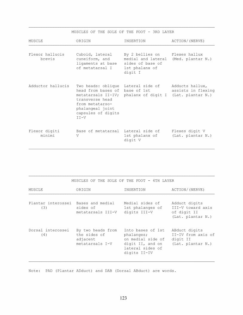
123
—————————————————————————————————————————————————————————————————————————————— MUSCLES OF THE SOLE OF THE FOOT - 3RD LAYER
MUSCLE ORIGIN INSERTION ACTION/(NERVE)——————————————————————————————————————————————————————————————————————————————
Flexor hallucis Cuboid, lateral By 2 bellies on Flexes hallux brevis cuneiform, and medial and lateral (Med. plantar N.) ligaments at base sides of base of of metatarsal I 1st phalanx of digit I
Adductor hallucis Two heads: oblique Lateral side of Adducts hallux, head from bases of base of 1st assists in flexing metatarsals II-IV; phalanx of digit I (Lat. plantar N.) transverse head from metatarso- phalangeal joint capsules of digits II-V
Flexor digiti Base of metatarsal Lateral side of Flexes digit V minimi V 1st phalanx of (Lat. plantar N.) digit V
——————————————————————————————————————————————————————————————————————————————
—————————————————————————————————————————————————————————————————————————————— MUSCLES OF THE SOLE OF THE FOOT - 4TH LAYER
MUSCLE ORIGIN INSERTION ACTION/(NERVE)——————————————————————————————————————————————————————————————————————————————
Plantar interossei Bases and medial Medial sides of Adduct digits (3) sides of 1st phalanges of III-V toward axis metatarsals III-V digits III-V of digit II (Lat. plantar N.)
Dorsal interossei By two heads from Into bases of 1st ABduct digits (4) the sides of phalanges; II-IV from axis of adjacent on medial side of digit II metatarsals I-V digit II, and on (Lat. plantar N.) lateral sides of digits II-IV
——————————————————————————————————————————————————————————————————————————————
Note: PAD (Plantar ADduct) and DAB (Dorsal ABduct) are words.
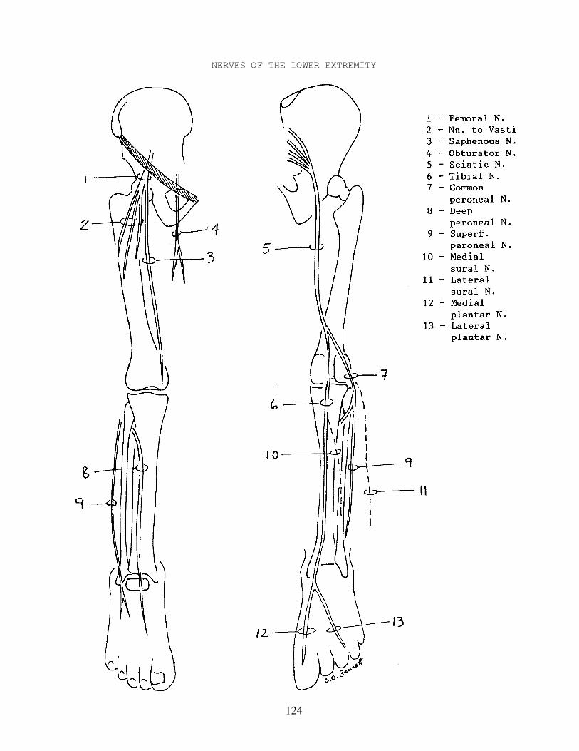
124
NERVES OF THE LOWER EXTREMITY
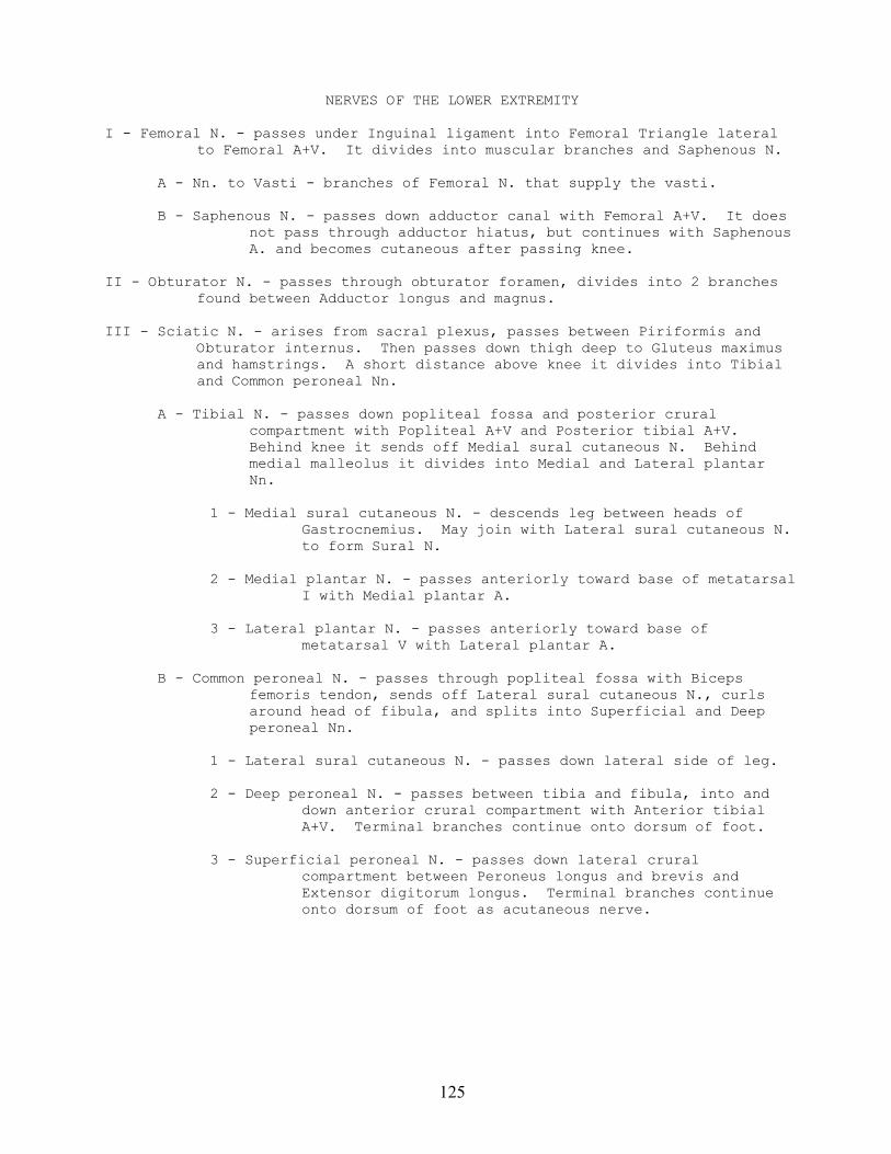
125
NERVES OF THE LOWER EXTREMITY
I - Femoral N. - passes under Inguinal ligament into Femoral Triangle lateralto Femoral A+V. It divides into muscular branches and Saphenous N.
A - Nn. to Vasti - branches of Femoral N. that supply the vasti.
B - Saphenous N. - passes down adductor canal with Femoral A+V. It doesnot pass through adductor hiatus, but continues with SaphenousA. and becomes cutaneous after passing knee.
II - Obturator N. - passes through obturator foramen, divides into 2 branchesfound between Adductor longus and magnus.
III - Sciatic N. - arises from sacral plexus, passes between Piriformis andObturator internus. Then passes down thigh deep to Gluteus maximusand hamstrings. A short distance above knee it divides into Tibialand Common peroneal Nn.
A - Tibial N. - passes down popliteal fossa and posterior cruralcompartment with Popliteal A+V and Posterior tibial A+V.Behind knee it sends off Medial sural cutaneous N. Behindmedial malleolus it divides into Medial and Lateral plantarNn.
1 - Medial sural cutaneous N. - descends leg between heads ofGastrocnemius. May join with Lateral sural cutaneous N.to form Sural N.
2 - Medial plantar N. - passes anteriorly toward base of metatarsalI with Medial plantar A.
3 - Lateral plantar N. - passes anteriorly toward base ofmetatarsal V with Lateral plantar A.
B - Common peroneal N. - passes through popliteal fossa with Bicepsfemoris tendon, sends off Lateral sural cutaneous N., curlsaround head of fibula, and splits into Superficial and Deepperoneal Nn.
1 - Lateral sural cutaneous N. - passes down lateral side of leg.
2 - Deep peroneal N. - passes between tibia and fibula, into anddown anterior crural compartment with Anterior tibialA+V. Terminal branches continue onto dorsum of foot.
3 - Superficial peroneal N. - passes down lateral cruralcompartment between Peroneus longus and brevis andExtensor digitorum longus. Terminal branches continueonto dorsum of foot as acutaneous nerve.
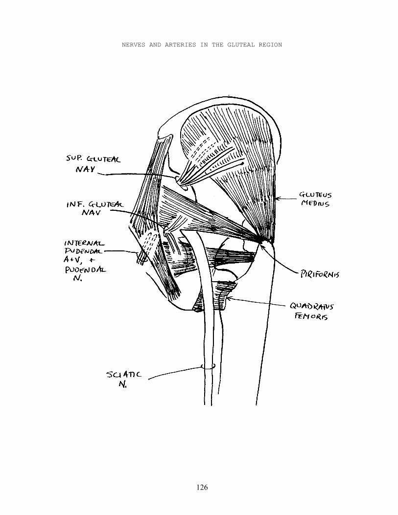
126
NERVES AND ARTERIES IN THE GLUTEAL REGION
127
NERVES AND ARTERIES IN THE GLUTEAL REGION
NERVES
I - Branches of sacral plexus in gluteal region
A - Superior gluteal N. - found with Superior gluteal A+V superior toPiriformis, then passes laterally with deep branches ofSuperior gluteal A+V between Gluteus minimus and medius.
B - Inferior gluteal N. - found with Inferior gluteal A+V betweenPiriformis and Obturator internus, and medial to Sciatic N.
C - Pudendal N. - found with Inferior pudendal A+V between Piriformis andObturator internus, medial to Superior gluteal NAV, andpassing deep to perineal region.
D - Sciatic N. - arises from sacral plexus, passes between Piriformis andObturator internus. Then passes down thigh deep to Gluteusmaximus and hamstrings.
ARTERIES
I - Branches of Internal Iliac A. in gluteal region
A - Superior gluteal A. - found with Superior gluteal N. superior toPiriformis. Deep branches pass laterally between Gluteusminimus and medius, and superficial branches pass laterallybetween Gluteus medius and maximus.
B - Inferior gluteal A. - found with Inferior gluteal N. betweenPiriformis and Obturator internus and medial to Sciatic N.
C - Internal pudendal A. - found with Pudendal N. between Piriformis andObturator internus and medial to Sciatic N. It goes deep toinguinal region.
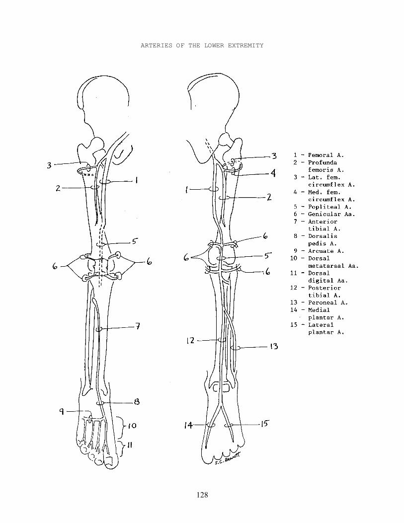
128
ARTERIES OF THE LOWER EXTREMITY
129
ARTERIES OF THE LOWER EXTREMITY
Ia - Femoral A. - continuation of External iliac A. after passing underInguinal ligament. Passes down femoral triangle, adductor canal,through adductor hiatus, and becomes Popliteal A.
A - Profunda femoris A. - branches off in Femoral triangle, goes deeptoward femur, and passes down thigh deep alongside femur.
1 - Medial femoral circumflex A. - branches off about 2 cm fromorigin of Profunda femoris A., passes medially betweenIliopsoas and Pectineus, around back of femur, andanastomoses with Lateral femoral circumflex A.
2 - Lateral femoral circumflex A. - branches off about 2 cm fromorigin of Profunda femoris A., splits into threebranches; one branch passes laterally around femur, andanastomoses with Medial femoral circumflex A.
Ib - Popliteal A. - continuation of Femoral A. on posterior thigh. It passesthrough popliteal fossa, and divides into Anterior and Posteriortibial Aa. shortly below knee.
A - Superior medial genicular A. ┐ Pass around knee from posterior to Superior lateral genicular A. ├ anterior and anastomose together. Inferior medial genicular A. │ Superiors branch off above heads of Inferior lateral genicular A. ┘ Gastrocnemius, inferiors below.
Bi - Anterior tibial A. - passes between tibia and fibula into and downanterior crural compartment on interosseus membrane. As itcrosses ankle it becomes Dorsalis pedis A.
Bii - Dorsalis Pedis A. - continuation of Anterior tibial A. Passesunder extensor retinacula toward base of metatarsal I.
1 - Arcuate A. - runs laterally along bases of metatarsals.
a - Dorsal metatarsal Aa. - four; each passes distallybetween metatarsals and divides into dorsaldigital Aa.
i - Dorsal digital Aa. - pass distallyalong digits.
C - Posterior tibial A. - passes down posterior crural compartment deepto Soleus, passes behind medial malleolus, and splits intoMedial and Lateral plantar Aa.
1 - Peroneal A. - branches off Posterior tibial A. about 6 cm belowknee, and passes deep along fibula.
2 - Medial plantar A. - passes anteriorly toward base of metatarsalI with Medial plantar N.
3 - Lateral plantar A. - passes anteriorly toward base ofmetatarsal V with Lateral plantar N.
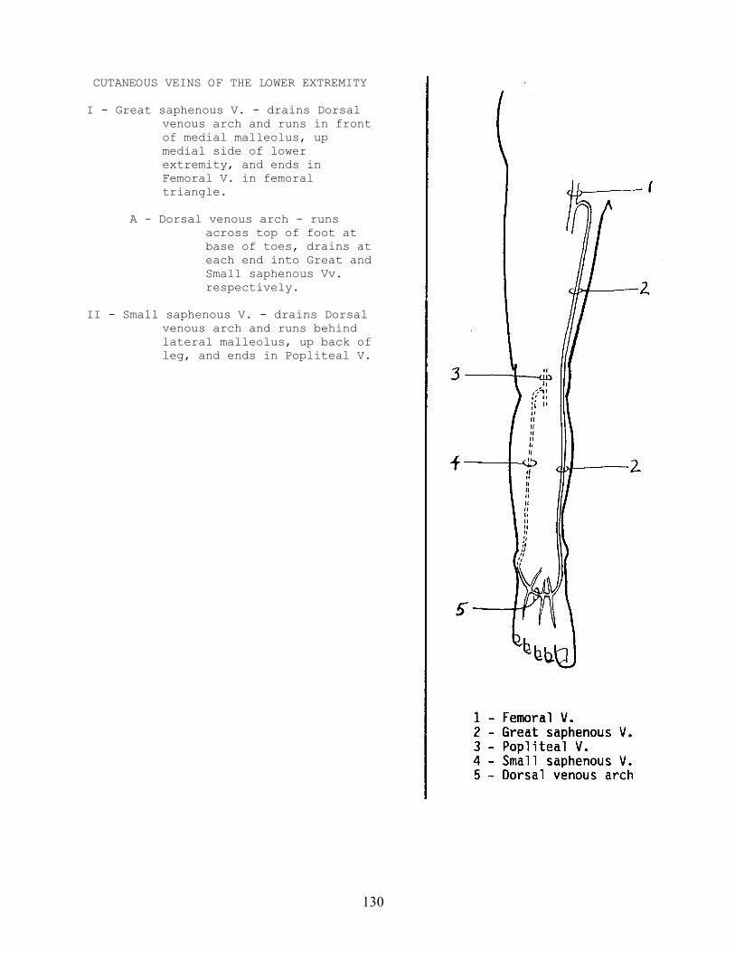
130
CUTANEOUS VEINS OF THE LOWER EXTREMITY
I - Great saphenous V. - drains Dorsalvenous arch and runs in frontof medial malleolus, upmedial side of lowerextremity, and ends inFemoral V. in femoraltriangle.
A - Dorsal venous arch - runsacross top of foot atbase of toes, drains ateach end into Great andSmall saphenous Vv.respectively.
II - Small saphenous V. - drains Dorsalvenous arch and runs behindlateral malleolus, up back ofleg, and ends in Popliteal V.
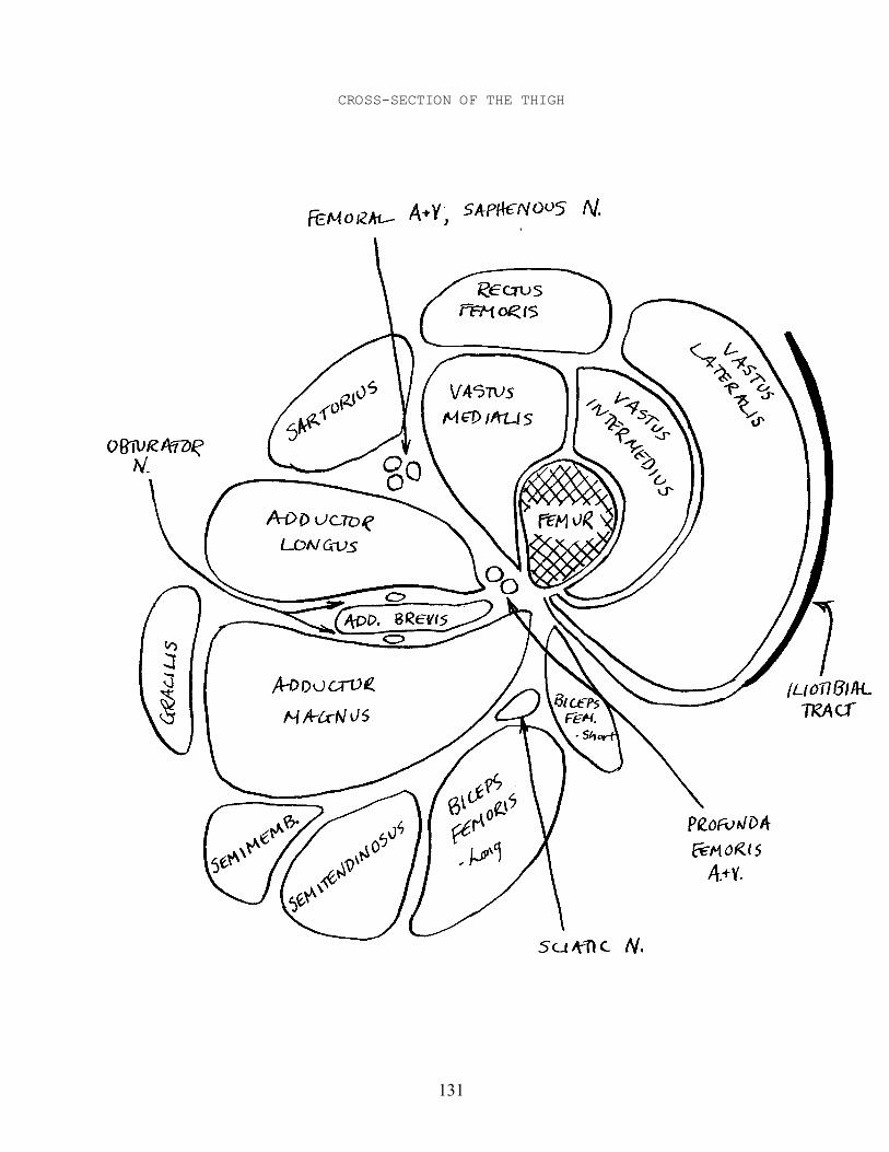
131
CROSS-SECTION OF THE THIGH
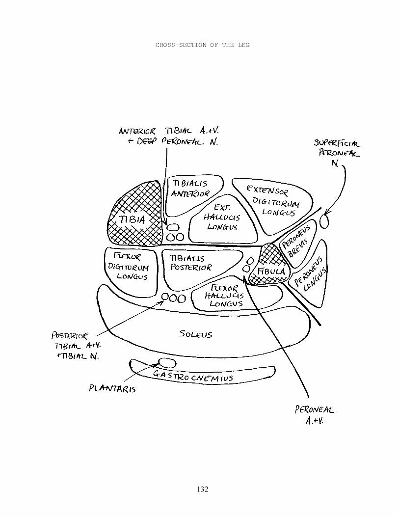
132
CROSS-SECTION OF THE LEG