A quantitative infection assay for human type I, II, and III
10
METHODOLOGY Open Access A quantitative infection assay for human type I, II, and III interferon antiviral activities Emily Voigt 1,2† , Bahar İnankur 1,2† , Ashley Baltes 1,2 and John Yin 1,2* Abstract Background: Upon virus infection, cells secrete a diverse group of antiviral molecules that signal proximal cells to enter into an antiviral state, slowing or preventing viral spread. These paracrine signaling molecules can work synergistically, so measurement of any one antiviral molecule does not reflect the total antiviral activity of the system. Results: We have developed an antiviral assay based on replication inhibition of an engineered fluorescent vesicular stomatitis virus reporter strain on A549 human lung epithelial cells. Our assay provides a quantitative functional readout of human type I, II, and III interferon activities, and it provides better sensitivity, intra-, and inter- assay reproducibility than the traditional crystal violet based assay. Further, it eliminates cell fixation, rinsing, and staining steps, and is inexpensive to implement. Conclusions: A dsRed2-strain of vesicular stomatitis virus that is sensitive to type I, II, and III interferons was used to develop a convenient and sensitive assay for interferon antiviral activity. We demonstrate use of the assay to quantify the kinetics of paracrine antiviral signaling from human prostate cancer (PC3) cells in response to viral infection. The assay is applicable to high-throughput screening for anti-viral compounds as well as basic studies of cellular antiviral signaling. Keywords: Antiviral activity assay, Reporter virus, Interferon, Paracrine signaling, Bioassay, Cytokine quantification Background Mammalian cells respond to virus infection by synthesiz- ing and secreting a host of antiviral molecules that are not only involved in recruitment of immune effector cells and activation of adaptive immunity, but also control localized spread of virus infection via mechanisms of “cell-intrinsic” innate immunity. These secreted antiviral molecules, first described collectively in 1957 as “interferon”, establish an anti-viral state within the infected cell, and signal other cells to react in an antiviral manner [1,2]. Antivirals secreted by non-immune cells comprise a diverse mixture of molecules which exert a combined paracrine effect on proximal cells. These include, for ex- ample, the classic type I interferons (IFNα/β, etc.) [3,4], type II interferon (IFNγ), and type III interferons (IFNλs) [5-9]. While type I interferons are known to play import- ant roles in host antiviral responses, for many viruses such as human rhinovirus and influenza infections of bronchial epithelial cells, other interferons such as the more recently identified IFNλs, may dominate [10,11]. Thus, measuring type I IFNs alone does not necessarily accurately assess cellular antiviral responses, and an assay that measures antiviral responses due to multiple types of interferons is required. The biological effects of interferons are often measured using reporter gene assays (RGAs), which use transgenic cell lines expressing a reporter gene driven by an IFN- responsive promoter [12-17]. These assays accurately measure the presence or upregulation of single molecules, such as Mx or various interferon-stimulated genes, and provide valuable information on specific components of an antiviral response. However, other assay methods are necessary for the quantification of the integrated antiviral effects of multiple-type interferon signaling, which is es- sential for studying inhibition of virus spread [18,19]. Moreover, functional measures of secreted antiviral * Correspondence: [email protected] † Equal contributors 1 Department of Chemical and Biological Engineering, University of Wisconsin, Madison, USA 2 Systems Biology Theme, Wisconsin Institute for Discovery, 330 N Orchard St., Madison, WI 53715, USA © 2013 Voigt et al.; licensee BioMed Central Ltd. This is an Open Access article distributed under the terms of the Creative Commons Attribution License (http://creativecommons.org/licenses/by/2.0), which permits unrestricted use, distribution, and reproduction in any medium, provided the original work is properly cited. Voigt et al. Virology Journal 2013, 10:224 http://www.virologyj.com/content/10/1/224
Transcript of A quantitative infection assay for human type I, II, and III

Voigt et al. Virology Journal 2013, 10:224http://www.virologyj.com/content/10/1/224
METHODOLOGY Open Access
A quantitative infection assay for human type I, II,and III interferon antiviral activitiesEmily Voigt1,2†, Bahar İnankur1,2†, Ashley Baltes1,2 and John Yin1,2*
Abstract
Background: Upon virus infection, cells secrete a diverse group of antiviral molecules that signal proximal cells toenter into an antiviral state, slowing or preventing viral spread. These paracrine signaling molecules can worksynergistically, so measurement of any one antiviral molecule does not reflect the total antiviral activity of thesystem.
Results: We have developed an antiviral assay based on replication inhibition of an engineered fluorescentvesicular stomatitis virus reporter strain on A549 human lung epithelial cells. Our assay provides a quantitativefunctional readout of human type I, II, and III interferon activities, and it provides better sensitivity, intra-, and inter-assay reproducibility than the traditional crystal violet based assay. Further, it eliminates cell fixation, rinsing, andstaining steps, and is inexpensive to implement.
Conclusions: A dsRed2-strain of vesicular stomatitis virus that is sensitive to type I, II, and III interferons was used todevelop a convenient and sensitive assay for interferon antiviral activity. We demonstrate use of the assay toquantify the kinetics of paracrine antiviral signaling from human prostate cancer (PC3) cells in response to viralinfection. The assay is applicable to high-throughput screening for anti-viral compounds as well as basic studies ofcellular antiviral signaling.
Keywords: Antiviral activity assay, Reporter virus, Interferon, Paracrine signaling, Bioassay, Cytokine quantification
BackgroundMammalian cells respond to virus infection by synthesiz-ing and secreting a host of antiviral molecules that are notonly involved in recruitment of immune effector cells andactivation of adaptive immunity, but also control localizedspread of virus infection via mechanisms of “cell-intrinsic”innate immunity. These secreted antiviral molecules, firstdescribed collectively in 1957 as “interferon”, establish ananti-viral state within the infected cell, and signal othercells to react in an antiviral manner [1,2].Antivirals secreted by non-immune cells comprise a
diverse mixture of molecules which exert a combinedparacrine effect on proximal cells. These include, for ex-ample, the classic type I interferons (IFNα/β, etc.) [3,4],type II interferon (IFNγ), and type III interferons (IFNλs)
* Correspondence: [email protected]†Equal contributors1Department of Chemical and Biological Engineering, University ofWisconsin, Madison, USA2Systems Biology Theme, Wisconsin Institute for Discovery, 330 N Orchard St.,Madison, WI 53715, USA
© 2013 Voigt et al.; licensee BioMed Central LCommons Attribution License (http://creativecreproduction in any medium, provided the or
[5-9]. While type I interferons are known to play import-ant roles in host antiviral responses, for many virusessuch as human rhinovirus and influenza infections ofbronchial epithelial cells, other interferons such as themore recently identified IFNλs, may dominate [10,11].Thus, measuring type I IFNs alone does not necessarilyaccurately assess cellular antiviral responses, and anassay that measures antiviral responses due to multipletypes of interferons is required.The biological effects of interferons are often measured
using reporter gene assays (RGAs), which use transgeniccell lines expressing a reporter gene driven by an IFN-responsive promoter [12-17]. These assays accuratelymeasure the presence or upregulation of single molecules,such as Mx or various interferon-stimulated genes, andprovide valuable information on specific components ofan antiviral response. However, other assay methods arenecessary for the quantification of the integrated antiviraleffects of multiple-type interferon signaling, which is es-sential for studying inhibition of virus spread [18,19].Moreover, functional measures of secreted antiviral
td. This is an Open Access article distributed under the terms of the Creativeommons.org/licenses/by/2.0), which permits unrestricted use, distribution, andiginal work is properly cited.

Voigt et al. Virology Journal 2013, 10:224 Page 2 of 10http://www.virologyj.com/content/10/1/224
signaling will be useful for advancing experimental andcomputational models of virus-cell interactions and viralinfection spread in monolayers and tissues [20-24]. Such anassay potentially has additional applications in the area ofhigh-throughput screening for antiviral compounds.The most traditional form of a functional antiviral assay
is the assay based on cytopathic effect (CPE), commonlyused to determine the potency of purified interferon stocks.In the CPE assay, antiviral activity is measured based on itsability to inhibit virus-induced cytopathology as measuredby a crystal violet live-cell stain [25]. While widely used,these types of assays are labor-intensive and contain manyhandling steps that can disturb cell layers and increase vari-ability. Additionally, successful infection that does not causemajor cytopathology is not detected by these assays. Theseshortcomings can be addressed through the developmentof reporter virus strains as robust readouts of virus replica-tion. Examples include a luciferase-expressing reporterstrain of the BSL3 Rift Valley Fever virus and a non-proliferative vesicular stomatitis virus (VSV) replicon alsoexpressing luciferase [2,26]. Here we create a replication-competent fluorescent reporter VSV strain. We use thisvirus in a simple, sensitive, and reproducible assay for de-tection of secreted antiviral signaling activity. The assaydoes not require the addition of expensive substrates elimi-nates cell fixing, rinsing, and staining steps, and signifi-cantly improves sensitivity, and reproducibility over thetraditional crystal violet CPE assay. Additionally, the broadtropism of VSV allows for potential future assay adaptationto a large range of host cell types [2,26].
ResultsSynthesis of reporter virus constructsWe sought to create an assay to quantify overall activity ofsecreted antiviral molecules with optimal sensitivity and re-producibility. We therefore compared four detectionmethods to determine antiviral activity: the traditionalcrystal violet live-cell stain, Sytox® orange, a fluorescentdead cell stain, and both a DsRed2-expressing and aZsGreen-expressing VSV reporter strain. Using fluorescentreporters of VSV replication may increase assay sensitivity,as inhibition of virus replication is a more direct measure
Figure 1 Genotype maps of wild-type and fluorescent reporter VSV sreverse-genetics, incorporating fluorescent reporter variants of GFP(ZsGreennucleocapsid protein (N), phosphoprotein (P), matrix protein (M), glycoprot
of antiviral activity than cytopathology alone. We createdstrains of DsRed2- or ZsGreen-encoding VSV with gen-omic organization as indicated in Figure 1 for use in ourantiviral assays. We tested the ability of these recombinantvirus strains to infect A549 lung epithelial cells, and foundtheir replication rates comparable to that of their non-fluorescent equivalent (Additional file 1: Figure S1) and amultiplicity of infection (MOI) of 5 pfu/cell sufficient tohomogenously infect A549 monolayers (data not shown).
Comparison of assay readout methodsWe compared the traditional virus-induced cytopathologymeasurement (crystal violet after 28-hour infection) with amore recently developed fluorescent dead cell stain(Sytox®) and our two fluorescent VSV reporter strains. Todo so, we incubated A549 cells under 2-fold dilutions ofan IFNβ standard solution in media for 24 hours. The cellswere then infected with either wild-type or one of thefluorescent reporter VSV strains as indicated in Figure 2,at an MOI of 5 pfu/cell, and the infection was allowed toprogress for 28 hours. WtVSV-infected plates were rinsedand stained as indicated. Assay results were quantified byfluorescent scanning at the appropriate wavelengths andsubsequent normalization to positive (no IFN) and nega-tive (no virus) controls, as shown in Figure 2.We found that the fluorescent signal from DsRed2 and
ZsGreen reporter viruses created reproducible gradientsindicating decreasing viral replication due to increasingIFN concentrations. These gradients were comparable tothose found by staining cells with crystal violet afterwtVSV replication and cell death. Sytox® fluorescent deadcell stain also produced quantifiable gradients, but with asignificantly poorer antiviral detection limit (IC50 = 7.87U/ml vs. <4 U/ml) than both crystal violet and fluorescentvirus assays.
Sensitivity and reproducibility comparison of fluorescentreporter virusesAs fluorescent signals are generally more sensitive thanreadouts of absorbance, and dead-cell stains require fixingand staining steps that can disturb cell layers and increasevariability, we tested the reproducibility and sensitivity
trains used in this study. Recombinant VSV strains were created by) and RFP(DsRed2) along with the five native VSV proteins:ein (G), and large protein (L).

Figure 2 Comparison of live/dead cell stains and fluorescent-protein expressing assay virus. A549 cells were incubated under serial 2-folddilutions of recombinant human IFNβ for 24 hours, then infected with either wild-type or recombinant VSV as indicated at a multiplicity of 5 pfu/cell.After 24 hours of infection, assay plates were stained, imaged, and quantified as discussed in the Methods section. Positive signal indicates cell survivalfor the crystal violet assay, virus replication for the fluorescent virus assays, and cell death in the Sytox assay. Positive control wells are cells untreated byantivirals and infected. Negative control wells are untreated, uninfected cells.
Voigt et al. Virology Journal 2013, 10:224 Page 3 of 10http://www.virologyj.com/content/10/1/224
of assays using ZsGreen and DsRed2 reporter virus andcompared it to that of the traditional crystal violetcytopathology-based assay. Fluorescence was read both ona standard fluorescent plate reader and also scanned witha high-resolution GE Typhoon FLA 9000 BiomolecularImager, to determine which reading method is more sensi-tive and reproducible. Sensitivity was defined as the limitof detection (LOD), the lowest antiviral starting dose that,upon two-fold serial dilution, would produce a dose–response curve crossing the 50% viral inhibition point andallow for accurate IC50 determination, as shown in Table 1.Calculation of IC50 values for both quantification typeswas done using linear interpolation within the linear dose-dependent range of the indicated interferon treatment asdescribed in Methods. A sigmoidal-fit IC50 calculationmethod was also tested, but showed no advantage overlinear interpolation.
Both fluorescent reporter viruses significantly de-creased the limit of detection and improved intra- andinter-assay reproducibility over the traditional crystalviolet staining method. Sensitivity differences betweenDsRed2 and ZsGreen were insignificant (p > 0.1). TheDsRed reporter virus showed better inter-assay repro-ducibility than the ZsGreen virus when quantified byfluorescent scanning. This may be due to fluorescentbackground in the green absorption/emission spectrumproduced by the plastic microtiter plate (Figure 3).There was no significant difference between the reporterviruses for the plate reader quantification (p-value = 0.44).
Optimization of fluorescent signalWe determined the optimal time for signal developmentby measuring a time course over 28 hours of signal de-velopment as shown in Figure 3. As time progresses, the
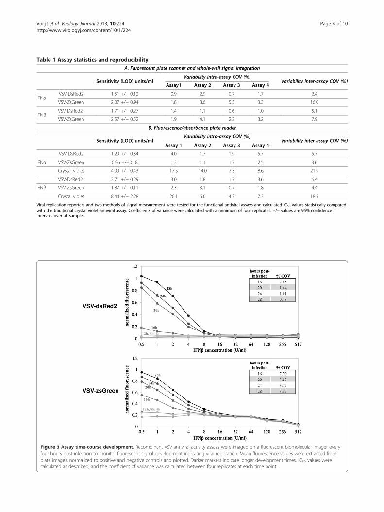
Table 1 Assay statistics and reproducibility
A. Fluorescent plate scanner and whole-well signal integration
Sensitivity (LOD) units/mlVariability intra-assay COV (%)
Variability inter-assay COV (%)Assay1 Assay 2 Assay 3 Assay 4
IFNαVSV-DsRed2 1.51 +/− 0.12 0.9 2.9 0.7 1.7 2.4
VSV-ZsGreen 2.07 +/− 0.94 1.8 8.6 5.5 3.3 16.0
IFNβVSV-DsRed2 1.71 +/− 0.27 1.4 1.1 0.6 1.0 5.1
VSV-ZsGreen 2.57 +/− 0.52 1.9 4.1 2.2 3.2 7.9
B. Fluorescence/absorbance plate reader
Sensitivity (LOD) units/mlVariability intra-assay COV (%)
Variability inter-assay COV (%)Assay 1 Assay 2 Assay 3 Assay 4
VSV-DsRed2 1.29 +/− 0.34 4.0 1.7 1.9 5.7 5.7
IFNα VSV-ZsGreen 0.96 +/−0.18 1.2 1.1 1.7 2.5 3.6
Crystal violet 4.09 +/− 0.43 17.5 14.0 7.3 8.6 21.9
VSV-DsRed2 2.71 +/− 0.29 3.0 1.8 1.7 3.6 6.4
IFNβ VSV-ZsGreen 1.87 +/− 0.11 2.3 3.1 0.7 1.8 4.4
Crystal violet 8.44 +/− 2.28 20.1 6.6 4.3 7.3 18.5
Viral replication reporters and two methods of signal measurement were tested for the functional antiviral assays and calculated IC50 values statistically comparedwith the traditional crystal violet antiviral assay. Coefficients of variance were calculated with a minimum of four replicates. +/− values are 95% confidenceintervals over all samples.
Figure 3 Assay time-course development. Recombinant VSV antiviral activity assays were imaged on a fluorescent biomolecular imager everyfour hours post-infection to monitor fluorescent signal development indicating viral replication. Mean fluorescence values were extracted fromplate images, normalized to positive and negative controls and plotted. Darker markers indicate longer development times. IC50 values werecalculated as described, and the coefficient of variance was calculated between four replicates at each time point.
Voigt et al. Virology Journal 2013, 10:224 Page 4 of 10http://www.virologyj.com/content/10/1/224

Voigt et al. Virology Journal 2013, 10:224 Page 5 of 10http://www.virologyj.com/content/10/1/224
dose-dependent signal gradient develops, where higherfluorescent intensity was observed at low interferon con-centrations where reporter virus replicates more pro-ductively. We found that a gradient sufficient for IC50
calculation first develops for the DsRed2 virus 16 hourspost-infection. The coefficient of variance decreasesuntil 28 hours post-infection, but met our goal of 1% co-efficient of variance as early as 24 hours post-infection.This gradient shows a linear range between 0.5 and 16units/ml. The minimum measurable IC50 for each set ofconditions was between 1–3 units/ml for IFNβ, compar-able to other published assays [2,6,12,14-17]. The coeffi-cient of variation for the ZsGreen IC50 improves until20 hours, then levels off but remains considerably higherthan for the VSV-DsRed2 assay. From these results, theVSV-DsRed2 assay developed for 24–28 hours appearsoptimal, providing excellent sensitivity, reproducibilityand lowest background. We chose a development timeof 24 hours as our standard for convenience. Our finalassay procedure and comparison to the traditional crys-tal violet assay is shown in Table 2.
Validation of assay against human type I, II, andIII interferonsWe tested the ability of the assay to detect antiviral activityof several human type I, II, and III interferons, using thefinal assay method as described in Table 2. Recombinanthuman interferons α1 and β were used to represent type IIFNs, IFNγ was tested as the sole type II IFN, and inter-ferons λ1 (IL-29), λ2 (IL-28A) and λ3 (IL-28B) representedtype III IFNs. Antiviral activity was successfully detectedand measured from all IFN samples using our assay, asshown in Figure 4. IC50 concentrations were lowest forIFNβ, and highest for IFNγ, as expected. As the antiviraleffects of IFNγ are largely due to cell-mediated and adap-tive immune responses absent in our assay, we measuredonly the direct antiviral effects of IFNγ which are far lesspotent than the type I and III interferons.
Demonstration of the assay to measure antiviralsproduced in response to VSV infectionFinally, we tested the suitability of our assay for measur-ing the kinetics of secretion of antiviral factors by an-other human cell type infected with virus. We infected
Table 2 Comparison of infection reporter assay and the tradi
Crystal violet assay
Antiviral incubation 24 hours, 67ul sample
Infection wt virus, 24 hour incubation
Fixation 4% PFA in 5% sucrose, 20 min
Rinse 2x PBS
Stain Addition of crystal violet, overnight incubation
Scan Microplate reader at 570 nm F
parallel wells of prostate cancer (PC3) cells at MOI 50with M51R-mutant VSV, a strain that is attenuated in itsability to block the cellular antiviral response to infec-tion [27]. Supernatants were sampled from parallel wellsover the course of infection. The titer of the supernatantsamples was measured by plaque assay. Separate aliquotsof supernatant were UV-irradiated to deactivate livevirus, serially diluted, and assayed (Figure 5).Extracellular antiviral activity was first detected be-
tween 6 and 8 hours post-infection (hpi), rapidly risingfrom 8 to 12 hpi, concurrent with the release of virusprogeny. The antiviral activity continued to increase at aslower rate from 12 to 24 hpi before reaching a plateau.The range of activity, which spanned four orders of mag-nitude, represents the collective effects of all secretedand extracellular factors that trigger antiviral responsesin A549 cells, independent of their specific pathways.This highlights the ability of this method to capture anintegrated picture of the antiviral response.
DiscussionWe have developed a functional antiviral assay, based onthe inhibition of fluorescence produced during infectionby an engineered RFP reporter strain of vesicular stoma-titis virus, which can be used to report the combined anti-viral activity of human interferons α, β, γ, λ1, λ2, and λ3.The assay shows an improvement in reproducibility overmost published assays, including more recently publishedassays using luciferase reporter viruses or cells. Sensitivityof the assay, as defined as the low limit of detection, is alsocomparable to luciferase-based antiviral assays. However,the linear range of luciferase assays has a higher saturationconcentration than the assay presented here, so further di-lution of antiviral sample may sometimes be necessarywhen using this method (see Additional file 2: Table S1 forassay comparisons). The fluorescent reporter virus used inthis assay is also easily propagated on standard laboratorycell lines, as opposed to non-replicative particles [28],leading to cheaper, more renewable and readily availableassay reagents. Additionally, the assay avoids the use of lu-ciferase reagents [2,12,14,28] which increase assay ex-pense, and eliminates many handling steps which canintroduce variability and error.
tional crystal violet antiviral assay
VSV-DsRed2 assay
24 hrs, 67ul sample
RFP reporter virus, 24 hour incubation
luorescent microplate reader 485/620 or fluorescent scanner 555/580 nm.

Figure 4 Validation of the assay’s ability to measure multiple interferon types. The final DsRed2 form of the assay was used to measureantiviral activity of human type I (IFNα1, IFNβ), type II (IFNγ), and type III (IFNλ1, IFNλ2, IFNλ3) recombinant bioactive interferon standards.IC50 values in pg/ml of added interferon were calculated as described, and are also shown in pmol/ml for molar comparisons.
Voigt et al. Virology Journal 2013, 10:224 Page 6 of 10http://www.virologyj.com/content/10/1/224
VSV is highly sensitive to interferon signaling, making ita good choice to assay for antiviral activities. However, ifthe ability of antiviral signaling molecules to inhibit specificviruses is of interest, or signaling in other tissue types is tobe more closely investigated, this assay could likelybe adapted for a multitude of virus/cell combinations.Fluorescent-expressing viruses are common throughout
virological research, allowing this method to be adaptedwith a minimum of reagent development. Additionally, theassay could potentially be usable for measuring antiviralparacrine signals from samples obtained in vivo, such as ir-radiated serum and nasal lavage. Due to its ease and lowexpense, it could also be applied to the high-throughputscreening of small molecules for antiviral properties.

Figure 5 Kinetics of functional antiviral signaling and virusprogeny release of PC3 cells in response to virus infection. Datapoints are averages of biological duplicates assayed in duplicate(titer) or quadruplicate (activity). Three antiviral activity data points(2, 4, and 6 hours post-infection) resulted in antiviral signal beneaththe assay detection limit (grey). Closed symbols: antiviral activity.Open symbols: viral titer. Error bars are +/− standard deviation.
Voigt et al. Virology Journal 2013, 10:224 Page 7 of 10http://www.virologyj.com/content/10/1/224
In contrast to most other published assays, our assayis non-specific to a particular signaling pathway or net-work, providing a measure that is complementary topathway-specific assays using bioengineered reportercells [12,14,16]. The assay can thus be used to detectthe collective effects of antiviral secreted factors fromvarious antiviral pathways. We note, however, that sinceantiviral responses can be cell-type dependent, the useof A549 cells makes this assay most suitable for studiesof antiviral secretions from respiratory cells. As such,our assay is best suited for studies of early cellular re-sponses to upper respiratory tract infections, such asinfluenza A, respiratory syncytial virus, and some rhinovi-ruses. Additionally, our assay is not limited to quantifyingthe antiviral activity of interferons. Other secreted poten-tially antiviral molecules include interferon-stimulated gene15 (ISG15) [29-31], inflammatory factors such as TNFαand IL-1β [32-34], and other species with antiviral functionsuch as various interleukins [35-37], interferon gamma-induced protein 10 (IP-10), and antiviral microRNAs[38-40]. While we have not specifically tested the ability ofour assay to detect these molecules, they may well contrib-ute to the antiviral signal reported by our assay when test-ing cellular or tissue responses to infection.Extrapolation and application of any quantitative, kinetic
results obtained in cultured transformed cells to an in vivosystem should only be done with discretion. However,characterizing the paracrine antiviral responses of culturedcells and the integrated effects of the multiple types of re-leased interferons is important to better understand the
complex interaction of antiviral signaling and virus spreadthroughout more intricate tissue systems.
ConclusionsThe assay presented here provides a functional measure-ment of antiviral activity of recombinantly expressed orcellullarly secreted human type I, III, and III interferons.It quantifies the potentially synergistic combination ofantiviral activities due to multiple types of interferons ina biological sample, and is therefore complementary topathway-specific measures of cell responses.
MethodsCell cultureHuman lung epithelial carcinoma (A549, ATCC CCL-185)and human prostate cancer (PC3, ATCC CRL-1435) cellswere obtained from American Type Culture Collectionand grown in RPMI 1640 medium (Gibco®) supplementedwith 10% fetal bovine serum (FBS) (Atlanta Biologicals,Lawrenceville, GA). Baby hamster kidney (BHK-21) cellsfor plaque assays, originally obtained from Isabel Novella(University of Toledo), were grown in minimal essentialmedium (MEM, Corning) with 10% FBS and 2 mMGlutamax I (Gibco®). All cell lines were cultured in a hu-midified incubator at 37°C in 5% CO2. Cells lines weretested for mycoplasma contamination on a monthly basis.
Cytokines and reagentsUniversal type I interferon (human interferon alpha A/D,IFNα) and recombinant human IFN beta 1a (IFNβ) werepurchased from PBL InterferonSource (Piscataway, NJ).Interferon antiviral activity levels in units/ml were con-firmed by comparison with NIH standard Human Inter-feron Beta (NR-3080) as obtained from BEI Resourcesusing the traditional interferon activity assay (VSV/A549)as described below. Sytox® Orange nucleic acid dead cellstain was obtained as a 5 mM solution in DMSO fromInvitrogen and used at a final concentration of 0.25 μM.Crystal violet was obtained from PML Microbiologicals.Recombinant human interferons alpha 1a, gamma,
lambda 1, lambda 2, and lambda 3 were obtained fromCell Signaling Technology (Danvers, MA).All experimental research in this work was done under
the oversight of the University of Wisconsin InstitutionalBiosafety Committee and Office of Biological Safety. Nohuman or animal subjects were used.
Virus strainsFluorescent VSV reporter virus strains incorporatingeither ZsGreen or DsRed2 into the fifth genomic pos-ition of VSV-Indiana were created using published re-verse genetics techniques [41,42]. Adapted plasmidspBS-N, pBS-P, pBS-L, and pVSVFL(+) [42], for the

Voigt et al. Virology Journal 2013, 10:224 Page 8 of 10http://www.virologyj.com/content/10/1/224
expression of VSV N, P, and L genes and antigenomicVSV RNA under a T7 promoter were generously pro-vided by Dr. Valery Grdzelishvili [43]. ZsGreen1-DRand DsRed2 genes (Clontech, Mountain View, CA)were PCR-amplified with the following primers andinserted into plasmid pVSVFL(+) in the fifth geneposition:
ZsGreen For 5′-aactcaaatcctgtatgaaaaaaactaacagatatccgtacggccaccatggcccagtcc-3′,DsRed2 For 5′-aactcaaatcctgtatgaaaaaaactaacagatatccgtacggccaccatggcctcctcc-3′,ZsGreen Rev 5′-gaagaatctggctaggagtcgcggccgcctacaca-3′,andDsRed2 Rev 5′-gaagaatctggctagcgctacaggaacaggtggtgg-3′.
These primers incorporated an overlap with plasmidpVSVFL(+) digested with NheI (overlap underlined) forIn-Fusion Cloning (Clontech) as well as an additionalVSV transcription unit (italicized) [44]. Successful inser-tion of the fluorescent protein genes into plasmidpVSVFL(+) was confirmed via Sanger sequencing.In addition to the fluorescent VSV reporter strains, a re-
combinant VSV strain with a well-studied mutation to theM protein was created. This methionine to arginine substi-tution at the 51st amino acid abolishes the ability of VSVM protein to inhibit host cell gene expression [45,46]. TheM51R mutation was introduced to the M protein region ofthe genome via multistep PCR site-directed mutagenesiswith the following primers (mutation in bold).
XbaI For 5′-ttgttctcatctagaggagagttcatctctgtcggaggtgac-3′M51R Rev 5′-attcggatcataggtgtccctctcgtcaactccaaa-3′M51R For 5′-tttggagttgacgagagggacacctatgatccgaat-3′NheI Rev 5′-gaagaatctggctagcaggatttgagttactttccaagtcgg-3′
PCR reaction A used primers XbaI For and M51R Rev,and PCR reaction B used primers M51R For and NheIRev. These PCR reactions both created fragments with thedesired mutation, and because primers M51R For andM51R Rev are reverse compliments of each other, theproducts from PCR reactions A and B overlap for 36 bases.Products A and B were mixed and used as templates withprimers XbaI For and NheI Rev in a third PCR reaction toproduce a DNA fragment that spanned between twounique restriction enzyme sites and contained the desiredM51R mutation. This fragment was then cloned intopVSVFL(+) digested with XbaI and NheI.The presence of the desired mutation was confirmed
in the plasmid and recovered infectious VSV via Sangersequencing.Infectious VSV was recovered from plasmid with T7
expressing vaccinia virus (VVT7), also from ValeryGrdzelishvili, on BHK cells at 36°C as previously
described [41,42,47]. Recovered VSV was separatedfrom VVT7 via filtration with a 0.22 μm Millex GV filterunit (Millipore, Billerica, MA), amplified for 2 days onBHK cells, and plaque purified. A master and subse-quent working stock of recovered recombinant VSVwere created from a single plaque. Growth curves con-firmed the recombinant VSV strains to have similargrowth rates as recombinant wtVSV (Additional file 1:Figure S1).
Antiviral activity assay67 μl/well of A549 cells were seeded into 96-well micro-titer plates at a density of 2.5×10⁵ cells/ml and culturedfor 24 h before antiviral treatment. Interferon was dilutedserially 1:2 in RPMI media supplemented with 2% FBS tofinal concentrations of 512 U/ml to 0.5 U/ml using anepMotion 5070 automated pipetting system. Culture mediawas vacuum aspirated from 96-well plates with confluentcell monolayers, 67 μl/well of antiviral dilution or controlmedia was added, and plates were again incubated underculture conditions for 24 hours. After 24-hour incubation,cells were challenged with virus (wtVSV, VSV-ZsGreen orVSV-DsRed2, as indicated) in 30 μl RPMI media + 2% FBSper well added to the antiviral dilution for a final multipli-city of infection (MOI) of 5 pfu/cell.In the standard antiviral assay with wtVSV infection, the
infection was allowed to progress until cytopathic effectswere readily apparent in unprotected control cells (16–28hpi, as indicated). The cell medium was discarded, andcells were fixed with a solution of 4% paraformaldehyde(w/v) and 5% sucrose (w/v) in PBS for 20 minutes. Thecells were rinsed twice with PBS (Sigma) and stained withcrystal violet (0.1% w/v) in 20% ethanol overnight.Alternatively, wtVSV-infected, unfixed assay plates
were treated with fluorescent dead cell stain (Sytox® Or-ange, Invitrogen) 28 hours post-infection as an endpointfluorescent readout of cell pathology. Fluorescent virusreplication was measured without stain or fixation.
ImagingCrystal violet staining was measured with a Synergy H4hybrid multi-mode microplate reader (BioTek) reading ab-sorbance at 570 nm, and scanned using a desktop scannerto obtain reference images. Sytox® Orange, ZsGreen, andDsRed2 were detected by the microplate reader in fluores-cence mode (485/620, 485/528, and 485/620, respectively).All fluorescent assay plates were also scanned with a GETyphoon FLA 9000 Biomolecular Imager (ZsGreen 489/508 nm, DsRed2 555/580 nm, Sytox® Orange 555/580 nm)under BSL 2 conditions.
Image quantification and analysisFluorescent scanning images were analyzed by using JEX, acustomized JAVA-based batch processing image analysis

Voigt et al. Virology Journal 2013, 10:224 Page 9 of 10http://www.virologyj.com/content/10/1/224
platform incorporating much of the functionality of ImageJ (Rasband, 1997–2012) that can be found as sharewareat <http://sourceforge.net/projects/jextools>. The meanfluorescent intensity of each well was extracted using JEX.Data for all assays were scaled using the following formula:
Sample read–average of uninfected control readsAverage of untreated; infected controls–average of uninfected control reads
IC50 value calculations for each dilution series werefound by linear least-squares regression through the threedata points in the linear range of the dose–responsecurves closest to half-maximum intensity. Subsequentinterpolation determined the standard interferon dilu-tion corresponding to a 50% decrease in signal abovebackground with respect to the positive (infected, un-treated) and negative (uninfected, untreated) controlwells. The limit of detection was defined as the mini-mum interferon concentration that resulted in an IC50
curve that included the 50% viral inhibition point.
Statistical analysisFor assay development the antiviral activity of interferonsamples were tested in quadruplicate. The intra-assay co-efficient of variance (COV) was calculated using the aver-age of the quadruplicate IC50 values and their standarddeviation. Inter-assay COV was calculated using averagedata from four separate assays and the standard deviationthereof. Comparisons between data sets were conductedusing a two-tailed Student’s t-test assuming unequal sam-ple variances.
One-step virus infection2 ml/well of PC3 cells were seeded in 6-well plates at adensity of 2.5 105 cells/ml and cultured for 24 h in RPMIsupplemented with 10% FBS, until cells formed 70-90%confluent monolayers. Cells were then infected with mu-tant VSV (M51R) at a multiplicity of 5 in 200 μl RPMIwith 2% FBS, and cells were incubated at 37°C for 1 h toallow for adsorption with rocking at 20 minute intervals.Mock-infected controls were incubated under 200 μl ofRPMI media with 2% FBS. All cells were then rinsed withPBS once to remove unbound virus and 2 ml of RPMIwith 2% FBS was added. Infection was allowed to progressunder standard culture conditions. At the indicated timespost-infection measured from the initial point of virusaddition, supernatants were removed from cells and storedat −80°C. The experiment was conducted with full bio-logical duplicates for every sample.Virus in 400 μl of each infection supernatant sample
was inactivated by exposure to 7000 J/m2 UVC irradi-ation in standard 24-well tissue culture plates withrocking over 20 minutes. Infection supernatants and cor-responding controls were serially diluted in RPMI mediawith 2% FBS and antiviral activity was quantified, with
technical duplicates, using the antiviral activity assaywith the DsRed2-VSV reporter of viral replication. Virustiters from each sample were quantified prior to irradi-ation using standard plaque assays on BHK monolayers.
Additional files
Additional file 1: Figure S1. Kinetics of VSV strain growth on A549cells. A549 cells were infected in parallel wells, MOI = 10, and parallelsupernatant samples were taken over time and titered byplaque assay.
Additional file 2: Table S1. A brief comparison of several publishedantiviral assays.
Competing interestsThe authors declare that they have no competing interests.
Authors’ contributionsEV and Bİ carried out the antiviral assays. AB generated the recombinant virusstrains. EV, Bİ, and JY designed the studies and were responsible for draftingand finalizing the manuscript. All authors read and approved the manuscript.
AcknowledgementsWe thank Dr. Valery Grdzelishvili and Megan Moerdyk-Schauwecker forsupplying plasmids, vaccinia virus, and invaluable advice in recombinantvirus production. E.V. is supported by the Department of Defense (DoD)National Defense Science & Engineering Graduate Fellowship (NDSEG) andthe National Science Foundation Predoctoral Fellowship. A.B. was supportedby an NHGRI Training Grant to the Genomic Sciences Training Program(5T32HG002760). We are grateful for support of this work from the NationalInstitutes of Health (AI091646).
Received: 25 February 2013 Accepted: 24 June 2013Published: 6 July 2013
References1. Isaacs A, Lindenmann J: Virus interference. I. The interferon. Proc Roy Soc
Lond Ser B 1957, 147:258–267.2. Kuri T, Habjan M, Penski N, Weber F: Species-independent bioassay for
sensitive quantification of antiviral type I interferons. Virol J 2010, 7:50.3. Kawaguchi S, Ishiguro Y, Imaizumi T, Mori F, Matsumiya T, Yoshida H, Ota K,
Sakuraba H, Yamagata K, Sato Y, et al: Retinoic acid-inducible gene-I isconstitutively expressed and involved in IFN-gamma-stimulated CXCL9-11 production in intestinal epithelial cells. Immunol Lett 2009, 123:9–13.
4. Randall RE, Goodbourn S: Interferons and viruses: an interplay betweeninduction, signalling, antiviral responses and virus countermeasures.J Gen Virol 2008, 89:1–47.
5. Ank N, West H, Bartholdy C, Eriksson K, Thomsen AR, Paludan SR: Lambdainterferon (IFN-lambda), a type III IFN, is induced by viruses and IFNsand displays potent antiviral activity against select virus infectionsin vivo. J Virol 2006, 80:4501–4509.
6. Kotenko SV, Gallagher G, Baurin VV, Lewis-Antes A, Shen ML, Shah NK,Langer JA, Sheikh F, Dickensheets H, Donnelly RP: IFN-lambda s mediateantiviral protection through a distinct class II cytokine receptor complex.Nat Immunol 2003, 4:69–77.
7. Meager A, Visvalingam K, Dilger P, Bryan D, Wadhwa M: Biological activityof interleukins-28 and-29: Comparison with type I interferons.Cytokine 2005, 31:109–118.
8. Sheppard P, Kindsvogel W, Xu WF, Henderson K, Schlutsmeyer S, WhitmoreTE, Kuestner R, Garrigues U, Birks C, Roraback J, et al: IL-28, IL-29 and theirclass II cytokine receptor IL-28R. Nat Immunol 2003, 4:63–68.
9. Zhou ZL, Hamming OJ, Ank N, Paludan SR, Nielsen AL, Hartmann R: Type IIIinterferon (IFN) induces a type I IFN-like response in a restricted subsetof cells through signaling pathways involving both the Jak-STATpathway and the mitogen-activated protein kinases. J Virol 2007,81:7749–7758.
10. Jewell NA, Cline T, Mertz SE, Smirnov SV, Flano E, Schindler C, Grieves JL,Durbin RK, Kotenko SV, Durbin JE: Lambda Interferon Is the Predominant

Voigt et al. Virology Journal 2013, 10:224 Page 10 of 10http://www.virologyj.com/content/10/1/224
Interferon Induced by Influenza A Virus Infection In Vivo. J Virol 2010,84:11515–11522.
11. Khaitov MR, Laza-Stanca V, Edwards MR, Walton RP, Rohde G, Contoli M,Papi A, Stanciu LA, Kotenko SV, Johnston SL: Respiratory virus induction ofalpha-, beta- and lambda-interferons in bronchial epithelial cells andperipheral blood mononuclear cells. Allergy 2009, 64:375–386.
12. Canosi U, Mascia M, Gazza L, SerlupiCrescenzi O, Donini S, Antonetti F, GalliG: A highly precise reporter gene bioassay for type I interferon.J Immunol Methods 1996, 199:69–76.
13. Seo YJ, Kim GH, Kwak HJ, Nam JS, Lee HJ, Suh SK, Baek KM, Sohn Y, HongSH: Validation of a HeLa Mx2/Luc Reporter Cell Line for theQuantification of Human Type I Interferons. Pharmacol 2009, 84:135–144.
14. Kugel D, Pulverer JE, Koster M, Hauser H, Staeheli P: Novel NonviralBioassays for Mouse Type I and Type III Interferon. J Interferon CytokineRes 2011, 31:345–349.
15. Larocque L, Bliu A, Xu RR, Diress A, Wang JZ, Lin RT, He RT, Girard M, Li XG:Bioactivity Determination of Native and Variant Forms of TherapeuticInterferons. J Biomed Biotechnol 2011, Article ID 174615, 11 pages.
16. Burgi MD, Prieto C, Etcheverrigaray M, Kratje R, Oggero M, Bollati-Fogolin M:WISH cell line: From the antiviral system to a novel reporter gene assayto test the potency of human IFN-alpha and IFN-beta. J Immunol Methods2012, 381:70–74.
17. Lewis JA: A Sensitive Biological Assay for Interferons. J Immunol Methods1995, 185:9–17.
18. Borderia AV, Hartmann BM, Fernandez-Sesma A, Moran TM, Sealfon SC:Antiviral-Activated Dendritic Cells: A Paracrine-Induced Response State.J Immunol 2008, 181:6872–6881.
19. Miller-Jensen K, Janes KA, Brugge JS, Lauffenburger DA: Common effectorprocessing mediates cell-specific responses to stimuli. Nat 2007, 448:604–608.
20. Bauer AL, Beauchemin CAA, Perelson AS: Agent-based modeling of host-pathogen systems: The successes and challenges. Inform Sci 2009,179:1379–1389.
21. Duca KA, Lam V, Keren I, Endler EE, Letchworth GJ, Novella IS, Yin J: Quantifyingviral propagation in vitro: Toward a method for characterization of complexphenotypes. Biotechnol Prog 2001, 17:1156–1165.
22. Haseltine EL, Lam V, Yin J, Rawlings JB: Image-guided modeling of virusgrowth and spread. Bull Math Biol 2008, 70:1730–1748.
23. Howat TJ, Barreca C, O'Hare P, Gog JR, Grenfell BT: Modelling dynamics ofthe type I interferon response to in vitro viral infection. J R Soc Interface2006, 3:699–709.
24. Lam V, Duca KA, Yin J: Arrested spread of vesicular stomatitis virusinfections in vitro depends on interferon-mediated antiviral activity.Biotechnol Bioeng 2005, 90:793–804.
25. Rubinstein S, Familletti PC, Pestka S: A convenient assay for interferons.J Virol 1981, 37:755–758.
26. Berger Rentsch M, Zimmer G: A Vesicular Stomatitis Virus Replicon-BasedBioassay for the Rapid and Sensitive Determination of Multi-SpeciesType I Interferon. PLoS One 2011, 6:e25858.
27. Ahmed M, McKenzie MO, Puckett S, Hojnacki M, Poliquin L, Lyles DS: Abilityof the matrix protein of vesicular stomatitis virus to suppress betainterferon gene expression is genetically correlated with the inhibitionof host RNA and protein synthesis. J Virol 2003, 77:4646–4657.
28. Rentsch MB, Zimmer G: A Vesicular Stomatitis Virus Replicon-BasedBioassay for the Rapid and Sensitive Determination of Multi-SpeciesType I Interferon. PLoS One 2011, 6(10):e25858.
29. Dcunha J, Ramanujam S, Wagner RJ, Witt PL, Knight E, Borden EC: In vitroand in vivo secretion of human ISG15, an IFN-inducedimmunomodulatory cytokine. J Immunol 1996, 157:4100–4108.
30. Malakhova OA, Yan M, Malakhov MP, Yuan YZ, Ritchie KJ, Kim KI, PetersonLF, Shuai K, Zhang DE: Protein ISGylation modulates the JAK-STATsignaling pathway. Genes Dev 2003, 17:455–460.
31. Zhao C, Denison C, Huibregtse JM, Gygi S, Krug RM: Human ISG15conjugation targets both IFN-induced and constitutively expressedproteins functioning in diverse cellular pathways. Proc Natl Acad Sci USA2005, 102:10200–10205.
32. Bose S, Kar N, Maitra R, DiDonato JA, Banerjee AK: Temporal activation ofNF-kappa B regulates an interferon-independent innate antiviralresponse against cytoplasmic RNA viruses. Proc Natl Acad Sci USA 2003,100:10890–10895.
33. Muruve DA, Petrilli V, Zaiss AK, White LR, Clark SA, Ross PJ, Parks RJ, TschoppJ: The inflammasome recognizes cytosolic microbial and host DNA andtriggers an innate immune response. Nat 2008, 452:103–107.
34. Poeck H, Bscheider M, Gross O, Finger K, Roth S, Rebsamen M,Hannesschlager N, Schlee M, Rothenfusser S, Barchet W, et al: Recognitionof RNA virus by RIG-I results in activation of CARD9 and inflammasomesignaling for interleukin 1 beta production. Nat Immunol 2010, 11:63–69.
35. Buttmann M, Berberich-Siebelt F, Serfling E, Rieckmann P: Interferon-beta is apotent inducer of interferon regulatory factor-1/2-dependent IP-10/CXCL10expression in primary human endothelial cells. J Vasc Res 2007, 44:51–60.
36. Hagele H, Allam R, Pawar RD, Reichel CA, Krombach F, Anders HJ: Double-Stranded DNA Activates Glomerular Endothelial Cells and EnhancesAlbumin Permeability via a Toll-Like Receptor-Independent CytosolicDNA Recognition Pathway. Am J Pathol 2009, 175:1896–1904.
37. Kumar A, Zhang J, Yu FSX: Toll-like receptor 3 agonist poly(I:C)-inducedantiviral response in human corneal epithelial cells. Immunol 2006,117:11–21.
38. Kelly EJ, Nace R, Barber GN, Russell SJ: Attenuation of Vesicular StomatitisVirus Encephalitis through MicroRNA Targeting. J Virol 2010, 84:1550–1562.
39. Lecellier CH, Dunoyer P, Arar K, Lehmann-Che J, Eyquem S, Himber C, SaibA, Voinnet O: A cellular microRNA mediates antiviral defense in humancells. Sci 2005, 308:557–560.
40. Mittelbrunn M, Sanchez-Madrid F: Intercellular communication: diversestructures for exchange of genetic information. Nat Rev Mol Cell Biol 2012,13:328–335.
41. Whelan SPJ, Ball LA, Barr JN, Wertz GTW: Efficient recovery of infectiousVesicular Stomatitis virus entirely from cDNA clones. Proc Natl Acad SciUSA 1995, 92:8388–8392.
42. Lawson ND, Stillman EA, Whitt MA, Rose JK: Recombinant VesicularStomatitis Virus from DNA. Proc Natl Acad Sci USA 1995, 92:4477–4481.
43. Grdzelishvili VZ, Smallwood S, Tower D, Hall RL, Hunt DM, Moyer SA:Identification of a new region in the vesicular stomatitis virus Lpolymerase protein which is essential for mRNA cap methylation.Virol 2006, 350:394–405.
44. Das SC, Nayak D, Zhou Y, Pattnaik AK: Visualization of intracellulartransport of vesicular stomatitis virus nucleocapsids in living cells. J Virol2006, 80:6368–6377.
45. Ferran MC, LucasLenard JM: The vesicular stomatitis virus matrix proteininhibits transcription from the human beta interferon promoter. J Virol1997, 71:371–377.
46. Ahmed M, Lyles DS: Effect of vesicular stomatitis virus matrix protein ontranscription directed by host RNA polymerases I, II, and III. J Virol 1998,72:8413–8419.
47. Fuerst TR, Niles EG, Studier FW, Moss B: Eukaryotic transient-expression systembased on recombinant vaccinia virus that synthesizes bacteriophage T7 RNApolymerase. Proc Natl Acad Sci USA 1986, 83:8122–8126.
doi:10.1186/1743-422X-10-224Cite this article as: Voigt et al.: A quantitative infection assay for humantype I, II, and III interferon antiviral activities. Virology Journal 2013 10:224.
Submit your next manuscript to BioMed Centraland take full advantage of:
• Convenient online submission
• Thorough peer review
• No space constraints or color figure charges
• Immediate publication on acceptance
• Inclusion in PubMed, CAS, Scopus and Google Scholar
• Research which is freely available for redistribution
Submit your manuscript at www.biomedcentral.com/submit


![Dx EBV ASSAY 96 37021 Direct Quantitative detection of ... · 1 [EN] Dx EBV ASSAY 96 37021 Direct Quantitative detection of Epstein-Barr virus DNA by Real-Time PCR EBV_rB0414 1-INTENDED](https://static.fdocuments.net/doc/165x107/5b3a45f67f8b9ace408b66d3/dx-ebv-assay-96-37021-direct-quantitative-detection-of-1-en-dx-ebv-assay.jpg)