Rice transcription factor OsMADS57 regulates plant height ...

MOLECULAR AND CELLULAR BIOLOGY,0270-7306/97/$04.0010
Jan. 1997, p. 69–80 Vol. 17, No. 1
Copyright q 1997, American Society for Microbiology
A Complex Containing Two Transcription Factors RegulatesPeroxisome Proliferation and the Coordinate Induction of
b-Oxidation Enzymes in Saccharomyces cerevisiaeIGOR V. KARPICHEV,† YI LUO, RUSSELL C. MARIANS, AND GILLIAN M. SMALL*
Department of Cell Biology and Anatomy, Mount Sinai Schoolof Medicine, New York, New York 10029
Received 27 June 1996/Returned for modification 15 August 1996/Accepted 10 October 1996
Expression of the POX1 gene, which encodes peroxisomal acyl coenzyme A oxidase in the yeast Saccharomycescerevisiae, is tightly regulated and can be induced by fatty acids such as oleate. Previously we have shown thatthis regulation is brought about by interactions between trans-acting factor(s) and an upstream activatingsequence (UAS1) in the POX1 promoter. We recently identified and isolated a transcription factor, Oaf1p, thatbinds to the UAS1 of POX1 and mediates its induction. A screening strategy has been developed and used toidentify eight S. cerevisiae mutants, from three complementation groups, that are defective in the oleateinduction of POX1. Characterization of one such mutant led to the identification of Oaf2p, a protein that is 39%identical to Oaf1p. Oaf1p and Oaf2p form a protein complex that is required for the activation of POX1 andFOX3 and for proliferation of peroxisomes. We propose a model in which these two transcription factorsheterodimerize and mediate this activation process. The mutants that we have isolated, and further identifi-cation of the corresponding defective genes, provide us with an opportunity to characterize the mechanismsinvolved in the coordinate regulation of peroxisomal b-oxidation enzymes.
Peroxisomes are organelles that play important roles in cel-lular respiration and lipid metabolism. In most organisms, per-oxisomes contain enzymes involved in fatty acid oxidation (b-oxidation) and catalase which decomposes the hydrogenperoxide generated from this process (for a review, see refer-ence 21). Peroxisomes are essential for human survival, asdemonstrated by the fact that the genetic disorder Zellwegersyndrome, in which there is a lack of functional peroxisomes, islethal (15, 22).The size, abundance, and enzyme content of peroxisomes
vary according to the cell type, organism, and metabolic re-quirements. In the yeast Saccharomyces cerevisiae, levels ofperoxisomal b-oxidation enzymes are regulated by the avail-able carbon source. The rate-limiting enzyme in the b-oxida-tion pathway is acyl coenzyme A (acyl-CoA) oxidase, which isencoded by a single-copy gene, POX1 (9). We have previouslyshown that POX1 expression is induced when S. cerevisiae isgrown in the presence of oleic acid and that this transcriptionalregulation is brought about by a protein, or proteins, binding toa specific upstream activating sequence (UAS1) in the POX1promoter (41, 42). UAS1-like sequences (also called oleateresponse elements [OREs]) (11, 14) are present in genes en-coding many peroxisomal proteins, including the other en-zymes of the peroxisomal b-oxidation cycle (10).We recently purified one protein, Oaf1p, that binds to UAS1
when cells are grown in oleate medium (25). Furthermore, bycloning and subsequently disrupting the gene encoding Oaf1p,we demonstrated that this protein is required for the oleateinduction of POX1. The deduced amino acid sequence ofOaf1p reveals a C6 zinc cluster motif, placing it in the samefamily of transcription factors as Gal4p and Cyp1p (7, 20).The genes RTG1 and RTG2 have been shown to be essential
for full oleic acid induction of POX1, as well as of genes
encoding peroxisomal catalase and a peroxisomal membraneprotein, Pmp27 (6). The RTG1 product is a basic helix-loop-helix transcription factor that binds upstream of the peroxiso-mal citrate synthase gene CIT2 (23, 29) and is essential for itsexpression (24). The function of the Rtg2 protein is unknown.The fact that there are a number of genes required for theoleate induction of peroxisomal enzymes leads us to believethat there is a multiplicity of factors involved in the signalingpathway that regulates expression of peroxisomal b-oxidationenzymes.To fully understand the mechanisms involved in the oleate
induction of peroxisomal proteins in S. cerevisiae, we havetaken a genetic approach to isolate mutants in which acyl-CoAoxidase is not induced when cells are grown in the presence ofoleate. A double-screening strategy (described in Materialsand Methods) has enabled us to screen for loss of POX1expression due to mutation in a trans-acting factor(s) involvedin the pathway leading to POX1 induction. Here we describethe use of this strategy to isolate oleate induction mutantsbelonging to three different complementation groups. Strainsfrom one of these groups carry a mutation in OAF2, whichencodes a transcription factor that is highly homologous toOaf1p. These two proteins appear to form a complex that isrequired for the activation of genes encoding peroxisomalb-oxidation enzymes. A model for the possible mode of actionof these two transcription factors in regulating peroxisomalenzymes is proposed.
MATERIALS AND METHODS
Yeast strains and media. The yeast strains used in this study are described inTable 1. Yeast strains were grown in either YPD (1% yeast extract, 2% peptone,2% glucose), SD (0.67% yeast nitrogen base without amino acids, 2% glucose),YPG (1% yeast extract, 2% peptone, 3% glycerol), YPGO (0.1% [wt/vol] oleicacid and 0.25% [vol/vol] Tween 40 added to YPG), or YPRO (1% yeast extract,2% peptone, 2% raffinose, 0.1% [wt/vol] oleic acid, 0.25% [vol/vol] Tween 40).Auxotrophic supplements were added to 20 mg/ml (40 mg/ml in the case ofleucine) as required. When 5-bromo-4-chloro-3-indolyl-b-D-galactopyranoside(X-Gal) and 5-fluoro-orotic acid (5-FOA) were used, they were added to givefinal concentrations of 0.02 and 0.1%, respectively.
* Corresponding author.† Permanent address: Centre of Bioengineering, Russian Academy
of Sciences, Moscow 117984, Russia.
69
Dow
nloa
ded
from
http
s://j
ourn
als.
asm
.org
/jour
nal/m
cb o
n 23
Dec
embe
r 20
21 b
y 27
.2.1
76.9
2.
Mutant isolation and genetic analysis. A double-screening strategy was usedto isolate mutants in which POX1 is not induced in the presence of oleate. Thefirst screen used the expression of the lacZ gene under the control of the POX1promoter (pPOX1lz), resulting in b-galactosidase induction when cells are grownon plates containing carbon sources such as glycerol or raffinose, together witholeate. This leads to formation of blue colonies when the plates contain X-Gal.Following mutagenesis, cells that fail to turn blue could carry mutations either inthe lacZ gene, in the POX1 promoter of the plasmid, or in trans-acting factorsinvolved in the pathway leading to POX1 induction. To distinguish among thesetypes of mutants, we created a plasmid which contains the URA3 gene under thecontrol of the POX1 promoter (pPOX1U). Yeast strains that contain a wild-typecopy of the URA3 gene are unable to grow in the presence of the pyrimidineanalog 5-FOA (1). Cells transformed with this plasmid and that are able to growon plates containing 5-FOA could carry mutations in the URA3 gene, in thePOX1 promoter of this plasmid, or in trans-acting factors in the oleate inductionpathway as described above. To enrich for this latter category of mutants, a strain(W303-PU) in which both of the plasmids described above were integrated intothe S. cerevisiae genome was prepared. When this strain is grown on YPGOplates containing X-Gal and 5-FOA, it grows poorly because the URA3 gene isexpressed from the POX1 promoter (Fig. 1). Following mutagenesis, strains thatare able to grow in the presence of 5-FOA, and are white or pale blue, wereselected as candidates for carrying mutations in genes encoding trans-actingfactors in the oleate induction pathway.Mutagenesis with 3% ethylmethanesulfonate was carried out with 60% mor-
tality as described previously (33). The cells were grown at 308C for 5 to 7 dayson plates containing YPRO and X-Gal. White and pale blue colonies wereselected and streaked, in duplicate, onto YPGO plates, one supplemented withX-Gal and the other supplemented with X-Gal and 5-FOA. Cells were grown at308C for 2 days, and putative mutants were selected (Fig. 1).Each of the mutant clones selected was crossed with a tester strain, W303B-P,
which was constructed as follows. W3031B, a strain isogeneic to W3031A but ofthe opposite mating type, was transformed with two plasmids. First, we inte-grated pPOX1lz into the TRP1 site of W3031B so that lacZ, under control of thePOX1 promoter, will be induced when this strain is grown in YPGO. In addition,we integrated the plasmid pRS303 (34) into the his3 locus of this strain so thatit would carry a wild-type copy of the HIS3 gene for future selection of diploids.Diploids resulting from the crossing of mutants with W303B-P were selected
on plates lacking leucine and histidine. They were then grown on YPRO platescontaining X-Gal. All diploids gave a control phenotype in that they produceddark blue colonies, indicating that the mutations in each mutant strain arerecessive. This was also confirmed by Northern blot analysis. The diploids ob-tained from each cross were sporulated, and 10 to 12 tetrads from each weredissected. All showed a 2:2 segregation pattern for POX1 induction by oleate,measured both by b-galactosidase activity expressed from the POX1-lacZ con-struct and by Western blot analysis using an antibody to acyl-CoA oxidase(generously supplied by Joel Goodman). This result demonstrates that there is asingle gene defect in each mutant strain.The mutants were assigned to complementation groups by crossing strains with
appropriate phenotypes, obtained from tetrad analysis performed as describedpreviously (33). Diploids were selected on minimal plates with the appropriateauxotrophic supplements. The diploids strains were then grown in YPGO me-dium and analyzed for acyl-CoA oxidase induction by Western blot analysis.RNA purification and Northern analysis. Total yeast RNA was isolated, re-
solved, and transferred to membranes as previously described (25). The mem-branes were prehybridized for 1 h and then hybridized overnight in the presenceof [a-32P]dATP-labeled random-primed probes (107 cpm/ml) at 658C, usingstandard conditions (32). After extensive washing, the filters were exposed to aPhosphorImager screen and scanned. The intensities of the corresponding bands
were determined by using Molecular Dynamics PhosphorImager software (Im-agequant); values were normalized by using 3-phosphoglycerate kinase (PGK1)mRNA expression levels as an internal control for loading.Plasmids (i) pPOX1lz. Plasmid p13570, which contains the POX1 promoter
fused, in frame, with the lacZ gene (42), was digested with KpnI and HindIII andcloned into KpnI/HindIII-digested YIp357 (26), to create a plasmid namedYIpKH. Next, YIpKH was digested with NcoI, the ends were rendered blunt bytreatment with T4 DNA polymerase, and the plasmid was then digested withKpnI. The resultant 4.5-kb fragment was cloned into pRS304 (34) which waspreviously digested with KpnI and NeaI.
FIG. 1. Strategy for the isolation of yeast strains that carry mutations in thepathway leading to the induction of peroxisomal acyl-CoA oxidase by oleate.Plasmid 1 consists of the lacZ gene under control of the POX1 promoter (PR),integrated at the trp1 locus. Plasmid 2 consists of the URA3 gene under controlof the POX1 promoter, integrated at the leu2 locus. (See text for details of thescreening strategy.)
TABLE 1. S. cerevisiae strains used
Strain Genotype Reference
W3031A MATa leu2 ura3 trp1 ade2 his3 38W3031B MATa leu2 ura3 trp1 ade2 his3 38OA1 MATa leu2 ura3 trp1 [pRS304-TRP1-POX1-lacZ ade2 his3 oaf1::HIS3 25OA2 MATa leu2 ura3 trp1, ade2 his3 oaf2::HIS3 This studyW303-PU MATa leu2 [YIp367R-LEU2-POX1-URA3] ura3 trp1 [pRS304-TRP1-POX1-lacZ] ade2 his3 This studyW303B-P MATa leu2 ura3 trp1 TRP1::POX1-lacZ ade2 his3 [pRS303-HIS3] This studym7OA1 MATa/a leu2/leu2 [YIp367R-LEU2-POX1-URA3] trp1 [pRS304-TRP1-POX1-lacZ]/trp1[pRS304-
TRP1-POX1-lacZ] ade2/ade2 his3 oaf1::HIS3/his3This study
m65OA2 MATa/a leu2 [YIp367R-LEU2-POX1-URA3/leu2 ura3/ura3 trp1 [pRS304-TRP1-POX1-lacZ]/trp1ade2/ade2 his3/his3 oaf2::HIS3
This study
OA1HA3 MATa leu2 [pRS305-LEU2-OAF1HA3] ura3 trp1 [pRS304-TRP1-POX1-lacZ] ade2 his3 oaf1::HIS3 This studyOA2HA3 MATa leu2 [pRS305-LEU2-OAF1HA3] ura3 trp1 [pRS304-TRP1-POX1-lacZ] ade2 his3 oaf2::HIS3 This studyOA2myc MATa leu2 ura3 [YIp357-URA3-POX1-lacZ] ade2 his3 oaf2::HIS3 trp1 [pRS304-TRP1-OAF2myc] This studyOA2HA3myc MATa leu2 [pRS305-LEU2-OAF1HA3] ura3 [YIp357-URA3-POX1-lacZ] his3 oaf2::HIS3 trp1
[pRS304-TRP1-OAF2myc] ade2This study
70 KARPICHEV ET AL. MOL. CELL. BIOL.
Dow
nloa
ded
from
http
s://j
ourn
als.
asm
.org
/jour
nal/m
cb o
n 23
Dec
embe
r 20
21 b
y 27
.2.1
76.9
2.

(ii) pPOX1U. The vector YIp367R (26) was digested with PstI and SmaI, anda PstI/SmaI fragment containing the S. cerevisiae URA3 gene was cloned into thisvector to create a plasmid named YIpURA3. Two DNA oligonucleotide primers,59-GCGCGCGAATTCAGATCTCGACCAA-39 and 59-GCGCGCGGATCCATCGCAATACTAATTT-39, were used in a PCR with plasmid DNA preparedfrom p13570. The amplified product was cloned into pT7Blue to create a plasmidnamed pR1T7B. Next, YIpURA3 was digested with PstI, and the ends wererendered blunt with T4 DNA polymerase. The plasmid was then digested withHindIII. A 450-bp HindIII/SmaI fragment, containing the POX1 promoter, wasexcised from pR1T7B and was subcloned into YIpURA3 to create a plasmidnamed pPOX1U.Epitope tagging. The 10-amino-acid epitope from human c-myc (EQKLISE
EDL), which can be recognized by the monoclonal antibody 9E10 (18), was usedfor tagging the OAF2 gene product. The myc epitope sequence was added to theextreme carboxyl terminus of the OAF2 gene by PCR amplification with primersy130 (59-CTACAAGTCTTCTTCAGAAATAAGCTTTTGTTCGTCGTTCTGGAAAAGTA-39; underlined bases are complementary to the plus strand en-coding the myc epitope, with the stop codon in boldface) and y131 (59-TTAAAAACTACTATGAC-39, nucleotides 2876 to 2896 of OAF2). The resultant 110-bp DNA fragment contained the 39 end of the OAF2 open reading frame (mi-nus its stop codon) followed by the myc sequence. This fragment was bluntended, digested with NcoI, and subcloned into the NcoI/blunt-ended SalI sites inpOAF27B to create pOAF2-myc. A DNA fragment encoding the completeOAF2-myc protein was released by digestion with SacI and PstI and was sub-cloned into the corresponding sites in pRS304 to create pRSOAF2-myc.To obtain a genomic copy of the OAF1 gene, we screened a yeast genomic
library (28) with a PCR product encoding OAF1 (25). A positive clone wasidentified, and a 4.8-kb HincII fragment containing the OAF1 gene was sub-cloned into the EcoRV site of pT7Blue (Novagen) to create pOAF1.The nine-amino-acid epitope of influenza virus hemagglutinin (HA) (YPYDV
PDYA), which can be recognized by monoclonal antibody 12CA5 (18), was usedfor tagging the OAF1 gene product. Three tandem copies of the HA epitopewere added to the carboxyl terminus of the OAF1 product by the PCR. Primersy124 (59-CATTCGGAAATGCTGTTCC-39) and y125 (59-TGGGACGTCGTATGGGTAAGCAAAGTCATTGCCAAACAAAA-39) were used to synthesize a345-bp DNA fragment containing the 39 open reading frame of OAF1 (minus itsstop codon) followed by the DNA sequence encoding six amino acids of the HAepitope. The underlined bases are complementary to the plus strand encodingHA. The PCR fragment was blunt ended, digested with EagI, and subcloned intothe EagI/SmaI sites in pOAF1. The resulting construct was then digested withAatII and HincII, and the released DNA was subcloned into the correspondingsites in pT7pebHA3 (45). This produced pOAF1-HA3 which contains the OAF1gene followed by DNA sequence encoding three copies of the HA epitope. ADNA fragment encoding OAF1-HA3 was released from this plasmid by digestionwithHindIII/SacI and was subsequently subcloned into the corresponding sites inpRS305 to create pRSOAF-1HA3.Coimmunoprecipitation. Cellular extract was prepared as described previously
(41), and 100 ml of the extract (approximately 500 mg of protein) was diluted with500 ml of buffer H (25 mM Tris-HCl [pH 7.4], 15 mMMgCl2, 1 mM EDTA, 0.1%Triton X-100, 1 mM dithiothreitol, 0.1 mg of sodium metabisulfite per ml, 1 mMphenylmethylsulfonylfluoride, 0.1 mg each of chymostatin, pepstatin, leupeptin,and antipain per ml). Two micrograms of monoclonal antibody 12CA5, whichrecognizes the HA epitope (18), or 9E10, which recognizes the myc epitope (18),was added to the extract. The mixture was incubated at 48C for 2 h with gentlerotation. Then 50 ml of protein G-agarose (Boehringer Mannheim) was added,and the mixture was incubated for a further 2 h. The agarose beads werecollected by centrifugation and were washed three times with buffer H. Boundproteins were eluted by boiling the sample in sodium dodecyl sulfate (SDS) gelloading buffer. The beads were pelleted by centrifugation; the supernatant wasloaded on an SDS-polyacrylamide gel and was then subjected to polyacrylamidegel electrophoresis (PAGE) and Western blotting.Construction of an OAF2 disruption strain. To disrupt the wild-type OAF2
gene, a 6.1-kb SalI/SacI fragment containing the OAF2 gene was subclonedinto the SalI/SacI sites of pT7Blue (Novagen), resulting in a plasmid namedpOAF27B. Next, a 1.7-kb BamHI/ClaI fragment containing the S. cerevisiae HIS3gene was ligated into the BamHI/ClaI-digested pOAF27B. This led to a deletionof 538 bp of the OAF2 open reading frame. The resulting plasmid, namedpOaf2::HIS37B, was digested with SalI and SacI to yield a 7.25-kb fragmentcomposed of the HIS3 gene flanked by OAF2 59 and 39 sequences (see Fig. 5a).This fragment was transformed into S. cerevisiae W3031B. Selected transfor-mants were screened for correctly targeted genomic integration by Southern blotanalysis of total DNA isolated from the transformants. The transformants werealso screened for oleate induction of acyl-CoA oxidase by preparing cell extractsfrom YPGO-grown cells and performing Western blot analysis. One OAF2-disrupted strain, named OA2, was selected for further studies.Immunoelectron microscopy. The electron microscope immunolocalization
with gold particles was carried out as described previously (43), using 10-324 at1:100 and protein A conjugated to 15-nm gold at 1:50. 10-324 is a rabbit anti-serum raised against total peroxisomal proteins from Candida tropicalis (36)which cross-reacts with a number of peroxisomal proteins from S. cerevisiae(unpublished data and reference 44).
Other methods. DNA band shift, protein, and b-galactosidase assays wereperformed as described previously (41). Western and Southern blot analyseswere carried out by using standard procedures (32). DNA recombination tech-niques were generally as described by Sambrook et al. (32), and yeast geneticswere as specified by Sherman et al. (33).
RESULTS
Isolation of oleate induction mutants. We used the mu-tagenesis and screening strategy described above (Fig. 1) toisolate mutants that were unable to induce POX1. Mutagen-ized W303-PU cells, in which both lacZ and URA3 are underthe control of the POX1 promoter, were grown on YPROplates in the presence of X-Gal. YPRO was used in place ofYPGO for this screen because POX1 expression in the pres-ence of raffinose is lower than in glycerol (unpublished data),resulting in more distinct differencies between white (loss ofthe POX1-lacZ plasmid), pale blue (possible mutants of inter-est), and dark blue (wild-type) colonies, due to oleate induc-tion.Of 105 colonies, approximately 500 appeared pale blue.
These colonies were then grown on YPGO plates containingX-Gal in the presence and absence of 5-FOA (Fig. 1). Thosestrains that were able to grow well in the presence of this drugand that were pale blue (approximately 100) were selected forfurther analysis. The selected colonies were then tested forlevels of b-galactosidase activity in extracts from cells grown ineither YPG or YPGO medium. Some of these candidatesdisplayed no b-galactosidase activity in either medium, someshowed low activity when grown in YPG medium and a 10-foldincrease in activity when grown in YPGO medium (represent-ing levels in parental strain), and others had low activity whengrown in YPG or in YPGO. The latter group were candidatesfor being defective in the oleate induction pathway. RNA wasextracted from each of these strains, and acyl-CoA oxidasemRNA levels were examined by Northern blotting. The eightmutants in which POX1 expression was not induced, or wasmarkedly lower than in the wild-type strain grown in the pres-ence of oleate, were further analyzed.Relative b-galactosidase activities in these eight mutants
compared to the control strain are shown in Table 2. The levelsof b-galactosidase activity in the mutants grown in YPG me-dium were variable and usually slightly lower than that of theparental strain. However, the activities in the mutant strainsgrown in the presence of oleate were induced only in m50,confirming that most of these mutants lacked the capacity foroleate induction of POX1. In the case of m50, the absolutelevel of b-galactosidase was lower than in the control (seven-fold in oleate-grown cells); however, the extent of induction byoleate was similar to the control value (Table 2).
TABLE 2. b-Galactosidase activities and assigned complementationgroups of the mutant strains
Strain
b-galactosidase activity(U/mg of protein) Complementation
groupYPG YPGO
m7 0.061 0.082 1m10 0.023 0.022 2m16 0.057 0.057 2m62 0.077 0.071 2m64 0.040 0.066 2m65 0.052 0.072 2m85 0.018 0.091 2m50 0.012 0.177 3W303-PU 0.128 1.67
VOL. 17, 1997 REGULATION OF PEROXISOMAL b-OXIDATION ENZYMES 71
Dow
nloa
ded
from
http
s://j
ourn
als.
asm
.org
/jour
nal/m
cb o
n 23
Dec
embe
r 20
21 b
y 27
.2.1
76.9
2.
All eight mutants selected carried single gene defects anddisplayed recessive phenotypes as determined by genetic anal-ysis (see Materials and Methods). The mutants were catego-rized into three complementation groups (Table 2).Expression of peroxisomal proteins in the oleate induction
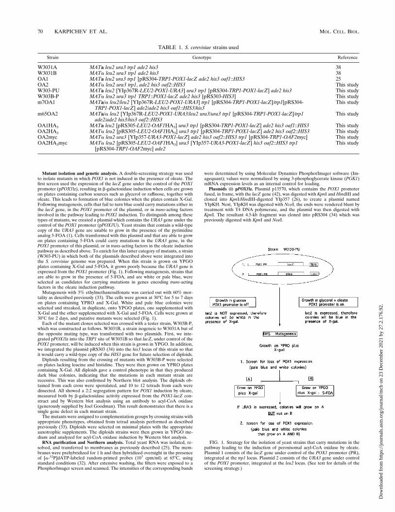
mutants. The levels of acyl-CoA oxidase, 3-ketoacyl-CoA thio-lase, and catalase A were determined by Northern blot analysisof total RNA isolated from mutant and control cells grown inYPG or YPGO medium (Fig. 2a). The intensity of signal foreach message was quantitated, and the fold induction wasnormalized to 3-phosphoglycerate kinase (PGK1) levels as de-scribed in Materials and Methods. Although expression ofPGK1 is known to be induced severalfold by growth of cells inglucose (5), we have found that, in general, the mRNA levelsare similar in cells grown in YPG or YPGO; therefore, we useit as an internal control for loading.In control cells, the POX1 message was induced approxi-
mately 14-fold in oleate-grown cells compared to the level incells grown in the presence of glycerol (Fig. 2a and b). Asshown in Fig. 2b, expression of POX1 in most of the YPG-grown mutants was approximately equal to the level in YPGO-grown cells. However, in the cases of m16 and m50, there wasapproximately a threefold increase in expression in YPGO-grown cells compared to those grown in YPG. These values aretaken from a single experiment. Northern analysis and quan-titation have been performed on each mutant at least twice,and while the actual numbers vary slightly, the differences inexpression levels are reproducible.The level of thiolase (FOX3) mRNA was increased eightfold
in control cells grown in oleate medium compared to thosegrown in glycerol medium (Fig. 2a and c). In mutants m7,m10, m16, and m62, the level of thiolase expression was thesame in cells grown in YPG or YPGO medium. In m64,m65, and m85, there appeared to be a slight induction ofthiolase message in cells grown in the presence of oleate. Inthe case of m50, the relative induction of thiolase was thesame as in control cells, but the absolute level of expressionwas fivefold lower.Catalase expression was induced fourfold in control cells
grown in oleate medium (Fig. 2a and d). The catalase expres-sion levels in mutants m10, m16, m50, m62, m64, and m85grown in glycerol were approximately the same as in controlcells. In m65 and m7, the expression levels of catalase messagein glycerol-grown cells were higher than in the control strain by
three- and fourfold, respectively. Catalase was marginally in-duced in mutants m7, m10, and m62 and was approximatelydoubled in the remaining mutants grown in the presence ofoleate.
FIG. 2. Levels of acyl-CoA oxidase, thiolase, and catalase mRNAs in wild-type and mutant strains. (a) Northern analysis of POX1, FOX3, CTA1, and PGK1expression in a control (W303-PU) and eight mutant strains grown in YPG (Gly)or YPGO (Ole) medium. (b to d) Quantitation of the mRNA levels of POX1 (b),FOX3 (c), and CTA1 (d) in the strains shown in panel a, using a PhosphorImagerand Imagequant software. In each case, the message was normalized by usingPGK1 levels as an internal control for loading. The results are expressed aspercentages of the level in the control strain grown in the presence of oleate, setat 100%.
72 KARPICHEV ET AL. MOL. CELL. BIOL.
Dow
nloa
ded
from
http
s://j
ourn
als.
asm
.org
/jour
nal/m
cb o
n 23
Dec
embe
r 20
21 b
y 27
.2.1
76.9
2.
In summary, all of the mutant strains except m50 were de-fective in the induction of POX1 and of FOX3. In m50, both ofthese genes are expressed at low levels in both media but thefold induction resembles that of wild-type cells. This is similarto the pattern seen for b-galactosidase activity in this strain.Expression of CTA1 in cells grown in either medium appears tobe variable.Morphology of the oleate induction mutants. The mutant
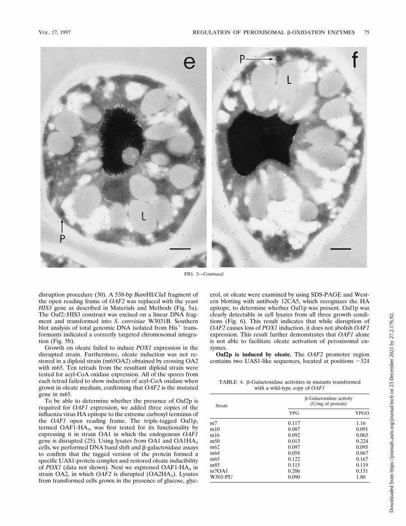
strains deficient in POX1 inducibility by oleate were examinedby immunoelectron microscopy in order to compare the mor-phologies of the peroxisomes between these strains and a con-trol strain. Samples were prepared by preculturing cells inYPD medium, then transferring them to either YPG orYPGO, and culturing them for a further 18 h. The number andsize of peroxisomes varied for the different samples. A randomsample of 100 cells was examined on three separate occasionsfor wild-type cells (W3031A) grown in YPG or YPGO and formutants m65 and m50 and a strain in which OAF1 was dis-rupted (OA1), each grown in YPGO medium (Table 3).In the wild-type strain grown in YPGO, numerous peroxi-
somes were easily identified and were often found in clusters(Fig. 3a). Upon immunostaining, the peroxisomes were la-beled, often densely, with gold particles (Fig. 3a and b). Incontrast, when this strain was grown in YPG medium, therewere fewer peroxisomes and the labeling was weak (Table 3;Fig. 3c). Mutant m50 closely resembled the wild-type strainmorphologically, in that peroxisomes were easily identifiableand were clearly labeled by the immunogold procedure, al-though not as densely as the wild-type strain (Fig. 3d).Strain OA1 contained very few identifiable peroxisomes
when grown in the presence of oleate (Table 3). Peroxisomesthat were visible were labeled with the immunogold procedurebut appeared to be much smaller than in wild-type cells (Fig.3e). Peroxisomes in m65, grown in the presence of oleate, wereslightly more numerous than in strain OA1; however, theywere also smaller than in wild-type cells (Table 3; Fig. 3f). Inboth OA1 and m65, there was a greater accumulation of lipiddroplets than seen in the wild-type strain (compare Fig. 3a withFig. 3e and f). This is not surprising, as it is expected that thesemutant strains would be unable to metabolize the oleic acidpresent in the growth medium.Complementation of mutants by OAF1. We tested the mu-
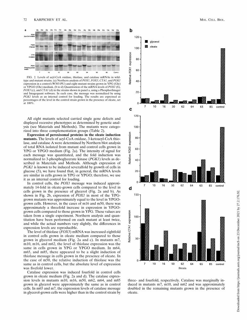
tants isolated by our screening strategy to determine whetherany of them carried a mutation in the OAF1 gene, which
encodes a transcription factor required for oleate induction ofPOX1 (25). For this purpose, a copy of the OAF1 gene wasintegrated into the his3 locus of each of the yeast mutants.Transformants were then analyzed for b-galactosidase activityin cells grown in YPG and YPGO media. In mutant m7, oleateinduction of the POX1-lacZ construct was restored (Table 4).A cross between an appropriate m7 segregant and strain OA1,in which OAF1 is disrupted, named m7OA1, had a mutantphenotype for POX1 induction by oleate (Table 4). This resultconfirms that OAF1 is the mutated gene in m7.In summary, of the mutants isolated thus far, m7, the single
member of complementation group 1, is defective in the OAF1gene. The remaining mutants, belonging to complementationgroups 2 and 3, are defective in different genes.Cloning of OAF2 by functional complementation. The mu-
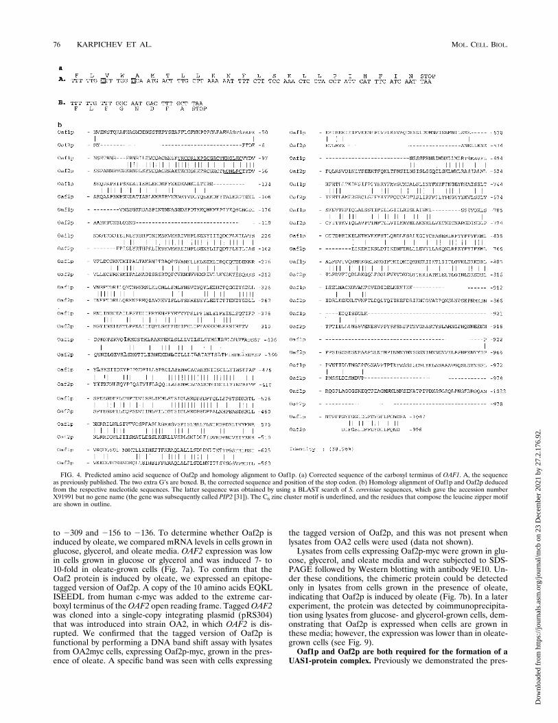
tant strain m65 was chosen as a representative of complemen-tation group 2 and was used to identify the defective genecarried by each member of this group. To clone this gene, m65was transformed with a genomic library (28). Approximately50,000 colonies grew, and these were replica plated ontoYPGO plates containing X-Gal. Fifty-six transformants thatformed dark blue colonies were selected and tested for b-ga-lactosidase activity. Of the transformants selected, three colo-nies had wild-type levels of activity when grown in YPG me-dium and a 10-fold induction in activity when grown in thepresence of oleate. Restriction analysis of the plasmid DNAisolated from these three clones demonstrated that they wereidentical. Further analysis was carried out on one of the plas-mids, named pm65. The plasmid contained a 16-kb insertwhich was able to restore POX1 induction by oleate whenreintroduced into m65 cells. To subclone the smallest DNAfragment that was able to complement the mutation in m65,pm65 DNA was partially digested with Sau3A and was re-solved by agarose gel electrophoresis. A region correspondingto a size of approximately 5 to 7 kb was excised, and thepurified DNA was subcloned into a centromeric plasmid.Pooled subclones were transformed back into m65 and testedfor POX1 induction by oleate. The smallest fragment with theability to restore POX1 induction was found to be 6.5 kb.Approximately 200 nucleotides at either end of this insert weresequenced and compared to known sequences in the yeastgenome database (Stanford Genomic Resources). The searchrevealed a perfect match with a genomic region of chromo-some XV, which contains a 2,988-bp open reading frame be-tween positions 1,020,217 and 1,023,204. The predicted 996-amino-acid sequence encoded by this open reading frame hashigh homology to Oaf1p, a transcription factor involved in theoleate-induction of POX1 (25).Upon sequencing the carboxy-terminal region of OAF1, we
noticed two discrepancies between our sequence and theOAF1sequence contained in the yeast genome database, which con-sisted of extra G’s at positions 3124 and 3130. Correction ofthese errors allowed us to conclude that the actual stop codonis located after the codon for amino acid 1047 (Fig. 4a); thus,Oaf1p is 15 amino acids shorter than reported by others (3).The predicted protein sequence of the gene that rescues m65is 39% identical to the corrected Oaf1p sequence (Fig. 4b).Thus, we refer to this gene as OAF2 and to the encodedprotein as Oaf2p. This protein contains a region homologousto the consensus sequence for the fungal C6 zinc cluster (aminoacids 23 to 52) and a leucine zipper motif (amino acids 320 to362); thus, it has the capacities both to bind to DNA and todimerize (19).Characterization of OAF2. To confirm that the mutation in
m65 is in the gene that we cloned, we prepared a strain (OA2)in which this gene was disrupted, using the one-step gene
TABLE 3. Number of cells containing visible peroxisomesin different strains
Sample Growth mediumNo. of cells withperoxisomes
(100 cells counted)
W3031A YPGO 676668
W3031A YPG 293731
OA1 YPGO 14108
m65 YPGO 293118
m50 YPGO 453343
VOL. 17, 1997 REGULATION OF PEROXISOMAL b-OXIDATION ENZYMES 73
Dow
nloa
ded
from
http
s://j
ourn
als.
asm
.org
/jour
nal/m
cb o
n 23
Dec
embe
r 20
21 b
y 27
.2.1
76.9
2.
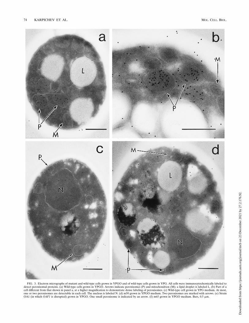
FIG. 3. Electron micrographs of mutant and wild-type cells grown in YPGO and of wild-type cells grown in YPG. All cells were immunocytochemically labeled todetect peroxisomal proteins. (a) Wild-type cells grown in YPGO. Arrows indicate peroxisomes (P) and mitochondrion (M); a lipid droplet is labeled L. (b) Part of acell different from that shown in panel a, at a higher magnification to demonstrate dense labeling of peroxisomes. (c) Wild-type cell grown in YPG medium. At most,one or two peroxisomes are detectable in each cell. The nucleus is labeled N. (d) m50 grown in YPGO medium. Two peroxisomes are marked with arrows. (e) StrainOA1 (in which OAF1 is disrupted) grown in YPGO. One small peroxisome is indicated by an arrow. (f) m65 grown in YPGO medium. Bars, 0.5 mm.
74 KARPICHEV ET AL. MOL. CELL. BIOL.
Dow
nloa
ded
from
http
s://j
ourn
als.
asm
.org
/jour
nal/m
cb o
n 23
Dec
embe
r 20
21 b
y 27
.2.1
76.9
2.
disruption procedure (30). A 538-bp BamHI/ClaI fragment ofthe open reading frame of OAF2 was replaced with the yeastHIS3 gene as described in Materials and Methods (Fig. 5a).The Oaf2::HIS3 construct was excised on a linear DNA frag-ment and transformed into S. cerevisiae W3031B. Southernblot analysis of total genomic DNA isolated from His1 trans-formants indicated a correctly targeted chromosomal integra-tion (Fig. 5b).Growth on oleate failed to induce POX1 expression in the
disrupted strain. Furthermore, oleate induction was not re-stored in a diploid strain (m65OA2) obtained by crossing OA2with m65. Ten tetrads from the resultant diploid strain weretested for acyl-CoA oxidase expression. All of the spores fromeach tetrad failed to show induction of acyl-CoA oxidase whengrown in oleate medium, confirming that OAF2 is the mutatedgene in m65.To be able to determine whether the presence of Oaf2p is
required for OAF1 expression, we added three copies of theinfluenza virus HA epitope to the extreme carboxyl terminus ofthe OAF1 open reading frame. The triple-tagged Oaf1p,termed OAF1-HA3, was first tested for its functionality byexpressing it in strain OA1 in which the endogenous OAF1gene is disrupted (25). Using lysates from OA1 and OA1HA3cells, we performed DNA band shift and b-galactosidase assaysto confirm that the tagged version of the protein formed aspecific UAS1-protein complex and restored oleate inducibilityof POX1 (data not shown). Next we expressed OAF1-HA3 instrain OA2, in which OAF2 is disrupted (OA2HA3). Lysatesfrom transformed cells grown in the presence of glucose, glyc-
erol, or oleate were examined by using SDS-PAGE and West-ern blotting with antibody 12CA5, which recognizes the HAepitope, to determine whether Oaf1p was present. Oaf1p wasclearly detectable in cell lysates from all three growth condi-tions (Fig. 6). This result indicates that while disruption ofOAF2 causes loss of POX1 induction, it does not abolish OAF1expression. This result further demonstrates that OAF1 aloneis not able to facilitate oleate activation of peroxisomal en-zymes.Oaf2p is induced by oleate. The OAF2 promoter region
contains two UAS1-like sequences, located at positions 2324
TABLE 4. b-Galactosidase activities in mutants transformedwith a wild-type copy of OAF1
Strain
b-Galactosidase activity(U/mg of protein)
YPG YPGO
m7 0.117 1.16m10 0.087 0.091m16 0.092 0.063m50 0.013 0.224m62 0.097 0.095m64 0.058 0.067m65 0.122 0.167m85 0.115 0.119m7OA1 0.206 0.151W303-PU 0.090 1.80
FIG. 3—Continued.
VOL. 17, 1997 REGULATION OF PEROXISOMAL b-OXIDATION ENZYMES 75
Dow
nloa
ded
from
http
s://j
ourn
als.
asm
.org
/jour
nal/m
cb o
n 23
Dec
embe
r 20
21 b
y 27
.2.1
76.9
2.
to 2309 and 2156 to 2136. To determine whether Oaf2p isinduced by oleate, we compared mRNA levels in cells grown inglucose, glycerol, and oleate media. OAF2 expression was lowin cells grown in glucose or glycerol and was induced 7- to10-fold in oleate-grown cells (Fig. 7a). To confirm that theOaf2 protein is induced by oleate, we expressed an epitope-tagged version of Oaf2p. A copy of the 10 amino acids EQKLISEEDL from human c-myc was added to the extreme car-boxyl terminus of theOAF2 open reading frame. TaggedOAF2was cloned into a single-copy integrating plasmid (pRS304)that was introduced into strain OA2, in which OAF2 is dis-rupted. We confirmed that the tagged version of Oaf2p isfunctional by performing a DNA band shift assay with lysatesfrom OA2myc cells, expressing Oaf2p-myc, grown in the pres-ence of oleate. A specific band was seen with cells expressing
the tagged version of Oaf2p, and this was not present whenlysates from OA2 cells were used (data not shown).Lysates from cells expressing Oaf2p-myc were grown in glu-
cose, glycerol, and oleate media and were subjected to SDS-PAGE followed by Western blotting with antibody 9E10. Un-der these conditions, the chimeric protein could be detectedonly in lysates from cells grown in the presence of oleate,indicating that Oaf2p is induced by oleate (Fig. 7b). In a laterexperiment, the protein was detected by coimmunoprecipita-tion using lysates from glucose- and glycerol-grown cells, dem-onstrating that Oaf2p is expressed when cells are grown inthese media; however, the expression was lower than in oleate-grown cells (see Fig. 9).Oaf1p and Oaf2p are both required for the formation of a
UAS1-protein complex. Previously we demonstrated the pres-
FIG. 4. Predicted amino acid sequence of Oaf2p and homology alignment to Oaf1p. (a) Corrected sequence of the carboxyl terminus of OAF1. A, the sequenceas previously published. The two extra G’s are boxed. B, the corrected sequence and position of the stop codon. (b) Homology alignment of Oaf1p and Oaf2p deducedfrom the respective nucleotide sequences. The latter sequence was obtained by using a BLAST search of S. cerevisiae sequences, which gave the accession numberX91991 but no gene name (the gene was subsequently called PIP2 [31]). The C6 zinc cluster motif is underlined, and the residues that compose the leucine zipper motifare shown in outline.
76 KARPICHEV ET AL. MOL. CELL. BIOL.
Dow
nloa
ded
from
http
s://j
ourn
als.
asm
.org
/jour
nal/m
cb o
n 23
Dec
embe
r 20
21 b
y 27
.2.1
76.9
2.

ence of a specific protein-DNA complex when extracts fromoleate-grown yeast cells were incubated with DNA containingthe UAS1 of POX1 in a DNA band shift assay (41). This shiftedband was not present when extracts from cells in which OAF1was disrupted were used in the assay (25). To determine ifOAF2 also binds to UAS1, we used extracts from wild-typecells and from cells carrying the OAF2 disruption in a bandshift assay with UAS1. Extracts from wild-type cells grown inoleate medium gave rise to the expected band containing theUAS1-protein complex (Fig. 8, lane 3). A weak band shift wasalso seen with extracts from cells grown in glycerol (lane 2). Weoccasionally see this shift with extracts from glycerol-growncells, but when present it is much weaker than the shift seenwith extracts from oleate-grown cells. No band shift is detectedwith extracts from glucose-grown cells (lane 1).In contrast to the results with wild-type cells, there was no
specific DNA band shift when extracts from m65 cells wereused (Fig. 8, lanes 4 to 6). The DNA bandshift was restoredwhen the assay was carried out with extracts from the m65strain transformed with plasmid pm65 (lane 9). In addition,there was no band shift when the assay was carried out withextracts from strain OA2 in which OAF2 is disrupted (lanes 10to 12). These results, together with our previous studies onOAF1 (25), suggest that Oaf1p and Oaf2p are both requiredfor the formation of a specific protein-DNA complex in ex-tracts from cells grown in oleate medium.Oaf1p and Oaf2p coimmunoprecipitate. Due to the similar-
ities between the Oaf1 and Oaf2 proteins, and to the factthat they are both required for the oleate induction ofPOX1, we postulated that they may interact with each otherto facilitate this induction. A strain expressing both OAF1-HA3 and OAF2-myc (OA2HA3myc) was grown in the pres-ence of glucose, glycerol, or oleate. Cell lysates were preparedand used for immunoprecipitation with monoclonal antibody12CA5. Following SDS-PAGE and Western blotting with9E10, tagged Oaf2p was present in immunoprecipitates fromall three growth conditions (Fig. 9a, lanes 1 to 3). As expected,no protein was detected in yeast strains that were not trans-formed with the OAF1-HA3 construct (Fig. 9a, lane 4). A si-milar result was obtained when lysates were immunoprecipi-tated with 9E10 followed by detection with 12CA5 (Fig. 9b).These data suggest that Oaf1p and Oaf2p form a complexwhich is present in all of the growth conditions tested.
DISCUSSION
We wish to gain an understanding of the mechanisms andfactors which regulate induction of peroxisomes and peroxiso-mal enzymes. Toward this end, we have initiated a geneticapproach to isolate mutants in this pathway in the yeast S.cerevisiae. Thus far, we have isolated eight mutant strains thatfall into three complementation groups. Strains belonging tocomplementation group 1 carry mutations in the gene encod-ing the oleate activation factor that we isolated previously (25).We predict that mutations in OAF1 should be isolated by ourscreening procedure; therefore, this result underscores the va-lidity of our approach.Electron microscope examination of the oleate activation
mutants grown in the presence of oleate revealed that all havea reduced number of peroxisomes. Furthermore, the peroxi-somes in most of these mutants appear to be smaller thanthose in wild-type cells. These observations, in combinationwith the peroxisomal enzyme mRNA expression levels re-ported in this study, suggest that the mechanisms involved inthe induction of peroxisomal enzymes and in proliferation ofthe organelle itself are coupled. Mutant m50 has a phenotypedifferent from those of the other mutants isolated. Peroxi-somes in this mutant are almost as numerous as in the wild-type strain; however, the peroxisomal enzyme activities are notfully induced by oleate. We are currently investigating thenature of the mutation in this strain.In recent years, peroxisome biogenesis mutants in S. cerevi-
siae have been described by several groups (12, 13, 40, 44),leading to the identification of a number of genes essential for
FIG. 5. Disruption of OAF2. (a) The OAF2DNA sequence (black box) encoding Oaf2p amino acid residues 321 to 501 was replaced by a DNA fragment containingthe S. cerevisiae HIS3 gene (stippled box) and integrated into the S. cerevisiae genome (white boxes) by homologous recombination. An arrow marks the start site anddirection of transcription of HIS3. Sc, SacI; X, XmnI; C, ClaI; B, BamHI; H, HindIII; Sl, SalI; Sa, ScaI. The dashed line represents 3.04 kb upstream of the OAF2 gene.(b) Correct targeting of the oaf2::HIS3 fragment is demonstrated by Southern blot analysis with BamHI/ScaI-digested genomic DNA of the wild-type (Wt) and twoseparate transformants in which OAF2 was disrupted (K1 and K2). The HindIII DNA fragment used as a probe is shown by a double-headed arrow in panel a. A ScaIfragment common to both wild-type and disrupted strains is marked by an asterisk.
FIG. 6. Oaf2p is not required for the expression of Oaf1p. Extracts fromOA2HA3 cells, in which endogenous OAF2 is disrupted, were cultured in YPD(glu), YPG (gly), or YPGO (ole) and separated by SDS-PAGE. The productswere then immunoblotted with monoclonal antibody 12CA5 against HA. Triple-tagged Oaf1p is marked with an arrow.
VOL. 17, 1997 REGULATION OF PEROXISOMAL b-OXIDATION ENZYMES 77
Dow
nloa
ded
from
http
s://j
ourn
als.
asm
.org
/jour
nal/m
cb o
n 23
Dec
embe
r 20
21 b
y 27
.2.1
76.9
2.

this process. These genes encode peroxisome targeting signalreceptors (2, 39, 46) as well as a peroxisomal integral mem-brane protein (16). The search for peroxisome biogenesis mu-tants has also led to the isolation of some mutants that havelow levels of peroxisomal enzyme activities. For example, per-oxisome assembly mutant 14 (pas14) was shown to be defectivein SNF1 (40), which encodes a protein kinase involved in theactivation of glucose-repressible genes (4). Pas19 was shown tocarry a mutation in the ADR1 gene (12), which was first iden-tified as encoding a protein that regulates transcription of thealcohol dehydrogenase gene ADH2 (8). Subsequently, ADR1was demonstrated to control transcription of several peroxiso-mal proteins, including catalase and thiolase (35). Thus, it isemerging that previously known regulators of glucose-repress-ible genes are also involved in the glucose repression andderepression of several peroxisomal proteins. There remainssome doubt as to whether these proteins are involved in theregulation of POX1 (37).Mutant selection procedures that specifically target the iso-
lation of peroxisome biogenesis mutants have not, to ourknowledge, led to the characterization of any genes involved inthe oleate induction of peroxisomal proteins. The RTG genes,known to affect oleate induction of some peroxisomal proteins,
were isolated in a screen for mutants in the retrograde regu-lation of CIT2 (24).Complementation group 2 contains six of the mutants iso-
lated using our screening strategy. The gene defective in thesestrains encodes a protein with a calculated molecular mass of114.7 kDa which is 39% identical to Oaf1p; thus, we named itOaf2p. During the purification of Oaf1p, we consistently coe-luted a doublet of proteins that ran with apparent molecularmasses of between 110 and 120 kDa on SDS-polyacrylamidegels (25). Oaf1p was the larger protein of this doublet; it ispossible that Oaf2p is the other. Oaf1p and Oaf2p are associ-ated with each other in all growth conditions and are bothrequired for the formation of an oleate-specific UAS1-proteincomplex. Due to the high homology in the predicted DNAbinding regions of these two proteins and their capacity toform a complex, it is likely that they would copurify in theDNA-affinity purification procedure used to isolate Oaf1p(25). We have further shown that Oaf2p is induced by oleate(Fig. 7). Interestingly, Oaf1p expression levels are similar inglycerol- and oleate-grown cells (unpublished data). Whetherinduction of Oaf2p is sufficient to mediate the activation ofb-oxidation enzymes and peroxisome proliferation remains tobe determined.The work reported here together with our previous studies
(25, 41) supports the idea that Oaf1p and Oaf2p are transcrip-tion factors and that both bind to the UAS1 of POX1. Wepropose a model in which Oaf1p and Oaf2p form a het-erodimer and bind to UAS1 as shown in Fig. 10. In this model,we have arbitrarily positioned Oaf1p and Oaf2p binding to thepalindromes of the POX1 UAS1 (they may, of course, bind in
FIG. 7. Oaf2p is induced in cells grown in the presence of oleate. (a) North-ern analysis of OAF2 expression. W303-PU cells were cultured in YPD (glu),YPG (gly), or YPGO (ole) medium. A poly(A)1 fraction was prepared from 500mg of total RNA, using Oligotex resin as specified by the manufacturer (Qiagen).The mRNA was resolved and transferred as described in Materials and Methods.The 1-kb HindIII fragment from the OAF2 coding region (Fig. 5a) was used asa probe. (b) Extracts from OA2myc cells cultured in YPD (glu), YPG (gly), orYPGO (ole) were separated by SDS-PAGE and immunoblotted with monoclo-nal antibody 9E10 against the human c-myc epitope. Extract from oleate-grownOA2 cells that were not transformed with OAF2-myc was used as a control fornonspecific bands (c). Tagged Oaf2p is marked with an arrow.
FIG. 8. DNA band shift assay with extracts from wild-type cells (lanes 1 to 3),m65 (lanes 4 to 6), m65 transformed with pm65 (lanes 7 to 9), and OA2 (lanes10 to 12). Labeled UAS1 was used as a probe with extracts from cells grown inglucose (lanes 1, 4, 7, and 10), glycerol (lanes 2, 5, 8, and 11), or oleate (lanes 3,6, 9, and 12) medium. An arrow marks the specific shifted band caused by proteinbinding to UAS1. An asterisk marks a nonspecific band seen in previous exper-iments (41).
FIG. 9. Oaf1p and Oaf2p form a protein complex. (a) Coimmunoprecipita-tion of Oaf2-myc and Oaf1p-HA3 with monoclonal antibody 12CA5. Proteinextract (500 mg of protein) prepared from OA2HA3myc cells cultured in YPD(glu), YPG (gly), or YPGO (ole) was incubated with 12CA5 and then precipi-tated with protein G-agarose. The immunoprecipitated material was separatedby SDS-PAGE and immunoblotted with 9E10. Extract from YPGO-grownOA2myc cells that were not transformed with HA-tagged Oaf1p was treated inthe same manner as a control (c). One hundred micrograms of cell lysate fromYPGO-grown OA2HA3myc cells was subjected to SDS-PAGE and immunob-lotted with 9E10 in order to detect the specific band corresponding to Oaf2p-myc(l). Tagged Oaf2p is marked with an arrow. (b) Coimmunoprecipitation ofOaf2p-myc and Oaf1p-HA3 with monoclonal antibody 9E10. Protein extract wasprepared as described above, incubated with 9E10, and then precipitated withprotein G-agarose. The immunoprecipitated material was resolved by SDS-PAGE and then transferred to membrane. Extract from YPGO-grown OA2 cellswere treated in the same manner as a control (c). One hundred micrograms oftotal OA2HA3myc cell lysate from YPGO-grown cells was run on the same gelas a positive control for Oaf2p-myc (l). Tagged Oaf1p is marked with an arrow.The membrane was first blotted with 9E10 to confirm that Oaf2p-myc wasdetectable (p). Oaf2pmyc is clearly seen in total cell lysate and is seen as a faintband in the immunoprecipitated material from each growth condition. Themembrane was subsequently blotted, without stripping, with 12CA5 to demon-strate that Oaf1p-HA3 was coprecipitated (arrow).
78 KARPICHEV ET AL. MOL. CELL. BIOL.
Dow
nloa
ded
from
http
s://j
ourn
als.
asm
.org
/jour
nal/m
cb o
n 23
Dec
embe
r 20
21 b
y 27
.2.1
76.9
2.
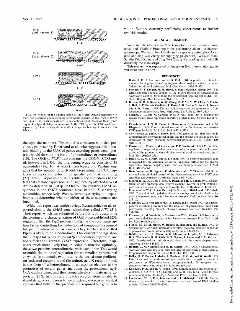
the opposite manner). This model is consistent with that pre-viously proposed by Einerhand et al., who suggested that pro-tein binding to the UAS of genes encoding peroxisomal pro-teins would do so in the form of a homodimer or heterodimer(10). The ORE of FOX3 also contains the CGGN17CCG mo-tif; however, in CTA1, the intervening sequence consists of 18nucleotides (Fig. 10). A report from Reece and Ptashne sug-gests that the number of nucleotides separating the CGG trip-lets is an important factor in the specificity of protein binding(27). Thus, it is possible that this difference contributes to thefact that catalase expression is not significantly affected in yeaststrains defective in Oaf1p or Oaf2p. The putative UAS1 se-quences in the OAF2 promoter have 10 and 15 separatingnucleotides, respectively. We are currently carrying out exper-iments to determine whether either of these sequences arefunctional.While this report was under review, Rottensteiner et al. re-
ported cloning the OAF2 gene, which they called PIP2 (31).Their report, which was submitted before our report describingthe cloning and characterization of Oaf1p was published (25),suggested that the Pip2 protein (Oaf2p) is the sole transcrip-tion factor controlling the induction of components requiredfor proliferation of peroxisomes. They further stated thatPip2p is likely to be a homodimer. Our current findings showthat Oaf1p-Oaf1p or Oaf2p-Oaf2p homodimers, if present, arenot sufficient to activate POX1 expression. Therefore, it ap-pears much more likely that, in order to function optimally,these two proteins heterodimerize with each other. This wouldresemble the mode of regulation for mammalian peroxisomalenzymes. In mammals, two proteins, the peroxisome prolifera-tor-activated receptor a and the retinoic acid X receptor, bind,in the form of a heterodimer, to a response element in thepromoters of several genes, including the peroxisomal acyl-CoA oxidase gene, and they cooperatively stimulate gene ex-pression (17). In this system, each receptor alone is able tostimulate gene expression to some extent, whereas in yeast, itappears that both of the proteins are required for gene acti-
vation. We are currently performing experiments to furthertest this model.
ACKNOWLEDGMENTS
We gratefully acknowledge Murl Casey for excellent technical assis-tance and Vladimir Protopopov for performing all of the electronmicroscopy. We thank Joel Goodman for supplying anti-acyl-CoA oxi-dase and Jing Wei Zhang for supplying pT7pebHA3. We also thankSerafın Pinol-Roma and Jing Wei Zhang for reading and helpfullydiscussing the manuscript.This research was supported by American Heart Association grants
95008910 and 92001690.
REFERENCES1. Boeke, J. D., F. Lacroute, and G. R. Fink. 1984. A positive selection formutants lacking orotidine-59-phosphate decarboxylase activity in yeast:5-fluoro-orotic acid resistance. Mol. Gen. Genet. 197:345–353.
2. Brocard, C., F. Kragler, M. M. Simon, T. Schuster, and A. Hartig. 1994. Thetetratricopeptide repeat-domain of the PAS10 protein of Saccharomycescerevisiae is essential for binding the peroxisomal targeting signal-SKL. Bio-chem. Biophys. Res. Commun. 204:1016–1022.
3. Bussey, H., D. B. Kaback, W. W. Zhong, D. T. Vo, M. W. Clark, N. Fortin,J. Hall, B. F. Francis Ouellette, T. Keng, A. B. Barton, Y. Su, C. J. Davies,and R. K. Storms. 1995. The nucleotide sequence of chromosome I fromSaccharomyces cerevisiae. Proc. Natl. Acad. Sci. USA 92:3809–3813.
4. Celenza, J. L., and M. Carlson. 1986. A yeast gene that is essential forrelease from glucose repression encodes a protein kinase. Science 233:1175–1180.
5. Chambers, A., J. S. H. Tsang, C. Stanway, A. J. Kingsman, and S. M.Kingsman. 1989. Transcriptional control of the Saccharomyces cerevisiaePGK gene by RAP1. Mol. Cell. Biol. 9:5516–5524.
6. Chelstowska, A., and R. A. Butow. 1995. RTG genes in yeast that function incommunication between mitochondria and the nucleus are also required forexpression of genes encoding peroxisomal proteins. J. Biol. Chem. 270:18141–18118.
7. Creusot, F., J. Verdiere, M. Gaisne, and P. P. Slonimski. 1988. CYP1 (HAP1)regulator of oxygen-dependent gene expression in yeast. I. Overall organi-zation of the protein sequence displays several novel structural domains. J.Mol. Biol. 204:263–276.
8. Denis, C. L., M. Ciriacy, and E. T. Young. 1981. A positive regulatory geneis required for the accumulation of the functional mRNA for the glucoserepressible alcohol dehydrogenase from Saccharomyces cerevisiae. J. Mol.Biol. 148:355–368.
9. Dmochowska, A., D. Dignard, R. Maleszka, and D. Y. Thomas. 1990. Struc-ture and transcriptional control of the Saccharomyces cerevisiae POX1 geneencoding acyl-coenzyme A oxidase. Gene 88:247–252.
10. Einerhand, A. W. C., W. T. Kos, B. Distel, and H. F. Tabak. 1993. Charac-terization of a transcriptional control element involved in proliferation ofperoxisomes in yeast in response to oleate. Eur. J. Biochem. 314:323–331.
11. Einerhand, A. W. C., I. Van Der Leij, W. T. Kos, B. Distel, and H. F. Tabak.1992. Transcriptional regulation of genes encoding proteins involved in bio-genesis of peroxisomes in Saccharomyces cerevisiae. Cell Biochem. Func. 10:185–192.
12. Elgersma, Y., M. Van den Berg, H. F. Tabak, and B. Distel. 1993. An efficientpositive selection procedure for the isolation of peroxisomal import andperoxisome assembly mutants of Saccharomyces cerevisiae. Genetics 135:731–740.
13. Erdmann, R., M. Veenhuis, D. Mertens, and W.-H. Kunau. 1989. Isolation ofperoxisome-deficient mutants of Saccharomyces cerevisiae. Proc. Natl. Acad.Sci. USA 86:5419–5423.
14. Filipits, M., M. M. Simon, W. Rapatz, B. Hamilton, and H. Ruis. 1993. ASaccharomyces cerevisiae upstream activating sequence mediates inductionof peroxisome proliferation by fatty acids. Gene 132:49–55.
15. Goldfischer, S., C. L. Moore, A. B. Johnson, A. J. Spiro, M. P. Valsamis,H. K. Wisniewski, R. H. Ritch, W. T. Norton, I. Rapin, and L. M. Gartner.1973. Peroxisomal and mitochondrial defects in the cerebro-hepato-renalsyndrome. Science 182:62–64.
16. Hohfeld, J., M. Veenhuis, and W. H. Kunau. 1991. PAS3, a Saccharomycescerevisiae gene encoding a peroxisomal integral membrane protein essentialfor peroxisome biogenesis. J. Cell Biol. 114:1167–1178.
17. Keller, H., C. Dreyer, J. Medin, A. Mahfoudi, K. Ozato, and W. Wahli. 1993.Fatty acids and retinoids control lipid metabolism through activation ofperoxisome proliferator-activated receptor-retinoid X receptor het-erodimers. Proc. Natl. Acad. Sci. USA 90:2160–2164.
18. Kolodziej, P. A., and R. A. Young. 1991. Epitope tagging and protein sur-veillance, p. 508–519. In C. Guthrie and G. R. Fink (ed.), Guide to yeastgenetics and molecular biology. Academic Press, San Diego, Calif.
19. Landschulz, W. H., P. F. Johnson, and S. L. McKnight. 1988. The leucinezipper: a hypothetical structure common to a new class of DNA bindingproteins. Science 240:1759–1764.
FIG. 10. Model for the binding action of the Oaf1p-Oaf2p heterodimer tothe UASs present in genes encoding peroxisomal proteins. In the UASs of POX1and FOX3, the CGG repeats are 17 nucleotides apart. Both of these genesrequire Oaf1p and Oaf2p for activation. In the CTA1 gene, the CGG motifs areseparated by 18 nucleotides; this may affect the specific binding of proteins to thisDNA.
VOL. 17, 1997 REGULATION OF PEROXISOMAL b-OXIDATION ENZYMES 79
Dow
nloa
ded
from
http
s://j
ourn
als.
asm
.org
/jour
nal/m
cb o
n 23
Dec
embe
r 20
21 b
y 27
.2.1
76.9
2.

20. Laughon, A., and R. F. Gesteland. 1984. Primary structure of the Saccharo-myces cerevisiae GAL4 gene. Mol. Cell. Biol. 4:260–267.
21. Lazarow, P. B., and Y. Fujiki. 1985. Biogenesis of peroxisomes. Annu. Rev.Cell Biol. 1:489–530.
22. Lazarow, P. B., and H. W. Moser. 1989. Disorders of peroxisomal biogenesis,p. 1479–1509. In C. R. Scriver, A. L. Beaudet, W. S. Sly, and D. Valle (ed.),The metabolic basis of inherited diseases. McGraw-Hill Co., New York.
23. Lewin, A. S., V. Hines, and G. M. Small. 1990. Citrate synthase encoded bythe CIT2 gene of Saccharomyces cerevisiae is peroxisomal. Mol. Cell. Biol.10:1399–1405.
24. Liao, X., and R. A. Butow. 1993. RTG1 and RTG2: two yeast genes requiredfor a novel path of communication from mitochondria to the nucleus. Cell72:61–71.
25. Luo, Y., I. V. Karpichev, R. A. Kohanski, and G. M. Small. 1996. Purification,identification and properties of a Saccharomyces cerevisiae oleate-activatedupstream activating sequence-binding protein that is involved in the activa-tion of POX1. J. Biol. Chem. 271:12068–12075.
26. Myers, A. M., A. Tzagoloff, D. M. Kinney, and C. J. Lusty. 1986. Yeast shuttlevectors with multiple cloning sites suitable for construction of lacZ fusions.Gene 45:299–310.
27. Reece, R. J., and M. Ptashne. 1993. Determinants of binding-site specificityamong yeast C6 zinc cluster proteins. Science 261:909–911.
28. Rose, M. D., P. Novick, J. H. Thomas, D. Botstein, and G. R. Fink. 1987. ASaccharomyces cerevisiae plasmid bank based on a centromere-containingshuttle vector. Gene 60:237–243.
29. Rosenkrantz, M., T. Alam, K.-S. Kim, B. J. Clark, P. A. Srere, and L. P.Guarente. 1986. Mitochondrial and nonmitochondrial citrate synthases inSaccharomyces cerevisiae are encoded by distinct homologous genes. Mol.Cell. Biol. 6:4509–4515.
30. Rothstein, R. 1983. One step gene disruption in yeast. Methods Enzymol.101:202–213.
31. Rottensteiner, H., A. J. Kal, M. Filpits, M. Binder, B. Hamilton, H. F. Tabak,and H. Ruis. 1996. Pip2: a transcriptional regulator of peroxisome prolifer-ation in the yeast Saccharomyces cerevisiae. EMBO J. 15:2924–2934.
32. Sambrook, J., E. F. Fritsch, and T. Maniatis. 1989. Molecular cloning: alaboratory manual, 2nd ed. Cold Spring Harbor Laboratory, Cold SpringHarbor, N.Y.
33. Sherman, F., G. R. Fink, and J. B. Hicks. 1986. Laboratory course manualfor methods in yeast genetics. Cold Spring Harbor Laboratory, Cold SpringHarbor, N.Y.
34. Sikorski, R. S., and P. Hieter. 1989. A system of shuttle vectors and yeast
host strains designed for efficient manipulation of DNA in Saccharomycescerevisiae. Genetics 122:19–27.
35. Simon, M., G. Adam, W. Rapatz, W. Spevak, and H. Ruis. 1991. The Sac-charomyces cerevisiae ADR1 gene is a positive regulator of transcription ofgenes encoding peroxisomal proteins. Mol. Cell. Biol. 11:699–704.
36. Small, G. M., T. Imanaka, H. Shio, and P. B. Lazarow. 1987. Efficientassociation of in vitro translation products with purified, stable Candidatropicalis peroxisomes. Mol. Cell. Biol. 7:1848–1855.
37. Stanway, C. A., J. Gibbs, and E. Berardi. 1995. Expression of the FOX1 geneof Saccharomyces cerevisiae is regulated by carbon source, but not by theknown glucose repression genes. Curr. Genet. 27:404–408.
38. Thomas, B. J., and R. Rothstein. 1989. Elevated recombination rates intranscriptionally active DNA. Cell 56:619–630.
39. Van Der Leij, I., M. Franse, Y. Elgersma, B. Distel, and H. F. Tabak. 1993.PAS10 is a tetratricopeptide-repeat protein that is essential for the import ofmost matrix proteins into peroxisomes of Saccharomyces cerevisiae. Proc.Natl. Acad. Sci. USA 90:11782–11786.
40. Van Der Leij, I., M. Van den Berg, R. Boot, M. Franse, B. Distel, and H. F.Tabak. 1992. Isolation of peroxisome assembly mutants from Saccharomycescerevisiae with different morphologies using a novel positive selection pro-cedure. J. Cell Biol. 119:153–162.
41. Wang, T., Y. Luo, and G. M. Small. 1994. The POX1 gene encoding perox-isomal acyl-CoA oxidase in Saccharomyces cerevisiae is under the control ofmultiple regulatory elements. J. Biol. Chem. 269:24480–24485.
42. Wang, T. W., A. S. Lewin, and G. M. Small. 1992. A negative regulatingelement controlling transcription of the gene encoding acyl-CoA oxidase inSaccharomyces cerevisiae. Nucleic Acids Res. 20:3495–3500.
43. Wright, R., and J. Rine. 1989. Transmission electron microscopy and immu-nocytochemical studies of yeast: analysis of HMG-CoA reductase overpro-duction by electron microscopy. Methods Cell Biol. 31:473–512.
44. Zhang, J. W., Y. Han, and P. B. Lazarow. 1993. Novel peroxisome clusteringmutants and peroxisome biogenesis mutants of Saccharomyces cerevisiae. J.Cell Biol. 123:1133–1147.
45. Zhang, J. W., and P. B. Lazarow. 1994. PEB1 (PAS7) in Saccharomycescerevisiae encodes a hydrophilic, intra-peroxisomal protein that is a memberof the WD repeat family and is essential for the import of thiolase intoperoxisomes. J. Cell Biol. 129:65–80.
46. Zhang, J. W., and P. B. Lazarow. 1996. Peb1p (Pas7p) is an intraperoxisomalreceptor for the NH2-terminal, type 2, peroxisomal targeting sequence ofthiolase: Peb1p itself is targeted to peroxisomes by an NH2-terminal peptide.J. Cell Biol. 132:325–334.
80 KARPICHEV ET AL. MOL. CELL. BIOL.
Dow
nloa
ded
from
http
s://j
ourn
als.
asm
.org
/jour
nal/m
cb o
n 23
Dec
embe
r 20
21 b
y 27
.2.1
76.9
2.