A Case Report with Plasmablastic Lymphoma of the Jejunum
5
INTRODUCTION Plasmablastic lymphoma (PBL) is a lymphoproliferative disorder that is considered a type of diffuse large B-cell non- Hodgkin’s lymphoma (NHL); it is a morphologic variant by the currently proposed World Health Organization (WHO) classification system (1). Due to its recent recognition as a unique disease entity, it has only been partially characterized, primarily on the basis of sporadic case reports (2-6). To date, PBLs have been found frequently in patients infected with the human immunodeficiency virus (HIV) (2, 3); these pati- ents characteristically present with extranodal disease involv- ing the oral cavity. In this report, we describe an unusual case of PBL that presented with a large mass originating from the jejunum in a HIV-seronegative patient. CASE REPORT A 60-yr-old man presented to the emergency department with complaints of dyspnea and dizziness. Two months ago, he underwent an upper endoscopy, colonoscopy and cardiac single photon emission computed tomography (CT) in another hospital for anemia and exertional dyspnea, and the results were all within normal limits. The patient also underwent a biopsy of a chest wall mass and a fine needle aspiration of a cervical lymphadenopathy that was incidentally detected by the CT of the chest. The results of the biopsy and fine needle aspiration were non-specific, and a follow-up appointment was made after 3 months. However, his exertional dyspnea progressively worsened over the past 1 month. On admission, he reported a weight loss of 7 kg during the last two months and melena five days ago. There were no conditions associat- ed with immunosuppression. On physical examination, the patient was found to have a 2.5 cm ovoid mass in the sub- mandibular area; similar but smaller lesions were present on the forehead, chest wall and right vestibule of the oral cavi- ty. There were a right supraclavicular lymphadenopathy and bilateral axillary lymphadenopathies without tenderness. The laboratory data on admission included a white blood cell count of 6,800/ mL (72% polymorphonuclear leucocytes, 18% lymphocytes and 8% monocytes), a hemoglobin con- centration of 6.2 g/dL and a platelet count of 397,000/ mL. The serum iron was 13 mL/dL, total iron-binding capacity 394 mL/dL and the serum ferritin was 5.4 ng/mL. The bio- chemical analysis of the blood for hepatic and renal function, 496 Jae Myung Cha 1 , Joung Il Lee 1 , Kwang Ro Joo 1 , Sung Won Jung 1 , Hyun Phil Shin 1 , Jae Jin Lee 1 , and Gyo Young Kim 2 Departments of Internal Medicine 1 and Pathology 2 , University of Kyunghee College of Medicine, Seoul, Korea Address for Correspondence Jae Myung Cha, M.D. Department of Internal Medicine, East-West Neo Medical Center, University of Kyunghee College of Medicine, 149 Sangil-dong, Gangdong-gu, Seoul 134-727, Korea Tel : +82.2-440-6113, Fax : +82.2-440-6295 E-mail : [email protected] J Korean Med Sci 2010; 25: 496-500 ISSN 1011-8934 DOI: 10.3346/jkms.2010.25.3.496 A Case Report with Plasmablastic Lymphoma of the Jejunum Plasmablastic lymphoma (PBL) is a recently identified entity that is considered to be a type of diffuse large B-cell lymphoma with a unique immunophenotype and a predilection for the oral cavity of patients with the human immunodeficiency virus (HIV). Although its clinical features may help in the differential diagnosis, an extra- oral location in a patient without HIV makes it more difficult to suspect clinically. This case report is the first to describe a patient with PBL originating from the jejunum in a 60-yr-old, HIV-seronegative man. Computed tomography of the face, chest and abdomen showed about a 9.4×9.0 cm mass of the proximal jejunum, multiple mass- es in the musculoskeletal soft tissue, and multiple lymphadenopathies. The histo- logical examinations demonstrated a large cell lymphoma with plasmablastic differ- entiation. The neoplastic cells were diffusely positive for MUM1, epithelial membrane antigen and lambda light chains, and focally positive for CD79a; but negative for CD3, CD20, CD30, CD34, CD45RO, CD56, CD99, and CD117. The proliferation index by Ki-67 immunohistochemistry was approximately 70%. These findings were compatible with the diagnosis of PBL. The findings in this case suggest that PBL should be included in the differential diagnosis of a small bowel mass even in a HIV-negative patient. Key Words : HIV-negative; Jejunum; Plasmablastic Lymphoma Received : 24 August 2008 Accepted : 12 November 2008 ⓒ 2010 The Korean Academy of Medical Sciences. This is an Open Access article distributed under the terms of the Creative Commons Attribution Non-Commercial License (http://creativecommons.org/licenses/by-nc/3.0) which permits unrestricted non-commercial use, distribution, and reproduction in any medium, provided the original work is properly cited.
Transcript of A Case Report with Plasmablastic Lymphoma of the Jejunum
INTRODUCTION
Plasmablastic lymphoma (PBL) is a lymphoproliferativedisorder that is considered a type of diffuse large B-cell non-Hodgkin’s lymphoma (NHL); it is a morphologic variant bythe currently proposed World Health Organization (WHO)classification system (1). Due to its recent recognition as aunique disease entity, it has only been partially characterized,primarily on the basis of sporadic case reports (2-6). To date,PBLs have been found frequently in patients infected withthe human immunodeficiency virus (HIV) (2, 3); these pati-ents characteristically present with extranodal disease involv-ing the oral cavity. In this report, we describe an unusual caseof PBL that presented with a large mass originating from thejejunum in a HIV-seronegative patient.
CASE REPORT
A 60-yr-old man presented to the emergency departmentwith complaints of dyspnea and dizziness. Two months ago,he underwent an upper endoscopy, colonoscopy and cardiacsingle photon emission computed tomography (CT) in another
hospital for anemia and exertional dyspnea, and the resultswere all within normal limits. The patient also underwent abiopsy of a chest wall mass and a fine needle aspiration of acervical lymphadenopathy that was incidentally detected bythe CT of the chest. The results of the biopsy and fine needleaspiration were non-specific, and a follow-up appointmentwas made after 3 months. However, his exertional dyspneaprogressively worsened over the past 1 month. On admission,he reported a weight loss of 7 kg during the last two monthsand melena five days ago. There were no conditions associat-ed with immunosuppression. On physical examination, thepatient was found to have a 2.5 cm ovoid mass in the sub-mandibular area; similar but smaller lesions were present onthe forehead, chest wall and right vestibule of the oral cavi-ty. There were a right supraclavicular lymphadenopathy andbilateral axillary lymphadenopathies without tenderness.
The laboratory data on admission included a white bloodcell count of 6,800/mL (72% polymorphonuclear leucocytes,18% lymphocytes and 8% monocytes), a hemoglobin con-centration of 6.2 g/dL and a platelet count of 397,000/mL.The serum iron was 13 mL/dL, total iron-binding capacity394 mL/dL and the serum ferritin was 5.4 ng/mL. The bio-chemical analysis of the blood for hepatic and renal function,
496
Jae Myung Cha1, Joung Il Lee1, Kwang Ro Joo1, Sung Won Jung1, Hyun Phil Shin1, Jae Jin Lee1, and Gyo Young Kim2
Departments of Internal Medicine1 and Pathology2, University of Kyunghee College of Medicine, Seoul,Korea
Address for CorrespondenceJae Myung Cha, M.D.Department of Internal Medicine, East-West Neo Medical Center, University of Kyunghee College ofMedicine, 149 Sangil-dong, Gangdong-gu, Seoul 134-727, KoreaTel : +82.2-440-6113, Fax : +82.2-440-6295 E-mail : [email protected]
J Korean Med Sci 2010; 25: 496-500 ISSN 1011-8934DOI: 10.3346/jkms.2010.25.3.496
A Case Report with Plasmablastic Lymphoma of the Jejunum
Plasmablastic lymphoma (PBL) is a recently identified entity that is considered tobe a type of diffuse large B-cell lymphoma with a unique immunophenotype and apredilection for the oral cavity of patients with the human immunodeficiency virus(HIV). Although its clinical features may help in the differential diagnosis, an extra-oral location in a patient without HIV makes it more difficult to suspect clinically. Thiscase report is the first to describe a patient with PBL originating from the jejunum ina 60-yr-old, HIV-seronegative man. Computed tomography of the face, chest andabdomen showed about a 9.4×9.0 cm mass of the proximal jejunum, multiple mass-es in the musculoskeletal soft tissue, and multiple lymphadenopathies. The histo-logical examinations demonstrated a large cell lymphoma with plasmablastic differ-entiation. The neoplastic cells were diffusely positive for MUM1, epithelial membraneantigen and lambda light chains, and focally positive for CD79a; but negative forCD3, CD20, CD30, CD34, CD45RO, CD56, CD99, and CD117. The proliferationindex by Ki-67 immunohistochemistry was approximately 70%. These findings werecompatible with the diagnosis of PBL. The findings in this case suggest that PBLshould be included in the differential diagnosis of a small bowel mass even in aHIV-negative patient.
Key Words : HIV-negative; Jejunum; Plasmablastic Lymphoma
Received : 24 August 2008Accepted : 12 November 2008
ⓒ 2010 The Korean Academy of Medical Sciences.This is an Open Access article distributed under the terms of the Creative Commons Attribution Non-CommercialLicense (http://creativecommons.org/licenses/by-nc/3.0) which permits unrestricted non-commercial use, distribution, and reproduction in any medium, provided the original work is properly cited.
A Case of Plasmablastic Lymphoma 497
lactate dehydrogenase and the urine analysis were all withinnormal limits. The serum CEA and CA 19-9 were normal,and the tests for HIV, which were performed at admissionand 3 months later, were all negative.
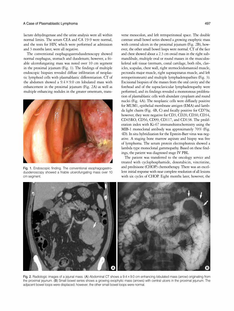
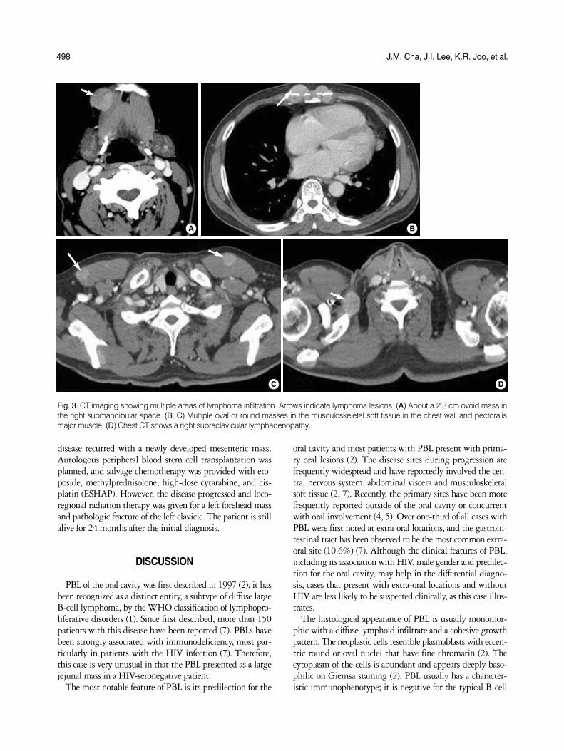
The conventional esophagogastroduodenoscopy showednormal esophagus, stomach and duodenum; however, a fri-able ulcerofungating mass was noted over 10 cm segmentin the proximal jejunum (Fig. 1). The findings of multipleendoscopic biopsies revealed diffuse infiltration of neoplas-tic lymphoid cells with plasmablastic differentiation. CT ofthe abdomen showed a 9.4×9.0 cm lobulated mass withenhancement in the proximal jejunum (Fig. 2A) as well asmultiple enhancing nodules in the greater omentum, trans-
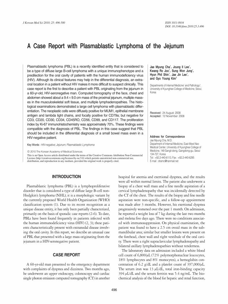
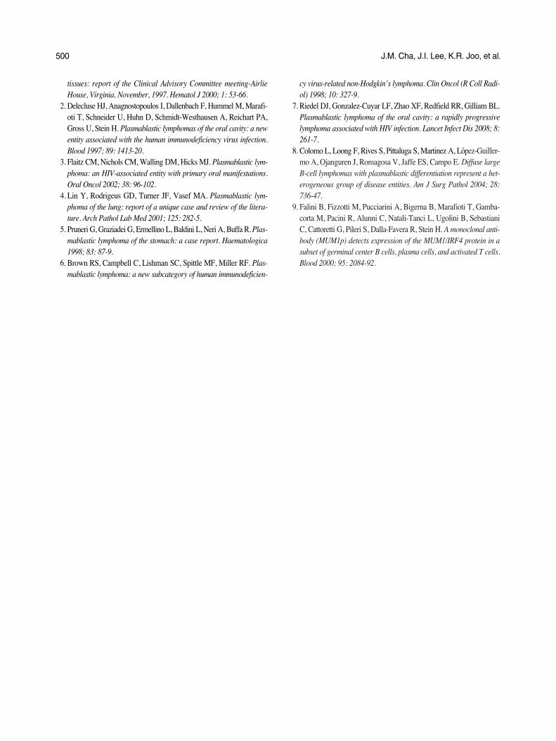
verse mesocolon, and left retroperitoneal space. The doublecontrast small bowel series showed a growing exophytic masswith central ulcers in the proximal jejunum (Fig. 2B), how-ever, the other small bowel loops were normal. CT of the faceand chest showed about a 2.3 cm ovoid mass in the right sub-mandibule, multiple oval or round masses in the musculos-keletal soft tissue (sternum, costal cartilage, both ribs, clav-icles, scapulas, chest wall, right sternocleidomastoid muscle,pectoralis major muscle, right supraspinatus muscle, and leftretroperitoneum) and multiple lymphadenopathies (Fig. 3).Excisional biopsies of the masses from the oral cavity and theforehead and of the supraclavicular lymphadenopathy wereperformed, and its findings revealed a monotonous prolifera-tion of plasmablastic cells with abundant cytoplasm and roundnuclei (Fig. 4A). The neoplastic cells were diffusely positivefor MUM1, epithelial membrane antigen (EMA) and lamb-da light chains (Fig. 4B, C) and focally positive for CD79a;however, they were negative for CD3, CD20, CD30, CD34,CD45RO, CD56, CD99, CD117, and CD138. The prolif-eration index with Ki-67 immunohistochemistry using theMIB-1 monoclonal antibody was approximately 70% (Fig.4D). In situ hybridization for the Epstein-Barr virus was neg-ative. A staging bone marrow aspirate and biopsy was freeof lymphoma. The serum protein electrophoresis showed alambda type monoclonal gammopathy. Based on these find-ings, the patient was diagnosed stage IV PBL.
The patient was transferred to the oncology service andtreated with cyclophosphamide, doxorubicin, vincristine,and prednisone (CHOP) chemotherapy. There was an excel-lent initial response with near complete resolution of all lesionswith six cycles of CHOP. Eight months later, however, the
Fig. 1. Endoscopic finding. The conventional esophagogastro-duodenoscopy showed a friable ulcerofungating mass over 10cm segment.
Fig. 2. Radiologic images of a jejunal mass. (A) Abdominal CT shows a 9.4×9.0 cm enhancing lobulated mass (arrow) originating fromthe proximal jejunum. (B) Small bowel series shows a growing exophytic mass (arrows) with central ulcers in the proximal jejunum. Theadjacent bowel loops were displaced; however, the other small bowel loops were normal.
A B
498 J.M. Cha, J.I. Lee, K.R. Joo, et al.
disease recurred with a newly developed mesenteric mass.Autologous peripheral blood stem cell transplantation wasplanned, and salvage chemotherapy was provided with eto-poside, methylprednisolone, high-dose cytarabine, and cis-platin (ESHAP). However, the disease progressed and loco-regional radiation therapy was given for a left forehead massand pathologic fracture of the left clavicle. The patient is stillalive for 24 months after the initial diagnosis.
DISCUSSION
PBL of the oral cavity was first described in 1997 (2); it hasbeen recognized as a distinct entity, a subtype of diffuse largeB-cell lymphoma, by the WHO classification of lymphopro-liferative disorders (1). Since first described, more than 150patients with this disease have been reported (7). PBLs havebeen strongly associated with immunodeficiency, most par-ticularly in patients with the HIV infection (7). Therefore,this case is very unusual in that the PBL presented as a largejejunal mass in a HIV-seronegative patient.
The most notable feature of PBL is its predilection for the
oral cavity and most patients with PBL present with prima-ry oral lesions (2). The disease sites during progression arefrequently widespread and have reportedly involved the cen-tral nervous system, abdominal viscera and musculoskeletalsoft tissue (2, 7). Recently, the primary sites have been morefrequently reported outside of the oral cavity or concurrentwith oral involvement (4, 5). Over one-third of all cases withPBL were first noted at extra-oral locations, and the gastroin-testinal tract has been observed to be the most common extra-oral site (10.6%) (7). Although the clinical features of PBL,including its association with HIV, male gender and predilec-tion for the oral cavity, may help in the differential diagno-sis, cases that present with extra-oral locations and withoutHIV are less likely to be suspected clinically, as this case illus-trates.
The histological appearance of PBL is usually monomor-phic with a diffuse lymphoid infiltrate and a cohesive growthpattern. The neoplastic cells resemble plasmablasts with eccen-tric round or oval nuclei that have fine chromatin (2). Thecytoplasm of the cells is abundant and appears deeply baso-philic on Giemsa staining (2). PBL usually has a character-istic immunophenotype; it is negative for the typical B-cell
Fig. 3. CT imaging showing multiple areas of lymphoma infiltration. Arrows indicate lymphoma lesions. (A) About a 2.3 cm ovoid mass inthe right submandibular space. (B, C) Multiple oval or round masses in the musculoskeletal soft tissue in the chest wall and pectoralismajor muscle. (D) Chest CT shows a right supraclavicular lymphadenopathy.
A B
C D

A Case of Plasmablastic Lymphoma 499
antigens (e.g., CD20 and CD45) and positive for plasma cellmarkers such as MUM1, EMA, CD38, and CD138 (2, 7-9).PBLs invariably have restricted immunoglobulin light chainsin the cytoplasm (positive staining for lambda, but not kappa),as in our case. All PBLs characteristically display a high rateof mitotic activity by the Ki-67 proliferation index (2), whichis also consistent with our findings. Although not specific,the combination of these histopathological features is char-acteristic of PBL; therefore, immunophenotypic studies mustbe performed to confirm the diagnosis. PBL should be differ-entiated from neoplastic plasma cells and other varieties ofNHL. Unlike PBL, a plasmacytoma typically consists of matureplasma cells without a high rate of mitotic activity. Diffuselarge B-cell lymphomas nearly always express CD20, CD45-RA and CD79a; and Burkitt’s lymphomas express CD20,CD45RA and CD79a, as well as the membrane-bound IgMheavy chain isotype, all of which are not typical for PBL (2, 7).
The general prognosis of PBL is very poor with a rapidlyprogressive clinical course (2, 7); half of the original 16 pati-ents died within one year (2). An optimal chemotherapy reg-imen has not been established; the initial therapy has includ-
ed administration of multi-agent systemic chemotherapy or,alternatively, local irradiation alone (2). Despite the advancedstage at presentation, this patient initially achieved a goodresponse to the lymphoma-specific combination chemother-apy and is still alive for 24 months after the initial diagnosis.
In conclusion, the clinical features of PBL including itsassociation with HIV, male gender and predilection for theoral cavity, may help in the differential diagnosis, however,an extra-oral location in a HIV-seronegative patient makesit more difficult to suspect clinically. This case highlights anunusual presentation of PBL with a large small bowel massin a HIV-seronegative patient. Hence, PBL may be includ-ed in the differential diagnosis of a small bowel mass evenin HIV-seronegative individuals.
REFERENCES
1. Harris NL, Jaffe ES, Diebold J, Flandrin G, Muller-Hermelink HK,Vardiman J, Lister TA, Bloomfield CD. The World Health Organi-zation classification of neoplasms of the hematopoietic and lymphoid
Fig. 4. Histopathology of plasmablastic lymphoma. (A) Haematoxylin and eosin section reveals a diffuse plasmablastic infiltrate with abun-dant cytoplasm, round nuclei, and occasionally central locating nucleoli (H&E stain, ×400). (B) The lymphoid infiltrate was positive forMUM1 (Polymer method, ×200). (C) Lambda light chain reactivity is seen in virtually all tumor cells (Polymer method, ×400). (D) Thestain for Ki-67 demonstrates nuclear staining in approximately 70% of the neoplastic cells (Polymer method, ×400).
A B
C D
500 J.M. Cha, J.I. Lee, K.R. Joo, et al.
tissues: report of the Clinical Advisory Committee meeting-AirlieHouse, Virginia, November, 1997. Hematol J 2000; 1: 53-66.
2. Delecluse HJ, Anagnostopoulos I, Dallenbach F, Hummel M, Marafi-oti T, Schneider U, Huhn D, Schmidt-Westhausen A, Reichart PA,Gross U, Stein H. Plasmablastic lymphomas of the oral cavity: a newentity associated with the human immunodeficiency virus infection.Blood 1997; 89: 1413-20.
3. Flaitz CM, Nichols CM, Walling DM, Hicks MJ. Plasmablastic lym-phoma: an HIV-associated entity with primary oral manifestations.Oral Oncol 2002; 38: 96-102.
4. Lin Y, Rodrigeus GD, Turner JF, Vasef MA. Plasmablastic lym-phoma of the lung: report of a unique case and review of the litera-ture. Arch Pathol Lab Med 2001; 125: 282-5.
5. Pruneri G, Graziadei G, Ermellino L, Baldini L, Neri A, Buffa R. Plas-mablastic lymphoma of the stomach: a case report. Haematologica1998; 83: 87-9.
6. Brown RS, Campbell C, Lishman SC, Spittle MF, Miller RF. Plas-mablastic lymphoma: a new subcategory of human immunodeficien-
cy virus-related non-Hodgkin’s lymphoma. Clin Oncol (R Coll Radi-ol) 1998; 10: 327-9.
7. Riedel DJ, Gonzalez-Cuyar LF, Zhao XF, Redfield RR, Gilliam BL.Plasmablastic lymphoma of the oral cavity: a rapidly progressivelymphoma associated with HIV infection. Lancet Infect Dis 2008; 8:261-7.
8. Colomo L, Loong F, Rives S, Pittaluga S, Martinez A, Lo@pez-Guiller-mo A, Ojanguren J, Romagosa V, Jaffe ES, Campo E. Diffuse largeB-cell lymphomas with plasmablastic differentiation represent a het-erogeneous group of disease entities. Am J Surg Pathol 2004; 28:736-47.
9. Falini B, Fizzotti M, Pucciarini A, Bigerna B, Marafioti T, Gamba-corta M, Pacini R, Alunni C, Natali-Tanci L, Ugolini B, SebastianiC, Cattoretti G, Pileri S, Dalla-Favera R, Stein H. A monoclonal anti-body (MUM1p) detects expression of the MUM1/IRF4 protein in asubset of germinal center B cells, plasma cells, and activated T cells.Blood 2000; 95: 2084-92.
′