A bioinformatics pipeline to search functional motifs ...
6
A bioinformatics pipeline to search functional motifs within whole-proteome data: a case study of poxviruses Haitham Sobhy 1 Received: 30 March 2016 / Accepted: 1 December 2016 / Published online: 20 December 2016 Ó The Author(s) 2016. This article is published with open access at Springerlink.com Abstract Proteins harbor domains or short linear motifs, which facilitate their functions and interactions. Finding functional motifs in protein sequences could predict the putative cellular roles or characteristics of hypothetical proteins. In this study, we present Shetti-Motif, which is an interactive tool to (i) map UniProt and PROSITE flat files, (ii) search for multiple pre-defined consensus patterns or experimentally validated functional motifs in large datasets protein sequences (proteome-wide), (iii) search for motifs containing repeated residues (low-complexity regions, e.g., Leu-, SR-, PEST-rich motifs, etc.). As proof of principle, using this comparative proteomics pipeline, eleven pro- teomes encoded by member of Poxviridae family were searched against about 100 experimentally validated functional motifs. The closely related viruses and viruses infect the same host cells (e.g. vaccinia and variola viruses) show similar motif-containing proteins profile. The motifs encoded by these viruses are correlated, which explains why poxviruses are able to interact with wide range of host cells. In conclusion, this in silico analysis is useful to establish a dataset(s) or potential proteins for further investigation or compare between species. Keywords Protein domain Á Protein function Á Protein annotation Á Functional genomics Á Comparative genomics Á Low-complexity regions (LCRs) Introduction Protein functions and interactions are facilitated by amino acid (aa) sequences, so-called functional motifs, or domains, which participate in various processes, including protein interactions, trafficking, pre- or post-translational regulation, or recruiting enzyme [1–5]. They are either short linear motifs (SLiM), 3–11 residues (e.g., RGD), or long domain, [ 30 residues (e.g., Zinc finger, ankyrin or tetratricopeptide repeats (TPR)). Motifs may contain repeated residue(s) or region(s) (e.g., L-, SR-, AR- or PEST-rich motifs). Number of databases were established to catalogue these motifs, including PROSITE, ELM, and Minimotif Miner (MnM) databases [6–8]. MnM, MEME Suite, QSLiMFinder, SLiMSearch, 3of5, MotifHound, and DoReMi tools can be used to predict motif(s), pattern(s), or shared consensus within input sequence(s) [9–14]. Another approach uses hidden Markov model (phylo-HMM) to search for evolutionarily conserved functional motifs [15]. These tools were previously reviewed in [13, 16]. Briefly, they offer arena for searching and parsing de novo or pre- defined motifs. They may require sequence alignment, uploading background sequences, or connection to third- party tools or databases. Statistics, based on background sequences to overcome false-positive results, were pro- vided. On the other hand, for finding sequences enriched with residues, EMBOSS provides a tool for finding PEST- rich motif within a query sequence (http://emboss.source forge.net/), whereas LCR-eXXXplorer is developed to visualize low-complexity regions (LCRs) [17]. Edited by Simon D. Scott. Electronic supplementary material The online version of this article (doi:10.1007/s11262-016-1416-9) contains supplementary material, which is available to authorized users. & Haitham Sobhy [email protected]; [email protected] 1 Department of Molecular Biology, Umea ˚ University, 901 87 Umea ˚, Sweden 123 Virus Genes (2017) 53:173–178 DOI 10.1007/s11262-016-1416-9
Transcript of A bioinformatics pipeline to search functional motifs ...
A bioinformatics pipeline to search functional motifswithin whole-proteome data: a case study of poxviruses
Haitham Sobhy1
Received: 30 March 2016 / Accepted: 1 December 2016 / Published online: 20 December 2016
� The Author(s) 2016. This article is published with open access at Springerlink.com
Abstract Proteins harbor domains or short linear motifs,
which facilitate their functions and interactions. Finding
functional motifs in protein sequences could predict the
putative cellular roles or characteristics of hypothetical
proteins. In this study, we present Shetti-Motif, which is an
interactive tool to (i) map UniProt and PROSITE flat files,
(ii) search for multiple pre-defined consensus patterns or
experimentally validated functional motifs in large datasets
protein sequences (proteome-wide), (iii) search for motifs
containing repeated residues (low-complexity regions, e.g.,
Leu-, SR-, PEST-rich motifs, etc.). As proof of principle,
using this comparative proteomics pipeline, eleven pro-
teomes encoded by member of Poxviridae family were
searched against about 100 experimentally validated
functional motifs. The closely related viruses and viruses
infect the same host cells (e.g. vaccinia and variola viruses)
show similar motif-containing proteins profile. The motifs
encoded by these viruses are correlated, which explains
why poxviruses are able to interact with wide range of host
cells. In conclusion, this in silico analysis is useful to
establish a dataset(s) or potential proteins for further
investigation or compare between species.
Keywords Protein domain � Protein function � Proteinannotation � Functional genomics � Comparative genomics �Low-complexity regions (LCRs)
Introduction
Protein functions and interactions are facilitated by amino
acid (aa) sequences, so-called functional motifs, or
domains, which participate in various processes, including
protein interactions, trafficking, pre- or post-translational
regulation, or recruiting enzyme [1–5]. They are either
short linear motifs (SLiM), 3–11 residues (e.g., RGD), or
long domain, [30 residues (e.g., Zinc finger, ankyrin or
tetratricopeptide repeats (TPR)). Motifs may contain
repeated residue(s) or region(s) (e.g., L-, SR-, AR- or
PEST-rich motifs). Number of databases were established
to catalogue these motifs, including PROSITE, ELM, and
Minimotif Miner (MnM) databases [6–8]. MnM, MEME
Suite, QSLiMFinder, SLiMSearch, 3of5, MotifHound, and
DoReMi tools can be used to predict motif(s), pattern(s), or
shared consensus within input sequence(s) [9–14]. Another
approach uses hidden Markov model (phylo-HMM) to
search for evolutionarily conserved functional motifs [15].
These tools were previously reviewed in [13, 16]. Briefly,
they offer arena for searching and parsing de novo or pre-
defined motifs. They may require sequence alignment,
uploading background sequences, or connection to third-
party tools or databases. Statistics, based on background
sequences to overcome false-positive results, were pro-
vided. On the other hand, for finding sequences enriched
with residues, EMBOSS provides a tool for finding PEST-
rich motif within a query sequence (http://emboss.source
forge.net/), whereas LCR-eXXXplorer is developed to
visualize low-complexity regions (LCRs) [17].
Edited by Simon D. Scott.
Electronic supplementary material The online version of thisarticle (doi:10.1007/s11262-016-1416-9) contains supplementarymaterial, which is available to authorized users.
& Haitham Sobhy
[email protected]; [email protected]
1 Department of Molecular Biology, Umea University,
901 87 Umea, Sweden
123
Virus Genes (2017) 53:173–178
DOI 10.1007/s11262-016-1416-9

Shetti-Motif was developed to help experimental biol-
ogists to mine for multiple (pre-defined or experimentally
validated) motifs, consensus patterns, or motifs enriched
with residues within a large dataset of protein sequences
(e.g., entire proteome). The tool is interactive, versatile,
and user-friendly, Fig. 1. It visualizes UniProt and PRO-
SITE flat files and maps them in a human-readable table.
Method
Shetti-Motif is standalone and portable program, which is
developed in C#.NET. The tool is free for academic uses.
The main purpose of the tool is to mine for data within
large dataset of sequences, and present them in a human-
readable table. The input file is FASTA sequences, UniProt
or PROSITE flat files, which are publically available in the
databases. All the sequences were downloaded from Uni-
Prot, GeneBank and PROSITE (prosite.expasy.org/) web-
sites during October 2015. Three modules were
implemented in Shetti-Motif tool.
The first module is searching for x-rich motifs (i.e.,
motifs enriched with a residue(s), where x is any residue,
e.g., Leu-, SR- or PEST-rich motifs) in multiple sequences
(entire proteome). Coverage of the residue(s) within motif
is the criterion to select the motif. The default coverage
value is 30% (e.g., if the length of P-rich motif is 10 aa, P is
enriched [3 aa) and can be modified by users. Using
sliding window, Shetti-Motif slides over the sequence until
residue coverage and motif length thresholds are fulfilled.
The tool reports proteins enriched with the input residues,
protein length, number of motifs in each protein, motif
length, and coverage (number) of residue(s), Figs. S1, S2.
Shetti-Motif has additional interactive feature, which
enables searching for one or multiple consensus pattern
among multiple protein sequences, Figs. S3–S6 [18].
Shetti-Motif provides two built-in databases; the first
obtained from PROSITE database, while the second
obtained from literature, which are validated experimen-
tally, Fig. S3, Tables 1, S1–S3. Users may select patterns
from the list, or third-party motif/pattern of interest.
Notably, the tool accepts PROSITE pattern syntax,
Table S1. The tool uses perfect (exact) text-search method,
including regular expression, to search for patterns. By this
option, large datasets of proteomes can be parsed effi-
ciently. The outputs are presented in a table or exported to
text file, Figs S4–S6. Protein names, number of proteins,
and enrichment of the proteins to total number of proteins
on the dataset are reported.
Third module can parse UniProt and PROSITE flat files
and convert them to human-readable tables, Figs. S7–S9.
Shetti-Motif maps them into one table, which includes
PROSITE IDs, patterns, and name of proteins harboring
these patterns, Fig. S9. The tables can be copied into
clipboard or can be exported into a tabulated text file.
Implementation
Shetti-Motif tool, sample files, and documentation are
available on http://sourceforge.net/projects/ShettiMotif/.
The tool runs and it was tested on windows 7 or higher,
without any preliminary installation. For Mac and Linux,
MonoDevelop (http://www.monodevelop.com/) are nee-
ded. For details, see program’s user guide.
Case study
As a proof of concept, we analyzed proteomes encoded by
eleven members of Poxviridae family (2251 proteins)
against experimentally validated built-in motifs (Walker
motifs, glycosylation, nuclear localization, SUMO-,
ESCRT- and integrin-binding motifs, etc.), Tables 1, S2,
Fig. 1 Screenshot of Shetti-Motif main window (a), and flowchart of features and method used in this study (b)
174 Virus Genes (2017) 53:173–178
123
Table
1Themotif-containingproteins(M
cPs)
profile
ofpoxviruses,table
S1–S3
Vaccinia
virusWR
Variola
virus
DNA
Monkeypoxvirus
strain
Zaire-96-I-16
Yabamonkey
tumorvirus
Fowlpox
virus
Canarypox
virus
Orf
virus
Cowpox
virus
Cam
elpox
virus
Myxomavirus
strain
Lausanne
Nile
crocodilepox
virus
GenBankID
AY243312
X69198
AF380138
AY386371
AF198100
AY318871
AY386264
AF482758
AF438165
AF170726
DQ356948
Number
ofproteins
218
197
191
140
260
328
130
233
211
170
173
Protein
interaction,thiol-disulfidetransfer
[25]
CxxxC
25
23
22
16
27
41
15
32
28
16
36
CxxC
35
33
32
27
48
64
30
44
35
31
28
Bindingto
integrins,RGD-related
motifs
(3–8%
ofwhole
proteome)
[26]
RGD
96
10
58
10
11
710
614
%4.1
35.2
3.6
3.1
38.5
34.7
3.5
8.1
Bindingto
phospholipids,lipid
raft-m
ediatedendocytosis(3–27%
ofproteome)
[27]
RxLR
12
810
612
19
36
14
11
538
%5.5
4.1
5.2
4.3
4.6
5.8
27.7
65.2
2.9
22
Glycosylationsites(58–81%
ofproteome)
-(http://prosite.expasy.org/PDOC00001)*
N{P}[ST]{P}
165
153
154
112
209
264
78
181
167
128
101
%75.7
77.7
80.6
80
80.4
80.5
60
77.7
79.1
75.3
58.4
Nuclearlocalizationsequence
(NLS;KR-rich)motifs
[28]
KRxR
11
10
10
68
18
917
13
10
19
KRx[10,12]
K[K
R][KR]
00
00
14
10
00
1
KRx[10,12]
K[K
R]X
[KR]
11
22
15
02
00
2
K[K
R]RK
33
22
68
03
32
5
KR[K
R]R
11
11
03
21
21
7
[PR]xxKR{DE}[K
R]
00
03
55
10
03
1
[RP]xxKR[K
R]{DE}
12
02
42
32
12
2
RKRP
11
10
20
01
00
0
Protein
folding,Rossmannfoldsmotifs,bindFAD
orNAD(P)[29]
Gx[1,2]GxxG
810
13
814
12
815
13
11
21
Gxxx[G
A]
110
96
101
54
108
146
106
116
99
94
128
SUMO
binding(40–58and40–61%
ofproteome)
[12]
[VI]x[V
I][V
I]105
102
98
78
141
191
53
122
107
78
72
%48.2
51.8
51.3
55.7
54.2
58.2
40.8
52.4
50.7
45.9
41.6
hKx[D
E]
119
110
112
82
147
194
52
128
116
104
74
%54.6
55.8
58.6
58.6
56.5
59.1
40
54.9
55
61.2
42.8
RecruitESCRTpathway
[30]
YxxL
129
120
128
90
162
222
61
149
133
119
111
Virus Genes (2017) 53:173–178 175
123
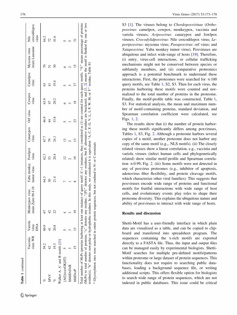
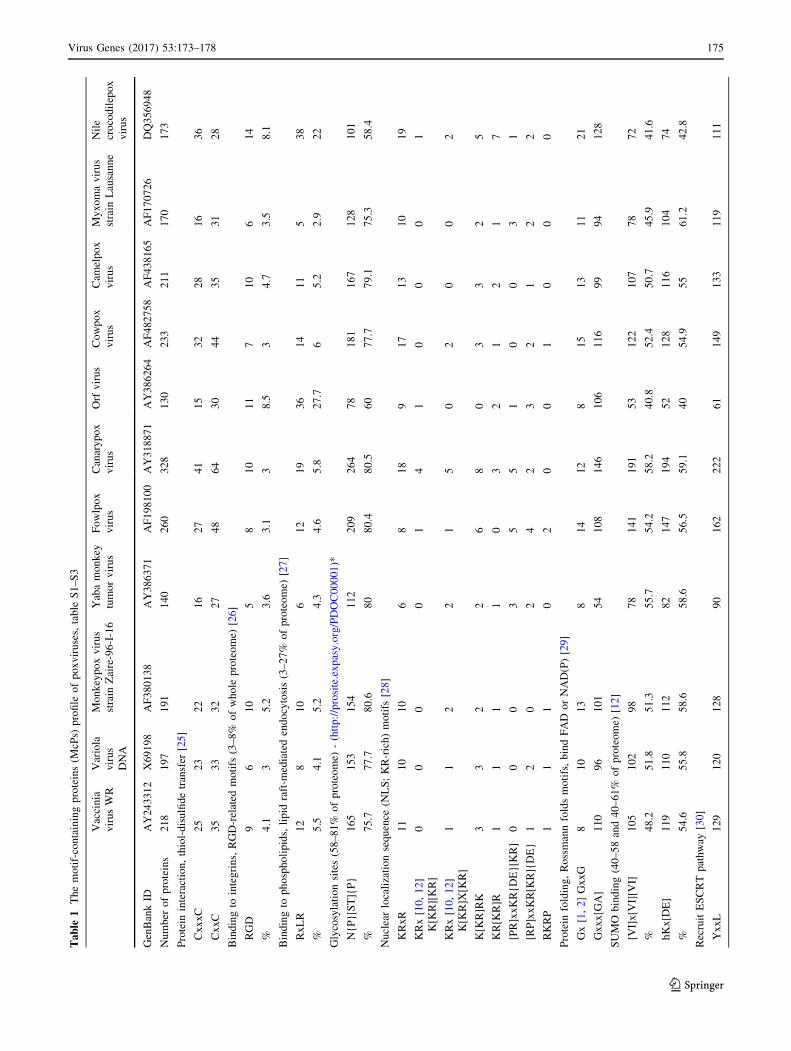
S3 [1]. The viruses belong to Chordopoxvirinae (Ortho-
poxvirus: camelpox, cowpox, monkeypox, vaccinia and
variola viruses; Avipoxvirus: canarypox and fowlpox
viruses; Crocodylidpoxvirus: Nile crocodilepox virus; Le-
poripoxvirus: myxoma virus; Parapoxvirus: orf virus; and
Yatapoxvirus: Yaba monkey tumor virus). Poxviruses are
ubiquitous and infect wide-range of hosts [19]. Therefore,
(i) entry, virus-cell interactions, or cellular trafficking
mechanisms might not be conserved between species or
subfamily members, and (ii) comparative proteomics
approach is a potential benchmark to understand these
interactions. First, the proteomes were searched for &100
query motifs, see Table 1, S2, S3. Then for each virus, the
proteins harboring these motifs were counted and nor-
malized to the total number of proteins in the proteome.
Finally, the motif-profile table was constructed, Table 1,
S3. For statistical analysis, the mean and maximum num-
ber of motif-containing proteins, standard deviation, and
Spearman correlation coefficient were calculated, see
Figs. 1, 2.
The results show that (i) the number of protein harbor-
ing these motifs significantly differs among poxviruses,
Tables 1, S3, Fig. 2. Although a proteome harbors several
copies of a motif, another proteome does not harbor any
copy of the same motif (e.g.,. NLS motifs). (ii) The closely
related viruses show a linear correlation, e.g., vaccinia and
variola viruses (infect human cells and phylogenetically
related) show similar motif-profile and Spearman correla-
tion &0.99, Fig. 2. (iii) Some motifs were not detected in
any of poxvirus proteomes (e.g., inhibitor of apoptosis,
adenovirus fiber flexibility, and protein cleavage motifs,
which characterize other viral families). This suggests that
poxviruses encode wide range of proteins and functional
motifs for fruitful interactions with wide range of host
cells, and evolutionary events play roles to shape their
proteome diversity. This explains the ubiquitous nature and
ability of poxviruses to interact with wide range of hosts.
Results and discussion
Shetti-Motif has a user-friendly interface in which plain
data are visualized as a table, and can be copied to clip-
board and transferred into spreadsheet program. The
sequences containing the x-rich motifs are exported
directly to a FASTA file. Thus, the input and output files
can be managed easily by experimental biologists. Shetti-
Motif searches for multiple pre-defined motifs/patterns
within proteome or large dataset of protein sequences. This
functionality does not require to searching public data-
bases, loading a background sequence file, or writing
additional scripts. This offers flexible option for biologists
to search wide range of protein sequences, which are not
indexed in public databases. This issue could be criticalTable
1continued
Vaccinia
virusWR
Variola
virus
DNA
Monkeypoxvirus
strain
Zaire-96-I-16
Yabamonkey
tumorvirus
Fowlpox
virus
Canarypox
virus
Orf
virus
Cowpox
virus
Cam
elpox
virus
Myxomavirus
strain
Lausanne
Nile
crocodilepox
virus
%59.2
60.9
67
64.3
62.3
67.7
46.9
63.9
63
70
64.2
hPxV
42
41
42
30
53
79
44
47
41
51
72
%19.3
20.8
22
21.4
20.4
24.1
33.8
20.2
19.4
30
41.6
Walker
A,A’andB
motifs
[31]
[AG]xxxxGK[ST]
55
47
12
13
56
56
5
hhhhDxDxR
33
31
22
13
32
5
hhhDxxP
15
13
19
819
13
23
18
17
15
31
Totalnumber
ofMcPs(proteinsharboringat
leastoneinstance
ofquerymotif;if[1instances,they
considered
as(1)arecountedforeach
querymotifs;‘‘%’’meanspercentageofproteins
(McPs)
tototalnumber
ofproteins;
‘‘x’’denotesanyresidue;
‘‘{P}’’denotesanyresidues,butP;alternativeresidues
arebracketed;and[1,2]meansthemotifis
flanked
byoneortwo
residue(s);‘‘h’’denotesnon-polarorhydrophobic
residues.In
thisstudy,weconsidered
hisequivalent‘‘A,C,F,G,V,L,I,P,W,M,orY’’residue,
Table
S1
*Glycosylationsitesweresearched
inentire
protein
sequences,butnotconfined
toN-orC-terminals
176 Virus Genes (2017) 53:173–178
123
when parsing proteome datasets of recently isolated
microbiological and metagenomics samples. To the best
our knowledge, this whole-proteome mining approach
cannot be achieved by similar tools. Shetti-Motif was used
to search for &100 experimentally validated patterns
against poxvirus proteomes. The results show variation in
enrichment of motif-containing proteins among the viruses,
which support that motifs are correlated with evolutionary
events, cellular interaction, or host-specificity.
LCRs are sequence repeats or extension of one or more
residue(s), e.g., 6xHis-tag. Despite their functional
importance, they are under-represented on publications,
reviewed in [1, 17, 20–22]. Their crystallization could be
difficult; thus, previous efforts attempted to mask them.
Another type of motifs, which are enriched with a
residue(s) but interrupted by others, e.g., Cys-rich, Gly-
rich or KR-rich motifs, reviewed in [1]. Notably, in lit-
erature, they are referred as x-rich motif, but not as LCRs.
This could be due to the following: (i) they may not be
considered as disordered repeats, (ii) may not conform to
a known pattern, and (iii) could be structurally important.
The difference between LCRs and x-rich motifs can be
noticed in some proteins (e.g., Q5UNS9, E3VZK9,
Q5UNX5, and Q5UQQ7), see SI-1, SI-2. Q5UNS9 har-
bors glycosylation sites LCRs, whereas the x-rich regions
in the others are not masked by NCBI-BLASTp. For this
reason, the criterion for finding x-rich motifs in Shetti-
Motif is the coverage of the residue(s) to the total motif
length. The x-rich proteins may share common bio-
chemical or molecular interactions, e.g., post-translational
modification for non-histone proteins. Therefore, it is
beneficial to establish a dataset of proteins rich with
particular residues, for investigating (experimentally) their
molecular functions.
Short motifs are subjected to evolutionary changes,
which could affect cellular processes, interactions, or pro-
tein characteristics [1–3]. Although proteins sharing func-
tional motifs might share similar function, the consensus
pattern is not the absolute measure for the protein func-
tions, and other factors could influence the function,
reviewed in [1]. Our bioinformatics approach may benefit
in predicting tropism and pathogenicity for emerging
infectious agents [23, 24], as well as determining potential
protein dataset(s) among whole proteome for designing
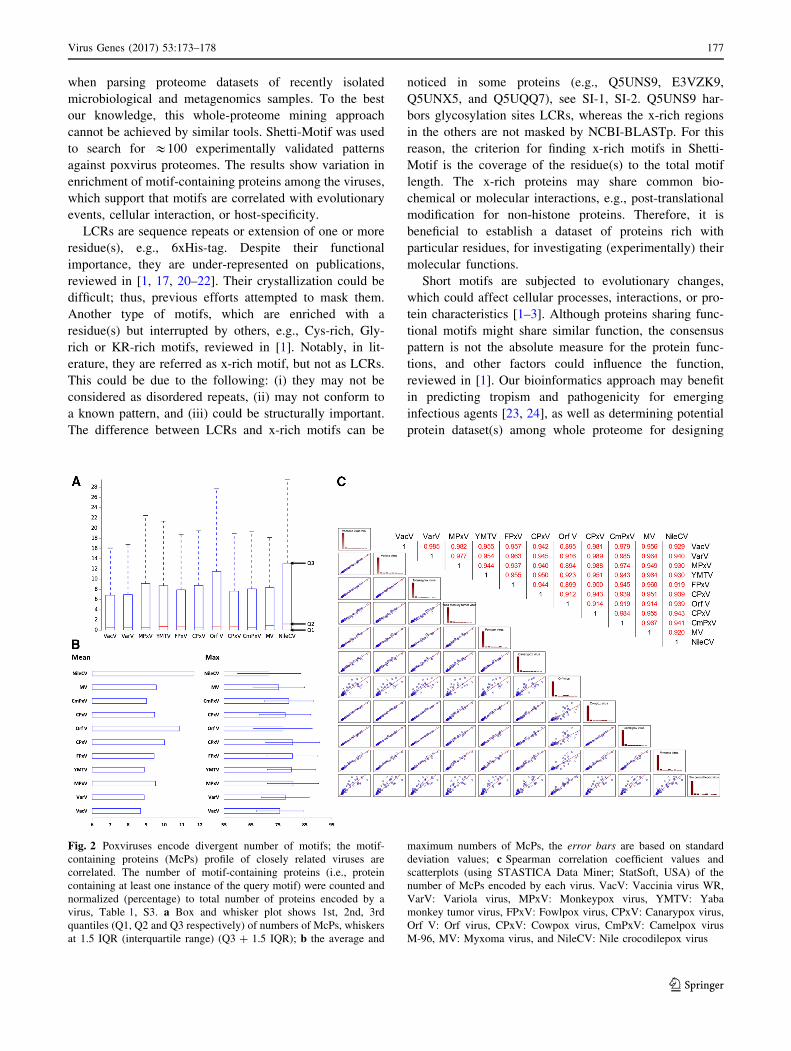
Fig. 2 Poxviruses encode divergent number of motifs; the motif-
containing proteins (McPs) profile of closely related viruses are
correlated. The number of motif-containing proteins (i.e., protein
containing at least one instance of the query motif) were counted and
normalized (percentage) to total number of proteins encoded by a
virus, Table 1, S3. a Box and whisker plot shows 1st, 2nd, 3rd
quantiles (Q1, Q2 and Q3 respectively) of numbers of McPs, whiskers
at 1.5 IQR (interquartile range) (Q3 ? 1.5 IQR); b the average and
maximum numbers of McPs, the error bars are based on standard
deviation values; c Spearman correlation coefficient values and
scatterplots (using STASTICA Data Miner; StatSoft, USA) of the
number of McPs encoded by each virus. VacV: Vaccinia virus WR,
VarV: Variola virus, MPxV: Monkeypox virus, YMTV: Yaba
monkey tumor virus, FPxV: Fowlpox virus, CPxV: Canarypox virus,
Orf V: Orf virus, CPxV: Cowpox virus, CmPxV: Camelpox virus
M-96, MV: Myxoma virus, and NileCV: Nile crocodilepox virus
Virus Genes (2017) 53:173–178 177
123
further experiments. Importantly, this approach includes
exact text search of experimentally validated motifs, which
increase the chances of true-positive results. However,
motif-containing proteins may still have different functions
from that being expected, which benefits studies on evo-
lution of protein function.
In conclusion, Shetti-Motif has simple, versatile, user-
friendly, and interactive features, which are useful for
experimental biologists lacking prior knowledge of bioin-
formatics, such as search for pattern(s) or x-rich motifs in
protein sequence(s) or entire proteome without loading
background files and user-friendly interface to visualize
UniProt and PROSITE flat files as tables.
We applied this pipeline to poxvirus proteomes, and we
observed that our pipeline is able to correlate the closely
related viruses. The results show that functional motifs are
conserved within evolutionary related viruses and/or viru-
ses that share similar molecular interactions. Therefore, we
conclude that the pipeline is useful to compare between
species; it will help in designing a dataset of candidate
proteins for further experimental investigations, either by
confirming the function or studying the evolution of protein
function.
Acknowledgements I would like to thank the reviewers. The author
receives fund from Kempestiftelserna (Kempe Foundations) and
Epigenetic Cooperation Norrland (EpiCoN) fellowships.
Compliance with ethical standards
Conflicts of interest The author declares no conflict of interest.
Ethical approval This article does not contain any studies with
human participants or animals performed by any of the authors.
Open Access This article is distributed under the terms of the
Creative Commons Attribution 4.0 International License (http://crea
tivecommons.org/licenses/by/4.0/), which permits unrestricted use,
distribution, and reproduction in any medium, provided you give
appropriate credit to the original author(s) and the source, provide a
link to the Creative Commons license, and indicate if changes were
made.
References
1. H. Sobhy, Proteomes 4, 3 (2016)
2. P. Tompa, N.E. Davey, T.J. Gibson, M.M. Babu, Mol. Cell. 55,161–169 (2014)
3. K. Van Roey, B. Uyar, R.J. Weatheritt, H. Dinkel, M. Seiler, A.
Budd, T.J. Gibson, N.E. Davey, Chem. Rev. 114, 6733–6778(2014)
4. K. Kadaveru, J. Vyas, M.R. Schiller, Front Biosci. 13, 6455–6471(2008)
5. A. Via, B. Uyar, C. Brun, A. Zanzoni, Trends Biochem. Sci. 40,36–48 (2015)
6. T. Mi, J.C. Merlin, S. Deverasetty, M.R. Gryk, T.J. Bill, A.W.
Brooks, L.Y. Lee, V. Rathnayake, C.A. Ross, D.P. Sargeant, C.L.
Strong, P. Watts, S. Rajasekaran, M.R. Schiller, Nucleic Acids
Res. 40, D252–D260 (2012)
7. H. Dinkel, K. Van Roey, S. Michael, N.E. Davey, R.J. Weatheritt,
D. Born, T. Speck, D. Kruger, G. Grebnev, M. Kuban, M. Stru-
millo, B. Uyar, A. Budd, B. Altenberg, M. Seiler, L.B. Chemes, J.
Glavina, I.E. Sanchez, F. Diella, T.J. Gibson, Nucleic Acids Res.
42, D259–D266 (2014)
8. C.J. Sigrist, E. de Castro, L. Cerutti, B.A. Cuche, N. Hulo, A.
Bridge, L. Bougueleret, I. Xenarios, Nucleic Acids Res. 41,D344–D347 (2013)
9. H. Horn, N. Haslam, L.J. Jensen, PeerJ 2, e315 (2014)
10. N.E. Davey, N.J. Haslam, D.C. Shields, R.J. Edwards, Nucleic
Acids Res. 39, W56–W60 (2011)
11. N. Palopoli, K.T. Lythgow, R.J. Edwards, Bioinformatics 31,2284–2293 (2015)
12. T.L. Bailey, J. Johnson, C.E. Grant, W.S. Noble, Nucleic Acids
Res. 43, W39–W49 (2015)
13. A. Kelil, B. Dubreuil, E.D. Levy, S.W. Michnick, PLoS ONE 9,e106081 (2014)
14. M. Seiler, A. Mehrle, A. Poustka, S. Wiemann, BMC Bioinfor-
matics 7, 144 (2006)
15. A.N.N. Ba, B.J. Yeh, D. van Dyk, A.R. Davidson, B.J. Andrews,
E.L. Weiss, A.M. Moses, Sci. Signal. 5, rs1 (2012)
16. R.J. Edwards, N. Palopoli, Methods Mol. Biol. 1268, 89–141(2015)
17. I. Kirmitzoglou, V.J. Promponas, Bioinformatics 31, 2208–2210(2015)
18. H. Sobhy, Microbial Genomics 1, 5 (2015)
19. B. Moss, Viruses 4, 688–707 (2012)
20. M.A. Huntley, G.B. Golding, Proteins 48, 134–140 (2002)
21. W. Haerty, G.B. Golding, Genome 53, 753–762 (2010)
22. H. Luo, H. Nijveen, Brief Bioinform 15, 582–591 (2014)
23. C.M. Robinson, X. Zhou, J. Rajaiya, M.A. Yousuf, G. Singh, J.J.
DeSerres, M.P. Walsh, S. Wong, D. Seto, D.W. Dyer, J. Chodosh,
M.S. Jones, MBio 4, e00595 (2013)
24. C.M. Robinson, G. Singh, C. Henquell, M.P. Walsh, H. Peigue-
Lafeuille, D. Seto, M.S. Jones, D.W. Dyer, J. Chodosh, Virology
409, 141–147 (2011)
25. T.G. Senkevich, C.L. White, E.V. Koonin, B. Moss, Proc. Natl.
Acad. Sci. U S A 99, 6667–6672 (2002)
26. J.G. Smith, C.M. Wiethoff, P.L. Stewart, G.R. Nemerow, Curr.
Top. Microbiol. Immunol. 343, 195–224 (2010)
27. D. Dou, S.D. Kale, X. Wang, R.H. Jiang, N.A. Bruce, F.D.
Arredondo, X. Zhang, B.M. Tyler, Plant Cell 20, 1930–1947
(2008)
28. S. Kosugi, M. Hasebe, N. Matsumura, H. Takashima, E. Miya-
moto-Sato, M. Tomita, H. Yanagawa, J. Biol. Chem. 284,478–485 (2009)
29. G. Kleiger, D. Eisenberg, J. Mol. Biol. 323, 69–76 (2002)
30. S. Wolff, H. Ebihara, A. Groseth, Viruses 5, 528–549 (2013)
31. C. Grangeasse, S. Nessler, I. Mijakovic, Philos. Trans. R. Soc.
Lond. B Biol. Sci. 367, 2640–2655 (2012)
178 Virus Genes (2017) 53:173–178
123