9977-18273-1-PB
10
288 Diagnosis and Molecular Marker Analysis of Bali’s Rabies Virus Isolates (DIAGNOSIS DAN ANALISIS PENANDA MOLEKULER VIRUS RABIES ISOLAT BALI) I Nyoman Dibia 1 , Bambang Sumiarto 2 , Heru Susetya 2 , Anak Agung Gde Putra 1 , I Gusti Ngurah Kade Mahardika 3 , Helen Scott-Orr 4 1 Animal Disease Investigation Center, Denpasar, Balai Besar Veteriner Denpasar, jln Raya Sesetan, Banjar Pegok, Denpasar, Bali 2 Faculty of Veterinary Sciences, University of Gadjah Mada, Yogyakarta, 3 Faculty of Veterinary Sciences, University of Udayana, Denpasar, Bali 4 University of Sidney, Sidney, Australia. E-mail : [email protected] ABSTRACT The direct fluorescent antibody test (dFAT) was recommended by both World Health Organization (WHO) and Office International des Epizooties (OIE) as a standard diagnostic technique for rabies. Since the outbreak of rabies in Bali, it was ascertain the importance to develop a reverse transcriptase-polymerase chain reaction (RT-PCR) technique with specific primers as an alternative diagnostic method. The aim of this study was to develop a RT-PCR technique for rabies diagnosis in animals and find out the molecular marker of Bali’s rabies virus (BRV) isolates based on the sequence of nucleoprotein (N) gene. Brain samples were obtained during 2009 from 14 suspected rabid dogs and one cattle, where rabies viruses were isolated. The dFAT was used to detect the presence of rabies viral antigen. Ribonucleic acid (RNA) of rabies viruses was extracted with TRIzol reagent. Fragment of N gene was amplified using one-step RT- PCR method with specifically-designed primer pairs and sequenced using ABI automatic sequencer. Multiple alignment of nucleotide and deduced amino acid sequences were analyzed using ClustalW of MEGA 4.0 program. This study found that twelve out of fifteen animal brain samples confirmed as rabies by dFAT. Similarly, a single band of 1215 bp PCR product for rabies virus was also detected in twelve out of twelve (100%) dFAT rabies positive samples. It is therefore evident that alternative diagnostic of rabies in animals can be established using RT-PCR technique. The results showed that the RT-PCR has a very high agreement with dFAT. Polymorphic sites of N gene of twelve BRV isolates were identified at the position 186, 501, 801, 840, 1068 and 1153. Bali’s rabies virus isolates have conserved amino acid (isoleucine) alterations at position 308 (open reading frame). Isoleucine distinguished between all Bali’s isolates and the all of isolates from other area of Indonesia and other part of the world. This finding significantly different as compared to other rabies virus isolates from other part of Indonesia or the world documented on the GenBank. Accordingly it is proposed that it can be used as molecular marker and believed to be the first study of molecular marker of rabies virus in Indonesia. Keywords : rabies virus, diagnosis, molecular marker, nucleoprotein gene, Bali ABSTRAK Uji direct fluorescent antibody test (dFAT) merupakan teknik diagnostik baku internasional untuk diagnosis rabies pada hewan dan direkomendasikan oleh Office International des Epizooties (OIE). Teknik reverse transcriptase-polymerasechain reaction (RT-PCR) dengan primer khas dapat digunakan sebagai alternatif. Penelitian ini bertujuan untuk mengembangan RT-PCR untuk diagnosis rabies pada hewan dan menemukan marka molekuler virus rabies Bali berdasarkan sekuens gen nukleoprotein (N). Otak hewan tersangka dikumpulkan dari kasus rabies di Bali tahun 2009. Sampel yang diuji adalah 14 otak anjing dan 1 otak sapi. Teknik dFAT digunakan untuk mendeteksi antigen rabies pada sampel. Total RNA diekstraksi dari otak tersebut dengan reagen TRIzol. Fragmen gen N diamplifikasi dengan metode one-step RT-PCRdengan pasangan primer spesifik dan disekuens dengan ABI automatic sequencer. Sekuens nukleotida dan asam amino turunannya disejajarkan dengan ClustalWdalam piranti lunak MEGA 4.0. Kita menemukan bahwa 12 dari 15 kasus tersangka itu positif dengan dFAT. Selanjutnya, satu pita Jurnal Veteriner September 2014 Vol. 15 No. 3 : 288-297 ISSN : 1411 - 8327
-
Upload
adi-paramartha -
Category
Documents
-
view
212 -
download
0
description
aaa
Transcript of 9977-18273-1-PB

288
Diagnosis and Molecular Marker Analysisof Bali’s Rabies Virus Isolates
(DIAGNOSIS DAN ANALISIS PENANDA MOLEKULERVIRUS RABIES ISOLAT BALI)
I Nyoman Dibia1, Bambang Sumiarto2, Heru Susetya2,Anak Agung Gde Putra1, I Gusti Ngurah Kade Mahardika3, Helen Scott-Orr4
1Animal Disease Investigation Center, Denpasar,Balai Besar Veteriner Denpasar, jln Raya Sesetan, Banjar Pegok, Denpasar, Bali
2Faculty of Veterinary Sciences, University of Gadjah Mada, Yogyakarta,3Faculty of Veterinary Sciences, University of Udayana, Denpasar, Bali
4University of Sidney, Sidney, Australia.E-mail : [email protected]
ABSTRACT
The direct fluorescent antibody test (dFAT) was recommended by both World Health Organization(WHO) and Office International des Epizooties (OIE) as a standard diagnostic technique for rabies. Sincethe outbreak of rabies in Bali, it was ascertain the importance to develop a reverse transcriptase-polymerasechain reaction (RT-PCR) technique with specific primers as an alternative diagnostic method. The aim ofthis study was to develop a RT-PCR technique for rabies diagnosis in animals and find out the molecularmarker of Bali’s rabies virus (BRV) isolates based on the sequence of nucleoprotein (N) gene. Brainsamples were obtained during 2009 from 14 suspected rabid dogs and one cattle, where rabies viruseswere isolated. The dFAT was used to detect the presence of rabies viral antigen. Ribonucleic acid (RNA) ofrabies viruses was extracted with TRIzol reagent. Fragment of N gene was amplified using one-step RT-PCR method with specifically-designed primer pairs and sequenced using ABI automatic sequencer. Multiplealignment of nucleotide and deduced amino acid sequences were analyzed using ClustalW of MEGA 4.0program. This study found that twelve out of fifteen animal brain samples confirmed as rabies by dFAT.Similarly, a single band of 1215 bp PCR product for rabies virus was also detected in twelve out of twelve(100%) dFAT rabies positive samples. It is therefore evident that alternative diagnostic of rabies inanimals can be established using RT-PCR technique. The results showed that the RT-PCR has a very highagreement with dFAT. Polymorphic sites of N gene of twelve BRV isolates were identified at the position186, 501, 801, 840, 1068 and 1153. Bali’s rabies virus isolates have conserved amino acid (isoleucine)alterations at position 308 (open reading frame). Isoleucine distinguished between all Bali’s isolates andthe all of isolates from other area of Indonesia and other part of the world. This finding significantlydifferent as compared to other rabies virus isolates from other part of Indonesia or the world documentedon the GenBank. Accordingly it is proposed that it can be used as molecular marker and believed to be thefirst study of molecular marker of rabies virus in Indonesia.
Keywords : rabies virus, diagnosis, molecular marker, nucleoprotein gene, Bali
ABSTRAK
Uji direct fluorescent antibody test (dFAT) merupakan teknik diagnostik baku internasional untukdiagnosis rabies pada hewan dan direkomendasikan oleh Office International des Epizooties (OIE). Teknikreverse transcriptase-polymerasechain reaction (RT-PCR) dengan primer khas dapat digunakan sebagaialternatif. Penelitian ini bertujuan untuk mengembangan RT-PCR untuk diagnosis rabies pada hewandan menemukan marka molekuler virus rabies Bali berdasarkan sekuens gen nukleoprotein (N). Otakhewan tersangka dikumpulkan dari kasus rabies di Bali tahun 2009. Sampel yang diuji adalah 14 otakanjing dan 1 otak sapi. Teknik dFAT digunakan untuk mendeteksi antigen rabies pada sampel. TotalRNA diekstraksi dari otak tersebut dengan reagen TRIzol. Fragmen gen N diamplifikasi dengan metodeone-step RT-PCRdengan pasangan primer spesifik dan disekuens dengan ABI automatic sequencer. Sekuensnukleotida dan asam amino turunannya disejajarkan dengan ClustalWdalam piranti lunak MEGA 4.0.Kita menemukan bahwa 12 dari 15 kasus tersangka itu positif dengan dFAT. Selanjutnya, satu pita
Jurnal Veteriner September 2014 Vol. 15 No. 3 : 288-297ISSN : 1411 - 8327

289
INTRODUCTION
Rabies is a fatal zoonotic disease caused byrabies virus. In Bali, rabies was firstly confirmedlaboratorically in November 2008. The diseasegives rise to a serious public health problem, with135 human deaths reported from 2008-2011.Human rabies characteristically follows a bitefrom a rabid dog. Currently, free-roaming dogsplay an important role as the reservoir andtransmitter of the disease to humans anddomestic animals. It is generally accepted thatmass vaccination program has been recognizedas an important tool to control the disease.
Rabies virus belongs to the genus Lyssavirusof the family Rhabdoviridae (Boldbaatar et al.,2010; Nguyen et al., 2011; Muleya et al., 2012).The rabies virus is non-segmented single-stranded RNA (Sato et al., 2005; Benedictis etal., 2011) and has a negative-sense (Metlin etal., 2007; Talbi et al., 2009). The viral genomecontains five genes, approximately 12 kb inlength (Wunner, 2007; Bourhy et al., 2008) andencodes five proteins namely: nucleoprotein (N),phosphoprotein (P), matrix protein (M),glycoprotein (G), and RNA polymerase or largeprotein (L) (Ito et al., 2001; Sugiyama and Ito,2007; Wunner, 2007; Yousaf et al., 2012).
The N gene of rabies virus genome contains1353 nucleotides and consists of 450 amino acids,when open reading frame (ORF) was translated(Ito et al., 2001). The antigenic sites of the Ngene were identified that involved the stretchesof amino acid at position 313–337 and 374–383(Tordo, 1996). The virus nucleoprotein playscritical role in replication and transcription.Nucleoprotein is produced abundantly duringviral replication and has been used as a targetfor diagnosis (Nicholson, 2000).
Ribonucleic acid viruses are oftencharacterized by abundant genetic variation(Pybus et al., 2007). Each RNA virus genomeexhibits a high degree of sequence variation(Khawplod et al., 2006). Even, Worobey andHolmes (1999) declared that RNA viruses
deserve their reputation as Nature’s swiftestevolvers. According to Murphy et al., (2007)nearly every progeny genome in infected cell willbe different from the parental genome at leastone nucleotide. Point mutation is the main forcein lyssavirus evolution and it is showed noevidence of recombination (Badrane and Tordo,2001). The changes found were distributedunevenly in the genome, which means that genescoding for different proteins evolved at differentrates (Fenner et al., 1993; Murphy et al., 2007).The amino acid sequence of N gene was highlyconserved, with homologies 99% (Ito et al., 2001).Kouznetzoff et al., (1998) identified that the mostconserved region of the N gene was at aminoacid position 298–352. Therefore, molecularcharacterization of the new-rabies virus isolatesis important to perform.
The direct flourescent antibody test (FAT)is one of main diagnostic tool for rabies inIndonesia. The method was recommended byboth World Health Organization (WHO) andOffice International des Epizooties (OIE) as astandard technique. In order to be able toundertake molecular characterization, it is anecessity to develop a reliable molecular assaysboth rabies virus diagnosis and characterizationsuch as reverse transcription-polymerase chainreaction/RT-PCR with specific primers. In thisarticle we present the diagnosis and molecularmarker of Bali’s rabies virus isolates in order toinvestigate dynamic of rabies virus and itsspreading.
RESEARCH METHODS
SamplesFifteen brain samples (14 dogs and one
cattle) were obtained during 2009 in this study.The dog brain samples were collected inDenpasar, Badung, Tabanan, Gianyar, Bangli,Karangasem, and Buleleng districts of the Baliprovince. Meanwhile, brain samples of cattlesuspected infected with rabies virus was collectedin Tabanan district.
tunggal dapat divisualisasikan dalam 12 dari 12 sampel yang positif itu. Hasil tersebut merupakanbukti bahwa peneguhan diagnosis rabies pada hewan dapat dilakukan dengan teknik RT-PCR sebagaialternative. RT-PCR menunjukkan kesesuaian dengan FAT sampai 100%. Situs polimorfik gen N dari 12isolat tampak pada posisi 186, 501, 801, 840, 1068, dan 1153. Yang unik, isolat rabies Bali mempunyaiasam amino khas pada posisi 308 yaitu isoleusin. I308 membedakan isolat Bali dengan isolat lain diIndonesia dan dunia. Dengan hasil tersebut, I308 dapat diguanakan sebagai marka molekuler virusisolat Bali.
Kata Kunci: virus rabies, diagnosis, marka molekuler, gen nukleoprotein, Bali
I Nyoma Dibia et al Jurnal Veteriner

290
anti-sense primer NR1251, 0.5 µL SuperScriptIII RT/Platinum Taq Mix (containingSuperScriptTM III Reverse Transcriptase andPlatinum Taq DNA Polymerase) and 5.1 µLultrapure aquabidest. Conditions of the thermalcycler for cDNA synthesis followed immediatelyby PCR amplification were as follows: one cyclecDNA synthesis at 50°C for one hour, one cycleinitial denaturation at 95°C for 45 seconds andfollowed by 40 cycles PCR amplification ofdenaturation at 94°C for 45 seconds, annealingat 50°C for 45 seconds, and extension at 72°Cfor one minute. The final steps were extensionat 72°C for five minutes. The PCR products werevisualized using electrophoresis on 1% ultrapureagarose gel (Invitrogen) containing ethidiumbromide. Expected amplification product was a1215-bp DNA fragment.
Nucleotide Sequencing and GeneticAnalysis
The RT-PCR products were excised from thegel and purified using QIAquick PCRPurification Kit (QIAGEN). The purifiedproducts (amplified cDNAs) were employed fordirect sequencing with the N gene–specificprimers NF36Y, NF303R, NF587, and NR1251.Cycle sequencing reaction was carried out witha Big Dye Terminator v3.1 Cycle Sequencing Kit(Applied Biosystems, USA). Sequencing productswere obtained using the ABI PRISM 3100 GeneticAnalyzer (Applied Biosystems, USA) at theEijkman Institute, Jakarta. The sequencingresults were analyzed and rabies virus wasdetermined by BLAST analysis (http://www.ncbi.nlm.nih.gov/blast/Blast.cgi) The Ngene target sequence was 1125-b correspondingto the nucleotides at position 40 to 1164.
Genetic analysis was performed to determinethe molecular marker of Bali rabies virus isolatesbased on the N gene. A nucleotide sequence ofthe N gene of the isolates from Bali and otherregions of Indonesia were compared geneticallywith those available in the GenBank data base.Multiple alignment of nucleotide and deducedamino acid sequences were analyzed usingClustalW of MEGA 4.0 software (Tamura et al.,2007).
RESULTS AND DISCUSSION
Flourescent Antibody TestThe direct flourescent antibody test was
conducted on 15 animal brain samples that rabid
Rabies Antigen Detection and VirusInactivation
The direct fluorescent antibody test was usedto confirm the presence of rabies viral antigen.Smears were prepared from composite sampleof original brain tissue, that includes thehippocampus. The brain smear slide was thenstained with specific conjugate (fluoresceinisothiocyanate-labeled rabbit anti-rabiesnucleocapsid immunoglobulins) (BioRad). In thedFAT, the specific aggregates of nucleocapsidprotein are identified by their fluorescence. Theantigen reacting with antibodies tagged withfluorescein isothiocyanate, appears underultraviolet light a brightly coloured apple-greenor greenish-yellow objects against a darkbackground (Dean et al., 1996). A negativecontrol using a healthy dog brain smear and apositive control using a rabid dog brain smearwere performed for an accurate diagnosis. dFAT-positive samples were made up as a 20% (wt/vol) suspension with phosphate-buffered saline(PBS) and inactivated with sodiumdodecylsulphate (500 µL of 20% original brainsuspension containing 50 µL of 10% SDS). Theinactivated virus samples were stored at –80oCfor further study.
Primer DesignThe specific primers were developed in-house
using Primer 3 software (http://biotool. umass-med.edu/bioapps/Primer3-www.cgi). They weredesigned based on nucleotide sequences for thenucleoprotein (N) gene that are available inGenBank. The primers are NF36Y (5’-TCAGGTGGTCTCYTTGAAGCC-3’) at positions36 to 56, NF303R (5’-CCGATGTRGAAGGGAGTTGG-3’) at positions 303 to 322, NF587 (5’-ACTCACAAGATGTGTGCCAA-3’) at positions587 to 606, and NR1251 (5’-CTTTAGTCGACCTCCGTTCA-3’) at positions 1232 to 1251.
RNA Extraction and RT-PCRTotal RNA from the original brain
suspension was extracted with TRIzol reagent(Invitrogen) according to the manufacturer’sinstructions. The RT-PCR were performedsimultaneously in a single-step reaction with theSuperScriptTM III One-Step RT-PCR System witha Platinum Tag DNA Polymerase Kit(Invitrogen, CA). Twenty µL of reaction mixturecontained 10 µL 2x reaction mix (a buffercontaining 0.4 mM of each dNTP and 3,2 mMMgCl2), 2 µL template RNA, 1.2 µL of 10 pmol/µL sense primer NF36Y, 1.2 µL of 10 pmol/µL
Jurnal Veteriner September 2014 Vol. 15 No. 3 : 288-297

291
Figure 2. Electrophoresis result of PCR products. M = DNA ladder 100bp (Marker), (+) = positivecontrol, (-) = negative control, and lanes 1-12 = Bali’s isolates. Spesific PCR products atposition 1215-bp are indicated by the arrow.
suspected. Of 15 brain samples tested, 12 sampleswere rabies positive. Some examples of dFATshown in Figure 1.
RT-PCR and Nucleotide Sequence of NGene (nt 40 -1164)
All of the dFAT-positive samples from thetwelve brain samples obtained from the seven
districts of Bali province (Table 1) were amplifiedsuccessfully using RT-PCR method. An expectedPCR product was 1215 bp (Figure 2).
The specific PCR products were used fordirect sequencing analysis. Nucleotide sequences(1125 bp) of the N genes (nt 40 – 1164) of 12rabies isolates were determined. BLAST analysis(http://www.ncbi.nlm.nih.gov/blast/Blast/cgi) in
Figure 1. The dFAT positive samples are indicated by aggregates of nucleocapsid protein withbrightly-coloured, apple-green or greenish yellow against a dark background.
I Nyoma Dibia et al Jurnal Veteriner
292
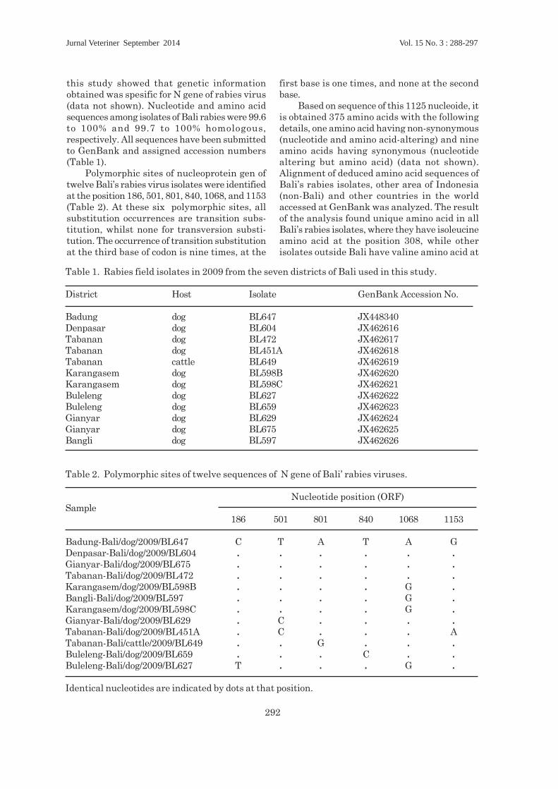
this study showed that genetic informationobtained was spesific for N gene of rabies virus(data not shown). Nucleotide and amino acidsequences among isolates of Bali rabies were 99.6to 100% and 99.7 to 100% homologous,respectively. All sequences have been submittedto GenBank and assigned accession numbers(Table 1).
Polymorphic sites of nucleoprotein gen oftwelve Bali’s rabies virus isolates were identifiedat the position 186, 501, 801, 840, 1068, and 1153(Table 2). At these six polymorphic sites, allsubstitution occurrences are transition subs-titution, whilst none for transversion substi-tution. The occurrence of transition substitutionat the third base of codon is nine times, at the
first base is one times, and none at the secondbase.
Based on sequence of this 1125 nucleoide, itis obtained 375 amino acids with the followingdetails, one amino acid having non-synonymous(nucleotide and amino acid-altering) and nineamino acids having synonymous (nucleotidealtering but amino acid) (data not shown).Alignment of deduced amino acid sequences ofBali’s rabies isolates, other area of Indonesia(non-Bali) and other countries in the worldaccessed at GenBank was analyzed. The resultof the analysis found unique amino acid in allBali’s rabies isolates, where they have isoleucineamino acid at the position 308, while otherisolates outside Bali have valine amino acid at
Table 1. Rabies field isolates in 2009 from the seven districts of Bali used in this study.
District Host Isolate GenBank Accession No.
Badung dog BL647 JX448340Denpasar dog BL604 JX462616Tabanan dog BL472 JX462617Tabanan dog BL451A JX462618Tabanan cattle BL649 JX462619Karangasem dog BL598B JX462620Karangasem dog BL598C JX462621Buleleng dog BL627 JX462622Buleleng dog BL659 JX462623Gianyar dog BL629 JX462624Gianyar dog BL675 JX462625Bangli dog BL597 JX462626
Table 2. Polymorphic sites of twelve sequences of N gene of Bali’ rabies viruses.
Nucleotide position (ORF)Sample
186 501 801 840 1068 1153
Badung-Bali/dog/2009/BL647 C T A T A GDenpasar-Bali/dog/2009/BL604 . . . . . .Gianyar-Bali/dog/2009/BL675 . . . . . .Tabanan-Bali/dog/2009/BL472 . . . . . .Karangasem/dog/2009/BL598B . . . . G .Bangli-Bali/dog/2009/BL597 . . . . G .Karangasem/dog/2009/BL598C . . . . G .Gianyar-Bali/dog/2009/BL629 . C . . . .Tabanan-Bali/dog/2009/BL451A . C . . . ATabanan-Bali/cattle/2009/BL649 . . G . . .Buleleng-Bali/dog/2009/BL659 . . . C . .Buleleng-Bali/dog/2009/BL627 T . . . G .
Identical nucleotides are indicated by dots at that position.
Jurnal Veteriner September 2014 Vol. 15 No. 3 : 288-297

293
Figure 3. Electropherogram of direct sequencing result of PCR products. Three nucleotides atposition 922 (A), 923 (T) and 924 (A) in box show codon for Isoleucine (I) at position 308of N gene of Bali’s isolates.
same position (Figure 4). Codon encoding specificamino acid (isoleucine) of N gene for Bali’s rabiesvirus isolates were shown in eletropherogram(Figure 3).
Analysis of the deduced amino acid sequencesof N gene (Figure 4) revealed that the Bali rabiesvirus isolates different from the non-Bali rabiesvirus isolates. Bali rabies virus has unique non-
I Nyoma Dibia et al Jurnal Veteriner

294
synonymous substitutions at position amino acid308 (Isoleucine) whilst all rabies viruses inIndonesia and all over the world have valine atthe same position. Amino acid alignment of Ngene of rabies field isolates in Bali obtained inthis study and accessed at GenBank shown inFigure 4.
Rabies diagnosis in animal brains is basedon the FAT as the standard technique(Wacharapluesadee et al., 2008) and it providesa reliable diagnosis in 98-100% of cases (OIE,2008). Rabies diagnostic methods based onmolecular biology approach have been developedone of them is RT-PCR. This method is also
Figure 4. Alignment of the deduced amino acid sequences of the rabies N protein from rabies fieldisolates in Bali and other rabies viruses accessed at GenBank. Identical residues areindicated by dots. Specific substitution at position 308 is indicated by arrowhead and asin box.
Jurnal Veteriner September 2014 Vol. 15 No. 3 : 288-297

295
much beneficial in further analyzing on geneticcharacterization of rabies viruses.
Twelve of twelve (100%) dFAT-positivesamples in this study were detected succesfullyusing one step RT-PCR (Figure 2). This conditionshowed that RT-PCR developed has a highagreement with dFAT, so it can be used as areliable confirmative diagnostic tool. The resultof this study supported similar study resultconducted by previous researchers. Superiorityof using RT-PCR is this method still able to detectviral antigen in decomposed brain specimen,which is not detected by using dFAT, as reportedDavid et al., (2002). Benedictis et al., (2011)reported that the one-step RT-PCR showed highrelative specificity 98.94% (CI of 97.55 to 99.65),sensitivity 99.71% (CI of 98.40 to 99.99) andaccuracy 98.90% values in comparison withthose obtained with the FAT used as a gold-standard method. It is also reported that theagreement between the one-step RT-PCRdeveloped and the gold-standard method (FAT)was calculated as 98.91% with a Cohen’s kappacoefficient of 0.977, which corresponds to nearlyperfect agreement between the two methods. Itis concluded that RT-PCR could be used as anadjunct to dFAT, especially to suspected rabiddog that died more than eight hours beforespecimen is taken. Polymorphic sites of nucleoprotein gen oftwelve Bali’s rabies virus isolates were identifiedat the position 186, 501, 801, 840, 1068, and 1153(Table 2). Nei and Kumar (2000) and Cann (1993)described that a transition substitution is thesubstitution of a purine (adenine or guanine) foranother purine or the substitution of apyrimidine (thymine or cytosine) for anotherpyrimidine. The occurrence of substitutiontoward six polymorphic sites in this study istransition substitution whist non transversionsubstitution. The happening of transitionsubstitution at the third base of codon is ninetimes, at the first base is one times, and none atthe second base. The result of this study refersto opinion of Murphy et al., (2007) who statedthat the most common mutation are singlenucleotide substitution that called pointmutation. Point mutation in third nucleotide ofa codon are often silent, so do not result analtered amino acid because of redundancy in thegenetic code. According to Yang et al. (2000) thatcomparison of relative fixation rates ofsynonymous (silent) and non-synonymous (aminoacid-altering) mutations provides a means forunderstanding the mechanisms of molecular
sequence evolution. The event of mutation ofBali’s rabies virus isolates corresponds to RNAviruses character. Murphy et al., (2007)explained that RNA viruses are characterizedby a high mutation rate during replication,because of the absence of a cellular proof readingmechanism and post replication error correctionby RNA polymerase. Ming et al., (2010) alsoreported that rabies virus evoluted continouslythrough genetic mutation. Hughes et al., (2005)showed that the evolutionary rate for N gene ofrabies virus in bats in North America wasestimated to be 2.32 x 10⁻⁴ substitutions per siteper year, meanwhile Talbi et al., (2009) reportedthat the mean rates of nucleotide substitutionfor the N gene of rabies virus isolates belongingto the Africa was 3.82 x 10⁻⁴ substitutions persite per year.
The deduced amino acid sequences of N geneof Bali rabies virus isolates were compared withthose of rabies virus isolates from other part ofIndonesia, Asia, the America, Erope, Africa, andrelated rabies virus isolates (Figure 4). A uniqueamino acid substitution between Bali’s rabiesvirus isolates (BRV) and non-BRV isolates(accessed in GenBank) was found in the mostconserved region at amino acid position 308 ofnucleoprotein gene and distinguished all ofisolates in Bali from the isolates in other part ofIndonesia and the world.
This study is believed to be the first studyto detect molecular marker of rabies virus inIndonesia. It was found unique amino acid(isoleucine) at the position 308 from all Baliisolates, which has not been possessed by rabiesisolates in all Indonesia and some other countriesall over the world, accessed at GeneBank (Figure4). In addition, this specific amino acid(isoleucine) is precise to most conserve region ofN gene of rabies virus. Thus, it can be said thatisoleucine at position 308 (open reading frame)of N gene is molecular marker of Bali’s rabiesvirus isolates and it can be used as anepidemiological marker in other to investigatedynamic and its spreading. The data obtainedin this study support opinion of Nagarajan etal., (2006) who stated that amino acid mutationsof gene might specifically carry a molecularmarker that can be used as an epidemiologicalmarker.
In the future, based on the result of thisstudy, it can be developed a RT-PCR techniqueto distinguish between Bali’s rabies virusisolates and non-Bali’s isolates. Ito et al., (2003)developed a technique to discriminate dog-related
I Nyoma Dibia et al Jurnal Veteriner

296
and vampire bat related rabies virus isolates(DRRV and VRRV, respectively) in Brazil usingstrain-spesific (SS) primers. All the DRRV andVRRV were successfully distinguished by RT-PCR with SS primers developed. Theoreticallyand referring to success of RT-PCR developmentconducted by Ito et al., (2003) that it is reallypossible and necessary to do. Strain-specificprimers for detection of Bali’s rabies virus isolateswere designed namely: Primer1 (5’-TACTCATCTAATGCAGTTGGTCACA-3’ atposition 898 to 922) and Primer2 (5’-TCCAACAAAGTGAATGAGATTGAATAT-3’ atposition 922 to 948).
CONCLUSION
This research finding showed thatconfirmative diagnostic of rabid animals can beestablished using RT-PCR technique withspecific primers and isoleucine at position 308 ofN gene of Bali’s rabies virus isolates identifiedas molecular marker.
ACKNOWLEDGMENT
This study was funded by ACIAR AH 2006-166 project. The authors would like to thankHeads of DIC Denpasar, for having providedsome of the animal brain specimens.
REFERENCES
Badrane H, Tordo N. 2001. Host Switching inLyssavirus History from the Chiroptera tothe Carnivora Orders. J Virol 75(17) : 8096-8104.
Benedictis PD, Battisti CD, Dacheux L,Marciano S, Ormelli S, Salomoni A,Caenazzo ST, Lepelletier A, Bourhy H,Capua I, Cattoli G. 2011. LyssavirusDetection and Typing UsingPyrosequencing. J Clin Microbiol 49(5) :1932-1938.
Boldbaatar B, Inoue S, Tuya N, Dulam P,Batchuluun D, Sugiura N, Okutani A, KakuY, Noguchi A, Kotaki A, Yamada A. 2010.Molecular Epidemiology of Rabies Virus inMongolia, 2005-2008. Jpn J Infect Dis 63 :358-363.
Bourhy H, Reynes J, Dunham EJ, Dacheux L,Larrous F, Huong VTQ, Xu G, Yan J,Miranda MEG, Holmes EC. 2008. The originand phylogeography of dog rabies virus. JGen Virol 89 : 2673-2681.
Cann AJ. 1993. Principles of molecular virology.San Diego, CA: Academic Press Inc.
David D, Yakobson B, Rotenberg D, Dveres N,Davidson I, Stram Y. 2002. Rabies virusdetection by RT-PCR in decomposednaturally infected brains. Vet Microbiol87(2) : 111-118.
Dean DJ, Abelseth MK, Anatasiu P. 1996. Thefluorescent antibody test. In Meslin FX,Kaplan MM, Koprowski H. (Ed). Laboratorytechniques in rabies. 4th ed. Geneva: WorldHealth Organization. Pp. 88-95.
Fenner FJ, Gibbs EPJ, Murphy FA, Rott R,Studdert MJ, White DO. 1993. VeterinaryVirology. 2nd ed. California: Academic PressInc.
Hughes GJ, Orciari LA, Rupprecht CE. 2005.Evolutionary timescale of rabies virusadaptation to North American bats inferredfrom the substitution rate of thenucleoprotein gene. J Gen Virol 86 : 1467-1474.
Ito M, Itou T, Shoji Y, Sakai T, Ito FH, Arai YT,Takasaki T, Kurane I. 2003. Discriminationbetween dog-related and vampire bat-relatedrabies viruses in Brazil by strain-specificreverse transcriptase-polymerase chainreaction and restriction fragment lengthpolymorphism analysis. J Clin Virol 26(3) :317-330.
Ito N, Kakemizu M, Ito KA, Yamamoto A,Yoshida Y, Sugiyama M, Minamoto N.2001. A comparison of complete genomesequences of the attenuated RC-HL strainof rabies virus used for production of animalvaccine in Japan, and the parentralNishigahara strain. Microbiol Immunol 45: 51-58.
Khawplod P, Shoji Y, Ubol S, Mitmoonpitak C,Wilde H, Nishizono A, Kurane I, MorimotoK. 2006. Genetic analysis of dog rabiesviruses circulating in Bangkok. Infect GenEvol 6 : 235-240.
Jurnal Veteriner September 2014 Vol. 15 No. 3 : 288-297

297
Kouznetzoff A, Buckle M, Tordo N. 1998.Identification of a region of the rabies virusN protein involved indirect binding to theviral RNA. J Gen Virol 79 : 1005-1013.
Metlin AE, Rybakov S, Grusdev K, NeuvonenE, Huovilainen A. 2007. Geneticheterogeneity of Russian, Estonian andFinnish filed rabies viruses. Arch Virol DOI10.1007/s00705-007-1001-6.
Ming P, Yan J, Rayner S, Meng S, Xu G, TangQ, Wu J, Luo J, Yang X. 2010. A historyestimate and evolutionary analysis of rabiesvirus variants in China. J Gen Virol 91:759-764.
Muleya W, Namangala B, Mweene A, Zulu L,Fandamu P, Banda D, Kimura T, Sawa H.Ishii A. 2012. Molecular epidemiology and aloop-mediated isothermal amplificationmethod for diagnosis of infection with rabiesvirus in Zambia. Virus Res 163 : 160-168.
Murphy FA, Gibbs EPJ, Horzinek MC, StuddertMJ. 2007. Veterinary Virology. 3rd ed. USA:Elsevier, Academic Press.
Nagarajan T, Mohanasubramanian B, SeshagiriEV, Nagendrakumar SB, SaseendranathMR, Satyanarayana ML, Thiagarajan D,Rangarajan PN, Srinivasan VA. 2006.Molecular Epidemiology of Rabies VirusIsolates in India. J Clin Microbiol 44(9) :3218–3224.
Nei M, Kumar S. 2000. Molecular Evolutionand Phylogenetics. New York: OxfordUniversity Press.
Nguyen AKT, Nguyen DV, Ngo GC, Nguyen TT,Inoue S, Yadama A, Dinh XK, Nguyen DV,Phan TX, Pham BQ, Nguyen HT, NguyenHTH. 2011. Molecular Epidemiology ofRabies Virus in Vietnam (2006-2009). JpnJ Infect Dis 64 : 391-396.
Nicholson KG. 2000. Rabies. In: Zukerman AJ,Banatvala JE, Pattison JR, (Ed). Principlesand Practice of Clinical Virology. Fourthedition. USA: Jhon Wiley & Sons Ltd. Pp.583-606.
OIE (Office International des Epizooties). 2008.Manual of Standards for Diagnostic testsand Vaccines. Chapter 2.1.13. Pp.304-323.
Pybus OG, Rambaut A, Belshaw R, FreckletonRP, Drummond AJ, Holmes EC. 2007.Phylogenetic Evidence for DeleteriousMutation Load in RNA Viruses and ItsContribution to Viral Evolution. Mol BiolEvol 24(3) : 845-852.
Sato G, Tanabe H, Shoji Y, Itou T, Ito FH, SatoT, Sakai T. 2005. Rapid discrimination ofrabies viruses isolated from various hostspecies in Brazil by multiplex reversetranscriptionpolymerase chain reaction. JClin Virol 33: 267-273.
Sugiyama M, Ito N. 2007. Control of rabies:epidemiology of rabies in Asia anddevelopment of new generation vaccines forrabies. Comp Immunol Microbiol Inf Dis30:273-286.
Talbi C, Holmes EC, Benedictis PD, Faye O,Nakoune E, Gamatie D, Diarra A, ElmamyBO, Sow A, Adjogua EV, Sangare O, DundonWG, Capua I, Sall AA, Bourhy H. 2009.Evolutionary history and dinamics of dograbies virus in western and central Africa.J Gen Virol 90 : 783-791.
Tamura K, Dudley J, Nei M, Kumar S. 2007.MEGA4: Molecular Evolutionary GeneticsAnalysis (MEGA) Software Version 4.0. MolBiol Evol 42(8) : 1596-1599.
Tordo N. 1996. Chracteristic and molecularbiology of the rabies virus. In: Meslin FX,Kaplan MM, Koprowski H. (Ed). Laboratorytechniques in rabies, 4th ed. Geneva. WorldHealth Organization. Pp. 28-51.
Wacharapluesadee S, Sutipanya J, Damrong-watanapokin S, Phumesin P, ChamnanpoodP, Leowijuk C, Hemachudha T. 2008.Development of a TaqMan real-time RT-PCRassay for the detection of rabies virus. J VirolMethods 151 : 317-320.
Worobey M, Holmes EC. 1999. Evolutionaryaspects of recombination in RNA viruses. JGen Virol 80 : 2535-2543.
Wunner WH. 2007. Rabies Virus. In: JacksonAC, Wunner WH. (Ed.). Rabies. 2nd ed. USA:Elsevier Inc. Pp. 23-68.
Yang Z, Nielsen R, Goldman N, Pedersen AMK.2000. Codon-substitution models forheterogeneous selection pressure at aminoacid sites. Gen 155 : 431-449.
Yousaf MZ, Ashfaq UA, Zia S, Khan MR, KhanS. 2012. Rabies moleculer virology,diagnosis, prevention and treatment. J Virol9(50) : Doi 10.1186/1743-422X-9-50.
I Nyoma Dibia et al Jurnal Veteriner