82 cystic pancreatic masses on ct and mri
17
82 Cystic Pancreatic Masses on CT and MRI
-
Upload
muhammad-bin-zulfiqar -
Category
Education
-
view
68 -
download
2
Transcript of 82 cystic pancreatic masses on ct and mri

82 Cystic Pancreatic Masses on CT and MRI

CLINICAL IMAGAGINGAN ATLAS OF DIFFERENTIAL DAIGNOSIS
EISENBERG
DR. Muhammad Bin Zulfiqar PGR-FCPS III SIMS/SHL

• Fig GI 82-1 True epithelial cysts. Contrast CT scan shows multiple unilocular cysts (arrows) scattered throughout an otherwise healthy-looking pancreas in this patient with von Hippel-Lindau disease.167
• Fig GI 82-2 Pseudocyst. (A) Axial contrast CT scan demonstrate a well-defined unilocular cyst (arrow) in the tail of the pancreas. (B) T2-weighted MR image shows the cyst (arrow) with homogeneously bright signal intensity, a finding that confirms that the lesion of a fluid-filled unilocular structure.167

• Fig GI 82-3 Multiple pancreatic pseudocysts. CT scan after the administration of contrast material demonstrates four sharply marginated, fluid-filled collections.
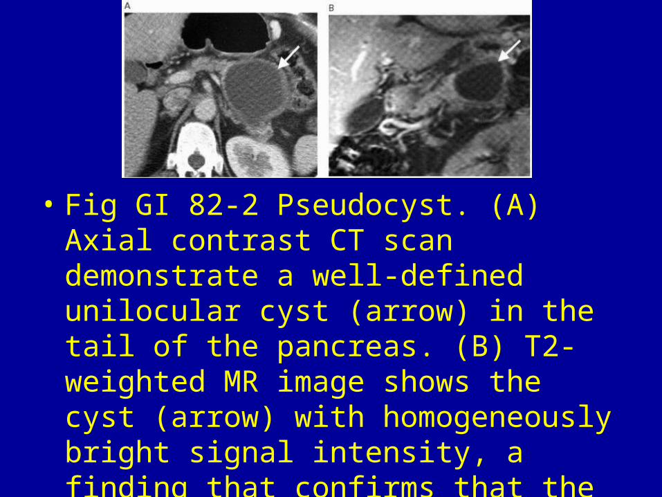
• Fig GI 82-4 Hemorrhagic pseudocyst. Contrast CT scan shows a cystic mass containing an area of high attenuation (arrow), a finding consistent with recent hemorrhage.168

• Fig GI 82-5 Ectopic pancreatic pseudocyst. The low-attenuation pseudocyst (PC) lies in the superior recess of the lesser sac posterior to the stomach (S). Note the dilated intrahepatic bile ducts (arrow).

• Fig GI 82-6 Serous cystadenoma. (A) CT scan shows a lobulated mass (arrow) of the pancreatic head with typical central scar (arrowhead) and lack of vascular encasement. (B) T2-weighted MR image shows the internal morphological features of the cyst, with high-signal-intensity microcysts (arrows) that are clearly distinguished from the dark central scar (arrowhead).167

• Fig GI 82-7 Mucinous cystic neoplasm. A contrast CT scan shows a large cystic mass (arrows) with internal septa in the head of the pancreas. The peripheral and septal calcification (arrowheads) indicate the malignant nature of the lesion. (B) In another patient, a contrast scan demonstrates a well-circumscribed, 18-cm palpable mass within the tail of the pancreas. There is enhancement on the thin external septa and peripheral wall.164
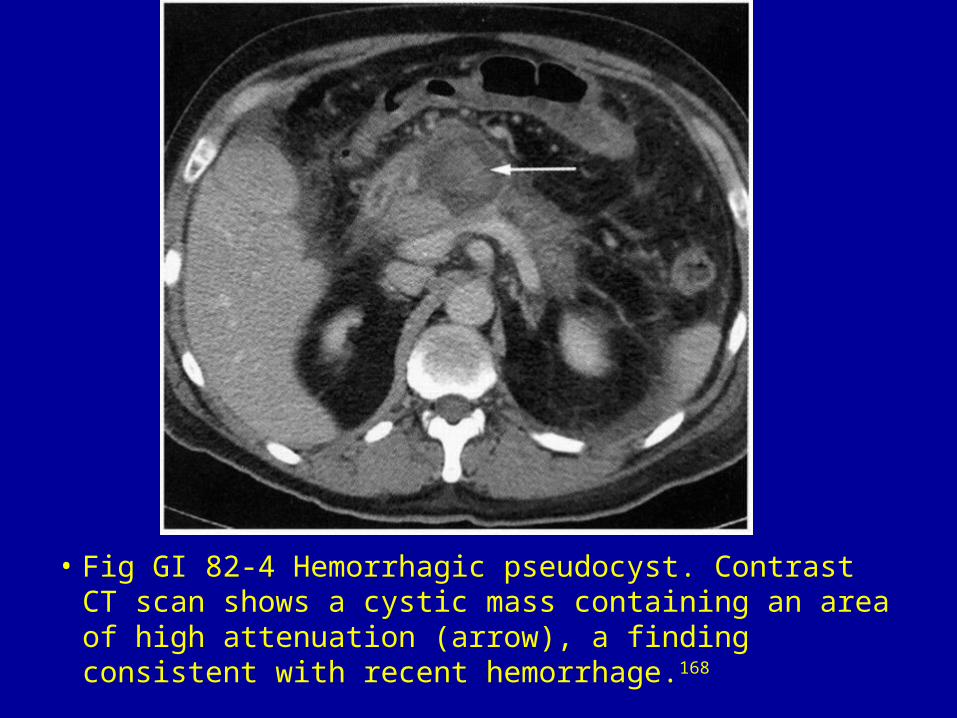
• Fig GI 82-8 Intraductal IPMN. (A) Contrast CT scan shows a small cyst (arrow) in the head of the pancreas. (B) Coronal oblique MRCP shows communication between the cyst (arrow) and the main pancreatic duct (arrowheads), a finding that helped establish the diagnosis.167

• Fig GI 82-9 Islet cell tumor. CT scan in a patient with a malignant primary neuroendocrine tumor of the pancreas demonstrates a cystic lesion in the pancreatic body with peripheral mural nodules (arrows).167

• Fig GI 82-10 Pseudopapillary tumor. Contrast CT scan shows a lesion in the body of the pancreas with cystic areas and a solid component or mural nodule (arrow).167
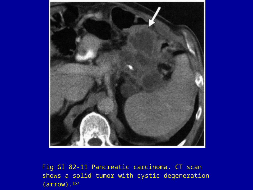
Fig GI 82-11 Pancreatic carcinoma. CT scan shows a solid tumor with cystic degeneration (arrow).167

• Fig GI 82-12 Malignant IPMN. CT scan demonstrates a multiseptated cyst with solid components (arrow).167

• Fig GI 82-13 Pancreatic abscess. CT scan shows a heterogeneous fluid collection with low attenuation and irregular margins in the body and tail of the pancreas. Note the high-attenuation debris (arrow) within the lesion.168