70. New Acute Otitis Externa - PharmaInfo Isolated from Acute Otitis Externa ... This study...
5
Synergistic Effect of Biosynthesized Silver Nanoparticles Combined with Antibiotics against Pseudomonas aeruginosa and Proteus mirabilis Isolated from Acute Otitis Externa Kassim R. Dekhil 1 , Adnan H. Aubaid 2 2 Professor, Department of Medical Microbiology, College of Medicine, University of Al-Qadisiyah, Iraq, 58001. 1 Department of Surgery, College of Medicine, University of Al-Qadisiyah, Iraq, 58001. Abstract Background: the acute external ear infections is an infectious disease recognize to be polymeric in most of the cases, usually associated with superadded by either fungus, bacteria or both of destroyed skin and it is underlying structure. Objective: To evaluate the synergistic effect of bio prepared silver nanoparticles(Ag-NPs) with antibiotics for Pseudomonas aeruginosa and Proteus mirabilis. Methods: In this study, we use the Proteus mirabilis ATCC 16404 & the standard strains of Pseudomonas aeruginosa ATCC 27853 to test the inhibitory effect of Ag-NPs alone or by using diffusion technique with antibiotics. The biosynthesis of Ag- NPs was done by using the basidiomycete, mushroom (Agaricus Bosporus). The synthesized Ag-NPs was characterized by UV/Vis spectroscopy, Fourier Transform Infrared Spectroscopy (FTIR) and Scanning electron microscopy (SEM). Antibacterial activity was determined by agar well diffusion by zones of growth inhibition and this activity was evaluated by calculating the increase in the folded area of inhibition. Results: The synthesized Ag-NPs were (4.5-35 nm) as confirmed by SEM. Spectrum detection of analysis showed peaks between 500-4000 cm-1. Biological formation of Ag-NPs was shown by changing the color of the intermixture of AgNPs (fungal cell filtrate with1mmol/ litter of silver nitrate) from clear yellow color to brown color at variable volumes and different concentration (20, 30, 40 and 50 μl) was evaluated in versus to bacterial isolates. The way it has been found that Ag-NPs was the most efficient solution in the inhibition of bacterial growth with the concentration of 50 μl. Estimation of the synergistic result was tested via method of disc diffusion opposite Pseudomonas aeruginosa and protues mirabilis. The results showed that Ag-NPs was the most efficient in the inhibition of bacterial growth with the concentration of (50 μl) and a significant combined influnece were revealed for all measured antibiotics combined with AgNPs at extremely little amount of both AgNPs and antibiotics. Conclusion: This study concluded that the nanoparticles synthesized from Agaricus bisporus have great potential as antimicrobial compound against tested pathogenic microorganisms. However, synthesis of nanoparticles can potentially eliminate the problem of chemical agents, which may have adverse effects on its application Keywords: synergistic effects; Ag-NPs; antibiotics; Pseudomonas aeruginosa; Proteus mirabilis; Acute otitis externa INTRODUCTION Otitis externa or external ear infection refers to a set of inflammatory diseases on the integumentary infection of the ear external canal, usually associated with superadded by either fungus, bacteria or both of destroyed skin and it is an underlying structure [1]. There is a multiple factors act, changing the skin layers mainly the superficial layers, making the way for infection to occur, resulting in bacterial otitis externa, the main reason of infection in the external ear [2]. The systematic disorders like chronic anemia, endocrine disorders - especially diabetes mellitus, low body concentration of vitamin and different type of skin lesion like seborrhea, any type of eczema and psoriasis resulting in decrease the protective mechanism against infectious assaults in the external canal of the ear, resulting in otitis externa [3]. Silver nanoparticles assume noteworthy part of the field for science and pharmaceutical. The silver nanoparticles antibacterial effect (AgNPs) is a very well-known and applied for technological and medical purposes [4]. The usual pathogens responsible for AOE are Pseudomonas aeruginosa, P. mirabilis, Staphylococcus spp. and various gram bacilli and a culture of the canal will usually demonstrate a mixed growth of theses organism [5]. P. aeruginosa leading to a nosocomial infection, it is a Gram- negative opportunistic pathogen in human. In particular, it may lead to a recurrent or chronic lung infection in a patient with cystic fibrosis, also causing an increase in death rate by the formation of high virulence factors and the incorrect or poor response of the host [6]. P. mirabilis causes 90% of Proteus infections. P. mirabilis causes 90% of Proteus infections and is believed to be the most common cause of infection-related otitis media [7]. The strong toxicity of silver against a wide range of microorganisms is well known and silver nanoparticles showed to be promising antimicrobial materials. So, the present study aimed to evaluate the synergistic effect of bio prepared silver nanoparticles using edible mushroom as bioreactance with antibiotics for Pseudomonas aeruginosa and Proteus mirabilis isolated from acute otitis media. Kassim R. Dekhil et al /J. Pharm. Sci. & Res. Vol. 9(12), 2017, 2644-2648 2644
Transcript of 70. New Acute Otitis Externa - PharmaInfo Isolated from Acute Otitis Externa ... This study...

Synergistic Effect of Biosynthesized Silver Nanoparticles Combined with Antibiotics
against Pseudomonas aeruginosa and Proteus mirabilis Isolated from Acute Otitis Externa
Kassim R. Dekhil1, Adnan H. Aubaid2
2 Professor, Department of Medical Microbiology, College of Medicine, University of Al-Qadisiyah, Iraq, 58001.
1Department of Surgery, College of Medicine, University of Al-Qadisiyah, Iraq, 58001.
Abstract Background: the acute external ear infections is an infectious disease recognize to be polymeric in most of the cases, usually associated with superadded by either fungus, bacteria or both of destroyed skin and it is underlying structure. Objective: To evaluate the synergistic effect of bio prepared silver nanoparticles(Ag-NPs) with antibiotics for Pseudomonas aeruginosa and Proteus mirabilis. Methods: In this study, we use the Proteus mirabilis ATCC 16404 & the standard strains of Pseudomonas aeruginosa ATCC 27853 to test the inhibitory effect of Ag-NPs alone or by using diffusion technique with antibiotics. The biosynthesis of Ag-NPs was done by using the basidiomycete, mushroom (Agaricus Bosporus). The synthesized Ag-NPs was characterized by UV/Vis spectroscopy, Fourier Transform Infrared Spectroscopy (FTIR) and Scanning electron microscopy (SEM). Antibacterial activity was determined by agar well diffusion by zones of growth inhibition and this activity was evaluated by calculating the increase in the folded area of inhibition. Results: The synthesized Ag-NPs were (4.5-35 nm) as confirmed by SEM. Spectrum detection of analysis showed peaks between 500-4000 cm-1. Biological formation of Ag-NPs was shown by changing the color of the intermixture of AgNPs (fungal cell filtrate with1mmol/ litter of silver nitrate) from clear yellow color to brown color at variable volumes and different concentration (20, 30, 40 and 50 µl) was evaluated in versus to bacterial isolates. The way it has been found that Ag-NPs was the most efficient solution in the inhibition of bacterial growth with the concentration of 50 µl. Estimation of the synergistic result was tested via method of disc diffusion opposite Pseudomonas aeruginosa and protues mirabilis. The results showed that Ag-NPs was the most efficient in the inhibition of bacterial growth with the concentration of (50 µl) and a significant combined influnece were revealed for all measured antibiotics combined with AgNPs at extremely little amount of both AgNPs and antibiotics. Conclusion: This study concluded that the nanoparticles synthesized from Agaricus bisporus have great potential as antimicrobial compound against tested pathogenic microorganisms. However, synthesis of nanoparticles can potentially eliminate the problem of chemical agents, which may have adverse effects on its application
Keywords: synergistic effects; Ag-NPs; antibiotics; Pseudomonas aeruginosa; Proteus mirabilis; Acute otitis externa
INTRODUCTION Otitis externa or external ear infection refers to a set of inflammatory diseases on the integumentary infection of the ear external canal, usually associated with superadded by either fungus, bacteria or both of destroyed skin and it is an underlying structure [1]. There is a multiple factors act, changing the skin layers mainly the superficial layers, making the way for infection to occur, resulting in bacterial otitis externa, the main reason of infection in the external ear [2]. The systematic disorders like chronic anemia, endocrine disorders - especially diabetes mellitus, low body concentration of vitamin and different type of skin lesion like seborrhea, any type of eczema and psoriasis resulting in decrease the protective mechanism against infectious assaults in the external canal of the ear, resulting in otitis externa [3]. Silver nanoparticles assume noteworthy part of the field for science and pharmaceutical. The silver nanoparticles antibacterial effect (AgNPs) is a very well-known and applied for technological and medical purposes [4]. The
usual pathogens responsible for AOE are Pseudomonas aeruginosa, P. mirabilis, Staphylococcus spp. and various gram bacilli and a culture of the canal will usually demonstrate a mixed growth of theses organism [5]. P. aeruginosa leading to a nosocomial infection, it is a Gram-negative opportunistic pathogen in human. In particular, it may lead to a recurrent or chronic lung infection in a patient with cystic fibrosis, also causing an increase in death rate by the formation of high virulence factors and the incorrect or poor response of the host [6]. P. mirabilis causes 90% of Proteus infections. P. mirabilis causes 90% of Proteus infections and is believed to be the most common cause of infection-related otitis media [7]. The strong toxicity of silver against a wide range of microorganisms is well known and silver nanoparticles showed to be promising antimicrobial materials. So, the present study aimed to evaluate the synergistic effect of bio prepared silver nanoparticles using edible mushroom as bioreactance with antibiotics for Pseudomonas aeruginosa and Proteus mirabilis isolated from acute otitis media.
Kassim R. Dekhil et al /J. Pharm. Sci. & Res. Vol. 9(12), 2017, 2644-2648
2644

MATERIALS AND METHODS Collection, isolation, and susceptibility of Clinical Samples Goldenberg's inclusion criteria used to select eighty patients with acute otitis externa for this study, these criteria include, no previous any treatment and tympanic membrane intact for diagnosis of otitis externa. All patients selected in this study had material gained from the infected ear via a swab through a skilled professional and it was elated in Stuart's culture medium. Samples were taken from patients of ages ranged between (20 -65) years old who were attending the clinical outpatient of Otorhinolaryngology of Ad-Diwaniyah teaching hospital and private clinics at Ad-Diwaniyah city during the period from November 2016 to April 2017. We take cultures and sent for investigation and the disc spread agar were used for susceptibility tests, and the outcome studied according to following laboratory result and clinical examination [8-9].
Preparation of Crude Extract of Agaricus bisporus Ag-NPs which were used synthesized by fresh edible mushroom Agaricus bisporus from commercial sources. About 20 gm of mushroom was weighted out and rinsed well with two times distilled water, then crushed plus transferred to a beaker of 100ml of sterile distilled water. This mixture is stirred for about 2 hours, filtered using Whatt man No.1 filter paper. The extract of mushroom can be preserved for further experiments by storing it at 40° C. [13]. Samples of different concentrations of the mushroom extract and AgNO3 was prepared to derive the most efficient preparatory method for efficient and faster synthesis of silver nanoparticles. Sample 1 was prepared using 50ml of extracted mushroom put with 50 ml of 1mM AgNO3 solution. A control sample was prepared by mixing 40 ml of 1mM AgNO3 (approximately 8.5 mg) directly to 10 ml of Distilled water [10].
Characterization of Ag-NPs 1. Visual detection and UV-Visible SpectroscopySynthesis of Ag-NPs using Agaricus bisporus extract wasobserved by the transformation of the color yellow color todark brown color within 12 hours. additionally, itsrecognized by UV-Visible Spectroscopy (UltraViolet-1600PC Shimadzu). The mushroom extract & AgNO3 responseprocess was monitored by Ultraviolet-Visible spectroscopyby a setting of 2.0 nm, amongst the wavelength twohundred to seven hundred nm. [11].
2. Scanning Electron Microscopy (SEM)The characterization of the size of AgNps was done byscanning electron microscope, the AgNps synthesizedusing mushroom extract was allowed to dry completely andgrounded well to a powder. SEM- specimen required to becompletely dry since the specimen is in high vacuum. Themorphology of AgNps is apparently spherical, wereobserved that are in AgNps size of 30 ± 15nm and poly-dispersed [11].
3- FTIR spectroscopy measurementsThe residual of silver nanaoparticles via Agaricus bisporusextract was centrifuged at 1000 rpm for 15 min to removethe unwanted impurities then supernatant again wasrepeated. Pellets obtained were washed with deionizedwater to get the pure AgNps. The sample completely airdried at room temperature; collected powdered AgNPswere taken (FTIR analysis) in the range of 250 to 4250 cm-1(14) [11].
Antibacterial activity and Ag-NPs Antibacterial activities of antibiotics were determined by disc diffusion method according to [9]. The Combination of Ag-NPs and antibiotics against bacterial isolates were done by disc diffusion method. The antibiotics used in our study belong to several classes and have various cellular targets, modes of action and bacterial resistance mechanisms penicillin G, ampicillin, cefotaxime, gentamycin, and rifampicin. To regulate the synergistic result of Ag-NPs the discs were saturated with newly set Ag-NPs and these discs were used for antibacterial action tests. Antibacterial action was quantified by the equation (B2 - A2)/A2, where A and B are the zone of inhibition of antibiotic and antibiotic with Ag-NPs, respectively [12].
Combination of Antibiotics and Ag-NPs Antibacterial activities of antibiotics were determined by disc diffusion method (Kirby-Bauer method) according to [8]. The mixture of Ag-NPs and antibiotics opposite bacterial isolates were done by disc diffusion method. To regulate the synergistic result of Ag-NPs the discs were saturated with newly set Ag-NPs and then these discs were used for antibacterial action tests [13].
RESULTS AND DISCUSSION Visual detection Ag-NPs were visually detected by changing color from yellow to dark brown figure (1). The reduction of silver ions to Ag-NPs (Ag+ to Ag0) lead to changing color from transparent or light yellow to brown [13]. The control did not show any change in its initial color when incubated under the same conditions [14].
Fig.1: Colloid of mushroom and AgNO3
Kassim R. Dekhil et al /J. Pharm. Sci. & Res. Vol. 9(12), 2017, 2644-2648
2645
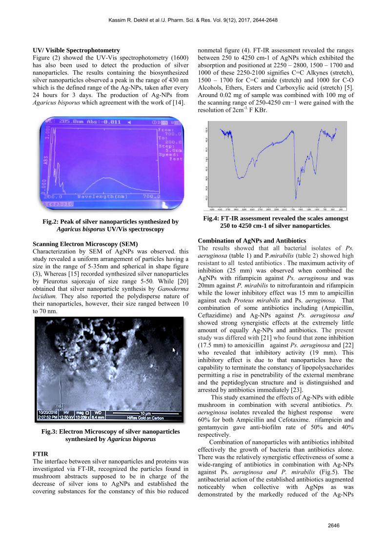
UV/ Visible Spectrophotometry Figure (2) showed the UV-Vis spectrophotometry (1600) has also been used to detect the production of silver nanoparticles. The results containing the biosynthesized silver nanoparticles observed a peak in the range of 430 nm which is the defined range of the Ag-NPs, taken after every 24 hours for 3 days. The production of Ag-NPs from Agaricus bisporus which agreement with the work of [14].
Fig.2: Peak of silver nanoparticles synthesized by Agaricus bisporus UV/Vis spectroscopy
Scanning Electron Microscopy (SEM) Characterization by SEM of AgNPs was observed. this study revealed a uniform arrangement of particles having a size in the range of 5-35nm and spherical in shape figure (3), Whereas [15] recorded synthesized silver nanoparticles by Pleurotus sajorcaju of size range 5-50. While [20] obtained that silver nanoparticle synthesis by Ganoderma lucidium. They also reported the polydisperse nature of their nanoparticles, however, their size ranged between 10 to 70 nm.
Fig.3: Electron Microscopy of silver nanoparticles synthesized by Agaricus bisporus
FTIR The interface between silver nanoparticles and proteins was investigated via FT-IR, recognized the particles found in mushroom abstracts supposed to be in charge of the decrease of silver ions to AgNPs and established the covering substances for the constancy of this bio reduced
nonmetal figure (4). FT-IR assessment revealed the ranges between 250 to 4250 cm-1 of AgNPs which exhibited the absorption and positioned at 2250 – 2800, 1500 – 1700 and 1000 of these 2250-2100 signifies C=C Alkynes (stretch), 1500 – 1700 for C=C amide (stretch) and 1000 for C-O Alcohols, Ethers, Esters and Carboxylic acid (stretch) [5]. Around 0.02 mg of sample was combined with 100 mg of the scanning range of 250-4250 cm−1 were gained with the resolution of 2cm-1. F KBr.
Fig.4: FT-IR assessment revealed the scales amongst 250 to 4250 cm-1 of silver nanoparticles.
Combination of AgNPs and Antibiotics The results showed that all bacterial isolates of Ps. aeruginosa (table 1) and P.mirabilis (table 2) showed high resistant to all tested antibiotics . The maximum activity of inhibition (25 mm) was observed when combined the AgNPs with rifampicin against Ps. aeruginosa and was 20mm against P. mirabilis to nitrofurantoin and rifampicin while the lower inhibitory effect was 15 mm to ampicillin against each Proteus mirabilis and Ps. aeruginosa. That combination of some antibiotics including (Ampicillin, Ceftazidime) and Ag-NPs against Ps. aeruginosa and showed strong synergistic effects at the extremely little amount of equally Ag-NPs and antibiotics. The present study was differed with [21] who found that zone inhibition (17.5 mm) to amoxicillin against Ps. aeruginosa and [22] who revealed that inhibitory activity (19 mm). This inhibitory effect is due to that nanoparticles have the capability to terminate the constancy of lipopolysaccharides permitting a rise in penetrability of the external membrane and the peptidoglycan structure and is distinguished and arrested by antibiotics immediately [23]. This study examined the effects of Ag-NPs with edible mushroom in combination with several antibiotics. Ps. aeruginosa isolates revealed the highest response were 60% for both Ampicillin and Cefotaxime. rifampicin and gentamycin gave anti-biofilm rate of 50% and 40% respectively. Combination of nanoparticles with antibiotics inhibited effectively the growth of bacteria than antibiotics alone. There was the relatively synergistic effectiveness of some a wide-ranging of antibiotics in combination with Ag-NPs against Ps. aeruginosa and P. mirabilis (Fig.5). The antibacterial action of the established antibiotics augmented noticeably when collective with AgNps as was demonstrated by the markedly reduced of the Ag-NPs
Kassim R. Dekhil et al /J. Pharm. Sci. & Res. Vol. 9(12), 2017, 2644-2648
2646

amount against the established bacteria. The combined effect of antibiotics and Ag-NPs was demonstrated at extremely low amount of Ag-NPs of (volume of 50 ml of mushroom extract with 50ml AgNO3) is the most effective against the growth of bacteria, on the other hand the results concluded that the inhibition zone diameters were increased by using 50 µl Ag-NPs by edible mushroom Agaricus bisporus. Species of mushroom exhibited a positive for the synthesis of Ag-NPs are protein rich and a pharmaceutically significant type of fungi. The precise mode of action behind the change of AgNO3 to Ag-NPs by mushroom abstract still not clear. Nevertheless, the
enzymes outside the cells are may be in charge of the procedure of nanoparticles biosynthesis [22]. The spectrum by giving higher inhibition zones against isolates ranges, as the highest inhibition zone obtained in bacterial isolates Ps. aeruginosa (15mm) and less inhibition zone it was P.mirabilis (13mm) because of the maximum resistantcapacity of the bacterial isolates. Though advantageous asantimicrobial agents, silver nanoparticles have adverseeffects on cells, for example, the generation of free radicalwhich are harmful to both eukaryotic cell and bacteria [23-24].
(Table 1) Zone of growth inhibition (mm) of P. aeruginosa against different antibiotics combined with or without Ag-NPs at content of 30 µl per disc)
(Table 2) Zone of inhibition (mm) of different antibiotics against P.mirabilis, (in absence and in presence of (Ag-NPs) at content of 30 µl per disc)
A B Fig.5: Inhibitory effect of different concentration and volumes of Ag-NPs against (A)P. aeruginosa and (B)
P.mirabilis.
Kassim R. Dekhil et al /J. Pharm. Sci. & Res. Vol. 9(12), 2017, 2644-2648
2647

CONCLUSION The current study concluded that mushroom (Agaricus bisporus) synthesized by Ag-NPs revealed significant inhibitory action and has great potential as antimicrobial compound against tested pathogens because of the tested resistant bacteria to an antibiotic, established their vulnerability to antibiotics combined with Ag-NPs. Though, production of nanoparticles may strongly terminate the chemical agent’s problem, which has possible side effects against its application.
REFERENCES 1. Enoz, M., Sevinc, I., Lapeña3, J.F. Bacterial and fungal organisms in
otitis externa patients without fungal infection risk factors inErzurum, Turkey. Braz J Otorhinolaryngol. 2009,75,721-5.
2. Fisher T. Synopsis of Causation: Otitis externa. Queen’s MedicalCentre Nottingham. September 2008.
3. Franke, G., An earful on treatment, Otitis Externa. PracticalOtolaryngology Conference. The Canadian Journal of CME. October20-Byron, J. Baily. 1998 H&N surgery-otolaryngology secondedition. USA. Pp.1965-1979.
4 Byron, J. Baily, H&N surgery-otolaryngology second edition. The USA. 1998, Pp.1965-1979.
5 Dakhil, A.S. Biosynthesis of silver nanoparticle (AgNPs) using Lactobacillus and their effects on oxidative stress biomarkers in rats. Journal of King Saud University - Science. 2017, 29, 462-467.
6 Bilal, A., Wani; Bodha, R. H., and Wani , A. H. Nutritional and medicinal importance of mushrooms. Medicinal Plants Res. 2010, 4, 2598-2604.
7 Marek, K., Tatsiana, D., Aleksandra, M., and Lidia R. Certain Aspects of Silver and Silver Nanoparticles in Wound Care: A Minireview Hindawi Publishing Corporation. Journal of Nanomaterials. 2016, ID 7614753, 10
8 Macfaddin, J.F. Biochemical test for identification of medical bacteria, 3nd.ed the Williams &Wilkin London. 2000.
9 CLSI. Performance Standards for Antimicrobial Susceptibility Testing; 24th Informational Supplement M100-S26. Wayne, PA: Clinical and Laboratory Standards Institute. 2016.
10 Sudhakar,T.; Nanda,A.; George,S.; Janani,B.S.; Evans, M.D.; Markose, T.K. Synthesis of Silver Nanoparticles from Edible Mushroom and Its Antimicrobial Activity against Human Pathogens. International Journal of PharmTech Research. 2014, 6, 1718-1723.
11 Narasimha,G.;Praveen,B.;Mallikarjuna.K.;Raju.B.B.D.Mushrooms (Agaricusbisporus) mediated biosynthesis of sliver nanoparticles,
characterization, and their antimicrobial activity Int.J.Nano Dim.2011, 2, 29-36.
12 Haq, M.U., Rathod,V., Singh,D., Singh,A.K, Ninganagouda,S., and Hiremath,J., Dried Mushroom Agaricus bisporus mediated synthesis of silver nanoparticles from Bandipora District (Jammu and Kashmir) and their efficacy against Methicillin-Resistant Staphylococcus aureus (MRSA) strains. Gulbarga University Kalaburagi, Karnataka Received 27 March 2015; accepted 22 April
13 Ahmad, A., Mukherjee, P., Senapati, S., Mandal, D., Khan M.I.,and Kumar R.. Extracellular biosynthesis of silver nanoparticles using fungus Fusarium oxysporum. Colloids Surf B. 2003, 28:313-18,
14 Rai, M., Yadav, A., Gade, A. Silver nanoparticles as a new generation of antimicrobials. Biotechnology Advances. 2009, 27, 76-83.
15 Nithya R and Ragunathan R. Synthesis of the silver nanoparticle using Pleurotus Sajor Cajun and its antimicrobial activity. Digest Journal of Nanomaterials and Biostructures. 2009, 4, 623-629.
16 Jaidev, L.R., G. Narasimha Fungal mediated biosynthesis of silver nanoparticles, characterization, and antimicrobial activity. Colloids and Surfaces B: Biointerfaces. 2010, 81: 430–433.
17 Pulit, J.; Banach, M.; Kowalski, Z. Nanosilver- Making difficult decisions; Ecological Chemistry and Engineering S-Chemia I Inzynieria Ekologiczna. 2011,18, 185.
18 Panáˇcek,A., Smékalová,M., Kilianová,M., Prucek ,P., Bogdanová,K., Veˇceˇrová, R., Havrdová ,K.,M.,et al. Strong and Nonspecific Synergistic Antibacterial Efficiency of Antibiotics Combined with Silver Nanoparticles at Very Low Concentrations Showing No Cytotoxic Effect: 2015, 28 December.
19 Bhosale, R.S., Hajare, K.Y., Mulay, B., Mujumdar, S., Kothawade, M., Biosynthesis, characterization, and study of the antimicrobial effect of silver nanoparticles by Actinomycetes spp. Int. J. Curr. Microbiol. Appl. Sci. 2015, 2, 144–151.
20 Devika, R., Elumalai, S., Manikandan, E., et al. Biosynthesis of silver nanoparticles using the fungus Pleurotus status and their antimicrobial activity. 2012, 10. 4172 / scientific reports.55734-
21 Elechiguerra J, Burt J, and Morones JR. Interaction of silver nanoparticles with HIV-I. J. Nanobiotechnol. 2005, 3, 6.
22 Nilda V, Ayala-Nunez ,Liliana del Carmen Ixtepan Turrent,Cristina Rodrıguez Padilla .Bactericidal effect of silver nanoparticles against multidrug-resistant bacteria. World J Microbiol Biotechnol. 2010, 26:615–621.
23 Bhat,R. Huh,D.S., Deshpande,R. and Venkataraman. Photo-Irradiated Biosynthesis of Silver Nanoparticles Using Edible Mushroom Pleurotusflorida and Their Antibacterial Activity Studies. Hindawi Publishing Corporation, Bioinorganic Chemistry, and Applications. 2011, 7 pages.
24 Carlson C, et al. Unique cellular interaction of silver nanoparticles: size-dependent generation of reactive oxygen species. J. Phys. Chem. 2008; B 112:13608–13619.
Kassim R. Dekhil et al /J. Pharm. Sci. & Res. Vol. 9(12), 2017, 2644-2648
2648