5IF FGGFDUT PG QMBUFMFU SJDI QMBTNB 131 JO … · iptu cpof jo mpdbmj[fe efgfdut pg uif bmwfpmbs...
18
I. Introduction Predictable regeneration of large alveolar defects with complex morphology can pose a significant clinical challenge, particularly when there is a signif- icant vertical component involved. The presence of sufficient bone volume is an important prerequisite for dental implant placement 1,2,3,4) . Guided bone regeneration (GBR) is an accepted surgical proce- dure intended to increase the quantity and quality of host bone in localized defects of the alveolar ridge 5) . Methods described to increase the rate of bone for- mation and to augment the bone quantity include the utilization of autografts, allografts, xenografts, and alloplastic bone substitutes 6) . Autogenous bone is considered the "gold stan- dard" for grafting alveolar defects. Despite being highly effective, these techniques subject patients to a second surgical site, which may increase morbidi- ty, hospital stay, recovery, and cost 7) . There is also a greater risk for wound infection, more blood loss, and a slower return to normal function. It is also reasonably challenging to contour and can undergo significant and unpredictable resorption 8) . For this reason, researchers continue to work toward the development of a graft materal that is osteogenic, osteoinduction, and osteoconductive. Allogenic bone is the most commonly used alter- native to the autogenous harvest. The allografts most commonly used are demineralized freeze- 217 The effects of platelet-rich plasma(PRP) in combination with anorganic bovine bone(Bio-Oss ) on the early wound healing of rabbit cranial defects Dong-Woong Lim 1 , Hyun-Seon Jang 1,4 , Ju-Chol Park 2,4 , Heoung-Jung Kim 3,4 , Jong-Woo Lee 1 , Chong-Kwan Kim 5,6 , Byung-Ock Kim 1,4 Dept. of Periodontology, College of Dentistry, Chosun University 1 Dept. of Oral Histology, College of Dentistry, Chosun University 2 Dept. of Oral Anatomy, College of Dentistry, Chosun University 3 Oral Biology Research Institute, Chosun University 4 Dept. of Periodontology, College of Dentistry, Yonsei University 5 Research Institute for Periodontal Regeneration, Yonsei University 6 대한치주과학회지 : Vol. 35, No. 1, 2005 *This study was supported by a grant of the Korea Health 21 R & D Project, Ministry of Health & Welfare, Republic of Korea (03-PJ1- PG1-CH08-0001). Corresponding author : Byung-Ock Kim , Department of Periodontology, College of Dentistry, Chosun University, 421 Seoseok- Dong, Dong-Ku, Gwang-ju, 501-759, Korea, Tel:+82-62-220-3856, Fax: +82-62-224-4664, E-mail: [email protected]
Transcript of 5IF FGGFDUT PG QMBUFMFU SJDI QMBTNB 131 JO … · iptu cpof jo mpdbmj[fe efgfdut pg uif bmwfpmbs...
I. Introduction
Predictable regeneration of large alveolar defectswith complex morphology can pose a significantclinical challenge, particularly when there is a signif-icant vertical component involved. The presence ofsufficient bone volume is an important prerequisitefor dental implant placement1,2,3,4). Guided boneregeneration (GBR) is an accepted surgical proce-dure intended to increase the quantity and quality ofhost bone in localized defects of the alveolar ridge5).Methods described to increase the rate of bone for-mation and to augment the bone quantity includethe utilization of autografts, allografts, xenografts,and alloplastic bone substitutes6).
Autogenous bone is considered the "gold stan-dard" for grafting alveolar defects. Despite beinghighly effective, these techniques subject patients toa second surgical site, which may increase morbidi-ty, hospital stay, recovery, and cost7). There is also agreater risk for wound infection, more blood loss,and a slower return to normal function. It is alsoreasonably challenging to contour and can undergosignificant and unpredictable resorption8). For thisreason, researchers continue to work toward thedevelopment of a graft materal that is osteogenic,osteoinduction, and osteoconductive.
Allogenic bone is the most commonly used alter-native to the autogenous harvest. The allograftsmost commonly used are demineralized freeze-
217
The effects of platelet-rich plasma(PRP) in combinationwith anorganic bovine bone(Bio-Oss ) on the early
wound healing of rabbit cranial defects
Dong-Woong Lim1, Hyun-Seon Jang1,4, Ju-Chol Park2,4, Heoung-Jung Kim3,4,Jong-Woo Lee1, Chong-Kwan Kim5,6, Byung-Ock Kim1,4
Dept. of Periodontology, College of Dentistry, Chosun University1
Dept. of Oral Histology, College of Dentistry, Chosun University2
Dept. of Oral Anatomy, College of Dentistry, Chosun University3
Oral Biology Research Institute, Chosun University4
Dept. of Periodontology, College of Dentistry, Yonsei University5
Research Institute for Periodontal Regeneration, Yonsei University6
대한치주과학회지 : Vol. 35, No. 1, 2005
*This study was supported by a grant of the Korea Health 21 R & D Project, Ministry of Health & Welfare, Republic of Korea (03-PJ1-PG1-CH08-0001).Corresponding author : Byung-Ock Kim , Department of Periodontology, College of Dentistry, Chosun University, 421 Seoseok-Dong, Dong-Ku, Gwang-ju, 501-759, Korea, Tel:+82-62-220-3856, Fax: +82-62-224-4664, E-mail: [email protected]

deried allograft (DFDBA) and freeze-dried boneallograft (FDBA), and controversy exists with respectto the osteoinductive potential of these materials. Ithas been shown that the inductive capacity variesfor DFDBA processed from different bone banks,and even different batches from the same bankrespond differently. Also, the bioactivity of DFDBAseems to be dependent on the age of the donor,since the younger the donor, the more osteoinduc-tive properties in the graft material9). Controversialresults and patient's concerns about disease trans-mission have encouraged the development ofxenografts and alternatives. Both have shown goodbiocompatibility and osteoconductive potential, butthe clinical outcomes with these bone substitutes areunpredictable10,11,12,13). The lack of predictability inosseous regenerative procedures using bone graftssuggests that improvement in the osteoinductiveand oeteoconductive properties of these materials ishighly desirable.
Clinicians are constantly attempting to improvethe results obtained with anorganic bovine bonealone by adding barrier membranes14), fibringlue15,16), platelet concentrates17), autogenousbone18,19), osteogenic protein 19), and other nonauto-genous grafting materials20). In 1990, Gibble andNess21) introduced fibrin glue, alternatively referredto as fibrin sealant or fibrin gel, a biomaterial thatwas developed in response to the necessity forimproved hemostatic agents with adhesive proper-ties. Platelet-rich plasma gel is an autogenous modi-fication of fibrin glue that has been described andused in various applications with apparent clinicalsuccess22). PRP is potentially useful as an adjunct toallograft and xenograft materials in oral and maxillo-facial bone and implant reconstructive surgery.
Platelets are very important in the wound healingprocess. They quickly arrive at the wound site andbegin the coagulation process. They release multi-
ple wound healing growth factors and cytokines,including platelet-derived growth factor (PDGF),transforming growth factor-β(TGF-β), insulin-likegrowth factor-1 (IGF-1), vascular endothelial cellgrowth factor (VEGF), platelet-derived epidermalgrowth factor (PDEGF), platelet factor 423,24). Thesegrowth factors are thought to contribute to initialbone regeneration and increase vascularity, and vitalfeatures of heal the bone graft25,26,27).
The aim of the this study is to evaluate the effectof PRP on the early wound healing of rabbit cranialdefects.
II. Materials and Methods
1. Animal surgical procedure
Fifteen New Zealand white male rabbits between2.8 and 4 kg were included in this randomized,blinded, and prospective study. Each rabbit wasanesthetized with Ketamine Hcl (5 mg/kg) andXylazine Hcl (1.5 ml/kg). The fur was shaved overthe cranium, which was prepared and draped in asterile fashion. An incision was made to the bonycranium and the periosteum was reflected. Bymeans of a trephine bur (external diameter : 8mm),four standardized 'through-and-through' bone defectwere created with copious irrigation. The four cra-nial defects were randomly grafted with Bio-Oss(Geistlich, Wolhusen/Switzerland) only, Bio-Osswith PRP, PRP only, and no graft as a control(Figure1). The four defects were covered with nonre-sorbable PTFE membrane (Tefgen , LifecoreBiomedical, Inc, U.S.A.). The wound was closedwith resorbable suture materials, and the rabbitswere extubated and allowed to recover(Table 1). Atthe end of the surgical procedure, all animalsreceived a single intramuscular injection of antibi-otics Gentamicin (0.1 ml/kg).
218

2. PRP Preparation
The 10 mL of autogenous blood drawn from eachrabbit was combined with 1.5 mL of Anticoagulantdextrose citrate (ACDC) to prevent coagulation. Theblood was spun in a centrifuge (Placon, Oscotec,Korea) at 2,000G for 3 minutes to separate the plas-ma containing the platelets from the red blood cells.The plasma was drawn off the top of the test tube,and centrifuged for an additional 5 minutes at5,000G to separate the platelets. The platelet-poorplasma (PPP) was separated from the PRP and thebuffy coat. The buffy coat and the PRP, approxi-mately 1 mL, were resuspended and added to thegrafting material within minutes. One thousnadunits of topical thrombin powder (Dirabine , KoreaUnited Pharm. inc, Korea) was reconstituted with 1mL of 10% calcium gluconate (Calmia , KoreaUnited Pharm. inc, Korea)(Figure 2).
Platelet counts were performed on six samples,including a peripheral blood count, a PPP count,and a PRP count. The platelets were counted with astandard hemocytometer (Sigma, USA), and the totalwas calculated for each sample.
3. Evaluation
Rabbits were killed using phentobarbital, 100mg/kg intravenously at 1,2, and 4 weeks. Therewere 5 rabbits in each group. The entire craniumwas removed with a reciprocating saw, withoutencroaching on the grafted areas(Figure 3, A).
1) Radiographic evaluation Radiographs were taken of the rabbit calvaria in
its entirety before histologic sections were per-formed. A aluminum step-wedge was used in eachradiograph for comparison(Figure 3, B). The radi-ographs were scanned and images were analyzedwith a ImageJ 1.31v software on a IBM computer.
2) Histologic evaluation The rabbit calvarias were fixed in 4% paraform-
aldehyde, and decalcified in hydrochloric aciddecalcifying solution (Fisher Scientific, Tustin, CA) at4℃ for 2-4 weeks. It was embedded in paraffin andcut into 6㎛ thickness. The sections were stainedwith H&E and observed by optical microscope.
3) Statistical methodsNumerical data was presented as mean plus one
standard deviation. One way analysis of variance(ANOVA) with fisher's Tukey test was used for mul-tiple comparisons to compare with the control. Theprobability level of p < 0.05 was regarded as statisti-cally significant.
III. Results
1. Platelet count
Platelet counts that the PRP preparation techniqueused in this study produced a highly concentratedsource of platelets. The mean peripheral bloodplatelet count was 150,500/㎜3, with a range of
219
Table 1. The four groups randomly grafted at the cranial defects.
Group n Graft materials Membrane
control 5 no graft PTFE (Tefgen )PRP 5 PRP PTFE (Tefgen )Bio-Oss 5 Bio-Oss PTFE (Tefgen )Bio-Oss with PRP 5 Bio-Oss with PRP PTFE (Tefgen )

112,000 to 174,000/㎜3. The mean platelet countPPP was 34,000/㎜3, with a range of 10,000 to60,000/㎜3. The mean platelet count in PRP was606,000/㎜3, with a range from 425,000 to 752,000/
㎜3. These values confirmed the platelet sequestra-tion ability of the process and quantified the con-centration as 402% of baseline platelet counts(Table2; Figure 4).
220
Figure 1. Photographs of the surgical sitesA, Rabbit cranium with surgical sites preparedB, Rabbit cranium with surgical sites grafted
Figure 2. Preparation of platelet-rich plasmaA, Blood was taken from the marginal ear veinB, Separation of three layers after second centrifuge

2. Radiographic evaluation
Figure 5 demonstrates the bone density as deter-mined radiographically. Bio-Oss group and Bio-Oss with PRP group showed a significant increasein radiographic bone density when compared to thecontrol and PRP group at all 3 time points(p<0.01).However, at no time was there a statistically signifi-cant difference in radiographic density when Bio-Oss alone was compared to Bio-Oss with PRPgroup(p>0.05). There is no significant difference inradiographic density when control was compared toPRP group (p>0.05) (Table 3).
3. Histologic evaluation
In all specimens, the defects were completelyclosed by the PTFE membrane and all groupshowed an increase in bone formation overtime(Figure 6,7,8). The perforated areas were filledwith loose fibrous tissue in control group and filled
with dense fibrous tissue in PRP group(Figure. 6A,B). There was active osteoblastic activity andimmature bone formation at the border of the defectin control and PRP group, it was similar histological-ly between control and PRP group. A slightlyincrease osteoblastic and osteoid layers was seenwhen PRP group was compared withcontrol(Figure. 6,7,8 A,B). A slightly increase inosteoblastic and osteoid layers was seen for Bio-Ossgroup compared with control and PRP group(Figure6,7,8 A,B,C). There were osteoblastic and osteoidlayers at the border of the defect and around graftedbone particles in Bio-Oss group and Bio-Osswith PRP group at 1 week(Figure 6 C,D), moreosteoblastic and osteoid layers of Bio-Oss withPRP group were seen than that of Bio-Oss group.There was newly formed bone at the border of thedefect and around grafted bone particles in Bio-Ossgroup and Bio-Oss with PRP group at 2, 4weeks(Figure 7,8 C,D). The grafted bone particleshave been incorporated in mature new bone and
221
Figure 3. Gross and radiographic examination of surgical site A, Rabbit cranium specimen was taken at 4 weeksB, Radiograph of a rabbit cranium harvested after 4 weeks of healing

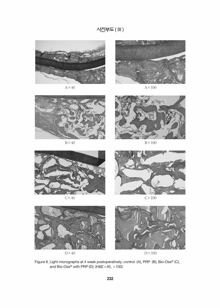
was resorbed during the remodeling process in Bio-Oss with PRP group at 4 weeks(Figure 8 D). Newbone formation was increased in Bio-Oss with PRPcompared those of Bio-Oss alone.
All group showed an increase in bone formationat 4 weeks as compared with 1,2 weeks (Figure6,7,8). There was no difference at newly formedbone when control group was compared with PRPgroup (Figure 6,7,8 A,B) and Bio-Oss group wascompared with Bio-Oss with PRP group (Figure6,7,8 C,D). The individual particles of the bovinebone material were clearly identifiable and theywere found to be surrounded by varying amountsof newly formed bone without being encapsulatedby loose fibrous connective tissue in Bio-Oss and
Bio-Oss with PRP group at all time.
IV. Discussion
In the scope of bone regeneration, the augmenta-tion of bone defects using autogenous bone to bethe gold standard. But, autogenous bone is notalways available in sufficient volume for grafting.The ideal grafting material remains the subject ofcontinued research. Researchers strive continuouslyto improve upon current bone grafting techniquesand provide faster and denser regeneration. Growthfactors are a realistic way to improve both soft tissueand bony wound healing. Platelets contain angio-genic, mitogenic, and vascular growth factors in
222
Table 2. Platelet Counts (platelet count/㎣): 402%increase
Blood PPP PRP
A 167,000 10,000 664,000B 148,000 10,000 425,000C 174,000 45,000 723,000D 112,000 60,000 752,000E 159,000 50,000 597,000F 143,000 28,000 475,000
평 균 150,500 34,000 606,000Blood : whole blood, PPP : platelet-poor plasma, PRP :platelet-rich plasma
Figure 4. Average platelet counts Blood : whole blood, PPP : platelet-poor plasma, PRP :platelet-rich plasma
Table 3. Amount of bone fill determined radi-ographically over the 4 weeks.
1 week 2 week 4 week
control 0.02 ± 0.01 0.04 ± 0.02 0.21 ± 0.05PRP 0.04 ± 0.01 0.13 ± 0.02 0.24 ± 0.04
Bio-Oss 0.65 ± 0.06* 0.94 ± 0.15* 1.16 ± 0.02*Bio-Oss + PRP 0.76 ± 0.17* 1.03 ± 0.08* 1.20 ± 0.06*mean ± SD (gram/square inch) analyzed by a ImageJ 1.31v
softwarestatistical analysis : one-way ANOVA with fisher's Tukey test ;P<0.05* : Significantly different from corresponding control ( P<0.05 )
Figure 5. Amount of bone fill determined radi-ographically over the 4 weeks.

their granules28). TGF-β, PDGF, and VEGF areknown to ve produced by platelets and releasedduring degranulation. TGF-βhas been shown tostimulate proliferation and collagen synthesis byosteoblasts and osteoblast precursors25). It also mayact as a chemotactic agent in recruiting pre-osteoblasts to the site of bone injury. PDGF alsostimulates mitogenesis of osteoblasticprecursors29,30). Although there are numerouscytokines and growth factors that play a role in thespecific temporal sequence that occurs during bonegraft healing, TGF-βand PDGF most likely con-tribute to the early influx of cells and stimulation ofproliferation. The biologic of autogenous andrecombinant growth factors such as TGF-βandPDGF and underlying mechanisms were investigat-ed in numerous studies31,32). These factors belong toa class of biologic mediators with an important stim-ulatory and regulatory function on cellular processessuch as mitogenesis, differentiation, and chemotaxis,as well as angioneogenesis during bone and soft tis-sue healing. Studies on application of single or com-bination growth factors using PDGF or PDGF/IGF-1have been shown to enhance the early cascade oftissue repair processes both in vitro33,34)and invivo35,36,37).
Marx and associates25) attributed the osteoregener-ative effect of PRP to an increased release of PDGFand TGF-βand the resultant enhanced stimulationof angiogenesis, mitogenesis of marrow stem cellsand preosteoblasts, and their activation and differen-tiation into mature osteoblasts. In vitro studiesshowed that the combination of certain cytokinesand growth factors increased osteoblast proliferationand differentiation38). Also Kawase and associates39)
suggested that growth factor might have a potentialfor enhancing collagen synthesis in periodontal liga-ment and osteoblastic cells. It is therefore a reason-able hypothesis that increasing the concentration of
platelets in a bone defects in a bony defect may leadto improved and faster healing. Recent reports havesuggested that more rapid epithelialization, moredense and mature bone with better organized tra-beculae, and greater bone regeneration take placewhen PRP is added to bone autografts andallografts25,40,41). Most of these reports also suggestthat PRP improves the handling properties of thegrafts material with which it is combined, facilitatinggraft placement and stability.
Marx and coworkers25) performed the first andmost compelling study available on the use of PRPin combination with bone grafts. The authorsclaimed that the bone grafts combined with PRPshowed a maturity index more than twice andslightly less than twice the actual maturity at 2 and 4months, respectively.
Anitua40) reported the results of the use of PRP ina series of patients who underwents tooth extractionbecause of root fracture or periodontitis. The sitestreated with PRP demonstrated more mature bone,with better organized trabeculae and greater boneregeneration. Also, the epithelialization wasdescribed based on subjective observations as verygood to excellent. PRP is thought to accelerate softtissue healing by promoting a more rapid revascu-larization and reepithelialization of flaps and cellproliferation. However, because of the limited abili-ty to extrapolate the data to human conditions, fur-ther investigations of PRP in combination withxenograft materials such as anorganic bovine boneis obviously necessary.
Bio-Oss is natural bovine bone that is complete-ly deproteinized to prevent a potential immuneresponse42,43). Electron microscopic evaluationshows that this material has a structural configura-tion similar to human bone. Its compressive strengthand modulus of elasticity are also similar to the val-ues for human bone44).
223

Khalid45) reported that the bovine bone materialsposseessed the best potential of osteoconductivegrafting material, followed by the biograss crystalsand the hydroxyapatite particles respectively. In thisstudy, the bovine bone materials showed significantincrease in newly formed bone when compared tothe no graft as a control. Also, there was morenewly formed bone area in the bovine bone groupthan in the control group at 1, 2, 4 weeks.
In this study, radiographic assessment did notshow any significant difference between Bio-Ossgroup and Bio-Oss with PRP group. It showed asignificant increase in bone density when Bio-Ossand Bio-Oss with PRP were grafted, compared toungrafted control and PRP group, at nearly everyevaluation point. The clinical significance of thesedata is difficult to determine because anyradiopaque bone grafting material will look moredense on a radiograph. However, this is not consis-tent with previous studies, which showed signifi-cantly greater bone density at 1 and 2 months whenevaluating digitized radiographs and computedtomography scans when PRP was added to Bio-Oss
in rabbit cranial defects17). Also, Marx and associ-ates25) showed a 1.62- to 2.16-fold increase in radio-logic bone density at 6 and 2 months, respectively,when PRP was added to autogenous bone, as evalu-ated with panoramic radiographs. The previouslyreport, however, studied human mandibular conti-nuity defects.
In the present study Bio-Oss group and Bio-Osswith PRP group showed a significant increase in
newly formed bone when compared to control andPRP group at all time. Also, adding PRP to Bio-Ossresulted in a significant increase in bone area at alltime periods compared with Bio-Oss alone. Subaand associates46) reported the results of the use ofPRR in extraction site of beagle dog. The combineduse with beta-tircalcium phosphate (Cerasorb ) and
PRP results in more intense bone regeneration,especially in early phase.
Aghloo47) showed a histomorphometric increasein bone formation with the addition of PRP to Bio-Oss in non-critical sized defects in the rabbit crani-um. These results conflict with those of Wiltfangand associates48). They reported that the bone rege-naration in critical-size defects of pig's frontal region.PRP did not add additional benefit when xenogenicbone substitute were used. however, a significanteffect on bone regeneration was found in the auto-genous group initially when PRP in added. Also,Furst49) reported that minipig sinus grafted core sam-ples were evaluated histological after grafting withbovine hydroxyapatite alone and bovine hydroxya-patite with PRP. The authors found no histologicbenefit by the addition of PRP.
These inconsistent results may be due to the dif-ferent experimental systems and different animalemployed in their experiments. Therefore the bioac-tive effects of PRP on bone wound healing and min-eralized tissue formation depend on the localosseous environment where PRP has been applied.Also, the majority of the bony regeneration tookplace within the first month of healing. Significantdifferences might have been seen in early woundhealing between bone alone and bone with PRP,had samples been evaluated before 1 month.
In conclusion, this study has clearly demonstratedthat the addition of PRP to Bio-Oss in the rabbitcranial defect model was shown to be potentiallybeneficial at early bone healing. Also, Deproteinizedbovine bone materials had the good osteoconduc-tive properties and served as a space maintainer suc-cessfully. However, this study was designed to eval-uated non-critical sized cranial defects. Further stud-ies need to evaluate the potential benefits of PRP inhealing critical sized defects.
224

V. Conclusion
Platelets are very important in the wound healingprocess. They quickly arrive at the wound site andbegin the coagulation process. They release multi-ple wonud healing growth factors and cytokines,including platelet-derived growth factor (PDGF),transforming growth factor-β(TGF-β), insulin-likegrowth factor-1, vascular endothelial cell growth fac-tor (VEGF), platelet-derived epidermal growth fac-tor, platelet factor 4. These growth factors arethought to contribute to initial bone regenerationand increased vascularity, vital features of a healingbone graft. This study is to evaluate the effect ofPRP on the early wound healing of rabbit cranialdefects.
Fifteen New Zealand white male rabbits between2.8 and 4 kg were included in this randomized,blinded, prospective study. By means of a trephinebur (external diameter : 8mm), four standardized'through-and-through' bone defect were createdwith copious irrigation. The four cranial defectswere randomly grafted with Bio-Oss , Bio-Ossmixed with PRP, PRP alone, and no graft as a con-trol. The Four defects were covered with nonre-sorbable PTFE membrane (Tefgen , LifecoreBiomedical, Inc., U.S.A.). The wound was closedwith resorbable suture materials. Rabbits were killedusing phentobarbital, 100 mg/kg intravenously at 1,2, and 4 weeks. Radiographs were taken of the rab-bit cranium in its entirety before histologic sectionswere performed. A aluminum step-wedge was usedin each radiograph for camparison. Specimens weretreated with hydrochloric acid decalcifying solution(Fisher Scientific, Tustin, CA) and sectioned bybisecting the 8-mm diameter defects. The histologicspecimens were prepared in the usual fashion withH&E staining at 6 ㎛ in thickness. Also, six speci-mens were sampled analysed for platelet count. The
following results were obtained through the in vivostudy.
1. In platelet count test, PRP group is 4 timeshigher platelet concentration than normal.
2. In radiographic evaluation, Bio-Oss groupand Bio-Oss with PRP group showed a signifi-cant increase in radiographic bone densitywhen compared to the control and PRP groupat all 3 time points (p<0.01). However, signifi-cant increase was not seen at all time whencontrol group was compared with PRP group(P>0.05). There was also no significant differ-ence between Bio-Oss and Bio-Oss withPRP group at 1, 2, and 4weeks (P>0.05).
3. In histologic evaluation, all grafting materialsshowed an increase in bone formation over atime. Bio-Oss and Bio-Oss with PRP groupshowed a increase in newly formed bonewhen compared to control and PRP group at 1,2 and 4 weeks. Also, a increase in new boneformation was seen when Bio-Oss with PRPwas compared with Bio-Oss alone.
The results has suggested that the addition of PRPto Bio-Oss in the rabbit cranial defects model wasshown to be potentially beneficial at early bonehealing. PRP might positively influence the earlybone wound healing.
VI. References
1. Shanaman, R. Filstein, M. R. Danesh-Meyer, M.J.. "Localized ridge augmentation using GBRand platelet-rich plasma: case reports." Int JPeriodontics Restorative Dent 2001;21:345-355.
2.Buser, D. Bragger, U. Lang, N. P. et. al.."Regeneration and enlargement of jaw boneusing guided tissue regeneration." Clin Oral
225

Implants Res 1990;1:22-32.3.Buser, D. Dula, K. Belser, U. et, al.. "Localized
ridge augmentation using guided bone regener-ation. I. Surgical procedure in the maxilla." Int JPeriodontics Restorative Dent 1993;13:137-179.
4.Becker, W. Dula, K. Belser, U. et. al.. "Localizedridge augmentation using absorbable pins ande-PTFE barrier membranes: A new surgical tech-nique. Case reports." Int J PeriodonticsRestorative Dent 1994;14:49-61.
5.Hammerle, C. H. Karring, T.. "Guided boneregeneration at oral implant sites."Periodontology 2000 1998;17:151-175.
6. Sanchez, A. R. Sheridan, P. J. Kupp, L. I.. "Isplatelet-rich plasma the perfect enhancementfactor? A current review." Int J Oral MaxillofacImp2003;18:93-103.
7.Nkenke, E. Schultze-Mosgau, S. Radespiel-Troger, M. et. al.. "Morbidity of harvesting ofchin grafts: a prospective study." Clin OralImplants Res 2001;12:495-502.
8.Yunger, E. M. Chapman, M. W.. "Morbidity atbone graft donor sites." J Orthop Trauma1989;3:192-195.
9. Schwartz, Z. Somers, A. Mellonig, J. T. et. al.."Ability of comercial demineralized freeze-driedbone allograft to induce new bone formation isdependent on donor age but not gender." JPeriodontol 1998;69:470-478.
10. Carmagnola, D. Berglundh, T. Araujo, M. et. al.."Bone healing around implants paced in a jawdefect augmented with Bio-Oss : An experi-mental study in dogs." J Cin periodontol2000;27:799-805.
11. Carmagnola, D. Berglundh, T. Lindhe, J.. "Theeffect of fibrin glue on the integration of Bio-Oss
with bone tissue: An experimental study inlabrador dogs." J Clin Periodontol 2002;29:377-381.
12. Zitzmann, N. U. Scharer, P. Marinello, C. P. et,al... "Alveolar ridge augmentation with Bio-Oss
: A histologic study in humans." Int JPeriodontics Restorative Dent 2001;21:288-295.
13. Tal, H.. "Autogenous masticatory mucosal graftsin extraction socket seal procedures: A compari-son between sockets grafted with demineralizedfreeze-dried bone and deproteinized bovinebone material." Clin Oral Implants Res1999;10:289-296.
14. Houser, B. E. Mellonig, J. T. Brunsvold, M. A.et. al.. "Clinical evaluation of anorganic bovinebone xenograft with a bioabsorbable collagenbarrier in the treatment of molar furcationdefects." Int J Periodontics Restorative Dent2001;21:161-169.
15. Hallman, M. Cederlund, A. Lindskog, S. et. al.."A clinical histologic study of bovine hydroxyap-atite in combination with autogenous bone andfibrin glue for maxillary sinus floor augmenta-tion. Results after 6 to 8 months of healing." ClinOral Implants Res 2001;12:135-143.
16. Yildirim, M. Spiekermann, H. Biesterfeld, S.."Maxillary sinus augmentation using xenogenicbone substitute material Bio-Oss in combina-tion with venous blood: A histlogic and histo-morphometric study in humans." Clin OralImplants Res 2000;11:217-229.
17. Kim, E. S. Park, E. J. Choung, P. H.. "Plateletconcentration and its effect on bone formationin calvarial defects: An experimental study inrabbits." J Prosthet Dent 2001;86:428-433.
18. Maiorana, C. Santoro, F. Rabagliati, M. et. al.."Evaluation of the use of iliac cancellous boneand anorganic bovine bone in the reconstructionof the atrophic maxilla with titanium mesh: Aclinical and histologic investigation." Int J OralMaxillofac Implants 2001;16:427-432.
19. Mcallister, B. S. Margolin, M. D. Cogan, A. G..
226

"Residual lateral wall defects following sinusgrafting with recombinant human osteogenicprotein-1 or Bio-Oss in the 7 chimpanzee." IntJ Periodontics Restorative Dent 1998;18:227-239.
20. Kim, S. G. Kim, H. K. Im, S. C.. "Combinedimplantation of particulate dentine, plaster ofParis, and a bone xenograft (Bio-Oss ) for boneregeneration in rats." J Craniomaxillofac Surg2001;29:282-288.
21. Gibble, J. Ness, P.. "Fibrin glue: The perfectoperative sealant?" Transfusion 1990;30:741-747.
22. Hood, A. G. Hill, A. G. Reeder, G. D.."Perioperative autologous sequestraction. III: Anew physiologic glue with wound healing prop-erties." Proc Am Acd Cardiovasc Perfusion1993;14::126-130.
23. Linder, B. L. Chernoff, A. Kaplan, K. L. et. al.."Release of PDGF from human platelets byarachidonic acid." Proc Natl Acad Sci USA1979;76:4107-4111.
24. Mohle, R. Green, D. Moore, M.A. et. al.."Constitutive production and thrombin-inducedrelease of VEGF by human megakaryocytes andplatelets." Proc Natl Acad Sci USA 1997;94:663-668.
25. Marx, R. E. Carlson, E. R. Eichsraedt, R. M. et.al.. "Platelet-rich plasma: Growth factorenhancement for bone grafts." Oral Surg OralMed Oral Pathol Oral Radiol Endod 1998;85:638-646.
26. Pierce, G. F. Mustoe, T. A. Altrock, B.W. et. al.."Role of platelet-derived growth factor in woundhealing." J Cell Biochem 1991;45:319-326.
27. Antoniades, H. N.. "Human platelet derivedgrowth factor (PDGF) : Purification of PDGF-Iand PDGF-II and separation of their reducedsub-units." Proc Natl Acad Sci USA 1981;78:7314-7317.
28. Maloney, J. P. Silliman, C. C. Ambruso, D. R. et.
al.. "In vitro release of vascular endothelialgrowth factor during platelet aggregation." Am JPhysiol 1998;2755:1054-1061.
29. Sandy, J. Davies, M. Prime, S. et. al.. "Signalpathways that transduce growth factor-stimulat-ed mitogenesis in bone cells." Bone 1998;23:17-26.
30. Horner, A. Bord, S. Kemp, P. et. al.."Distribution of platelet-derived growthfactor(PDGF): A chain mRNA, protein, andPDGF-alpha receptor in rapidly forming humanbone." Bone 1996;19:353-62.
31. Lind, M.. "Growth factor stimulation of bonehealing. Effects onosteoblasts, Osteomies, andimplant fixation." Acta Orthop Scand Suppl1998;328:2-37.
32. Solheim, E.. "Growth factors in bone." IntOrthops 1998;22:410-416.
33. Canalis, E,. McCarthy, T. L. Centrella, M.."Effects of platelet - derived growth factor onbone formation in vitro." J Cell Physiol1989;140:530-537.
34. Stephan, E.B. Renjen, R. Lynch, S.E. et. al.."Platelet-derived growth factor enhancement ofa mineral-collagen bone substitute." JPeriodontol 2000;71:1887-1892.
35. Lynch, S. E. Ruiz de Castilla, G. Williams, R.C.et. al.. "The effects of short-term application of acombination of platelet-derived and insulln-likegrowth factors on periodontal wound healing." JPeriodontol 1991;62:458-467.
36. Lynch, S. E. Buser, D. Hernandez, R. A. et al.."Effects of PDGF/IGF-1 combination on boneregeneration around titanium dental implants.Results of a pilot study on beagle dogs." JPeriodontol 1991;62:710-716.
37. Stefani, C. M. Machado, M. A. Sallum, E. A. et.al.. "Platelet-derived growth factor/insullin-likegrowth factor-1 around implants placed into
227
extraction sockets: A histometric study in dogs."Implant Dent 2000;9:126-132.
38. Lind, M. Overgaard, S. Nguyen, T. et. al.."Transforming growth factor-beta stimulatesbone on growth Hydroxyapatite-coated implantsstudied in dogs." Acta Orthopaedica Scandinavia1996;67:611-616.
39. Kawase, T. Okuda, K. Yoshie, H.. "Platelet-richplasma derived fibrin clot formation stimulatescollagen synthesis in periodontal ligament andosteoblastic cells in vitro" J Periodontol2003;74:858-864.
40. Anitua, E.. "Plasma rich in growth factors:Preliminary results of use in the preparation offuture sites for implats." Int J Oral MaxillofacImplants 1999;14:529-525.
41. Kassolis, J. D. Rosen, P.S. Reynilds, M. A.."Alveolar ridge and sinus augmentation utilizingplatelet-dried bone allograft: Case series." JPeriodontol 2000;71:1654-1661.
42. Hislop, W.S. Finlay, P. M. Moos, K. F.. "A pre-liminary study into the uses of anorganic bone inoral and maxillofacial surgery" Br J OralMaxillofac Surg 1993;31:149-153.
43. Indovina, A. Block, M. S.. "Comparison of 3bone substitutes in canine extraction sites." JOral Maxillofac Surg 2002;60:53-58.
44. Thaller, S. R. Hoyt, J. Borieson, K..
"Reconstruction of calvarial defects with anor-ganic bovine bone mineral (Bio-Oss ) in a rab-bit model." J Craniofac Surg 1993;4:79-84.
45. Khalid, A. Ruhaimi, A. L.. "Bone graft substi-tutes: A comparative qualitative histologic reviewof current osteoconduction grafting materials."Int J Oral Maxillofac Implants 2001;16:105-114.
46. Suba, Z. Takacs, D. Kovacs, K. et. al.. "Alveolarbone regeneration stimulated by a combinationof platelet-rich plasma and Cerasorb graft inBeagle dogs. Histological and histomorphomet-ric studies." Fogorv Sz 2004;97:143-149.
47. Aghaloo, T. L. Moy, P. K. Freymiller, E. G.."Evaluation of platelet-rich plasma in combina-tion with anorganic bovine bone in the rabbitcranium: A pilot study." Int J Oral MaxillofacImplants 2004;19:59-65.
48. Wiltfang, J. Kloss, F. R. Kessler, P. et. al.."Effects of platelet-rich plasma on bone healingin combination with autogenous bone and bonesubstitutes in critical-size defects. An animalexperiment." Clin Oral Implants Res2004;15:187-193.
49. Furst, G. Gruber, R. Tangl, S. et. al.. "Sinusgrafting with autogenous platelet-rich plasmaand bovine hydroxyapatite. A histomorphomet-ric study in minipigs." Clin Oral Implants Res2003;14:500-508.
228

사진부도설명
Figure 6. (A) A light micrograph of control at 1 week postoperatively: The perforated areas were filled withloose fibrous tissue. There was osteoblastic and osteoid layers from cortical bone margin.
(B) A light micrograph of PRP group at 1 week postoperatively : The perforated areas were filledwith dense fibrous tissue. There was osteoblastic and osteoid layers from cortical bone margin.
(C) A light micrograph of Bio-Oss group at 1 week postoperatively : There were osteoblastic andosteoid layers at the border of the defect and around deproteinized bovine bone material parti-cles.
(D) A light micrograph of Bio-Oss with PRP group at 1 week postoperatively: There was formationof new bone from cortical bone margin. Osteoprogenitor cells and preosteoblasts were seenon the periphery of the graft materials.
Figure 7. (A) A light micrograph of control at 2 week postoperatively : The perforated areas were filled withdense fibrous tissue. There was formation of new bone from cortical bone margin.
(B) A light micrograph of PRP group at 2 week postoperatively : The perforated areas were filledwith dense fibrous tissue. There was formation of new bone from cortical bone margin.
(C) A light micrograph of Bio-Oss group at 2 week postoperatively : There was formation of newbone from cortical bone margin. Osteoprogenitor cells and preosteoblasts were seen on theperiphery of the graft materials.
(D) A light micrograph of Bio-Oss with PRP group at 2 week postoperatively : There was forma-tion of new bone from cortical bone margin. The graft materials have been incorporated intothe newly formed bone matrix.
Figure 8. (A) A light micrograph of control at 4 week postoperatively : The perforated areas were filled withdense fibrous tissue. There was formation of new bone from cortical bone margin.
(B) A light micrograph of PRP group at 4 week postoperatively : The perforated areas were filledwith dense fibrous tissue. There was formation of new bone from cortical bone margin.
(C) A light micrograph of Bio-Oss group at 4 week postoperatively : There was formation of newbone from cortical bone margin. The graft materials have been incorporated into the newlyformed bone matrix.
(D) A light micrograph of Bio-Oss with PRP group at 4 week postoperatively : There was forma-tion of new bone from cortical bone margin. The graft materials have been incorporated inmature new bone and was resorbed during the remodeling process.
229

230
사진부도 ( I )
A×40 A×100
B×40 B×100
C×40 C×100
D×40
Figure 6. Light micrographs at 1 week postoperatively; control (A), PRP (B), Bio-Oss (C),and Bio-Oss with PRP (D). (H&E×40, ×100)
D×100

231
사진부도 ( II )
A×40 A×100
B×40 B×100
C×40 C×100
D×40
Figure 7. Light micrographs at 2 week postoperatively; control (A), PRP (B), Bio-Oss (C),and Bio-Oss with PRP (D). (H&E×40, ×100)
D×100
232
사진부도 ( III )
A×40 A×100
B×40 B×100
C×40 C×100
D×40
Figure 8. Light micrographs at 4 week postoperatively; control (A), PRP (B), Bio-Oss (C),and Bio-Oss with PRP (D). (H&E×40, ×100)
D×100

-국문초록-
혈소판농축혈장과혼합된이종골이식재(Bio-Oss )가가토두개골결손부초기치유에미치는 향
임동웅1, 장현선1,4, 박주철2,4, 김흥중3,4, 이종우1, 김종관5,6, 김병옥1,4
조선대학교 치과대학 치주과학교실1
조선대학교 치과대학 구강조직학교실2
조선대학교 치과대학 구강해부학교실3
조선대학교 치과대학 구강생물학연구소4
연세대학교 치과대학 치주과학교실5
연세대학교 치과대학 치주조직 재생연구소6
혈소판 농축 혈장은 구강과 안면부 재건수술에 새로이 사용되는 유용한 첨가물이다. 혈소판은 상처 치유과정에서 매우 중요하며, 혈소판은 상처부위에 빠르게 도달하여 응고를 형성한다. 그리고 다양한 성장인자를 분비한다. 이러한 성장인자는 골의 형성과 혈관의 증가, 골 이식재의 치유에 관여하는 것으로 생각된다. 본 연구의 목적은 실험 동물을 통하여 혈소판 농축 혈장에 함유된 혈소판의 정량화를 통한 성장인자 함유량을 추정하고, 방사선학적, 조직학적 평가를 통해 혈소판 농축 혈장이 초기의 골형성에 미치는 향에 대한 평가를 하는데있다.
15마리의 가토 두개골에 6 mm trephine bur(외경 8 mm)를 이용하여 경뇌막의 손상을 주지 않도록 하면서 4개의 결손부를 형성하 다. 각각의 두개골 결손부는 Bio-Oss 만 이식한 군, PRP만 이식한 군, PRP와 Bio-Oss를 혼합하여 이식한 군, 그리고 아무것도 이식하지 않은 군을 대조군으로 설정하 다. 각각의 재료를 이식한 후비흡수성 차폐막(Tefgen )을 위치시키고 흡수성 봉합사로 일차봉합을 시행하 다. 각 군 당 술 후 1, 2, 4주의치유기간을 설정하 다. 동물을 희생시키고 두개골을 절제하 다. 먼저 방사선학적인 골 도 측정을 시행하고, 조직학적 평가를 위해 통법에 따라 조직 표본을 제작한 후 광학현미경으로 관찰하 다. 또한 가토 귀 변연정맥에서 채취한 10 ㎖의 혈액을 원심분리하여 혈소판 함유량을 평가하여 다음과 같은 결과를 얻었다.
1. 혈소판 농축 혈장은 일반 혈액에 비해 약 4.02배 많은 수의 혈소판이 함유되어 있었다. 2. 방사선적인 평가에서 1, 2, 4주 사이에 대조군과 비교하여 Bio-Oss 에 PRP를 이식한 군에서 골의 도는
큰 차이를 보이고 있다(p<0.01). 하지만, 동일한 시기에 PRP만 이식한 군과 대조군의 차이는 발견할 수 없었으며(p>0.05), Bio-Oss 만 이식한 군과 Bio-Oss 에 PRP를 이식한 군의 차이 또한 발견할 수 없었다(p>0.05).
3. 조직학적 평가에서 모든 이식재는 시간이 경과할수록 골 형성이 증가함을 알 수 있었다. 대조군에 비해PRP만 이식한 군에서 더 두꺼운 섬유성 결합을 보이고 있다. 대조군과 PRP만 이식한 군과 비교해 Bio-Oss와 Bio-Oss 에 PRP를 혼합 이식한 군에서 골의 형성이 더 진행됨을 알 수 있었다. Bio-Oss 에 PRP를 혼
233
합 이식한 군이 Bio-Oss 만 이식한 군에서보다 더 많은 신생골 형성을 관찰할 수 있다.
이상의 결과에서 가토의 두개골 결손부에 Bio-Oss 에 PRP를 혼합 이식하 을 경우 결손부의 초기 골 형성을촉진 할 수 있음을 시사하 다.
주요어 : 혈소판 농축 혈장, Bio-Oss ,PRP
234