57 year-old male with hypokalemia
49
By Anoopa Koshy, M.D. Endorama 5/10/2012
Transcript of 57 year-old male with hypokalemia

By Anoopa Koshy, M.D.Endorama5/10/2012
HPI
57‐year‐old Bosnian male with a 20 year hx of lung mass
(for which he has previously refused worked up) presents to the ER on 3/13 with generalized weakness and found to have hypokalemia (K= 2.7).
3 weeks ago, he presented to an OSH with SOB, dyspnea
on exertion, and was treated for “a throat infection”‐K was noted to be as low as 1.7.‐discharged home on K supplements and antibiotics
Re‐presented to the OSH 5 days prior to admission with
DOE/SOB after walking only 10 feet, polyuria, polydipsia, generalized weakness,and a 15‐lb weight loss in 2 weeks.
‐left AMA before work‐up could be completed

HPI
Progressive, generalized weakness for 3‐4 months.
~30 lbs of unintentional weight loss over that same time
frame, with associated poor appetite
Reports poor sleep and poor energy
Has recently noticed polyuria. Denies polydipsia
Positive vision change in R eye (describes as blurriness,
without field defects).
No HA or numbness.
No fevers, chills, or sweats
No CP, cough, pleuritic chest pain, hemoptysis, or wheezes
No abdominal pain, nausea, vomiting, diarrhea,
constipation, or blood in his stools
No heat/cold intolerance
He has not noticed any skin changes

HPI
Given his reported history of an abnormal CXR and
history of lung mass, he was sent for a CXR
Show image

Chest X‐ray
IMPRESSION: Large mass along the right inferior mediastinal border, likely arising from
the posterior mediastinum. Small right pleural effusion.

HPI continued…
Patient admitted to General Medicine for further
evaluation of his hypokalemia and his intrathoracic mass.
Despite aggressive potassium repletion (280 mEq in
one 24 hour period), his potassium remained < 3
CT chest/abdomen/pelvis ordered for further
evaluation.

CT chest/abdomen/pelvis
1.) There is a 12.0 x 8.8 cm right infrahilar mass with mass effect on the bronchus intermedius and is adjacent to the rightpulmonary artery. There is post obstructive atelectasis .
2.) There is a 2.8 x 2.1 cm left adrenal nodule, which mayrepresent adrenal gland thickening. The right adrenal glandappears uniformly hyperplastic.
CT chest/abdomen/pelvis: Official Read

HPI continued…
Pulmonary, Oncology, and Thoracic Surgery services
consulted for further evaluation of lung mass.
Random cortisol value from 3/15 returned at 87.3
mcg/d/L at 6:29 AM
Endocrinology consulted for further evaluation

Past Medical History, MedsPMHXLung mass x 20 yearsHypertension Depression (recently
diagnosed after death of his wife)
Meds (at discharge from
OSH)
Potassium supplement 40
MEQ BIDHCTZ/triamtereneLevaquinClindamycin Megestrol 800 mg daily Sertraline 50 mg po daily
Allergies: NDKA

SOCIAL HXImmigrated from
Bosnia in 1998 and worked briefly in retail
Currently on disability due to lung mass
Wife died recently in February, 2012
Denies smoking, EtOH, illicit drugs
Physical ExamVitals: Temp 37.0C BP 154/90 HR 84 RR 12 Sat 93% RAWeight‐160 lbs, Height 6’
1’’
BMI‐21
GEN: Thin, male in NADHEENT: Sclerae anicteric. Oropharynx with poor dentition, otherwise clearNECK: Supple. CV: RRR. No m/r/g. No JVD. No LE edema. Extremities warm, well perfused. PULM: Breathing unlabored. Decreased BS with dullness to percussion in the R
posterior lung field
ABD: Flat. +BS. Soft. Non‐tender. No hepatosplenomegallyEXT: Muscle atrophy in bilateral arms and legs. No edema. NEURO: A+O X3. CN 2‐12 grossly intact. +proximal muscle weakness. Distal
sensation intact. Normal finger to nose testing bilaterally. Gait normal
bilaterally
SKIN: no rash, no striae, normal pigmentationPSYCH: Sad affect. Tearful


3/13/2012 (labs on presentation)
143 | 101 | 8 / 1352.7
| 28 | 0.6\
3/14/2012Urine electrolytes: Na 12 mEq/L, K 33
mEq/L , Cl <20 mEq/LAldosterone‐
<4 ng/dlRenin‐
7.5 ng/ml/hrNormal (Na deplete): 2.9‐10.8Normal (Na replete): 0.6‐3
3/16/2012 at 6:29 AMACTH‐
311 (<50 pg/dl )Cortisol‐
97.3 mcg/dlTSH‐
0.62 (0.3‐4 mcU/ml)
AST‐30ALT‐53Alk phos‐69Tbili‐1.3Tprotein‐5.3Albumin‐3.5
Calcium‐
7.5 (8.4‐10.2 mg/dl)Albumin 3.5 (3.5‐5 g/dl)Corrected calcium‐
7.9 mg/dlMagnesium 2 (1.6‐2.5 mg/dl)Phosphorus‐
1.9 (2.5‐4.4 mg/dl)PTH‐
86 (15‐75 pg/ml)1, 25 OH vit D‐
71 (18‐64 pg/ml)25 OH vit D‐
8 (10‐52 ng/ml)
13.9 \ 14.8 / 124/42.3 \

What are your thoughts and what tests would you order next?


Endo Recs
Started Spironolactone at 100 mg po BID, eventually
titrated to 200 mg po BID
Started Amiloride 5 mg po daily, eventually titrated to
10 mg po daily
Aggressive IV and po K replacement with K checks
q6h
Started ergocalciferol 4,000 units daily and calcium
carbonate 1,250 mg QID. Recommended DEXA scan.
Started lantus 10 units SC daily along with a low dose
novolog sliding scale

Ilias I, Torpy DJ, Pacak K, Mullen N,
Wesley RA, Nieman LK 2005
Cushing’s syndrome due to ectopic corticotropin secretion: twenty years’
experience at the National Institutes
of Health. J Clin Endocrinol Metab 90:4955–
4962.

Hypertension and Hypokalemia in Cushing’s Syndrome
Increased peripheral vascular sensitivity to adrenergic agonists
Increased hepatic production of renin substrate
(angiotensinogen)
Activation of renal tubular type 1 (mineralocorticoid) receptors
by cortisol (due to severe hypercortisolism, which is usually due
to ectopic ACTH secretion)
High serum cortisol concentrations overwhelm the ability of the
kidneys to convert cortisol to cortisone, resulting in activation
of mineralocorticoid receptors.
Hypokalemia may also result from adrenal hypersecretion of
mineralocorticoids such as deoxycorticosterone and
corticosterone.
Bone loss and Glucose intolerance
Bone loss‐
Osteoporosis common in patients with Cushing's
syndrome‐caused by decreased intestinal calcium absorption ‐decreased bone formation‐increased bone resorption‐
decreased renal calcium reabsorption
Glucose intolerance‐
common in Cushing's syndrome‐primarily due to stimulation of gluconeogenesis by
cortisol and peripheral insulin resistance caused by obesity‐
direct suppression of insulin release also may contribute

Neuropsychological changes and cognition
Emotional lability
Agitated depression
Irritability
Anxiety
Panic attacks
Mild paranoia
Insomnia
Depression (occurs in 2/3 of patients with Cushing's
syndrome)
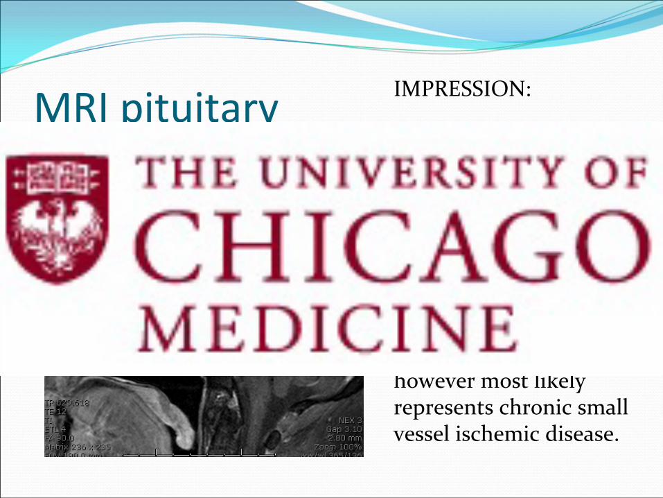
MRI pituitary IMPRESSION:
1. Normal appearance of the pituitary gland and
hypothalamic region.
2. Multifocal abnormal signal intensity within the
subcortical and periventricular white
matter, nonspecific however most likely represents chronic small
vessel ischemic disease.

What would you do next?

CRH Stimulation Test
Time
(min)
‐15 0 15 30 60 90 120 180
ACTH 373 420 445 435 386 273 401 431
Cortisol 114 113 112 110 113 115 113

Summary of findings
The urinary cortisol in excess of >11,000 ug/24 hr is
extraordinary, and is diagnostic of Cushing’s syndrome.
The diagnosis of ectopic ACTH Syndrome is
confirmatory with the results of the CRF stimulation test (elevated cortisol and ACTH unresponsive to
CRH stimulation).
The high 5‐HIAA and dopamine levels are consistent
with an active neuroendocrine tumor.


Account for 25% of neuroendocrine tumors, but only 1% of lung cancers
Arise from enterochromaffin cells (a bronchial mucosal cell that
is part of
the Diffuse Neuroendocrine System, or DNES)
Presentation: usually present with pulmonary symptoms (obstructive
pneumonia, cough, wheeze, hemoptysis)
Carcinoid syndrome is rare
25‐30% of patients are asymptomatic (found incidentally or post‐mortem)
Imaging
Characteristic CT imaging findings: well defined, centrally located tumors
involving the airway with calcification
Roles for other imaging studies (PET, MRI, somatostatin receptor
scintigraphy) are not well defined.
Pathology
Can be broken down into typical and atypical carcinoids based off
histologic appearance
Pulmonary carcinoid tumors

Staging
Follows TNM guidelines used for other NSCLCs
Can spread to regional nodes, bone, adrenal glands, liver, and brain
Atypical carcinoids at higher risk of metastasis than typical carcinoids
(one series shows 25% of pts developing distant disease)
Prognosis
Variably reported, ranging from 5 year surival of 44‐97%
Dependent on histologic subtype and stage of disease at presentation
Treatment
Surgery is treatment of choice and only curative option
For patients with unresectable or metastatic disease, there is currently
limited data regarding chemotherapy or XRT
Regimens previously reported: interferon alpha, etoposide based
regimens, streptozocin, 5‐FU, octreotide (if carcinoid syndrome)
Under study: VEGF inhibitors, mTOR inhibitors, tyrosine kinase
inhibitors
Pulmonary carcinoid tumors

Clinical Course continued…
Thoracic Surgery requests whole body PET scan prior
to surgical removal of lung mass

PET Scan• Markedly hypermetabolic
large R infrahilar pulmonary
mass (SUV max 15.2),
compatible with tumor
activity.
• Both enlarged adrenal
glands are hypermetabolic
(SUV max 14.1). This is
suspicious for tumor
involvement, or
alternatively could represent
benign uptake from marked
bilateral adrenal hyperplasia
• No FDG avid tumor
elsewhere

Adrenal mets versus hyperplasia?
Thoracic surgery reluctant to operate given PET scan
results.
Is the intense uptake in the adrenal glands evidence
of metastatic disease?
Intense bilateral uptake would be unusual for mets
The CT scan of the adrenals suggest bilateral
hyperplasia (as one would expect in ectopic ACTH syndrome)
2 papers indicate that SUVmax in adrenal hyperplasia
can be 3.5‐5.5 (metastatic disease is usually >3.1 also):
We recommended surgical removal of lung mass

Surgery
He underwent a right thoracotomy and
pneumonectomy on 3/27 by Dr. Ferguson.
No complications with surgery
Extubated without difficulties
After surgery, he was monitored closely for adrenal
insufficiency with q2h cortisol checks for 24 hours
Did not develop overt clinical or biochemical signs of
adrenal insufficiency.

ACTH and Cortisol Trend After Surgery
Surgery
Hydrocortisone 20 mg/10 mg started

He was discharged home on hydrocortisone 20 mg
PO q am, 10 mg PO q afternoon until further follow‐ up.
His potassium remained normal without
supplementation.
His blood sugars normalized and his insulin was
stopped.
Clinical Course

Post‐op 24‐hr urine free cortisol
Note: 24 hour urine cortisol down from 11,640 mcg prior to surgery (nl < 35)

Post‐op 5‐HIAA
Note: 24 hour urine 5‐hydroxyindoleacetic Acid down from 18 mg prior to surgery (nl < 8)
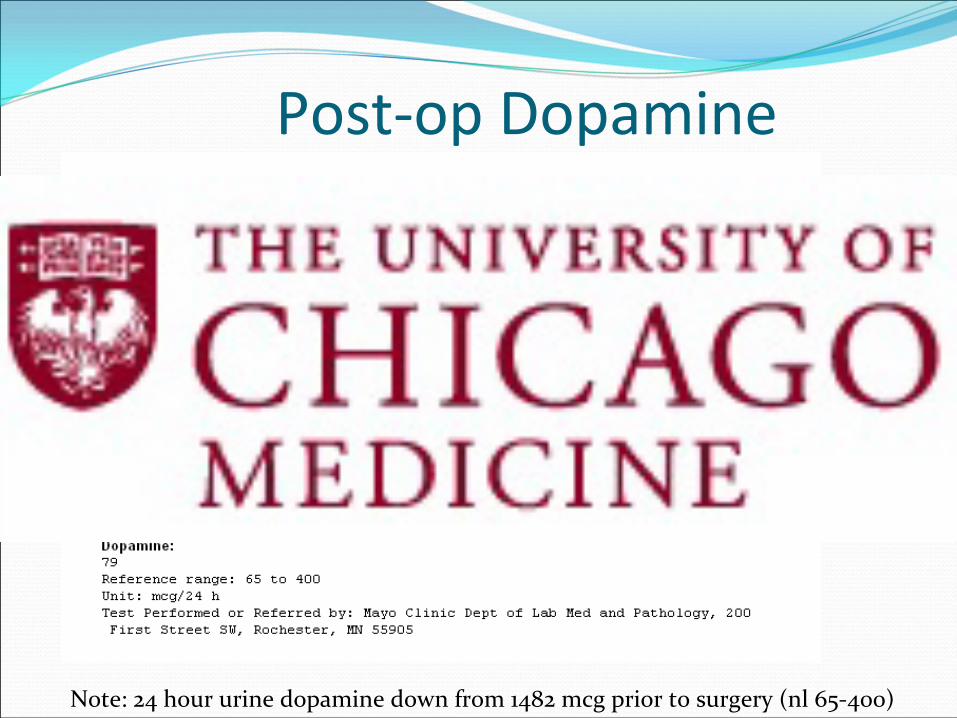
Post‐op Dopamine
Note: 24 hour urine dopamine down from 1482 mcg prior to surgery
(nl 65‐400)

Pathology
Invite Dr. Li to present pathology findings.

Additional Questions
Why was the staining for ACTH negative?
Could there have been ectopic CRH production?
A Case ReportA 56 year old woman presented with clinical and laboratory features consistent with
ACTH‐dependent CS.
Pituitary imaging was normal and cortisol did not suppress with a high dose
dexamethasone test, consistent with a diagnosis of ectopic ACTH.
CT imaging did not reveal any discrete lung lesions but there were mediastinal and
abdominal lymphadenopathy and multiple liver lesions suspicious for metastatic
disease.
Laboratory testing was positive for elevated serum carcinoembryonic antigen and
chromogranin A.
Serum markers of carcinoid, medullary thyroid carcinoma, and pheochromocytoma
were in the normal range. Because the primary tumor could not be
identified by
imaging, biopsy of the presumed metastatic liver lesions was performed.
Immunohistochemistry was consistent with a neuroendocrine tumor, specifically small
cell carcinoma.
Immunostaining for ACTH was negative but was strongly positive for CRH and
laboratory testing revealed a plasma CRH of 10 pg/ml (normal 0 to 10 pg/ml) which
should have been suppressed in the presence of high cortisol.
CONCLUSIONS:
This case illustrates the importance of considering the ectopic production of CRH in the
differential diagnosis for presentations of ACTH‐dependent Cushing's Syndrome.
Shahani S, Nudelman RJ, Nalini R, Kim HS, Samson SL. Ectopic corticotropin‐releasing hormone (CRH)
syndrome from metastatic small cell carcinoma: a case report and review of the literature. Diagn Pathol.
2010 Aug 31; 5:56.

Shahani
S, Nudelman
RJ, Nalini
R, Kim HS, Samson SL. Ectopic corticotropin‐releasing hormone (CRH) syndrome from metastatic small cell carcinoma: a case report and review of the literature. Diagn
Pathol. 2010 Aug 31; 5:56.

Outpatient Follow‐up
Patient failed to show‐up for his Endocrinology
appointment after discharge.
Had f/u with Thoracic Surgery on 4/20
Is still taking HC 20/10 mg
Random cortisol at 16:15 returned at 21.7 mcg/dl and
ACTH of 29.5 pg/ml
Chromogranin A level returned at 170 (nl <225 ng/ml,
previously 2560 ng/ml)
Recommended follow‐up with Dr. Salgia in Oncology,
has scheduled appointment on 5/11.

Take Home Points1. Cushing’s syndrome should be on the differential for
a patient presenting with refractory hypokalemia.
2. The CRH stim test can be used to differentiate pituitary from ectopic ACTH secretion in Cushing’s
syndrome.
3. Ectopic CRH production can be a cause of Cushing’s syndrome, although rare.

References
1. Orth DN. Cushing's syndrome. N Engl J Med. 1995 Mar 23;332(12):791‐803.
2. Tani Y, Sugiyama T, Hirooka S, Izumiyama H, Hirata Y. Ectopic ACTH syndrome caused by
bronchial carcinoid tumor indistinguishable from Cushing's disease. Endocr J. 2010;57(8):679‐86.
Epub 2010 Jun 15.
3. Isidori AM, Lenzi A. Ectopic ACTH syndrome. Arq Bras Endocrinol Metabol. 2007 Nov;51(8):1217‐
25.
4. Shahani S, Nudelman RJ, Nalini R, Kim HS, Samson SL. Ectopic corticotropin‐releasing hormone
(CRH) syndrome from metastatic small cell carcinoma: a case report and review of the literature.
Diagn Pathol. 2010 Aug 31; 5:56.
5. Isidori AM, Kaltsas GA, Pozza C, Frajese V, Newell‐Price J, Reznek RH, Jenkins PJ, Monson JP,
Grossman AB, Bessner GM. The ectopic adrenocorticotropin syndrome: clinical features, diagnosis,
management, and long‐term follow‐up. J. Clin Endocrinol Metab. 2006 Feb;91(2):371‐7. Epub 2005
Nov 22.
6. Reimondo G, Paccotti P, Minetto M, Termine A, Stura G, Bergui
M, Angeli A, Terzolo M 2003 The
corticotrophin‐releasing hormone test is the most reliable noninvasive method to differentiate
pituitary from ectopic ACTH secretion in Cushing’s syndrome. Clin Endocrinol (Oxf) 58:718–724.
7. Ilias I, Torpy DJ, Pacak K, Mullen N, Wesley RA, Nieman LK 2005 Cushing’s syndrome due to
ectopic corticotropin secretion: twenty years’
experience at the National Institutes of Health. J Clin
Endocrinol Metab 90:4955–4962.

References8.
Susan G. Coe,
Winston W. Tan, and Thomas P. Fox. Cushing’s Syndrome Due to Ectopic
Adrenocorticotropic Hormone Production Secondary to Hepatic Carcinoid: Diagnosis, Treatment,
and Improved Quality of Life.
J Gen Intern Med. 2008 June; 23(6): 875–878.
9. Alencar, A et al. 18F‐FDG‐PET/CT Imaging of ACTH‐Independent Macronodular Adrenocortical
Hyperplasia (AIMAH) Demonstrating Increased 18F‐FDG Uptake J Clin Endo Metab 2011, 96:3300.
10. Lila, A et al. Localization of Remnant and Ectopic Adrenal Tissues with Cosyntropin‐Stimulated
18F‐FDG‐PET/CT in a Patient with Nelson Syndrome with Persistent Hypercortisolism
J Clin Endo
Metab 2010, 95:5172.