5. necrosis mdzah- sp sinhasan
15
-
Upload
kciapm -
Category
Health & Medicine
-
view
47 -
download
3
Transcript of 5. necrosis mdzah- sp sinhasan


Necrosis:
“Necrosis refers to a spectrum of morphologic changes
that follow cell death in living tissue, largely resulting
from progressive degradative action of enzymes on the
lethally injured cells”.

Necrosis Apoptosis
1. Cell size: enlarged.
2. Nucleus: Pyknosis-Karyorrhexis- Karyolysis.
3. Plasma membrane: disrupted
4. Cellular contents: leak out, enzymatic digestion.
5. Inflammation +++
6. Role: always pathologic.
7. Agarose gel electrophoresis: diffuse smearing pattern
1. Reduced.
2. Fragmentation.
3. Intact; altered structure.
4. Intact; released into apoptotic bodies.
5. No.
6. Physiologic/ pathologic.
7. Ladder pattern

Why Inflammation around it???
Necrotic cells are unable to maintain the membrane
integrity and their contents often leak out.
This will elicit inflammation in the surrounding tissue.

Morphology:
Increased Eosinophilia: attributable to loss of normal
basophilia imparted by RNA in the cytoplasm.
More glassy homogeneous appearance.
Cytoplasmic vacuolation: due to digestion of cytoplasmic
organelles –moth eaten appearance.

Nuclear changes: 3 forms--
1. Pyknosis: Nuclear shrinkage and increased basophilia---
DNA is condensed to solid, basophilic mass.
2. Karyorrhexis: Fragmentation of the pyknotic nucleus.
3. Karyolysis: Basophilia of the chromatin may fade—DNAase
activity++
Morphology: ……

Dead cells –replaced by:
Large, whorled phospholipid masses called Myelin figures
Calcification
Phospholipids are degraded into fatty acids which undergo
calcification to yield Calcium soaps.
Morphology: ……
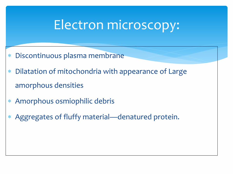
Electron microscopy:
Discontinuous plasma membrane
Dilatation of mitochondria with appearance of Large
amorphous densities
Amorphous osmiophilic debris
Aggregates of fluffy material—denatured protein.

1. Coagulative Necrosis
2. Liquefactive Necrosis
3. Caseous Necrosis
4. Enzymatic Fat Necrosis

1. Coagulative Necrosis:
Implies preservation of basic outline of the coagulated cell
at least for some days.
Example: Myocardial infarction—acidophilic, coagulated,
anucleate cells persisting for few weeks.
Ultimately it is removed by phagocytosis by scavenger
leukocytes.

2. Liquefactive Necrosis:
MC hypoxic death in CNS.
Transformation of tissue into a liquid viscous mass.
Seen in fungal/ bacterial infections also.
Pus: creamy yellow material containing dead white
cells.

3. Caseous Necrosis:
Most often seen in TUBERCULOSIS.
Cheesy white- gross appearance of the area of necrosis.
Amorphous, granular debris enclosed within a distinctive
inflammatory border known as Granulomatous
inflammation.

4. Fat Necrosis:
Due to release of activated pancreatic lipases—into
pancreatic substances and peritoneal cavity–destroying the
fat.
MC seen in acute pancreatits.
Lipases split triglycerides into fatty acids that combine with
calcium to produce grossly visible chalky white areas- FAT
SAPONIFICATION.