48B 2012 9693-9699 - Elixir International Journal (2012) 9693-9699.pdf · Sunil N. Bhamare et al./...
7
Sunil N. Bhamare et al./ Elixir Appl. Biology 48B (2012) 9693-9699 9693 Introduction Anoplophrya was reported by Stein (1860). Genus Anoplophrya belong to order Astomatida of Class Oligohymenoporea which is representative of the primitive and most controversial Ciliophoran. Anoplophrya belongs to Subclass Astomatia (Schewiakoff 1896), or Hymenostomatia (Levin 1980). Ciliates belongs to this subclass are ‘mouth less’ symbionts (sometimes Parasites) living in guts of annelids especially oligochaetes Somatic monokinetids like those of other Oligohymenophorea; with a divergenic post ciliary ribbon (absent in some genera) distinct anteriorly directed kinetodesmal fibril originating near triplets 5-7; a radial transverse ribbon near triplets 3,4 (reduced to distinct) and extending laterally towards adjacent kinety; cortical cytoskeleton in thigmotactic region may be conspicuously developed as anterior attachment structure. The order Astomatida, composed of entirely endosymbiotic form, has long puzzled protozoan phylogeneticists, although progress has certainly been made in recognizing that the loss of a mouth here is a secondary and thus regressive character, not a primitive one and that features of the infraciliature senso lato may be used to advantage in trying to determine the most likely origin of the group. From an overall approach the Astomes may continue to be characterized as relatively large uniformly ciliated mouth less ciliates. Unique characters include an almost universal infraciliary endoskeleton is of varying complexity and, in many species, the areas thigomotatic ciliature or even elaborately developed ‘hold-fast’ organelles in the form of hooks, spines, spicules or suckers. Fission is straightward, particularly in that it is uncomplicated by Stomatogenesis, but separation of the product is often incomplete, result in formation of cateniod colony (Corliss 1979). The typical body form is ovoid to elongate, with a flattening apparently related to the habit of lying up against the intestinal epithelium of the host’s digestive system. Rows of somatic cilia converge at the poles in characteristic patterns. The suture area or ‘air secante’ at anterior end pole is of particular diagnostic importance. Contractile vacuoles are sometime straight out in long line, and mucocysts are present. Feeding is by osmotrophy. The parasitic mode of life tends to simplication of the structural differentiation of any persisting organ system by reduction in numbers of repeated element and restriction structural modifications among parts of series of organs. The astomatous ciliate such as Anoplophrya, lives in the fluids of the digestive tract of earthworms and absorb food from the medium in which they are immersed, exhibit the loss of pharyngeal or organelles consisting in a other ciliates, of especially differentiated cilia and even membranelles the function of which in the ancestor of the astomatous ciliates was to pick up organisms such as bacteria and pass them in to and down the pharynx into the endoplasm often shaping them into food ball at the inner end of this circumscribed route. Ciliates serving this function, there are structural differentiation and their specialized groupings have been described by Lund (1933) in Paramecium and Rosenberg (1937) in Nyctotherus. Tele: E-mail addresses: [email protected] © 2012 Elixir All rights reserved Prevalence of Intestine dwelling ciliates and Morphological details of Anoplophrya infundibulii (n. sp.) from Earthworm, Pheretima posthuma from Nashik district of Maharashtra, India Sunil N. Bhamare 1 , Susheel V. Nikam 2 and Bhimrao N, Jadhav 3 1 Department of Zoology, K.R.A. College Deola, Nashik (M.S.) India. 2 Department of Zoology, Dr. B.A.M. University, Aurangabad, Maharashtra, India. 3 Department of Zoology, Shri. Muktanand College Gangapur, Aurangabad (M.S.), India. ABSTRACT Many ciliates species are the common parasites found in Indian earthworm Pheretima posthuma. During the period of two years (Jan. 2007 to Dec. 2008) total 2609 number of earthworm animals were examined. In the first year (Jan. 2007 to Dec. 2007) 1146 P. posthuma were examined, 693 of these were positive for ciliate infection. The percentage of prevalence being (60.47%), in second year (Jan. 2008 to Dec. 2008) total 963 animals were examined, 560 of these were positive with ciliates. The percentage of prevalence was 58.15%. A month wise analysis of the percentage of prevalence of ciliates during the first year (Jan. 2007 to Dec. 2007) was maximum in June to August (83.33%, 81.88%, 84.32%), minimum in April and May (36.19%, 36.67%) and moderate in remaining months. While In second year (Jan. 2008 to Dec. 2008) the maximum percentage of prevalence showed during August and September (84.72%, 81.33%), minimum in February to May (49.43%, 45.45%, 50.67%, 50.98%) and moderate in remaining months. While the observation of gut of the host P. posthuma it is seen that the four species of genus Anoplophrya were investigated. Out of that the two species are new to the sciences which are A. chakravartii, A. krishnamurthii, A. nikamai n. sp. and A. infundibulii n. sp. The parasite ciliates were first observed in 0.6% saline solution fixed in Schauddin’s fixative and then stained with phaspho-tungastic acid hematoxyline stain. © 2012 Elixir All rights reserved. ARTICLE INFO Article history: Received: 24 May 2012; Received in revised form: 20 July 2012; Accepted: 30 July 2012; Keywords P. posthuma, Percentage prevalence, Anolplophrya, N.sp. Elixir Appl. Biology 48B (2012) 9693-9699 Applied Biology Available online at www.elixirpublishers.com (Elixir International Journal)
-
Upload
hoanghuong -
Category
Documents
-
view
230 -
download
1
Transcript of 48B 2012 9693-9699 - Elixir International Journal (2012) 9693-9699.pdf · Sunil N. Bhamare et al./...

Sunil N. Bhamare et al./ Elixir Appl. Biology 48B (2012) 9693-9699
9693
Introduction Anoplophrya was reported by Stein (1860). Genus
Anoplophrya belong to order Astomatida of Class
Oligohymenoporea which is representative of the primitive and
most controversial Ciliophoran. Anoplophrya belongs to
Subclass Astomatia (Schewiakoff 1896), or Hymenostomatia
(Levin 1980). Ciliates belongs to this subclass are ‘mouth less’
symbionts (sometimes Parasites) living in guts of annelids
especially oligochaetes Somatic monokinetids like those of other
Oligohymenophorea; with a divergenic post ciliary ribbon
(absent in some genera) distinct anteriorly directed kinetodesmal
fibril originating near triplets 5-7; a radial transverse ribbon near
triplets 3,4 (reduced to distinct) and extending laterally towards
adjacent kinety; cortical cytoskeleton in thigmotactic region may
be conspicuously developed as anterior attachment structure.
The order Astomatida, composed of entirely endosymbiotic
form, has long puzzled protozoan phylogeneticists, although
progress has certainly been made in recognizing that the loss of
a mouth here is a secondary and thus regressive character, not a
primitive one and that features of the infraciliature senso lato
may be used to advantage in trying to determine the most likely
origin of the group. From an overall approach the Astomes may
continue to be characterized as relatively large uniformly
ciliated mouth less ciliates. Unique characters include an almost
universal infraciliary endoskeleton is of varying complexity and,
in many species, the areas thigomotatic ciliature or even
elaborately developed ‘hold-fast’ organelles in the form of
hooks, spines, spicules or suckers. Fission is straightward,
particularly in that it is uncomplicated by Stomatogenesis, but
separation of the product is often incomplete, result in formation
of cateniod colony (Corliss 1979). The typical body form is
ovoid to elongate, with a flattening apparently related to the
habit of lying up against the intestinal epithelium of the host’s
digestive system.
Rows of somatic cilia converge at the poles in characteristic
patterns. The suture area or ‘air secante’ at anterior end pole is
of particular diagnostic importance. Contractile vacuoles are
sometime straight out in long line, and mucocysts are present.
Feeding is by osmotrophy. The parasitic mode of life tends to
simplication of the structural differentiation of any persisting
organ system by reduction in numbers of repeated element and
restriction structural modifications among parts of series of
organs. The astomatous ciliate such as Anoplophrya, lives in the
fluids of the digestive tract of earthworms and absorb food from
the medium in which they are immersed, exhibit the loss of
pharyngeal or organelles consisting in a other ciliates, of
especially differentiated cilia and even membranelles the
function of which in the ancestor of the astomatous ciliates was
to pick up organisms such as bacteria and pass them in to and
down the pharynx into the endoplasm often shaping them into
food ball at the inner end of this circumscribed route. Ciliates
serving this function, there are structural differentiation and their
specialized groupings have been described by Lund (1933) in
Paramecium and Rosenberg (1937) in Nyctotherus.
Tele:
E-mail addresses: [email protected]
© 2012 Elixir All rights reserved
Prevalence of Intestine dwelling ciliates and Morphological details of
Anoplophrya infundibulii (n. sp.) from Earthworm, Pheretima posthuma from
Nashik district of Maharashtra, India Sunil N. Bhamare1, Susheel V. Nikam2 and Bhimrao N, Jadhav3
1Department of Zoology, K.R.A. College Deola, Nashik (M.S.) India.
2Department of Zoology, Dr. B.A.M. University, Aurangabad, Maharashtra, India.
3Department of Zoology, Shri. Muktanand College Gangapur, Aurangabad (M.S.), India.
AB STRACT
Many ciliates species are the common parasites found in Indian earthworm Pheretima
posthuma. During the period of two years (Jan. 2007 to Dec. 2008) total 2609 number of
earthworm animals were examined. In the first year (Jan. 2007 to Dec. 2007) 1146 P.
posthuma were examined, 693 of these were positive for ciliate infection. The percentage of
prevalence being (60.47%), in second year (Jan. 2008 to Dec. 2008) total 963 animals were
examined, 560 of these were positive with ciliates. The percentage of prevalence was
58.15%. A month wise analysis of the percentage of prevalence of ciliates during the first
year (Jan. 2007 to Dec. 2007) was maximum in June to August (83.33%, 81.88%, 84.32%),
minimum in April and May (36.19%, 36.67%) and moderate in remaining months. While In
second year (Jan. 2008 to Dec. 2008) the maximum percentage of prevalence showed during
August and September (84.72%, 81.33%), minimum in February to May (49.43%, 45.45%,
50.67%, 50.98%) and moderate in remaining months. While the observation of gut of the
host P. posthuma it is seen that the four species of genus Anoplophrya were investigated.
Out of that the two species are new to the sciences which are A. chakravartii, A.
krishnamurthii, A. nikamai n. sp. and A. infundibulii n. sp. The parasite ciliates were first
observed in 0.6% saline solution fixed in Schauddin’s fixative and then stained with
phaspho-tungastic acid hematoxyline stain.
© 2012 Elixir All rights reserved.
ARTICLE INF O
Artic le h istory: Received: 24 May 2012;
Received in revised form:
20 July 2012;
Accepted: 30 July 2012;
Keywords
P. posthuma,
Percentage prevalence,
Anolplophrya,
N.sp.
Elixir Appl. Biology 48B (2012) 9693-9699
Applied Biology
Available online at www.elixirpublishers.com (Elixir International Journal)

Sunil N. Bhamare et al./ Elixir Appl. Biology 48B (2012) 9693-9699
9694
Since Anoplophrya has neither mouth or pharynx, it is a
matter of interest to find out how far its parasitic mode of life
has reduced the pharyngeal apparatus and the motorium, found
near the pharynx and from which fibrils are sent out to the
pharyngeal apparatus and to other parts of ciliary mechanism.
Because of the relatively simple structure of the astomatous
ciliates their systematic has been in a state of confusion.
Heidenreich (1935) described few species of Anoplophrya.
Several later described species of Anoplophrya are A. allure
Ceped, A. complanata and a species by Exsemplerskaja. He
considered the commonly accepted name. A. striata, Dujardin is
as a synonym of the earlier A. lumbrici (Shrank 1803).
Genus Anoplophrya was also redscribed by Claparede
(1860), Leidy (1877); Kent (1881), Balbiani (1885) and Butschli
(1888). The general body form is elongate, cylindrical or slightly
flattened, with rounded ends. The posterior end of the body is
tapering in some species. The body is striated with clearly
defined often depressed line which runs longitudinally and
sometimes spirally. The contractile vacuoles are usually placed
in rows upon the edges. The macronucleus is almost always long
and band form, and generally extending through the entire
length of the body. Micronucleus may be distinct in some
species where as absent in other. Reproduction is affected by
simple cross division or by budding at the posterior end, and is
frequently combined with chain formation. The main
characteristics are the entire absence of mouth. A number of
workers consider this mouth- less forms representing the most
primitive order of holotrichs (Class Oligohymenophorea, de
Puytorac et al., 1974). It is also not unlikely that are secondarily
degenerate forms showing some specialization which may be
associated with their entirely symbiotic mode of life (Corliss
1956).
There is a single compact macronucleus with a single
lenticular micronucleus. Macronucleus is of elongated or ribbon
shaped where as in some cases it is spherical. Taxonomically,
the Astomatida is dividing in three major groups. Du Puytorac
(1972) has recognized super families. Genus Anoplophrya has
super family Anoplophryodae.
Family Anoplophryidae erected by Cepede (1910). He also
recognized in his monograph several families like
Discophryidae, Kofoidellidae, Haptophryidae and
Maupasellidae for ciliates from different invertebrate groups.
Subsequently several contributions have been made by Ghosh
(1918). Heidenreich (1935) Georgevitch (1941), de Puytorac
(1972, 1974) has contributed a series of papers on astomates.
Ghosh (1918) described a new species A. lloydi from the seminal
vesicle of Indian earthworm from Pheretima posthuma and A.
pheretimi. Raychaudhuri, Haldar and Chakravarty (1969) from
alimentary canal of same host.
Genus Anoplophrya has following species.
A. lumbrici (Shrank,1803)
A. lloydi (Ghosh,1918)
A. marylandensis (Conklin, 1935)
A. orchestii (Summer, 1936)
A. chakravartii (Lalpotu,1976)
A. perionychis (Lalpotu, 1976) and (T.T. Shaikh, 2006)
A. foldii (T.T. Shaikh, 2006)
A. bifoldii (Bhandari, 2010)
A. feretimi (Bhandari, 2010)
Anoplophrya infundibulii (n. sp.)
Material and methods
The hosts were collected from different parts of hilly
regions of Nashik dist (Deola, Nandgaon; Surgana, Kalwan
Satana) of Maharashtra state. Due care was taken and the hosts
were collected in moist soil with decaying leaves were present
and the temperature was maintained by using ice bags around
them. Mostly the hosts were collected during morning and
evening.
During a period of two years (Jan. 2007 to Dec. 2008) the
earthworm species Pheretima posthuma, were examined for
ciliate infection. In which four species of ciliates were
investigated from the gut of earthworms, all the four species of
ciliates are belongs to Genus Anoplophrya, they are,
1. Anoplophrya chakravartii
2. Anoplophrya krishnamurthii
3. Anoplophrya nikamai n. sp.
4. Anoplophrya infundibulii (n. sp.)
Out of four the two species are new to the science. The
present morphological study is concern with Anoplophrya
infundibulii (n. sp.). The hosts were collected from Nashik
district of Maharashtra. Earthworms were examined for the
ciliates. Entire alimentary canal of host animal was examined.
For the observation or detection the faecal matter was mixed
with 0.6% saline solution and observed under microscope. When
sample found positive it was treated with permanent preparation,
for that tungsten phosphoric haematoxyline method was used
along with Lugol’s solution and hyposolution. For fixation of
ciliates and gregarines Schaudinn’s fixative was used. Dry silver
impregnation method was also used for ciliates.
Topography-
Plate- 1 showing Map of Nashik District of Maharashtra,
India
Result and Discussion The Percentage Prevalence ciliates in Pheretima Posthuma
are counted for the period of two years, January 2007 to
December 2008 which is shown year wise in table No. 1 and 2,
with the graphs.
During the period of two years (Jan. 2007 to Dec. 2008)
total 2169 earthworms were examined. In the first year (Jan.
2007 to Dec. 2007) 1146 individuals were examined, 693 of
these were positive for ciliate infection. The percentage of
prevalence was found 60.47%. In second year (Jan. 2008 to Dec.
2008) total 963 P. posthuma species were examined 560 of these
were positive. The percentage of prevalence was 58.15%.
A month wise analysis of the prevalence in first the year
(Jan. 2007 to Dec. 2007) showed the maximum percentage of
prevalence during June to August (83.33%, 81.88%, 84.32%),
minimum in April and May (36.19%, 36.67%) and moderate in
remaining months. In second year (Jan. 2008 to Dec. 2008) the
maximum percentage of prevalence showed during August and
Sunil N. Bhamare et al./ Elixir Appl. Biology 48B (2012) 9693-9699
9695
September (84.72%, 81.33%), minimum in February to May
(49.43%, 45.45%, 50.67%, 50.98%) and moderate in remaining
months. The details of the number of earthworms
examined and the month wise prevalence are shown in Table No. 1, and 2.
Morphological details:
Description of the species:
This species of Anoplophrya was found in the intestine of
earthworm Pheretima posthuma. Hosts were collected besides
the Girna River and Deola (Nashik) college campus. The
parasites were usually numerous in the infected worms. They
were found only in the posterior part of the intestine.
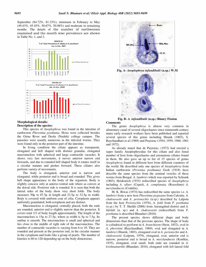
In living condition the ciliate appears as transparent,
elongated and bell shaped with distinct granular, elongated
macronucleus with spherical and large contractile vacuoles. It
shows very fast movements, it moves anterior narrow end
forwards, and due to rounded bell shaped body it rotates itself in
a circular manner and pushes forward. These ciliates also
perform variety of movements.
The body is elongated; anterior end is narrow and
elongated, while posterior end is broad and rounded. This gives
bell shape appearance to the body of the organism. Body is
slightly concave only at anterio-ventral side where as convex at
the dorsal side. Posterior side is rounded. It is seen that both the
lateral sides of the body show very short folds. The body
measures 50µ to 87.5µ in length and 22.5µ to 32.5µ in width.
Body is covered with uniform coat of cilia. Cytoplasm appears
uniformly granulated; both ectoplasm and are distinct.
Macronucleus is elongated, centrally placed, both the ends
are rounded, anterior end is slightly narrow than the posterior. It
covers total 1/3 of body length approximately. The length of the
macronucleus is 15µ to 27.5µ, where as width is 5µ to 7.5µ. Its
outline is smooth. The micronucleus is small and spherical and
lies close to the middle of right side of the macronucleus. The
number of contractile vacuoles is varying from 6 to 10. They are
rounded and present at the posterior end, in the circular manner
in the cytoplasm and looks like a group of pearls. The number of
kineties is 60 to 120 depending up on the body dimensions.
Fig. A- Anoplophrya infundibulii (n.sp.) W.M
Fig. B- A. infundibulii (n.sp.) Binary Fission
Comments The genus Anoplophrya is almost very common in
alimentary canal of several oligochaetes since nineteenth century
many early research workers have been published and reported
several species of this genus including Shrank (1803), S.
Raychaudhari et al (1969) and Puytorac (1954, 1959, 1960, 1961
and 1972).
As already stated that de Puytorac, (1972) had erected a
super family Anoplophryodae for this ciliate and also listed
number of host from oligochaetes and astomatous ciliates found
in them. He also gave an up to list of 15 species of genus
Anoplophrya found in different host from different countries of
the world. He described only one species of Anoplophrya from
Indian earthworm (Peretima posthuma) Gosh (1918) have
describe the same species from the seminal vesicles of these
worms from Bengal. A. lumbrici which was reported by Schrank
(1803), Heidenrich (1935) rediscribed species of Anoplophrya
including A. allure (Ceped), A. complanata, (Rossolimo) A.
merylandnesis (Conklin).
M. K. Biwas (1974) has rediscribed the same species i.e. A.
lumbrici from a new host Pheretima peguyana from Calcutta. A.
chakravartii and A. perionychis (n.sp.) described by Lalpotu
from the host Perionychis (1976), A. foldi from P. posthuma
(n.sp.) by T. T. Sheikh (2006) from Aurangabad district and A.
bifoldii (n. sp.) and A. chakravartii (redescribed) from P.
posthuma is described Bhandari (2010).
The present species shows different shape and body
dimensions than that of the previous species. The shape of body
is cylindrical to pyriform in A. branchiarm (Stein, 1852), oval in
A. pheretimi (Raychaudhari, 1969), oval and elongated in A.
lumbrici (Sharnk, 1803), elongated oval in A. perionychis and A.
chakravartii (Lalpotu, 1979), elongated, oval, anterior end is
narrow, posterior end is broad in A. merylandnesis (Konlkin,
1935), elongated, oval small, both ends are rounded in A.
krishnamurthii (Bhandari, 2010), elongated with left lateral fold

Sunil N. Bhamare et al./ Elixir Appl. Biology 48B (2012) 9693-9699
9696
in A. foldii (Shaikh, 2006), elongated with both lateral folds in A.
Bifoldii (Bhandari, 2010), leaf like in A. nikamai (n. sp. by
present author), while in present species body shape is elongated
and bell shaped.
The number contractile vacuoles is single in A.
branchiarm, 3 to 6 in two rows in A. pheretimi, 3 in posterior
end in A. lumbrici, 5 very small in two rows in A. perionychis, 3
to 7 in A. chakravartii, 4 in A. merylandnesis, 4 arranged in two
rows in A. krishnamurthii, 7 to 18 in A. foldii, 19 to 24 scattered
irregularly in A. bifoldii, single large and oval in A. nikamii
while 6 to 10 arranged in circular disc at the posterior end in
present described species.
Macronucleus is long and showing banding pattern in A.
branchiarm, long and ribbon like in A. pheretimi, ribbon shaped
with irregular outline in A. perionychis & A. krishnamurthii,
elongated in A. chakravartii, ribbon like with several fine
projection in A. merilandnesis, ribbon like in A. foldii, elongated
and ribbon like in A. bifoldii, long and S-shaped in A. nikamai
(n. sp.) while in the present species it is shorter than all the
above species with narrow anterior end and broad & rounded
posterior end. Comparative analysis of various species of
Anoplophrya is shown in Table No. 3.
The species described by present author is compared with
previously described species. It is seen that some distinct
features are found in this species which are as follows.
1. Body is bell shaped, narrow anterior end and broad rounded
posterior end.
2. Number of contractile vacuoles is 6 to 10 and are rounded
arranged in circular manner at the rounded posterior end in the
cytoplasm.
3. Macronucleus is ribbon like in A. pheretimi, A. lumbrici, A.
perionychis, A. merylandnesis, A. krishanamurthii, A. foldii and
in A. bifoldii. It is long with bands in A. branchiarm, elongated
in A. chakravertii while it is elongated with narrow anterior end
and broad rounded posterior end in the present species.
In view of its distinct features this species is considered new
to the science, it looks very beautiful because its bell shape. It is
designated as Anoplophrya infundibulii (n.sp.).
Reference:
Balbiani, E. G. (1885): sur un infusoire cilie parasite du sang de
I’aselle aquatique (Anoplophrya circulans). Zool. Suisse., 2:
277-305.
Ball, G. H. (1959): Some gregarines from crustaceans taken
near Bombay India J. Protozool; 6 (1): 8-13.
Ball, G.H. 1969: Organisms living on and in protozoa. In. Chen,
T. ed., Research in Protozoology. Vol. 3. Pergamon Press.
Oxford. Pp. 565-718.
Bary, Brian M. (1950): Studies on New Zealand freshwater
ciliates: Part II. An annotated list of species from the
neighbourhood of Wellington. Trans Roy. Soc. N.Z. 78 (2-3), pp.
311-323.
Beers and Dhale (1935): Structure and divisions in the
Astomatus ciliates Metaradiophrya asysmmetrica n. sp. J.
Mitchell Society., 54: 111-1276.
Bhandari J. C. (2010): Studies on some Ciliate and Gregarine
parasites of Annelida and Arthropoda
Bhatia, B. L. (1936): The Fauna of British India, Protozoa,
Ciliophora. Taylor and Francis, London.
Bhatia, B. L. And Gulati A. N. (1972): On some parasitic
ciliates from Indian frogs,toads earthworms and cockroaches.
Arch Protistenk; 57: 85-120.
Bhatia, B. L. and setna S. (1924): On some new cephaline
gregarines parasitol; 16: 279-288.
Bhatia, B. L., (1936): The Fauna of British India: Protozoa,
Ciliophora. London.
Borror, A. C. (1963): Morphology and ecology of some
uncommon ciliates from alligator harbor, Florida. Trans amer
micros., soc 82:125-131.
Borror, A. C. (1968): Systematic of Eqplotes (cilionora)
Hypotrichida toward union of the old and the new J.protozool;
15: 802-800.
Borror, A. C. (1972): Revision of the order hypotrichida
(ciliophora, protozoa). J. protozol; 19: 1-23.
Calkins, G. N. (1901): Protozoa. Macmillan, New York.
Calkins, G. N. (1911): Protozoology Nauka, M. (in Russian).
Chakravrty, M. M. Raychaudhuri, S. And Ghosh, C. (1910): Observations on astomate ciliates from earthworm. proch. 5
th
Ind. Sci. Cong; Part III: 500.
Cheng, T. C. 1986: General parasitology. 2nd Ed. Academic
Press. Charleston. USA.
Clapared, E and Lachmann J. (1958): Etudes sur les
infusories et les rhizopodes mem. Inst natn genev slyear; 1857:
1-260 (81).
Cole, F.J. (1919): The history of Protozoology, London.
Collin, B. (1909): La conjugations d’Anoplophrya branchiarum
(Stein) Arch Zool-Exper.Et gen.5th series, 1: 345.
Conklin, C. (1930): Anoplophrya marylandensis n. sp., Ciliates
from the Intestine of Earthworm Bio. Bull., 68: 176-181
Corliss, J. O. (1956): On the evolution and systematics of
ciliated protozoa, Syst. Zool. 5: 68-91, 121-140.
Corliss, J. O. (1959): An illustrated key to the higher group of
the ciliated protozoa, with definition of terms J. Protozool; 205-
28.
Corliss, J. O. (1961): An illustrated key to the higher group of
ciliated Protozoa with definition of terms J. Protozool; 6: 265-
281.
Corliss, J. O. (1962): Taxonomic nomenclature practices in
Protozoology and the new international code of zoological
nomenclature. J. Protozool; 9: 30-324.
Corliss, J. O. (1975): Three centuries of Protozoology: a brief
tribute to its funding father, A. van Leeuwenhoek of Delft. J.
Protozool; 22: 3-7.
Corliss, J. O. (1977): Annotated assignment of families and to
the order and classes currently comprising the Corlissan scheme
of higher classification for the Phylum Ciliophora. Trans. Am.
Micros. Soc. 96: 104-140.
Corliss, J. O. (1978): A salute to fifty- four great microscopist
of the past: a pictorial to the history of Protozoology. Part 1.
Trans. Amer. Micros. Soc. 97: 419-458.
Corliss, J. O. (1978): Protozoan ecology: a note on its current
status. Amer Zool; 13: 145-148.
Corliss, J. O. (1979): The Ciliated Protozoa: characterization
classification and guide to the literature 2nd
ed. Pergtamon Press
Oxford.
Corliss, J. O. (1991): Historically important events, discoveries
and works in Protozoology from the mid 17th
to mid 20th
century. Rev. Soc. Mex. Hist. nat. 42: 45-81.
Corliss, J. O. (1997): Some important anniversaries in the
history of Protozoology. Rev. Soc. Mex. Hist. Nat. 47: 5-17
Corliss, J.O. (1959): The ciliated protozoa: Characterization
Classification and Guide to the Literature. Oxford, New York
etc; Pergamon Press. 455pages.

Sunil N. Bhamare et al./ Elixir Appl. Biology 48B (2012) 9693-9699
9697
Ghosh E. (1918): Studies on Infusoria I. on a new species of
Anoplophrya Stein emend Ceped Rec. Ind. Mus; 15(3): 129-134.
Griffiths, B.S., Kuan, H.L., Ritz, K., Glover, K.A, McCaig,
A.E., Fenwick, C. 2004: The relationship between microbial
community structure and functional stability, tested
experimentally in an upland pasture soil. Microbial Ecology
47(1):104-113.
Griffiths, B.S., Ritz, K., Wheatley, R., Kuan, H.L., Fenwick,
C., Christensen, S., Ekelund, F., Sorensen, S.J., Muller, S. and Bloem, J. 2001: An examination of the biodiversity-
ecosystem function relationship in arable soil microbial
communities. Soil Biology and Biochemistry 33:1851-1858.
Hausmann, K. Hiilsmann, N and Rader, R. (2003): Protistology (3
rd) E schweizerbart, sche, varlagsbrichandlung.
Hymen, L. H. (1940): The invertebrates.Vol.1.protozoa through
Ctenophora Mc.Graw Hill Book Co. U.S.A.727.
Katashima R. (1952): Studies on the Astomata (Ciliata,
Protozoz.) II zool. Mag. 62 (1): 22.
Kijenskij, G. (1925): Cliiates in the intestinal cavity of
Oligochaeata from the surrounding of prangue. Vest. vsl. zool.
Spol. Praha; 1925: 1-32.
Kirby, Harold, (1950): Material and Methods in the Study of
Protozoa. Univ. of Calif. Press.
Kitching, J. A. (1956a): Food vacuoles. Ibid. 3:D3b.
Kitching, J.A. 1956: Contractile vacuoles of protozoa.
Protolasmatologia. 3:D3b.
Klein, B. M. (1958): The “dry” silver method and its proper
use. J. Protozool. 5:99.
Klein, B. M. (1927): The dry silver metod and its proper use.
J.Protozool; 5(2): 99-13.
Klein, B. M. (1958): The dry silver method and its proper use.
J. Protozool. 5:99.
Kudo, R. R. (1977): Protozoology 5th
ed. Springfield, III:
Chane, C.C. Thomas (1774).
Kuznick, L. (1982): Protozoology in Poland past and present, In
: Progress in Protozoology. Proc. VI Intern. Conger. Protozool.
Acta Protozool. (Special volume), 75-111
Kuznick, L. (2003): Protozoologia w polsce 1861-2001
warszawa: polska Akadmia Nauk (in Polish).
Labbe, (1899): Sporozoa. Das Tierreich.5:1-180.
Lalpotu (1976): Observation on some Protozoan Parasites of
invertebrates, Ph. D. thesis of Dr. B.A.M. University
Aurangabad.
Lom, J. (1959): A contribution to the knowledge of astomatous
ciliates. Mem. Soc. Zool. Tche. Cosl; 23: 200-210.
Lom, J. (1961): A contribution to the Knowledge of astomatos
ciliates Mem. Soc zool. tche cosl., 23: 200-210.
Lynn, D. H. and Small, E. B. (2000): Phylum Ciliophora In An
illustrated guide to the protozoa.2-nd ed.(Eds. Lee J. J. Hunter S.
H. and Bovee E. C.) Allen press, Lawrence, 371-655.
Meire, M. (1954): parasitische ciliaten bei Oligochaeten Arch
Protistenk, 100: 212-245.
Puytorac, (1974) : Proposition d’une. Classification du phylum
Ciliophora Doflein, 1901. C. R. Acad. Sci Paris., 278: 2799-
2802.
Puytorac, P. de, (1960): Obsevations sur quelque Ciliates
astomes des Oligochetes Du lac d’ ochrid (Youngoslavie). II
Famille des Holitophryidae (Hoplitophryinae et Maupasellinae),
des Intoshellinidae et des Anoplophryidae. J. Protozool., 7: 278-
289.
Puytorac, P. de, (1963): Comparison de la fauna infusorienne
endoparasite des Oligochaetes du lac d’Ohrid et de ceux du
Baical. Rec. Trav. Station. Biol. Ohrid; 6(2): 1-8.
Puytorac, P. de, (1968): structure et ultrastructure de
Sicuophora xenopi n. gen. n. sp. Cilie Heterotriches parasite du
Batracien xenopus fraseri Boul. Parasitologica; 4(3): 405-414.
Puytorac, P. de, (1971): Les ciliate astomes Hoplitophryidae II
Revision de la syst matique dece group. Protistologica., 8 (1):
5-42.
Puytorac, P. de, (1972): Les ciliate astomes Hoplitophryidae II
Revision de la syst. matique dece group. Protistologica; 8 (1): 5-
42.
Q. M. 1931: Morphological variation in Coleps octospinus. Tr.
Am. Micr. Soc. 50:136.
Raabe, (1949): Recherches sur les cilies Thigmotriches
(Thigmotricha Ch. Lw) III Development non-parallel de deux
especes du genera Sphenophrya Ch. Lw. Ann. Univ. Mariae
Curie-Sklodowska Lublin, Sect C; 4: 19-135.
Raabe, (1972): Ordo Thgmotricha (Ciliata Holotricha) V.
Familiae Hysterocinetidae et proto anoplophryidae. Acta
Protozool 10: 115-184.
Raychaudhuri, S. Halder, D. P. And Chakravarty, M. M.
(1969): Observation on new astomate ciliates, Anoplophrya
pheretimi, from the earthworm Pheretima posthuma (L. Vall).
ArchProtistenk., 111: 228-235.
Schewiakoof, W. T. 1896: Organisetion and systematics of
Infusoria Aspirotricha (Holotricha auctorum). Mem. Imp.
Acad. Sci. SPb. Ser VII, 4, 1-395 (in Russian).
Seethalakshmi Ayer (1979): Studies on Some Parasitic
Protozoa from some Invertebrates, Ph. D. thesis of Dr. B. A. M.
University Aurangabad.
Sergei I. Fokin, (2004): A brief history of ciliate studies (late
XVII _ the first third of the XX century) Protistology 3 (4),
283_296 (2004).
Sharnk (1830): The neuromotor system of Anoplophrya
lumbrici, degenerate ciliate from the earthworm Octolasium
cyaneum Americam Philosophical Socity; 78 (4): 595-614.
Sheikh, T. T. (2006): Studies on some free living and parasitic
ciliates Ph. D. thesis of Dr. B. A. M. University Aurangabad.
Vickerman K, Sleigh M. Leadeaterv B. and Mc Cready S. (2000): A century of Protozoology in Britain Brit. Sect. Intern.
Soc. Protozool. London.
Wenrich, D.H. 1956: Some American pioneers in protozoology.
J. Protozool. 3:1.

Sunil N. Bhamare et al./ Elixir Appl. Biology 48B (2012) 9693-9699
9698
TABLE 1.
SHOWING THE MONTHWISE PREVALENCE OF CILIATES IN EARTHWORMS
(Pheretima Posthuma) during the period from Jan.2007 To Dec.2007
Sr Total No. of No. of % of Total
No Months
Hosts Examined .+ve samples Prevalence
1 Jan-07 140 65 46.43
2 Feb-07 75 38 50.67
3 Mar-07 102 45 44.12
4 Apr-07 105 38 36.19
5 May-07 90 33 36.67
6 Jun-07 150 125 83.33
7 Jul-07 138 113 81.88
8 Aug-07 112 95 84.82
9 Sep-07 95 65 68.42
10 Oct-07 44 27 61.36
11 Nov-07 47 23 48.94
12 Dec-07 48 26 54.17
Total 1146 693 60.47
TABLE 2.
SHOWING THE MONTHWISE PREVALENCE OF CILIATES IN EARTHWORMS
(Pheretima posthuma) during the period Jan.2008 To Dec.2008
Sr Total No. of No. of % of Total
No Month
Hosts Examined .+ve samples Prevalence
1 Jan-08 45 25 55.56
2 Feb-08 87 43 49.43
3 Mar-08 77 35 45.45
4 Apr-08 75 38 50.67
5 May-08 102 52 50.98
6 Jun-08 104 60 57.69
7 Jul-08 110 58 52.73
8 Aug-08 72 61 84.72
9 Sep-08 75 61 81.33
10 Oct-08 65 41 63.08
11 Nov-08 47 29 61.70
12 Dec-08 104 57 54.81
Total 963 560 58.15

Sunil N. Bhamare et al./ Elixir Appl. Biology 48B (2012) 9693-9699
9699
Table 3- Comparison of the present species with the species of Anoplophrya
Sr, No. 1 2 3 4 5 6 7 8 9 10
particulars A.
branchiarm
stein
(1852)
A. pheretimi
S.
Raychaudhari
(1969)
A. lumbrici sharnk
(1803 )
A.
perionychis
Lalpotu
(1979 )
A.
chakravertii
Lalpotu
(1979 )
A.
merylandnesis
Konlkin
(1935 )
A.
kirshnamurthii
(n. sp.)
Bhandari
(2010)
A. foldii
T T Shaikh
(2006 )
A. bifoldii (n. sp.)
Bhandari
(2010)
A.
infundibulii
(n. sp.)
present
author
(2010)
Body shape and
dimensions
Cylindrical to pyriform
L – 104 to
136 µ
Oval shape L – 49.5 to
188.8µ
W – 23.1 to
46.5µ
Oval elongated L-
180 to 250µ
W-50 to
70µ
Elongated oval L-67
to 120µ
W-28 to
43µ
Elongated oval ant.
End.
Narrow
post. End
broad
L-63 to 179.6µ
W-28.4 to
94.6µ
Elongated oval ant. End.
Narrow post.
End broad
L-36.72µ
W-16.42µ
Elongated oval small
both ends
rounded L-
25.63 to 39.6µ
W-16.2 to
20.97µ
Elongated with left
lateral fold
L-58 to 112µ
W-30 to 48µ
Elongated with both
left lateral
folds L-
132.81 to
186.4µ
W- 32.62 to 55.92µ
Elongated bell shaped
ant. End
narrow
post. End
broad
L-50-87.5µ W-22.5-
32.5µ
Contractile vacuoles
Single Two, rows each side of
macronucleus
3 to 6 in each
3, post. End 5, two very small
3-7 4, arranged in longitudinal
rows
Four, arranged in two rows, 2
in each row
Two rows each side of
macronucleus
7-18 in each
rows
19-24 scattered
irregularly
6 to 10 circularly
arranged at
the post.
end
macronucleus Long band
formed 30-89µ
Long ribbon
shaped 33-92µ
Ribbon
shaped 100-120µ
Ribbon
shaped irregular
outline
Elongated
ant. end pointed
post. end
broad
Ribbon
shaped several fine
projections
Ribbon
shaped with irregular
outline 23.3 to
69.9µ
Ribbon
shaped
Elongated
ribbon shaped
L-93.21 to
51.45µ
W-4.6 to
6.99µ
Somewhat
elongated ant. end
narrow
rounded
post end
broad
rounded L-15 to
27.5µ
W-5 to
7.5µ
micronucleus …… …… …… …… Spherical Spherical Elongated
somewhat banana like
Spherical Spherical Spherical
Kineties ….. ……… ……. ……. …… ……. …… ……. ….. 60 to 120
Host ….. P. posthuma Octoplasium
cyancum
Perionychis Perionychis Lumbricus
trrestris
P. posthuma P. posthuma P.
posthuma
P.
posthuma
Locality ….. West Bengal California Aurangabad
Dist.
Parbhani
Dist.
Baltimore Partur Jalna Aurangabad
Dist.
Aurangabad
Dist.
Nashik
Dist.