37H s/B/ /M. IMl SHORT TERM EFFECTS OF EXTERNAL ELECTRIC ...
49
37H s/B/ /M. IMl SHORT TERM EFFECTS OF EXTERNAL ELECTRIC FIELDS ON ELECTRICAL ACTIVITY OF THE PINEAL GLAND IN RATS THESIS Presented to the Graduate Council of the University of North Texas in Partial Fulfillment of the Requirements For the degree of MASTER OF SCIENCE By Hung Q. Vu, B. S Denton, Texas May, 1996
Transcript of 37H s/B/ /M. IMl SHORT TERM EFFECTS OF EXTERNAL ELECTRIC ...
37H s/B/
/M. IMl
SHORT TERM EFFECTS OF EXTERNAL ELECTRIC FIELDS
ON ELECTRICAL ACTIVITY OF THE PINEAL GLAND
IN RATS
THESIS
Presented to the Graduate Council of the
University of North Texas in Partial
Fulfillment of the Requirements
For the degree of
MASTER OF SCIENCE
By
Hung Q. Vu, B. S
Denton, Texas
May, 1996

Vu, Hung Q., Short term effects of external electric
fields on electrical activity of the pineal gland in rats.
Master of Science (Biology), May, 1996, 43 pp., 1 table, 12
figures, bibliography, 28 titles.
The effects of short term exposure (5 minutes) to EEFs
at relatively high dosages (10, 25, 39, kV/m) on the
electrical activity in rat pineal glands was studied.
Daytime and nighttime recordings were taken from an
implanted microelectrode in the gland. The data show that
(1) both the activity and frequency were enhanced when the
animals were exposed to EEFs at 39 kV/m continuously and
discontinuously; (2) the later condition yielded a sustained
increase (36%) whereas the former a brief (10 sec) increase.
This enhancement was statistically significant under both
conditions (day and night). The effects observed were
thought to be due to membrane alterations either in the
pineal gland itself or in the neural inputs to the gland.

TABLE OF CONTENTS
Page
List of Tables iv
List of Illustrations v
Chapter
I. INTRODUCTION 1
II. MATERIALS AND METHODS 5
Electrode preparation and implantation Experimental Recordings
III. RESULTS 15
IV. DISCUSSION 36
BIBLIOGRAPHY 41
i n

LIST OF TABLES
Table Page
1. Summary of the effects of short term EEF
on the mean activity in rat pineal gland. . . 35
IV

LIST OF ILLUSTRATIONS
Figure Page
1. Experimental Set-up 11
2. Stereotaxic Apparatus 12
3. Faraday Cage 13
4. Diagram of Experimental Apparatus . . . 14
5. Mean Pineal Activity in Sham-Exposed Rats Day and Night 19
6. Effects of 10 kV/m Continuous EEF on Daytime and Nighttime Pineal Activity . . 21
7. Effects of 25 kV/m Continuous EEF on Daytime and Nighttime Pineal Activity. . . 23
8. Effects of 39 kV/m Continuous EEF on Daytime and Nighttime Pineal Activity . . 25
9. Typical Tracing of Electrical Activity Recorded from the Pineal Gland for Day Night 27
10. Effects on 39 kV/m Discontinuous EEF on Daytime and Nighttime 29
11. Effects of Blindfolding on Daytime and Light on at Nighttime on Pineal Activity . 31
12. Daytime and Nighttime Pineal Gland Activity in Sham-Control Male and Female Rats 33

CHAPTER I
INTRODUCTION
Over the past few decades, there has been a persistent
controversy over whether external electric fields (EEF),
such as those formed under and near overhead high power
lines, are potentially dangerous to one's health [Miller et
al., 1978; Marino et al., 1977]. In the 1980's numerous
epidemiological reports indicated that the incidence of
cancer was significantly higher in humans and in particular,
children who resided near overhead high power lines.
Behavioral and CNS changes during [Lott et al., 1973] and
following exposure to EEF have also been studied in depth
[Hjeresen et al., 1980; Gavalas et al., 1970].
Several workers have calculated the EEF directly under
a 765 kV power line and have found it to be about 10,000 V/m
[Marino et al., 1978]. Earlier evidence of EEF's effects on
humans came from Russian studies involving electric utility
personnel working around 500-700 kV/m substations.
Headaches, malaise, abnormal fatigue and sleepiness were
reported from a majority of the workers; Some young men even
complained of reduced sexual potency [Korobokova et al.,
1972] . As a consequence, a restriction area under power
lines and safety regulations was established. In addition,
Wever's experiment [197 0] involving humans indicated that,
1

in an underground bunker where subjects were isolated from
environmental cues, an EEF at 3 00 V/m shortened and
desynchronized the circadian periods.
The pineal gland in mammalians is the end organ of the
visual system, and produce primarily a neurohormone,
melatonin (MT) that has been involved in regulating
circadian rhythms. Not only do MT levels but also
spontaneous electrical activity in the pineal gland exhibit
a circadian rhythm (i.e. they increase at night and decrease
during daytime [Reuss et al., 1984; Reiter, 1991]). Several
reports have shown that exposing rats to EEF interferes with
the usual increase of nocturnal MT levels in pineal glands.
In 1981, Wilson [1981] exposed rats to a 65 kV/m electric
field in a grounded system for 30 days; he found that the
normal rise in nighttime pineal levels of MT and the
activity of N-acetyltransferase (NAT), the enzyme involved
in MT synthesis, were significantly suppressed. In 1986,
Wilson et al. [1986] again exposed rats to a lower EEF. They
observed the same effects of reduction in nocturnal MT and
NAT activity. Reiter [1988, 1993] exposed rats to 60-Hz
EEFs of 10, 65 and 13 0 kV/m from conception to 23 days of
age. He observed that all electric field strengths decreased
the nocturnal level of MT. Later, Reiter and Grota [1994]
performed another experiment in which rats were exposed to a
65 kV/m field for 30 days. The MT in the blood serum, the
pineal gland and activity of NAT and Hydroxyindole-o-

methyltransferase (HIOMT) were then analyzed. They
discovered that the EEF did not reduce the normal rise in MT
levels, NAT and HIOMT activity in pineal glands. Other
investigators [Lerchl et al., 1990; Wilson et al., 1990;
Lerchl et al., 1991] also studied the effects of both
magnetic field (MF) and EEF on MT production in the pineal
gland and came up with contradictory results.
Ruess [1987] in his review described the electrical
activity of the pineal gland under varying conditions and
listed several factors that influence pineal activity. Such
factors include superior cervicle ganglia input, age, the
habencular nuclei, the paraventricular nucleus (PVN) of the
hypothalamus, sciatic nerve stimulation, acoustic stimuli,
light and magnetic stimuli. This study will involve another
possible external stimulus: an external electric field.
Most of the recent work on EEF effects on the pineal
gland had been biochemical in nature and have involved long-
term exposures (weeks/ months) at relatively high dosages
(10 - 100 kV/m). The literature regarding the effects of
short-term exposure to EEFs on the electrical activity in
the pineal gland is currently sparse if not non-existent.
The general scope of this study, therefore, was
electrophysiological in nature since the electrical activity
of pineal glands was recorded from microelectrodes implanted
within the pineal gland in rats.
The specific purpose of this study was to measure the

electrical activity of rat pineal glands before, during and
following short-term exposure to varying dosages of EEF (10,
25, and 39 kV/m), and under varying conditions (e.g day
versus night; continuous versus discontinuous exposure).

CHAPTER II
MATERIALS AND METHODS
A total of 120 (60 males and 60 females) Sprague-Dawley
rats with average weights of 2 00 grams were used in this
study. In this weight range the brain size does not vary.
The age of the rats ranged between 8 to 11 weeks old. This
number excludes those animals used in developing the
experimental technique or those that died, or those in which
the electrodes were incorrectly implanted. They were housed,
one per cage, given lab chow and water ad libitum, and were
kept on a 12 hour light/dark cycle (light on at 6 AM, off at
6 PM). Room temperature was kept at approximately 26 degrees
Celsius. Every effort was made to minimize acoustic,
olfactory, air currents and motion cues to the animals
during the experiments. The experiments were carried out
between April and November 1995
The experiments were divided into two major series,
night and day. Each series consisted of six groups; each
group included 10 rats (5 of each sex).
I. Daytime Series
A. Sham-controls (15 minutes)
B. 10 kV/m - 5 minutes continuous exposure
C. 25 kV/m - 5 minutes continuous exposure
D. 39 kV/m - 5 minutes continuous exposure
5

E. 39 kV/m - 5 minutes discontinuous (on-off at 5
second intervals)
F. Sham exposed - blindfolded (15 minutes)
II. Nighttime Series
A. Sham-controls
B. 10 kV/m - 5 minutes continuous exposure
C. 25 kV/m - 5 minutes continuous exposure
D. 39 kV/m - 5 minutes continuous exposure
E. 39 kV/m - 5 minutes discontinuous (on-off at 5
second intervals)
F. Sham exposed - light on (15 minutes)
Electrode preparation and implantation
Preparation of the electrodes for implantation into the
pineal gland was carried out in the following manner. A
liquid insulator (Epoxylite Corp., El Monte California) was
used to insulate the stainless steel microelectrodes (#00,
Clay Adams Co., New York), as outlined by Hines [1985].
Before implantation, the diameter of the electrode was about
0.4 mm and the distal end was scraped so as to expose 0.5 mm
of the tip.
Prior to implantation, the subject was anesthetized by
intraperitoneal injection of sodium pentobarbital (10mg/100
g body weight). The animal was then placed in a stereotaxic
apparatus (Model-SMA-1 Baltimore Instrument Co., Baltimore,
Maryland) and a midline incision on the surface of the skull
was made. The skull was exposed and positioned so that

bregma and lambda were on a level plane (Fig.2). The
coordinates used for positioning the recording
microelectrode in the pineal gland were taken from the text,
Rat Brain in Stereotaxic Coordinates [Paxinos et al., 1986].
A small hole (0.7 mm in diameter) was drilled 8.3 mm
posterior to bregma on the midline to accommodate the
recording electrode, and another small hole was drilled 4.0
mm posterior to the bregma to accommodate a small jeweler's
screw. The plastic capped electrode was then lowered through
the hole in the skull to a distance of 2.0 mm. The electrode
opening in the skull was surrounded with soft acrylic. When
the electrode was in place, the acrylic connection between
the electrode cap and the skull was allowed to dry. The
small jeweler's screw was used to maintain the rigidity of
the electrode and the acrylic cap. When the cap was firmed,
the recording electrode was clipped at about 1.5 mm above
the plastic cap and the animal was allowed to recover for at
least two days prior to experimentation.
Experimental
A photo of the room with the entire laboratory assembly
is in Fig. 1 and its schematic set-up is in Fig. 4. A
diagram of the Faraday cage ensemble with dimensions is
depicted in Fig. 3. The purpose of the copperized Faraday
cage was to negate as much as possible any stray electric
fields produced by other electrical items in the room. The
Faraday cage itself was grounded to stabilize the electrical

recordings from the implanted electrode. The suspended anode
electrode ( 2.5 mm x 38 mm) consisted of a thin piece of
light metal completely covered with a coating of Bakelite, a
hard resinous insulating material. Therefore, the smooth
surface was entirely free of any pointed dirt particles that
might produce corona discharge during the exposures.
Recordings
Recordings were always taken in the morning between
8:00 AM and 10:00 AM and at night between 12:00 AM and 2:00
AM. All experiments were carried out under relatively
constant conditions (i.e. lighting, sound and temperature).
In the daytime experiments, the light sources in the
experimental room came from three overhead 100 W light-
bulbs. In the night experiments, a photographic dark-room
ruby red light was used. Schapiro [1971] and Reiter [1985]
found that red was the only wave-length that did not affect
pineal gland activity. The room temperature ranged between
24 and 27 degrees Celsius.
The electric field produced in the experimental chamber
was determined from the charge put on the electrode by the
field generator (Electrofields Inc. Miami, Florida) and the
distance to the top of the wooden box. Calculation of the
field was as follow:
Vf = V / d
Vf,= Voltage desired
V = Voltage needed from generator
d = Distance from suspended electrode to the wooden
platform
Every experiment lasted 15 minutes and consisted of a 5
minute pre-test, a 5 minute test (exposure) and a 5 minute
post-test (recovery) period. With this procedure, in each
experiment the animal was allowed to serve as its own
control. Moreover, such a set-up allowed uninterrupted
recording, before, during and following exposure to the EEF.
For those experiments involving blindfolding the rat
during the daytime and running the nighttime experiments in
the light, the electrical activity was continuously recorded
from the electrodes for a period of 15 minutes. Recording
from these experiments involved simply allowing the rat to
lie in the Faraday cage for the entire 15 minute period.
On the day of each experiment, the animal was
anesthetized with the same dose of sodium pentobarbital as
in implantation. When the animal did not show a corneal
reflex following the injection of the anesthetic, it was
assumed that the anesthesia was complete. A shielded
alligator clip reference electrode was attached to the back
of the neck and the Faraday cage was grounded. The rat was
then placed on a wooden platform in the middle of the cage;
therefore, the animal was "floating" inside the cage between
the field generating anode and a copper cathode. Prior to
the recording, an equilibration period of at least 2 0
minutes allowed the instruments and the animal to stabilize.

10
Test recordings were not began until trial tracings were
stabilized and presented valid pineal gland electrical
activity.
The electrical data obtained from the implanted
microelectrode were first amplified (Cold Springs
Instruments Corp., New York) before being fed into a
Physiograph (Desk Model Type Dmp-4a: Narco Instruments Inc,
Houston, Texas), and simultaneously into a digital
integrating device (Model 23 EEG Integrator: Cold Springs
Instruments Corp., New York) attached to a digital drive
recorder (Digital Data Recorder: Cold Springs Instruments
Corp., New York). These latter instruments were capable of
integrating the areas under the electrical tracing over a
given period of time and converting this activity into a
numerical printout every 60 seconds throughout the
experiment. Mean values were then obtained from this
printout and plotted accordingly. The electrical data from
the integrator unit were also fed into a pulse counting unit
(Universal Electronic Counter, Model Simpson 7026: Simpson
Electronic Co, Elgin, Illinois) where the response frequency
was measured and plotted.

II
a'"̂
Figure. 1: Experimental set-up

"l
1 z
< Figure. 2: Stereotaxic Apparatus

13
suspended electrode (anode)
copper mesh
acrylic platform
2.5 cm
38 cm
31.3 cm
38.5 cm
Figure. 3: Faraday cage

14
Faraday Cage "Animal"
Pre Amp
Physiograph
Integ ;rator
Digital
Recorder
Universal Electronic Counter
Figure. 4: Diagram of experimental apparatus

CHAPTER III
RESULTS
The data presented here came from 120 male and female
Sprague-Dawley rats, 8-11 weeks old, that were maintained in
an environment in which light, temperature, food and water
intake, sounds, humidity and handling were kept constant.
The sham-control animals were implanted with pineal gland
microelectrodes and recordings made in the absence of an
EEF.
The data are presented in the form of 12 Figures and
one Summary Table. The Figures contain curves that depict
the electrical activity from the pineal gland before, during
and after varying dosages of EEF (10, 25 and 39 kV/m). The
EEF were applied continuously and discontinuously (off-on
every 5 sec) for five minutes. The Summary Table contains
the percentage change in response in terms of overall
activity and frequency during pre-test (5 minutes), test (5
minutes), and post-test period of 5 minutes. Thus, each
experiment was a 15 minute duration. The change between the
mean pre-test and the mean test period were analyzed
statistically using Student's t test. The placement of the
electrodes was verified at autopsy.
Fig. 5A shows the mean electrical activity in ten sham-
control rats during the day. It is evident that no
15

16
significant change in activity or frequency occurred in the
15 minute experiments. In Fig. 5B the mean activity and
frequency from 10 sham-control rats also failed to show
changes in the entire experimental period. There was a clear
difference in the level of activity and frequency between
night and day that was expected. The nighttime activity was
about 47% higher than the daytime activity and the frequency
at night was 50 % higher.
Fig.6 contain curves showing the effects of 10 kV/m
continuous EEF (5 minutes) on the mean electrical activity
of the pineal gland in 10 rats during the daytime A and
during the night B. There were no measurable effects in
either the daytime activity or the nighttime recording. The
overall records were similar to those in the sham-control in
both activity and frequency.
Almost identical findings were observed in Fig. 7A and
B, in those rats receiving 25 kV/m EEF continuously for five
minutes. In Fig. 8 the effects of 39 kV/m continuous EEF on
the pineal gland activity are depicted. In Fig. 8A, a
distinct but short-lived increase in both the mean activity
(12%) and frequency (51%) occurred during the daytime
exposure. As shown in Fig. 8B, there was a clear but short-
lived increase in both the activity (15%) and frequency
(46%) when 39 kV/m EEF was given continuously at night. As
shown in Fig. 8. Recovery was complete in all animals within
one minute.

17
Fig.9A and B contain actual physiographic tracings that
show clearly the effects of exposing the animals to a
discontinuous EEF of 39 kV/m (on-off at 5 sec intervals for
5 minutes) in the day and in the night. The effects were
sustained during the entire period of exposure. Note the
differences in the amplitude between the day and night
recordings.
Fig. 10A and B show the effects of discontinuous (on-
off at 5 sec intervals for 5 minutes) day and night exposure
to 39 kV/m EEF. These findings were totally unexpected. As
shown in Fig. 10A, exposure to 39 kV/m intermittent EEF
during the day resulted in an increase of 36% in the mean
overall activity and over 136% in mean frequency. Fig. 10B
shows similar effects in the dark i.e. a 26% increase in the
mean activity and a 81% increase in frequency. The observed
increase in activity and frequency lasted the entire length
of exposure (5 min) and recovery was slightly slower during
the post-test periods.
Fig. 11A contains curves depicting the effect of
blindfolding the animals during daylight but no EEF
exposure. Recording was 15 minutes. No significant changes
were observed in the activity nor the frequency. Fig. 11B
curves also show no changes in activity or frequency when
the animals were in a lighted room at night and in the
absence of an EEF.
Fig. 12A curves show almost identical electrical

18
activity and frequency in male and female rat pineals either
during the day or during the night (Fig. 12B).
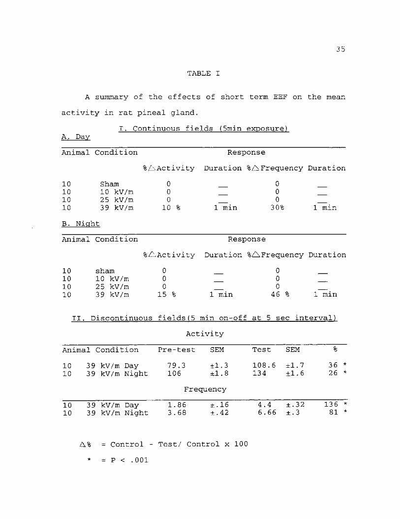
Table. I A summary of the effects of short-term
exposure to EEF on mean activity of pineal gland in rats. As
shown in the table, the greatest response in both the mean
activity and frequency occurred when the animals were
exposed to 39 kV/m discontinuously, although a significant
yet momentary response also occurred when the animals
received the same dosage (39 kV/m) continuously. Moreover,
in both the day and night discontinuously exposed group,
recovery did not occur until the field was turned off. The
differences in the activity in the interrupted field were
statistically significant.

19
FIGURE. 5
Mean pineal gland activity in sham-exposed rats, day and
night. (Each circle and square represent the mean electrical
activity and frequency of ten animals).

20
150
130
110
A: DAYTIME
70
50
30
Activity
Frequency
W ^
i ®- Hi m-
14 N
12 I
10 O c 8 <D 8 3 ) O" 6 £
uZ 4
'2
1 2 3 4 5 6 7 8 9 10 11 12 13 14 15 Time (min)
B: NIGHTTIME 150
130
110 110
" > •••• 90 o <
70 70
Activity
•" Frequency
1 2 3 4 5 6 7 8 9 10 11 12 13 14 15 Time (min)

21
FIGURE. 6
Effects of 10 kV/m continuous EEF on the daytime and
nighttime pineal gland activity in rats. (Each circle and
square represent the mean activity and frequency in ten
animals).

22
150
130
^ 1 1 0 mmmm
« 90 O
< 70
50i
30
A:DAYTIME Field on Field off s~ Activity
Frequency
-i i ..2
14 "n"
12 x
10 5 c
8 ®
f6
4
1 2 3 4 5 6 7 8 9 10 11 12 13 14 15
Time (min)
3 CT 0
150
130
110,
B: NIGHTTIME
o—
~ 90 O
< 70
50
30
Fie
lr n>—
d on Field off "®" Activity
"•"Frequency
l <V-
- I i i 6-__i—I i J_
14
1 2 3 4 5 6 7 8 9 10 11 12 13 14 15
Time (min)
12 N
12 X 10 >s
O c 8 <D 8 3
6 O"
6 £ u!
4
2
0

23
FIGURE. 7
Effects of 25 kV/m continuous EEF on the daytime and
nighttime pineal gland activity in rats. (Each circle and
square represent the mean electrical activity and frequency in
ten animals).

24
150
130
110
A:DAYTIME
~ 90 O <
70
50.
30
Field on
-i i i_
Field off Activity
Frequency
4 — £
• — s -
14
nT 12 X
i -4-
8
—
- - 1 2
1 2 3 4 5 6 7 8 9 10 11 12 13
Time (min) 14 15
O c <D 3 O* a>
LL.
150
130
110̂
•— 90
B: NIGHTTIME
a < 7°
i
50
30
Fie d on Field off "•" Activity
Frequency
14
-N 1 2
£
10 Q* c
8 ® O"
6 0) i -
1 2 3 4 5 6 7 8 9 10 11 12 13 14 15
Time (min)

25
FIGURE. 8
Effects of 39 kV/m continuous EEF on the daytime and
nighttime pineal gland activity in rats. (Each circle and
square represent the mean electrical activity and frequency in
ten animals).

26
150
130
110
A: DAYTIME
90 o <
70
50
30
o—J-
Field on Field off "^"Activity
Frequency
— ^ — i f — < 5 6
_i—1-
14
12 E 10 S*
c 8 ® 3
cr
0 1 2 3 4 5 6 7 8 9 10 11 12 13 14 15
Time (min)
150
130
< 110
•— 90
B: NIGHTTIME
4 - 1
o < 70
1
50
30
Fie
K /
Activity
Frequency
1
14
IT 12 I -Jl 610 5"
c 8 ®
O" 6 0
^ i"
d on Held off
•4
2
U-
1 2 3 4 5 6 7 8 9 10 11 12 13 14 15
Time (min)

27
FIGURE. 9
Typical tracing of electrical activity recorded from the
pineal gland for day and night sham-control and exposed to
discontinuous 39 kV/m field rats

28
ELECTRICAL ACTIVITY FROM A PINEAL GLAND
EXPOSED TO A DISCONTINUOUS
39 kV/M EEF RATS
A. DAYTIME
5 Sec 5 Sec
PRE-TEST FIELD ON
B. NIGHTTIME
5 Sec 5 Sec
PRE-TEST FIELD ON

29
FIGURE. 10
Effects of 39 kV/m discontinuous EEF on day and night
pineal gland activity (EEF interrupted 1/5 sec during
exposure. Each circle and square represent the mean electrical
activity and frequency in ten animals).

30
150
130
110
•— 90
A: DAYTIME
o < 70
50
30
- i
Field on Field
-i>-
-i 3f
l-1—
/
off Activity
\
14
12 N
12 X
10 5* O c
8 <D 8 3 cr
*6 <D Xm IL.
4
ii2
-'o 1 2 3 4 5 6 7 8 9 10 11 12 13 14 15
Time (min)
150
130
> 1 1 0 '
8 M B
90 O
< ™
50
30
B: NIGHTTIME
" A ~L
V
./
Field on
h Activity
Frequency
\ \
Field off
14
12
0 o c
8 ©
cr 6 <|)
U.
1 2 3 4 5 6 7 8 9 10 11 12 13 14 15
Time (min)

31
FIGURE. 11
Effects of blindfolding on daytime and light on at
nighttime pineal gland activity in rats. (Each circle and
square represent the mean electrical activity and frequency in
ten animals).

32
150 A:DAYTIME
Activity
Frequency
1 2 3 4 5 6 7 8 9 10 11 12 13 14 15
Time (min)
150
130
110
•— 90
B: NIGHTTIME
o <
Activity
*" Frequency
10 Q
1 2 3 4 5 6 7 8 9 1 0 1 1 1 2 1 3 1 4 1 5
Time (min)

33
FIGURE. 12
Daytime and nighttime pineal gland activity in sham-
control male and female rats. (Each circle and square
represent the mean electrical activity and frequency in five
animals).

34
150
130
>* 110
* -
> 90-90-
o <
70 70
5 ° i ,
30
A:DAYTIME Female activity
Female frequen<
* Male activity
"*"Male frequency
14 /
12 N
12 I
10 >% O
c 8 0 8
3 a *
Y6 a> *_ LL.
4
f — » — f 2
0 1 2 3 4 5 6 7 8 9 10 11 12 13 14 15
Time (min)
B: NIGHTTIME
150
130
> 110
o < 90
$ /
^ Male activity
Male frequency
^ Female activity
Female frequency Female jrequen
X I
50
14
N" 12 I
L. >% 5 f o 10
t %
c a>
T
8 3 T
8 O" 0 v.
6 U.
1 2 3 4 5 6 7 8 9 10 11 12 13 14 15
Time (min)
35
TABLE I
A summary of the effects of short term EEF on the mean
activity in rat pineal gland.
I. Continuous fields (5min exposure) A. Day
Animal Condition Response
Activity Duration %Z^,Frequency Duration
10 10 10 10
B. Night
Sham 10 kV/m 25 kV/m 39 kV/m
0 0 0
10 % 1 min
0 0 0 30% 1 min
Animal Condition Response
%AActivity Duration %Z\Frequency Duration
10 10 10 10
sham 10 kV/m 25 kV/m 39 kV/m
0 0 0 15 % 1 min
0 0 0 46 % 1 min
II. Discontinuous fields(5 min on-off at 5 sec interval)
Activity
Animal Condition Pre-test SEM Test SEM %
10 39 kV/m Day 79.3 ±1.3 108.6 ±1.7 36 * 10 39 kV/m Night 106 ±1.8 134 + 1.6 26 *
Frequency
10 39 kV/m Day 1.86 ±.16 10 39 kV/m Night 3.68 ±.42
4.4 ±.32 6 . 6 6 ± . 3
136 * 81 *
A% = Control - Test/ Control x 100
* = P < .001

CHAPTER IV
DISCUSSION
During this study, several problems concerning arose.
A technical problem involved grounding of the various
electronic devices. Proper grounding was essential so as not
to form ground loops that would alter the extremely
sensitive physiographic tracings.
The effect of anesthesia was another problem. It was
very easy to overdose the animal with sodium barbital
(Nembutal); on the other hand, some animals had a high
tolerance for Nembutal, they recovered during the experiment
causing that experiment to be aborted. Accuracy in
electrode implantation was at times another problem. Some of
the electrodes would bend slightly upon entry and deviate
from desired location. Occasionally, the animal would on the
cage strike the electrode while walking around following
recovery. Another problem arose concerning how to position
the animal in the Faraday cage, i.e. whether to allow the
animal to be part of the ground or let it "float" on a non-
conductive wooden platform. Poznanick [1978] reported that
in some experiment by other workers, a free moving animal
was allowed to remain on the grounded copper mesh floor and
thus when it reached for a drink of water from a cup
attached to the copper wall, it received a "mini shock." To
36

37
avoid this, it was decided to float the animal between the
generating field anode and the grounded cage.
It was difficult to compare the data here with other
workers regarding the effects of short term exposures to
EEFs on pineal activity due simply to the fact, there were
none to be found in the literature. Most if not all of the
reported EEF data have concerned long-term or chronic
exposures and at much higher dosages [Wilson et al., 1986;
Reiter, 1988; Grota et al., 1994]. Moreover most if not all
of these experiments were biochemical in nature rather than
electrophysiological.
It appears that both EEFs and MFs reduce the overall
nighttime activity as reflected by the reduction in
circulation and pineal gland MT levels and the enzymes
involved with MT production. No attempts were made in this
study to run MT and enzyme analysis of the pineal gland nor
plasma levels of these substances following the exposures.
Therefore, only the neuroelectrical consequences of exposure
to EEF will be discussed here.
First the data in this study confirmed the finding of
others regarding the increased pineal activity at night over
the daytime [Schapiro et al., 1971; Ruess et al., 1984;
Ruess ae al., 1986]. Moreover the lower daytime activity was
not changed when the animal was blindfolded during recording
(Fig. 11A) whereas the higher activity at night was
distinctly lowered by recording the activity in a lighted
room (Fig. 11B) which were in agreement with Taylor and
Wilson [1970], Schapiro and Salas [1971], Reiter [1985].
Therefore, whatever mechanisms are involved, the pineal
gland "knows" about the presence of an external light and
its role as an external "cue."
Secondly, there were no indications of a gender factor
in the results of this study. Male and female activity were
almost identical both in the daytime and at nighttime (Fig.
12) .
Third, the effective dosage of 39 kV/m was in agreement
with that used in the biochemical works [Wilson et al.,
1986]. Higher dosages were not used in this study due to the
concern of producing corona discharge on the hairs of the
animal or possibly creating acoustic cues [Kaune, 1981] to
the animal. That the effects observed were not the result of
current conduction in the recording electrodes coming from
the animal: no shock artifacts were observed at the lower
dosages. In addition, the dosage used had to be calculated
rather than measured directly with a suitable field meter.
Therefore, it is difficult to make any statements on what
the real EEFs were in the cage. The shape of the target
animal and the various current densities in the various body
tissues are all the factors that prevent the investigator
from obtaining an accurate measurement. The results reported
here, however, were replicable as indicated by the
relatively low SEM's in the data.

39
The distinct but brief increase in both activity and
frequency in response to 39 kV/m applied continuously
strongly indicate that the pineal gland is not only
"magneto-sensitive" [Lerchl et al., 1991] but may be also
"electro-sensitive."
Similar electro-sensitivity has been reported for the
paraventricular nucleus (PVN) of the hypothalamus in rats
[Lott et al., 1973]. Since there are many neural inputs into
the pineal gland, including the PVN, one may conclude that
the EEF had at least an indirect action on the pineal gland.
The disadvantage of studying external factors on whole
intact animals is that all of the complex response systems
are intact. Therefore determining whether given external
change such as an EEF has a direct or indirect action is
difficult.
The distinct and prolonged response to the interrupted
exposure to the EEF followed by a rapid recovery again
indicate extreme electro-sensitivity when an effective
dosage EEF was applied. One wonders what the real EEF
threshold is in regard to pineal gland activity.
Moreover, the rapidity of the response and recovery
suggested a distinct neural as opposed to biochemical
changes action brought about an EEF. It would be interesting
to know whether similar electrical responses also occurred
in the optic nerve, the suprachiasmatic nucleus, and the
superior cervical ganglia that form the major pathways

40
between the eye and the pineal gland that results in MT
production.
In summary, then the data in this study indicate
clearly that (1) external electric fields do alter the
electrical activity in rat pineal glands as reflected by the
observed increase in both activity and frequency when
exposed to both continuous and discontinuous EEF, (2) the
responses to the EEF are not altered by the light-dark
periods, (3) there are no gender differences in the
responses, (4) that interrupted exposure to EEF brought a
more profound and prolonged increase in activity than
continuous exposure, (5) that the dosage threshold for EEF
was relatively high (39 kV/m), (6) that the nature of the
EEF response was probably neural rather than biochemical,
and finally (7) that the pineal gland in rats has a distinct
electro-sensitive component.

BIBLIOGRAPHY
1. Gavalas, R., 0. Walter, R. Adey (1970) Effect of low-level, low frequency electric fields on EEG and behavior in Macaca Nemestrina. Brain Research 18:491-501.
2. Grota, L., R. Reiter et al.(1994) Electric field exposure alters serum melatonin but not pineal melatonin synthesis in male rats. Bioelectromagnetics 15:427-437.
3. Hines, G. (1985) Brain activities in rats exposed to short term external electric fields. Master thesis, North Texas State University, Denton, Texas.
4. Hjeresen, D., W. Kaune, J. Decker et al. (1980) Effects of 60-Hz electric field on avoidance behavior and activity of rats. Bioelectromagnetics 1:299-313.
5. Kaune, W. (1981) Power-frequency electric fields averaged over the body surfaces of grounded humans and animals. Bioelectromagnetics 2:403-406.
6. Korobokova, V. , Y. Morozov et al. (1972) Influence of the electric field in 500 and 750 kV switch yards on maintenance staff and means for its protection. International Conference on Large High Tension Electric Systems, (Cigre), Paris, August-September.
7. Lerchl, A., K. Nonaka, R. Reiter et al. (1990) Marked rapid alteration in nocturnal pineal serotonin metabolism in mice and rats exposed to weak intermittent magnetic field. Biochem. Biophys. Re. Commun. 169:102-108.
8. Lerchl, A., K. Nonaka, R. Reiter (1990) Pineal gland "magnetosensitivity" to static magnetic field is a consequence of induced electric currents (Eddy). Journal of Pineal Research 10:109-116.
9. Lott, J., H. McCain (1973) Some effects of continuous and pulsating electric field on brain wave activity in rats. Int. J. Biometeorol. 17:221-225
10. Marino, A., R. Becker (1978) High voltage lines hazard at a distance. Environment 20, no.9:6-15.
41

42
11. Miller, M., G, Kaufman (1978) High voltage overhead. Environment 20, no.1:6-15
12. Marino, A., R. Becker (1977) Biological effects of extremely low frequency electric and magnetic field: a review. Physio. Chem. & Physics. 9:131-147.
13. Paxinos, G., C. Watson (1986) Rat brain in stereotaxic coordinates. New York, Academic Press, Inc.
14. Poznanick, T., G. Johnson, et al. (1978) Biological effects of extremely low frequency electromagnetic fields. Proceedings of the 18th Annual Hansford Life Sciences Symposium, Richland, Washington, 158.
15. Reiter, R. (1985) Action spectra, dose-response relationships, and temporal aspects of light's effects on the pineal gland. Ann. NY. Acad. Sci. 453:215-230.
16. Reiter, R. (1988) Reduction of nocturnal rise in pineal melatonin levels in rats exposed to 60-Hz electric field in utero and for 23 days after birth. Life Science 42:2203-2206.
17. Reiter, R. (1991) The chemical expression of darkness. Mol. Cell. Endocrinol. 79:C153-C158.
18. Reiter, R. (1993) Impact of the electromagnetic environment on the neurohormone melatonin. Proceeding of the 13th International Congress of Biometerology 12-18 September, Calgary, Alberta, Canada.
19. Ruess, S., L. Vollarth (1984) Electrophysiological properties of rat pinealocyte: evidence for circadian and ultracardian rhythm. Exp. Brain. Res. 55:455-461.
20. Ruess,S. L. Vollarth (1986) Electrophysiological and endocrinological aspects of aging in the rat pineal gland. Neuroendocrinology 43z:466-470.
21. Ruess, S. (1987) Electrical activity of the mammalian pineal gland. Pineal Research Reviews 5:153-189.
22. Schapiro, S. M. Salas (1971) Effects of age, light and sympathetic innervation on electrical activity of the rat pineal gland. Brain Research 28:47-55.
23. Taylor, A, R. Wilson (1970) Electrophysiological evidence for the action of light on the pineal gland in the rat. Experienta 26:267-269.

43
24. Wilson, B., C. Wright, et al. (1990) evidence for an effect of ELF electromagnetic fields on human pineal gland function. Journal of pineal research 9:259-269.
25. Wilson, B, L. Anderson, et al. (1981) Chronic exposure to 60-Hz electric field: effects on pineal function in the rat. Bioelectromagnetics 2:371-380.
26. Wilson, B., E. Chess, et al. (1986) 60-Hz electric field effects on pineal melatonin rhythms. Electromagnetics 7:239-242.
27. Wever, R. (1970) The effects of electric fields on circadian rhythmicity in men. Life Sciences and Space Research VIII. North-Holland Publ. Co.