2ed lecture In Anatomy For 1st Class dr.Ibtisam Khalaf
36
2ed lecture In Anatomy For 1st Class dr.Ibtisam Khalaf
Transcript of 2ed lecture In Anatomy For 1st Class dr.Ibtisam Khalaf
2ed lecture
In
Anatomy
For
1st Class
dr.Ibtisam Khalaf
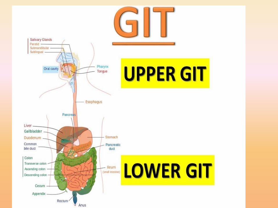

• Consist mainly two parts:
1. Tube or tract consist:a). Oral cavity
b). Pharynx
c). Oesophagus
Upper GIT
d). Stomach
e). Small intestine
f). Large intestine Lower GIT
g). Anus
2. Accessory organsa). Salivary glands
b). Liver and gall bladder
c). pancreas

General Structure of the Digestive
Tract
The wall of GIT is made up 4 principal layers:
1. Mucosa
2. Submucosa
3. Muscularis
4. Serosa or Advetitia

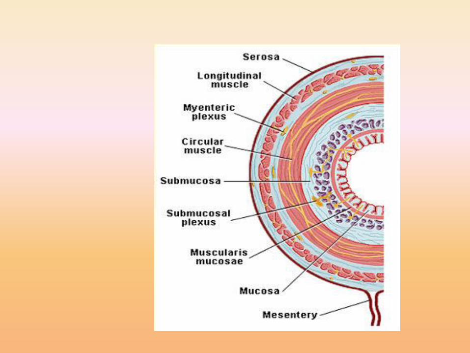

1.Mucosa (Mucous Membrane)
It consists of:
1. Epithelial lining
2. Lamina properia (loose connective tissue rich
in blood and lymph vessels, and sometimes
containing glands smooth muscles).
3. Mascularis mucosae thin muscular layers
saperate the mucosa from submucosa.

2. Submucosa
• It is composed of dense connective tissue with
many blood and lymph vessels and also nerve
plexus which called “Meissner’s plexus”.

3. Muscularis
• It is composed
• Inner circular smooth muscular layer
• Myenteric nerve plexus (Auerbach’s nerve
plexus).
• Outer longitudinal smooth muscular layer.
4. Serosa
It is composed
• A thin layer of loose connective, rich in blood
and lymph vessels and adipose tissue
• Layer of simple squamous epithelium.

Anatomy of oral cavity
• Oral cavity is divided into two parts:
1. Vestibule
a). Lateral: cheeks and lips
b). Medial: upper and lower row of teeth.
c). Posterior: reto-molar area.
2. Buccal cavity:
Roof: Hard palate (palatine) and Soft palate
which ended with uvula.
Floor: tongue.

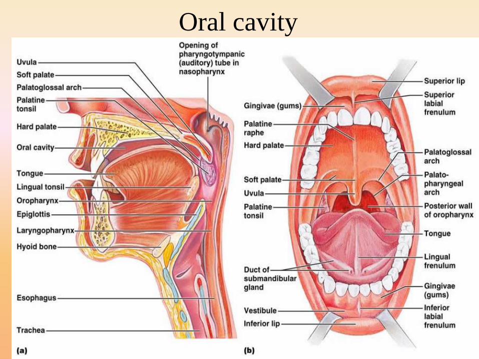
Oral cavity


Structures of the oral cavity
1. Tongue: Muscular organ has different types ofpapillae. These papillae are important for taste.
2. Gum and teeth
3. Salivary glands consist three major glands a).Parotid glands (pairs), b). Sub-mandibular glands(pair) and c). Sublingual gland.
4. Lymphoid tissues: Tonsils ( lingual tonsils,Palatine tonsils).

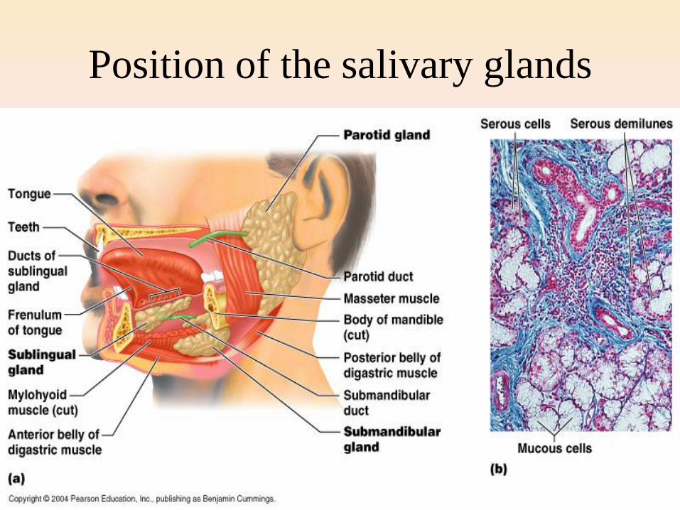
Position of the salivary glands
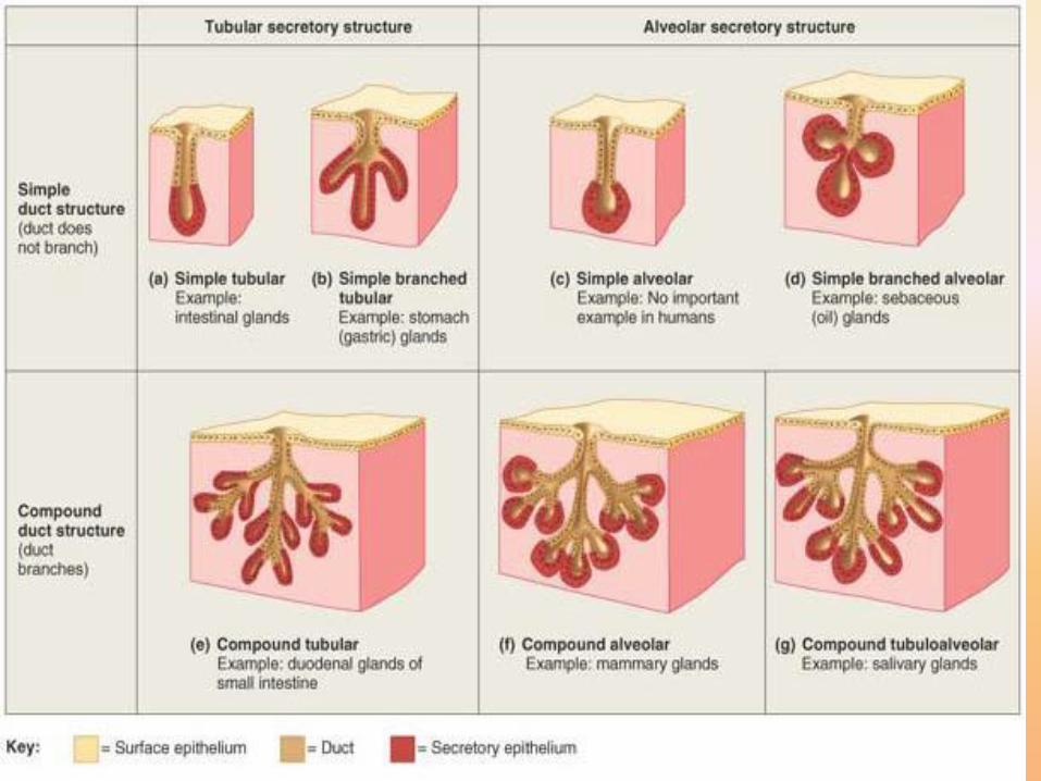
Salivary Glands• Glands are organized arrangement of secretary cells.
• Exocrine glands are organized as acini or tubule, exocrine gland has ducts therefore its secretion reaches by ducts to the affected part.
• All salivary glands are exocrine glands.
• Secretion of salivary glands may be serous, or mucous or mixed.
• Saliva in the mouth has digestive , lubricating, protective functions.
• Each salivary gland receives parasympathatic and sympathaticinnervation.
• Parasympathatic increases the secretion of saliva by all salivary glands.
• Sympathatic innervation remains uncertain.


Pharynx and esophagus
• The pharynx is divided into three parts:
1. Nasopharynx
2. Oropharynx
3. Laryngopharynx
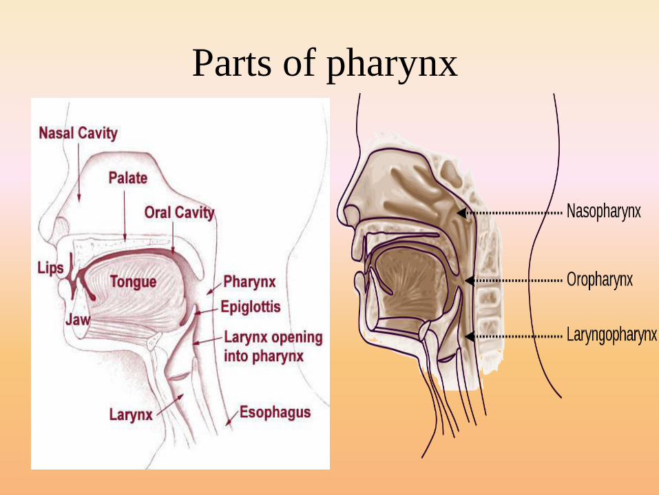
Parts of pharynx
Esophagus
• It is a muscular tube whose function to
transport foodstuff from the mouth to the
stomach.
• It descends toward thoracic cavity , posterior
to the trachea, and enters the abdominal cavity
through the oesophageal hiatus (an opening in
the diaphram, to empty to the stomach.

Histological Structure of Esophagus
• Mucosa and submucosa project into large folds
• Submucosa contains small mucous secreting glands (esophageal glands) their secretion facilitates the transport the foodstuff and protects mucosa).
• Superior third of esophagus muscular layers contains skeletal muscle fiber.
• Middel third of esophagus muscular layers compose from mixture of both skeletal and smooth muscle fiber
• Inferior third of esophageal muscular layers composes only from smooth muscle fibers.
• Serosa covers only the end part of oesophagal wall
Sphincters of esophagus
1. Upper esophageal sphincter composed fromskeletal muscle and located in the upper partof esophagus just below pharynx. Prevent airto inter esophagus.
2. Lower esophageal sphincter (cardiacsphincter) composed of smooth muscle,which normally remain in state of activecontraction to prevent backflow of materialsfrom the stomach into esophagus.

The esophagus and its sphincters
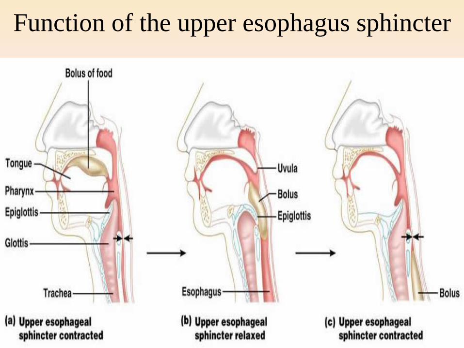
Function of the upper esophagus sphincter

Esophageal movement
Stomach
• It is a muscular organ has ability to digest foods and convert them to chyme.
• The major regions of the stomach include: cardia, fundus, body, and pyelorus regions.
• There are two curvature in the stomach: greater curvature (lateral surface) and lesser curvature (medial surface).
• Extending from curvatures are the lesser omentum and greater omentum which help to tie the stomach to other digestive organs.

Regions of the stomach and their histologic structure.

Anatomy of the Stomach

• The mucosa and submucosa of empty stomach
make longitudinal folds known as (rugae).
• Invagination of epithelial lining formed gastric
pits.
• Lining epithelia and gastric pit cells secrete an
alkaline mucus to protect them from stomach
acidity.
• The muscular layer contains an extra layer in
addition to the circular and longitudinal layers.

• Stomach has exocrine and endocrine secretions.
• The main secretary cells in the stomach are:
1. parietal cells (oxyntic): secret
- HCL (actually H+ and CL-)
-potassium chloride
-traces of electrolytes
-intrinsic factor is essential for absorption of
vitamin B12
2. Chief cells (zymogenic cells) secret Pepsinogen (inactive form of
pepsin enzyme). Pepsin is an enzyme digests proteins.
3. Mucous cells secret mucus
4. Enteroendocrine cells like
G- cells secret gastrin.(enhance of acid by parietal cells)
D- cells secret somatostatin (acts by inhibit release other
hormones like gastrin).
Innervations of the stomach
By sympathatic and parasympathatic
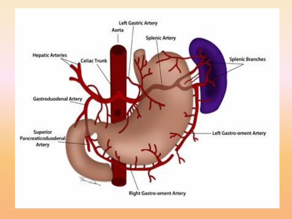
The Blood Supply to the Stomch
The greater curvature is supplied by the right
gastroepiploic artery inferiorly and the left
gastroepiploic artery superiorly. The fundus
of the stomach, and also the upper portion of
the greater curvature, is supplied by the short
gastric artery which arises from the splenic
artery