2019: CHRONIC LIVER DISEASE MANAGEMENT FOR THE ... · 2019: Chronic Liver Disease Management for...
149
2019: CHRONIC LIVER DISEASE MANAGEMENT FOR THE GASTROENTEROLOGIST 2019 NASPGHAN SINGLE TOPIC SYMPOSIUM TO BE HELD IN CONJUNCTION WITH 2019 ANNUAL MEETING SHERATON GRAND CHICAGO * CHICAGO, IL OCTOBER 16, 2019 1
Transcript of 2019: CHRONIC LIVER DISEASE MANAGEMENT FOR THE ... · 2019: Chronic Liver Disease Management for...
2019: CHRONIC LIVER DISEASE MANAGEMENT FOR THE GASTROENTEROLOGIST
2019 NASPGHAN SINGLE TOPIC SYMPOSIUM TO BE HELD IN CONJUNCTION WITH 2019 ANNUAL MEETING
SHERATON GRAND CHICAGO * CHICAGO, IL
OCTOBER 16, 2019
1
WEDNESDAY, OCTOBER 16 – SINGLE TOPIC SYMPOSIUM 2019: Chronic Liver Disease Management for the Gastroenterologist
Sheraton Chicago Ballroom – Level 4
Directors:
Saeed Mohammad, MD
Mercedes Martinez, MD
Advised by the Hepatology Committee
Objective: The objective of this program is to provide guidance on practical aspects of the management of children with chronic liver disease and liver transplantation, including updates on new therapies for chronic liver diseases.
2
WEDNESDAY, OCTOBER 16 – SINGLE TOPIC SYMPOSIUM 2019: Chronic Liver Disease Management for the Gastroenterologist
Table of Contents
How do I best evaluate a cholestatic infant? Sanjiv Harpavat MD Texas Children’s Hospital
How do I interpret genetic results? Saul J. Karpen MD, PhD, Emory University School of Medicine/Children’s Healthcare of Atlanta
What do abnormal liver enzyme levels mean in a tween? William F. Balistreri MD, Cincinnati Children’s Hospital Medical Center
What do I do with this abnormal radiology finding? Jean Molleston MD, Riley Children’s Hospital
SESSION II - FRONTIERS IN LIVER THERAPEUTICS
Keynote Speaker: Outcomes for the future: How do we improve on the status quo? Ronald J. Sokol, MD, FAASLD, Children’s Hospital Colorado
Recognition and stabilization of the pediatric patient with acute liver failure Robert Squires MD Children’s Hospital of Pittsburgh at UPMC
Should I offer treatment for my patients with Hepatitis B or Hepatitis C? Regino P. Gonzalez-Peralta MD, AdventHealth for Children
Are there any medical therapies for NASH? Marialena Mouzaki, MD, Cincinnati Children's Hospital Medical Center
SESSION III - UPDATE ON PORTAL HTN: ASSESSMENT AND MANAGEMENT
When there is good function, but the flow is all wrong: Approach to non-cirrhotic portal hypertension Evelyn Hsu, MD, Seattle Children’s Hospital
What do I do now? The management of portal hypertensive complications: Varices, ascites, and encephalopathy Rene Romero, MD, Children’s Hospital of Atlanta
3
The role of the interventional radiologist in the treatment of portal HTN: How can I help you?
Jared R. Green, MD, Ann and Robert H. Lurie Children’s Hospital
When to consider surgery in the treatment of portal HTN? Riccardo Superina, MD, FRCS(C), FACS, Northwestern University
SESSION IV - LIVER TRANSPLANT: PRE- AND POST-TRANSPLANT CONSIDERATIONS
Referring your patient for liver transplantation Shikha S. Sundaram, MD MSCI, FAASLD, Children’s Hospital Colorado
Where will we get our organs from in 2020? Jean Emond MD, Columbia University Medical College
What should I do if my liver transplant patient has elevated liver tests? Udeme Ekong MD, Georgetown University Hospital
What is a “normal” childhood after liver transplantation? Estella Alonso MD, Ann and Robert H Lurie Children’s Hospital
4
CONTINUING EDUCATION AND MOC PART II
ACCREDITATION STATEMENT The North American Society for Pediatric Gastroenterology, Hepatology and Nutrition (NASPGAN) is accredited by the Accreditation Council for Continuing Medical Education to provide continuing medical education for physicians.
Satisfactory Completion For MOC credit, learners must pass the post-test with a score of 60% or higher and complete an evaluation form to receive a certificate of completion. If you are seeking continuing education credit for a specialty not listed below, it is your responsibility to contact your licensing/certification board to determine course eligibility for your licensing/certification requirement.
Physician The North American Society for Pediatric Gastroenterology, Hepatology and Nutrition (NASPGHAN) is accredited by the Accreditation Council for Continuing Medical Education to provide continuing medical education for physicians. NASPGHAN designates this live activity for a maximum of 8 AMA PRA Category 1 Credit(s)TM. Physicians should claim only the credit commensurate with the extent of their participation in the activity.
Nurses
In support of improving patient care, this activity has been planned and implemented by Amedco LLC and NASPGHAN. Amedco LLC is jointly accredited by the Accreditation Council for Continuing
Medical Education (ACCME), the Accreditation Council for Pharmacy Education (ACPE), and the American Nurses Credentialing Center (ANCC), to provide continuing education for the healthcare team.
Amedco LLC designates this live activity for a maximum of 36.5 contact hours for nurses (21.0 Max for the NASPGHAN Annual Meeting / 8.75 Max for APGNN / 7.5 for the Post Graduate Course / 8.0 for the Single Topic Symposium). Learners should claim only the credit commensurate with the extent of their participation in the activity.
Disclosure of Conflict of Interest The following table of disclosure information is provided to learners and contains the relevant financial relationships that each individual in a position to control the content disclosed to NASPGHAN. All of these relationships were treated as a conflict of interest, and have been resolved. (C7 SCS 6.1--6.2, 6.5)
5
SUPPORTERS Thank you for the generous support through educational grants from:
Albireo Pharma, Inc
Alexion Pharmaceuticals, Inc
Gilead Sciences, Inc
Mirum Pharmaceuticals
New York Presbterian Morgan Stanley Children's Hospital
Retrophin, Inc
Takeda Pharmaceuticals America, Inc.
6
FACULTY SINGLE TOPIC SYMPOSIUM
Estella Alonso MD Medical Director, The Siragusa Transplantation Center Professor of Pediatrics (Gastroenterology, Hepatology, and Nutrition) and Medical Social Sciences Northwestern University Feinberg School of Medicine Sally Burnett Searle Professorship in Pediatric Transplantation Ann and Robert H. Lurie Children’s Hospital Chicago, IL
William F. Balistreri MD Dorothy M. M. Kersten Professor of Pediatrics Director Emeritus, Pediatric Liver Care Center Medical Director Emeritus, Liver Transplantation Program Director Emeritus Fellowship in Transplant Hepatology Professor, UC Department of Pediatrics Cincinnati Children’s Hospital Medical Center Cincinnati, OH
Udeme D. Ekong MD, MPH, FAASLD Associate Professor of Pediatrics Georgetown University Medstar Georgetown University Transplant Institute Washington, DC
Jean C Emond MD Thomas S. Zimmer Professor of Surgery Chief of Transplantation Services Columbia University and The New York Presbyterian Hospital Past President American Society of Transplant Surgeons New York, NY
Regino P. Gonzalez-Peralta, MD Pediatric Gastroenterology, Hepatology and Liver Transplant AdventHealth for Children AdventHealth Transplant Institute Orlando, Florida
Jared R. Green MD Assistant Professor of Radiology Northwestern University Feinberg School of Medicine Division Head, Interventional Radiology Ann and Robert H. Lurie Children’s Hospital Chicago, IL
Sanjiv Harpavat MD, PhD Assistant Professor, Department of Pediatrics Baylor College of Medicine Texas Children’s Hospital Houston, TX
Evelyn Hsu MD Associate Professor of Pediatrics University of Washington School of Medicine Director, Hepatology Fellowship Program Medical Director, Liver Transplant Program Seattle Children’s Hospital Seattle, WA
7
Samar Ibrahim MB, ChB Assistant Professor Pediatric Gastroenterologist Pediatric Transplant Hepatologist Mayo Clinic Rochester, MN
Saul J. Karpen, MD, PhD, FAASLD Raymond F. Schinazi Distinguished Biomedical Chair Professor of Pediatrics Emory University School of Medicine Division Chief, Pediatric Gastroenterology, Hepatology and Nutrition Children’s Healthcare of Atlanta Atlanta, GA
Simon Ling MB, ChB, MRCP (UK) Division Head, Gastroenterology, Hepatology and Nutrition The Hospital for Sick Children Associate Professor, Pediatrics University of Toronto Toronto, Canada
Cara Mack MD Professor, Pediatrics-Gastroenterology, Hepatology and Nutrition University of Colorado, School of Medicine Children’s Hospital Colorado Aurora, CO
Parvathi Mohan MD Director of Hepatology Children’s National Medical Center Professor of Pediatrics The George Washington School of Medicine Washington DC
Jean P. Molleston MD Division Chief, Pediatric Gastroenterology, Hepatology, and Nutrition Riley Hospital for Children Professor of Clinical Pediatrics Indiana University School of Medicine Indianapolis, IN
Marialena Mouzaki, MD MSc Associate Professor of Pediatrics University of Cincinnati Medical Director, Nutrition Services Division of Gastroenterolgy, Hepatology and Nutrition Cincinnati Children’s Hospital Medical Center Cincinnati, OH
Rene Romero MD Professor of Pediatrics Joseph H. Moss Chair in Pediatrics, Hepatology and Liver Transplantation Clinical Director, Pediatric Hepatology Medical Director, Pediatric Liver Transplant Program Children’s Hospital of Atlanta Emory University School of Medicine Atlanta, GA
Ronald J. Sokol MD, FAASLD Professor, Pediatrics-Gastroenterology, Hepatology and Nutrition Section Head, Gastroenterology, Hepatology and Nutrition Director, Colorado Clinical and Translational Sciences Institute Associate Medical Director, Pediatric Liver Center and Liver Transplantation Program Vice Chair, Clinical and Translational Research Assistant Vice Chancellor for Clinical and Translational Science University of Colorado, School of Medicine Children’s Hospital Colorado Aurora, CO
8
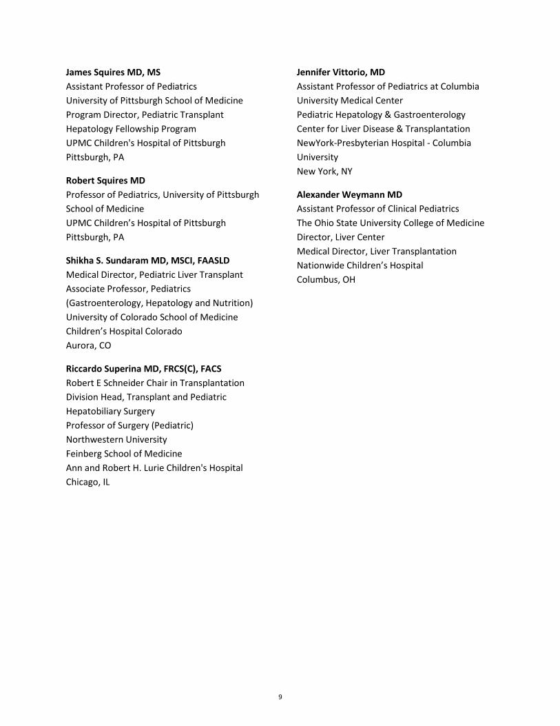
James Squires MD, MS Assistant Professor of Pediatrics University of Pittsburgh School of Medicine Program Director, Pediatric Transplant Hepatology Fellowship Program UPMC Children's Hospital of Pittsburgh Pittsburgh, PA
Robert Squires MD Professor of Pediatrics, University of Pittsburgh School of Medicine UPMC Children’s Hospital of Pittsburgh Pittsburgh, PA
Shikha S. Sundaram MD, MSCI, FAASLD Medical Director, Pediatric Liver Transplant Associate Professor, Pediatrics (Gastroenterology, Hepatology and Nutrition) University of Colorado School of Medicine Children’s Hospital Colorado Aurora, CO
Riccardo Superina MD, FRCS(C), FACS Robert E Schneider Chair in Transplantation Division Head, Transplant and Pediatric Hepatobiliary Surgery Professor of Surgery (Pediatric) Northwestern University Feinberg School of Medicine Ann and Robert H. Lurie Children's Hospital Chicago, IL
Jennifer Vittorio, MD Assistant Professor of Pediatrics at Columbia University Medical Center Pediatric Hepatology & Gastroenterology Center for Liver Disease & Transplantation NewYork-Presbyterian Hospital - Columbia University New York, NY
Alexander Weymann MD Assistant Professor of Clinical Pediatrics The Ohio State University College of Medicine Director, Liver Center Medical Director, Liver Transplantation Nationwide Children’s Hospital Columbus, OH
9
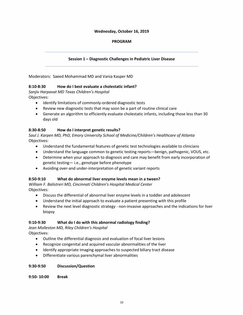
Wednesday, October 16, 2019
PROGRAM
Session 1 – Diagnostic Challenges in Pediatric Liver Disease
Moderators: Saeed Mohammad MD and Vania Kasper MD
8:10-8:30 How do I best evaluate a cholestatic infant? Sanjiv Harpavat MD Texas Children’s Hospital Objectives:
• Identify limitations of commonly-ordered diagnostic tests • Review new diagnostic tests that may soon be a part of routine clinical care • Generate an algorithm to efficiently evaluate cholestatic infants, including those less than 30
days old 8:30-8:50 How do I interpret genetic results? Saul J. Karpen MD, PhD, Emory University School of Medicine/Children’s Healthcare of Atlanta Objectives:
• Understand the fundamental features of genetic test technologies available to clinicians • Understand the language common to genetic testing reports—benign, pathogenic, VOUS, etc. • Determine when your approach to diagnosis and care may benefit from early incorporation of
genetic testing— i.e., genotype before phenotype • Avoiding over-and under-interpretation of genetic variant reports
8:50-9:10 What do abnormal liver enzyme levels mean in a tween? William F. Balistreri MD, Cincinnati Children’s Hospital Medical Center Objectives:
• Discuss the differential of abnormal liver enzyme levels in a toddler and adolescent • Understand the initial approach to evaluate a patient presenting with this profile • Review the next level diagnostic strategy - non-invasive approaches and the indications for liver
biopsy 9:10-9:30 What do I do with this abnormal radiology finding? Jean Molleston MD, Riley Children’s Hospital Objectives:
• Outline the differential diagnosis and evaluation of focal liver lesions • Recognize congenital and acquired vascular abnormalities of the liver • Identify appropriate imaging approaches to suspected biliary tract disease • Differentiate various parenchymal liver abnormalities
9:30-9:50 Discussion/Question 9:50- 10:00 Break
10
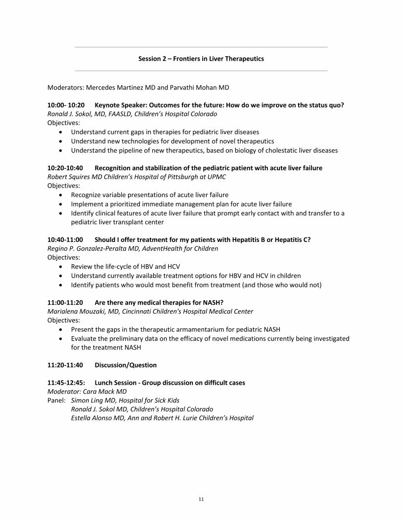
Session 2 – Frontiers in Liver Therapeutics
Moderators: Mercedes Martinez MD and Parvathi Mohan MD 10:00- 10:20 Keynote Speaker: Outcomes for the future: How do we improve on the status quo? Ronald J. Sokol, MD, FAASLD, Children’s Hospital Colorado Objectives:
• Understand current gaps in therapies for pediatric liver diseases • Understand new technologies for development of novel therapeutics • Understand the pipeline of new therapeutics, based on biology of cholestatic liver diseases
10:20-10:40 Recognition and stabilization of the pediatric patient with acute liver failure Robert Squires MD Children’s Hospital of Pittsburgh at UPMC Objectives:
• Recognize variable presentations of acute liver failure • Implement a prioritized immediate management plan for acute liver failure • Identify clinical features of acute liver failure that prompt early contact with and transfer to a
pediatric liver transplant center 10:40-11:00 Should I offer treatment for my patients with Hepatitis B or Hepatitis C? Regino P. Gonzalez-Peralta MD, AdventHealth for Children Objectives:
• Review the life-cycle of HBV and HCV • Understand currently available treatment options for HBV and HCV in children • Identify patients who would most benefit from treatment (and those who would not)
11:00-11:20 Are there any medical therapies for NASH? Marialena Mouzaki, MD, Cincinnati Children's Hospital Medical Center Objectives:
• Present the gaps in the therapeutic armamentarium for pediatric NASH • Evaluate the preliminary data on the efficacy of novel medications currently being investigated
for the treatment NASH 11:20-11:40 Discussion/Question 11:45-12:45: Lunch Session - Group discussion on difficult cases Moderator: Cara Mack MD Panel: Simon Ling MD, Hospital for Sick Kids
Ronald J. Sokol MD, Children’s Hospital Colorado Estella Alonso MD, Ann and Robert H. Lurie Children’s Hospital
11
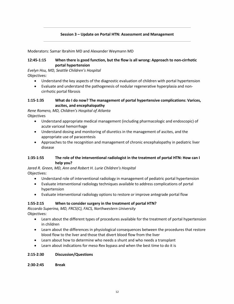
Session 3 – Update on Portal HTN: Assessment and Management
Moderators: Samar Ibrahim MD and Alexander Weymann MD 12:45-1:15 When there is good function, but the flow is all wrong: Approach to non-cirrhotic portal hypertension Evelyn Hsu, MD, Seattle Children’s Hospital Objectives:
• Understand the key aspects of the diagnostic evaluation of children with portal hypertension • Evaluate and understand the pathogenesis of nodular regenerative hyperplasia and non-
cirrhotic portal fibrosis 1:15-1:35 What do I do now? The management of portal hypertensive complications: Varices, ascites, and encephalopathy Rene Romero, MD, Children’s Hospital of Atlanta Objectives
• Understand appropriate medical management (including pharmacologic and endoscopic) of acute variceal hemorrhage
• Understand dosing and monitoring of diuretics in the management of ascites, and the appropriate use of paracentesis
• Approaches to the recognition and management of chronic encephalopathy in pediatric liver disease
1:35-1:55 The role of the interventional radiologist in the treatment of portal HTN: How can I help you? Jared R. Green, MD, Ann and Robert H. Lurie Children’s Hospital Objectives:
• Understand role of interventional radiology in management of pediatric portal hypertension • Evaluate interventional radiology techniques available to address complications of portal
hypertension • Evaluate interventional radiology options to restore or improve antegrade portal flow
1:55-2:15 When to consider surgery in the treatment of portal HTN? Riccardo Superina, MD, FRCS(C), FACS, Northwestern University Objectives:
• Learn about the different types of procedures available for the treatment of portal hypertension in children
• Learn about the differences in physiological consequences between the procedures that restore blood flow to the liver and those that divert blood flow from the liver
• Learn about how to determine who needs a shunt and who needs a transplant • Learn about indications for meso Rex bypass and when the best time to do it is
2:15-2:30 Discussion/Questions 2:30-2:45 Break
12
Session 4 – Liver Transplant: Pre- and Post-Transplant Considerations
Moderators: James Squires MD and Jennifer Vittorio MD 2:45-3:05 Referring your patient for liver transplantation Shikha S. Sundaram, MD MSCI, FAASLD, Children’s Hospital Colorado Objectives
• Understand when to refer a patient for a transplant evaluation • Understand what happens during a transplant evaluation • Understand indications/contraindications for liver transplantation • Understand how to help your patient choose a transplant program
3:05-3:25 Where will we get our organs from in 2020? Jean Emond MD, Columbia University Medical College Objectives:
• Understand the role of living donation and split livers in a pediatric program • Understand current data on PHS increased risk donations • Be familiar with updates in xeno and bioengineered organs
3:25-3:45 What should I do if my liver transplant patient has elevated liver tests? Udeme Ekong MD, Georgetown University Hospital Objectives:
• Recognize the differential diagnoses of elevated liver tests in a pediatric liver transplant recipient
• Become familiar with testing to consider in the setting of liver allograft dysfunction • Become familiar with proposed diagnostic criteria for acute and chronic antibody mediated
rejection 3:45-4:05 What is a “normal” childhood after liver transplantation? Estella Alonso MD, Ann and Robert H Lurie Children’s Hospital Objectives:
• Be able to identify the common physical and psychosocial challenges children experience following liver transplantation
• Be able to identify risk factors for lower than expected physical function and school performance following liver transplantation
• Be able to design screening programs to implement in a post-transplant ambulatory care setting that will identify children with high risk for lower psychosocial outcomes
4:05-4:25 Discussion/Questions 4:25-4:30 Closure Mercedes Martinez MD, Columbia University School of Medicine 4:30-6 pm Reception
13

14
How do I best evaluate a cholestatic infant? Sanjiv Harpavat, MD [email protected]
NASPGHAN 2019 Annual Meeting Single Topic Symposium Chronic Liver Disease Management for the Gastroenterologist
Disclosures
I have no relevant financial relationships or affiliations with commercial interests to disclose.
Supported by K23DK109207
Case
A 3 week former full-term infant is referred for persistent jaundice and an elevated serum direct bilirubin level.
• Born full-term without complications• PCP noticed jaundice at two weeks• PCP measured fractioned serum bilirubin levels*
• Labs: AST 43, ALT 32, GGT 453, Direct bili 2.3• Exam: 50th percentile for weight, yellow stools
*Joint NASPGHAN/ESPGHAN guidelines: Fawaz et al. (2017) JPGN 64: 154-168.
15
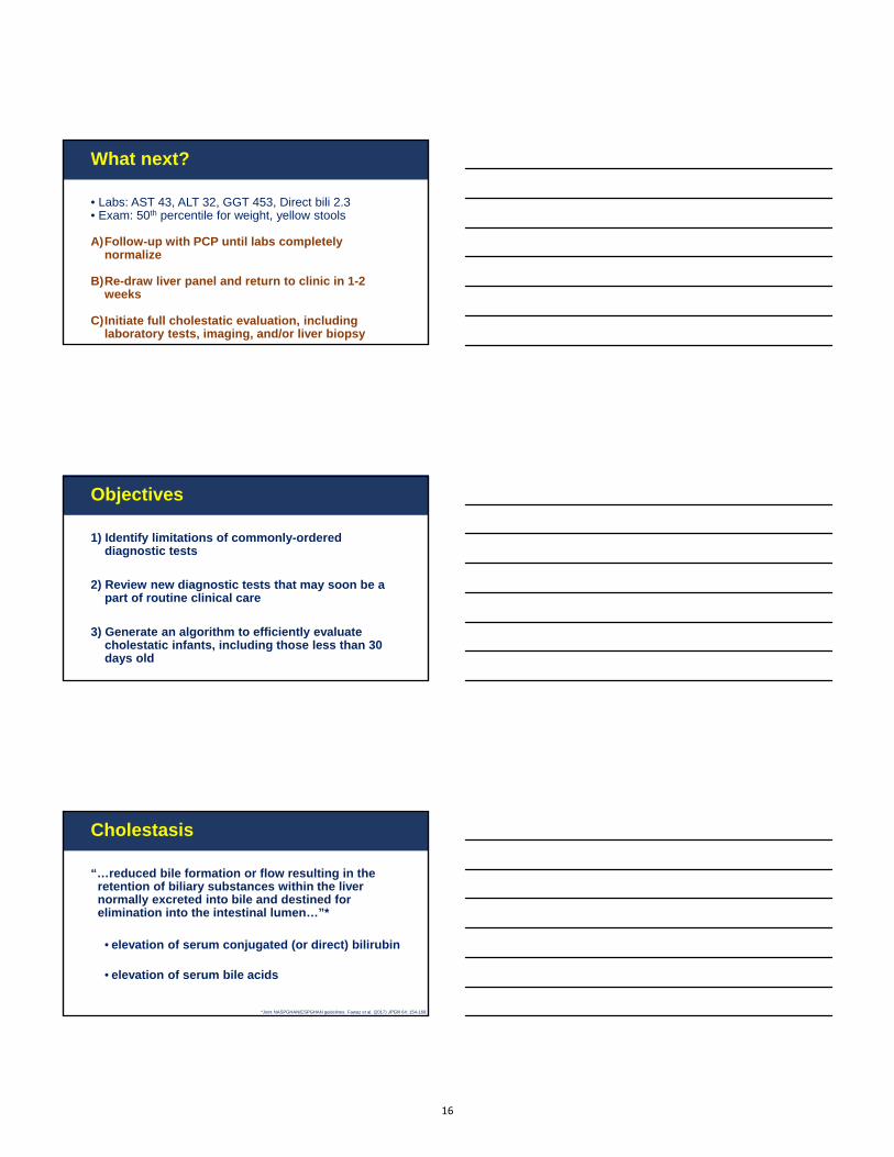
What next?
• Labs: AST 43, ALT 32, GGT 453, Direct bili 2.3• Exam: 50th percentile for weight, yellow stools
A)Follow-up with PCP until labs completely normalize
B)Re-draw liver panel and return to clinic in 1-2 weeks
C)Initiate full cholestatic evaluation, including laboratory tests, imaging, and/or liver biopsy
Objectives
1) Identify limitations of commonly-ordered diagnostic tests
2) Review new diagnostic tests that may soon be a part of routine clinical care
3) Generate an algorithm to efficiently evaluate cholestatic infants, including those less than 30 days old
Cholestasis
“…reduced bile formation or flow resulting in the retention of biliary substances within the liver normally excreted into bile and destined for elimination into the intestinal lumen…”*
• elevation of serum conjugated (or direct) bilirubin
• elevation of serum bile acids
*Joint NASPGHAN/ESPGHAN guidelines: Fawaz et al. (2017) JPGN 64: 154-168.
16
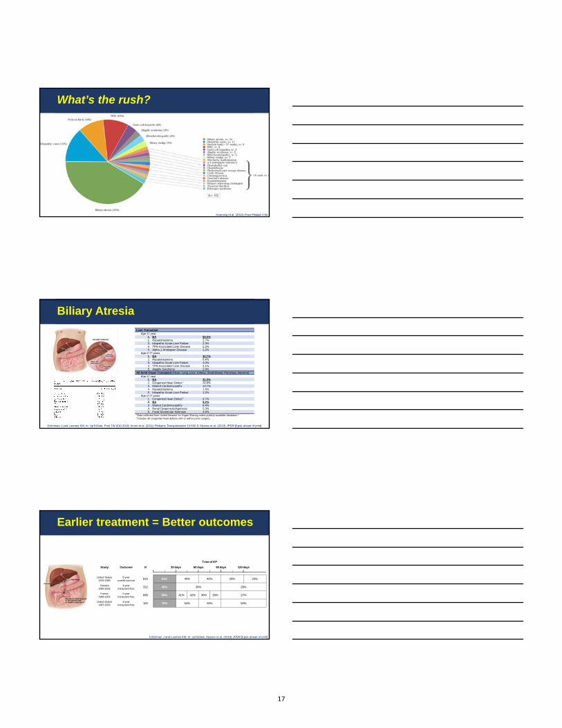
What’s the rush?
Hoerning et al. (2014) Front Pediatr 2:65.
Biliary Atresia
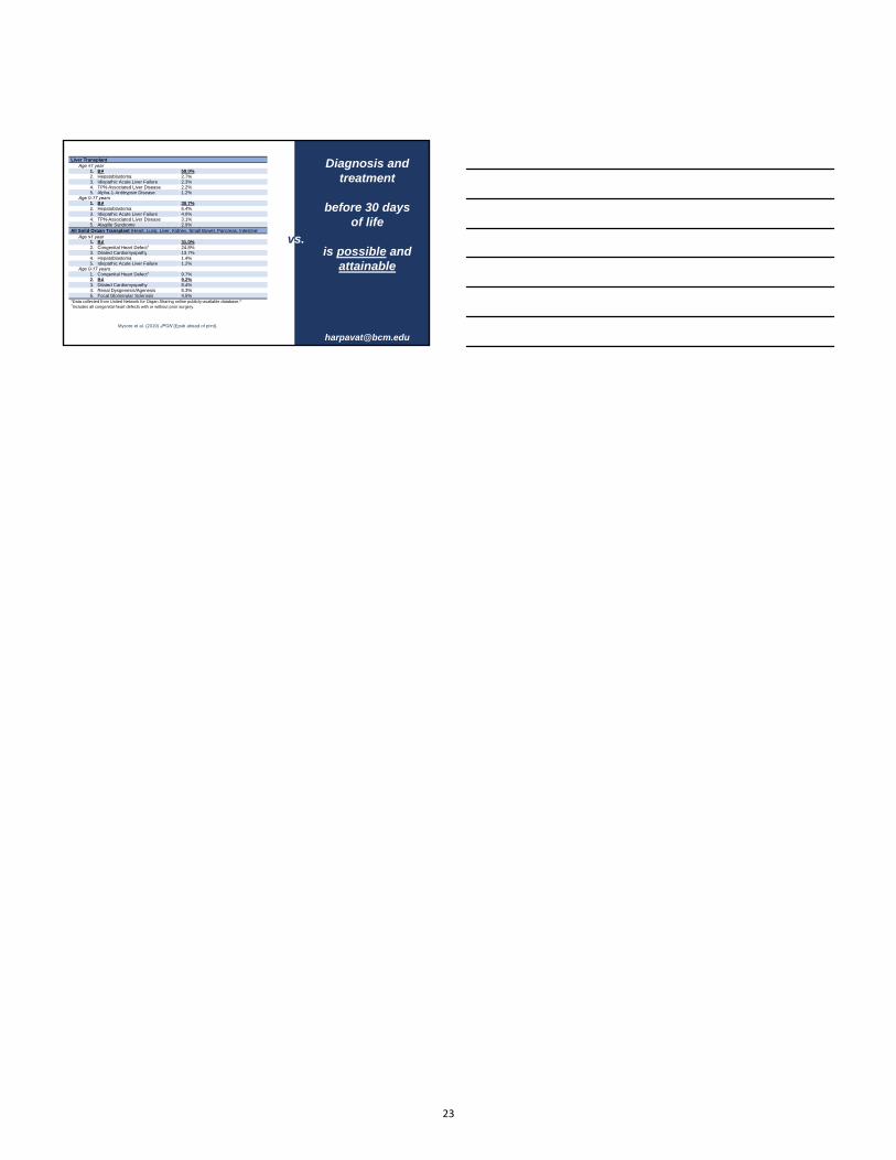
Erlichman J and Loomes KM, In: UpToDate, Post TW (Ed) 2018; Arnon et al. (2011) Pediatric Transplantation 15:650-8; Mysore et al. (2019) JPGN [Epub ahead of print].
Liver Transplant Age ≤1 year
1. BA 59.0% 2. Hepatoblastoma 2.7% 3. Idiopathic Acute Liver Failure 2.3% 4. TPN-Associated Liver Disease 2.2% 5. Alpha-1-Antitrypsin Disease 1.2%
Age 0-17 years 1. BA 30.7% 2. Hepatoblastoma 6.4% 3. Idiopathic Acute Liver Failure 4.0% 4. TPN-Associated Liver Disease 3.1% 5. Alagille Syndrome 2.9%
All Solid Organ Transplant (Heart, Lung, Liver, Kidney, Small Bowel, Pancreas, Intestine) Age ≤1 year
1. BA 31.0% 2. Congenital Heart Defect† 24.8% 3. Dilated Cardiomyopathy 13.7% 4. Hepatoblastoma 1.4% 5. Idiopathic Acute Liver Failure 1.2%
Age 0-17 years 1. Congenital Heart Defect† 9.7% 2. BA 9.2% 3. Dilated Cardiomyopathy 8.4% 4. Renal Dysgenesis/Agenesis 5.3% 5. Focal Glomerular Sclerosis 4.5%
*Data collected from United Network for Organ Sharing online publicly-available database.3 †Includes all congenital heart defects with or without prior surgery.
Earlier treatment = Better outcomes
Time of KP
Study Outcome N 30 days 60 days 90 days 120 days. 5
United States1976-1989
5-yearoverall survival
816 63% 44% 40% 29% 29%
Canada1985-2002
4-yeartransplant-f ree
312 49% 36% 28%
France 1986-2002
5-year transplant-f ree
695 58% 41% 42% 36% 26% 27%
United States1997-2000
2-year transplant-f ree
100 70% 54% 50% 50%
Erlichman J and Loomes KM, In: UpToDate; Mysore et al. (2019) JPGN [Epub ahead of print].
17
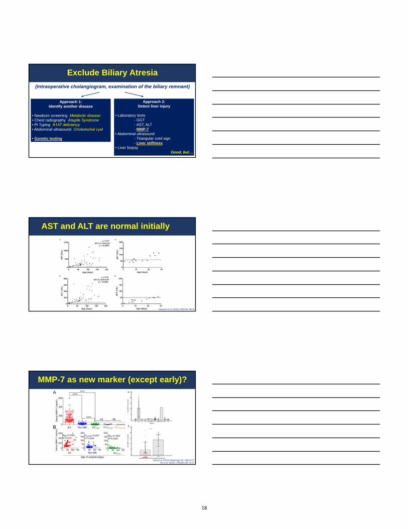
Exclude Biliary Atresia
(Intraoperative cholangiogram, examination of the biliary remnant)
Approach 1:Identify another disease
• Newborn screening Metabolic disease• Chest radiography Alagille Syndrome• PI Typing A1AT deficiency• Abdominal ultrasound Choledochal cyst
• Genetic testing
Approach 2:Detect liver injury
• Laboratory tests- GGT- AST, ALT- MMP-7
• Abdominal ultrasound- Triangular cord sign- Liver stiffness
• Liver biopsyGood, but…
AST and ALT are normal initially
Harpavat et al. (2018) JPGN 66: 850-6.
MMP-7 as new marker (except early)?
Yang et al. (2018) Hepatology 68: 2069-2077.Wu et al. (2019) J Pediatr 208: 30-37.
18
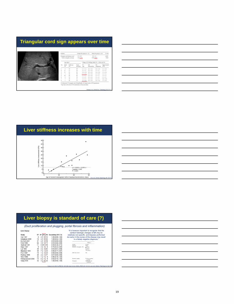
Triangular cord sign appears over time
Hwang et al. (2018) Eur J Radiology 28:1771-7.
Liver stiffness increases with time
Wu et al. (2018) Hepatology 68: 616-24.
Liver biopsy is standard of care (?)
(Duct proliferation and plugging, portal fibrosis and inflammation)
“It is however important to recognize that the earliest histologic changes of BA may be
relatively non-specific, and biopsies performed too early in the course of the disease may result
in a falsely negative diagnosis.”
Fawaz et al. (2017) JPGN 64: 154-168; Azar G et al. (2002) JPGN 134: 212-15; Lee et al. (2016) J Ped Surg 51:753-761.
19
What next?
• Labs: AST 43, ALT 32, GGT 453, Direct bili 2.3• Exam: 50th percentile for weight, yellow stools
A)Follow-up with PCP until labs completely normalize
B)Re-draw liver panel and return to clinic in 1-2 weeks
C)Initiate full cholestatic evaluation, including laboratory tests, imaging, and/or liver biopsy
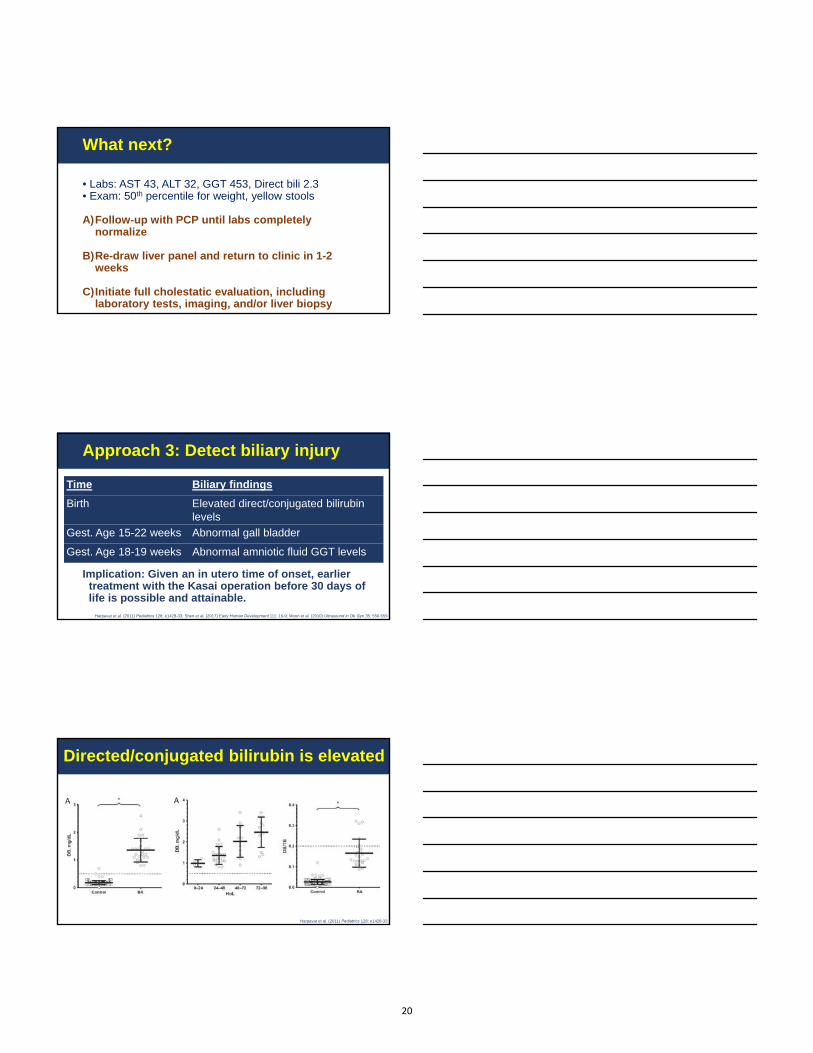
Approach 3: Detect biliary injury
Implication: Given an in utero time of onset, earlier treatment with the Kasai operation before 30 days of life is possible and attainable.
Time Biliary findings
Birth Elevated direct/conjugated bilirubinlevels
Gest. Age 15-22 weeks Abnormal gall bladder
Gest. Age 18-19 weeks Abnormal amniotic fluid GGT levels
Harpavat et al. (2011) Pediatrics 128: e1428-33; Shen et al. (2017) Early Human Development 111: 16-9; Moon et al. (2010) Ultrasound in Ob Gyn 35: 556-559.
Directed/conjugated bilirubin is elevated
Harpavat et al. (2011) Pediatrics 128: e1428-33.
20
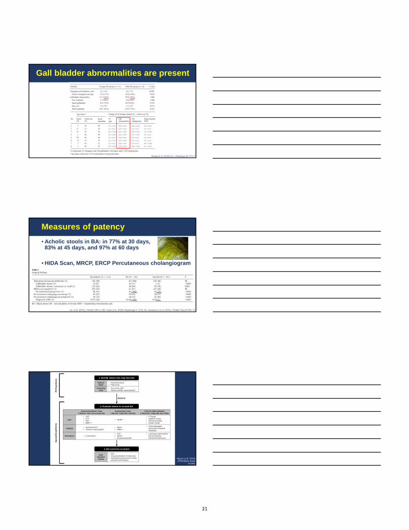
Gall bladder abnormalities are present
Hwang et al. (2018) Eur J Radiology 28:1771-7.
Measures of patency
• Acholic stools in BA: in 77% at 30 days, 83% at 45 days, and 97% at 60 days
• HIDA Scan, MRCP, ERCP Percutaneous cholangiogram
Gu et al. (2015) J Pediatr 166:e1–902; Hsiao et al. (2008) Hepatology 47:1233–40; Jancelewicz et al. (2015) J Pediatr Surg 50:363–70.
1. Identify infants who may have BA
2. Evaluate infants to exclude BA
3. BA cannot be excluded
History/Exam
• Persistent icterus• Pale stools
ScreeningTests
• Stool color card*• Newborn DB/Bc measurements*
Commonly Ordered Tests (“Normal” does not exclude BA)
Exclusionary Tests(“Normal” makes BA unlikely)
Tests for Other Diseases (“Abnormal” makes BA less likely)
Labs
• AST‡
• ALT‡
• GGT• MMP-7‡
• DB/Bc*
• PI Typing• Newborn screen• Infectious studies• Genetic tests§
Imaging• Abdominal US*‡
• Transient elastography‡• HIDA*• MRCP*
• Chest radiographabnormal for butterf ly vertebrate
Procedures • Liver Biopsy ‡• PTC*• ERCP*• Duodenal aspirate*
• Liver biopsy abnormal for bile duct paucity, transporter expression
Gold-Standard Testing
• IOC*• Visual examination of biliary tree• Histological assessment of biliary
remnant (conf irmation)
PC
P p
erf
orm
sS
pec
ialis
t p
erf
orm
s
Referral
Mysore et al. (2019) JPGN [Epub ahead
of print].
21
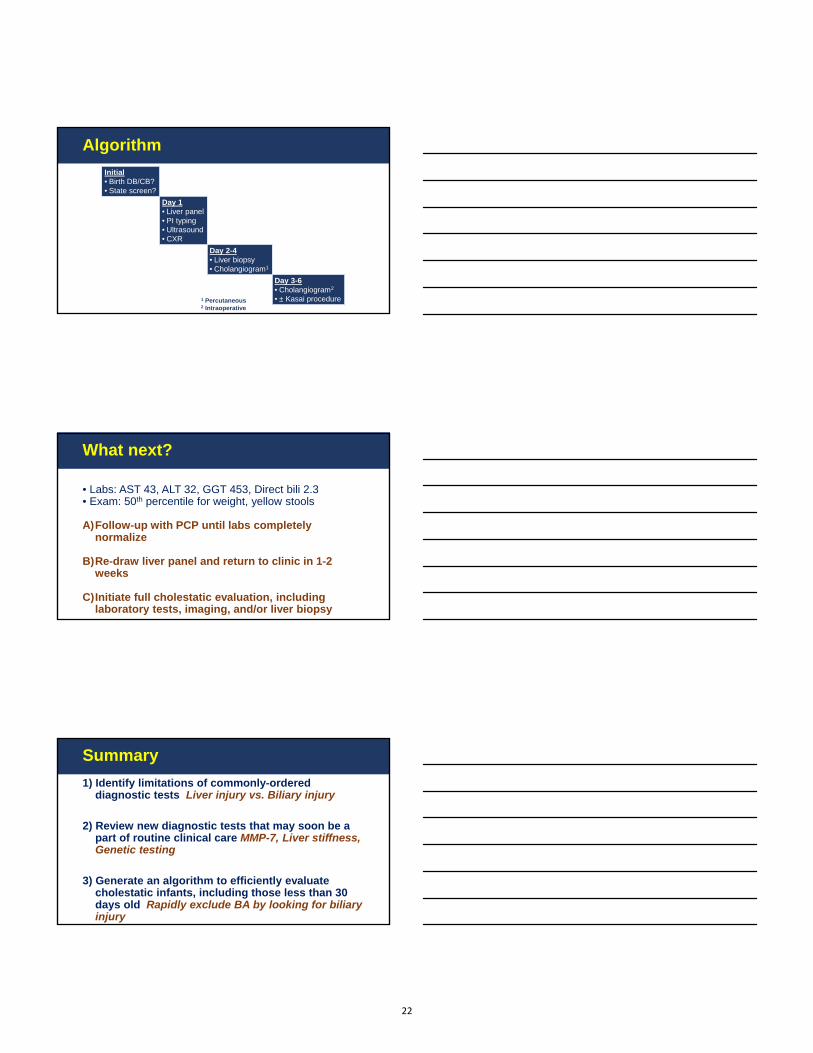
Algorithm
Initial• Birth DB/CB?• State screen?
Day 1• Liver panel• PI typing• Ultrasound• CXR
Day 2-4• Liver biopsy• Cholangiogram1
Day 3-6• Cholangiogram2
• ± Kasai procedure1 Percutaneous2 Intraoperative
What next?
• Labs: AST 43, ALT 32, GGT 453, Direct bili 2.3• Exam: 50th percentile for weight, yellow stools
A)Follow-up with PCP until labs completely normalize
B)Re-draw liver panel and return to clinic in 1-2 weeks
C)Initiate full cholestatic evaluation, including laboratory tests, imaging, and/or liver biopsy
Summary
1) Identify limitations of commonly-ordered diagnostic tests Liver injury vs. Biliary injury
2) Review new diagnostic tests that may soon be a part of routine clinical care MMP-7, Liver stiffness, Genetic testing
3) Generate an algorithm to efficiently evaluate cholestatic infants, including those less than 30 days old Rapidly exclude BA by looking for biliary injury
22
Liver Transplant Age ≤1 year
1. BA 59.0% 2. Hepatoblastoma 2.7% 3. Idiopathic Acute Liver Failure 2.3% 4. TPN-Associated Liver Disease 2.2% 5. Alpha-1-Antitrypsin Disease 1.2%
Age 0-17 years 1. BA 30.7% 2. Hepatoblastoma 6.4% 3. Idiopathic Acute Liver Failure 4.0% 4. TPN-Associated Liver Disease 3.1% 5. Alagille Syndrome 2.9%
All Solid Organ Transplant (Heart, Lung, Liver, Kidney, Small Bowel, Pancreas, Intestine) Age ≤1 year
1. BA 31.0% 2. Congenital Heart Defect† 24.8% 3. Dilated Cardiomyopathy 13.7% 4. Hepatoblastoma 1.4% 5. Idiopathic Acute Liver Failure 1.2%
Age 0-17 years 1. Congenital Heart Defect† 9.7% 2. BA 9.2% 3. Dilated Cardiomyopathy 8.4% 4. Renal Dysgenesis/Agenesis 5.3% 5. Focal Glomerular Sclerosis 4.5%
*Data collected from United Network for Organ Sharing online publicly-available database.3 †Includes all congenital heart defects with or without prior surgery.
Diagnosis and treatment
before 30 days of life
is possible and attainable
Mysore et al. (2019) JPGN [Epub ahead of print].
vs.
23
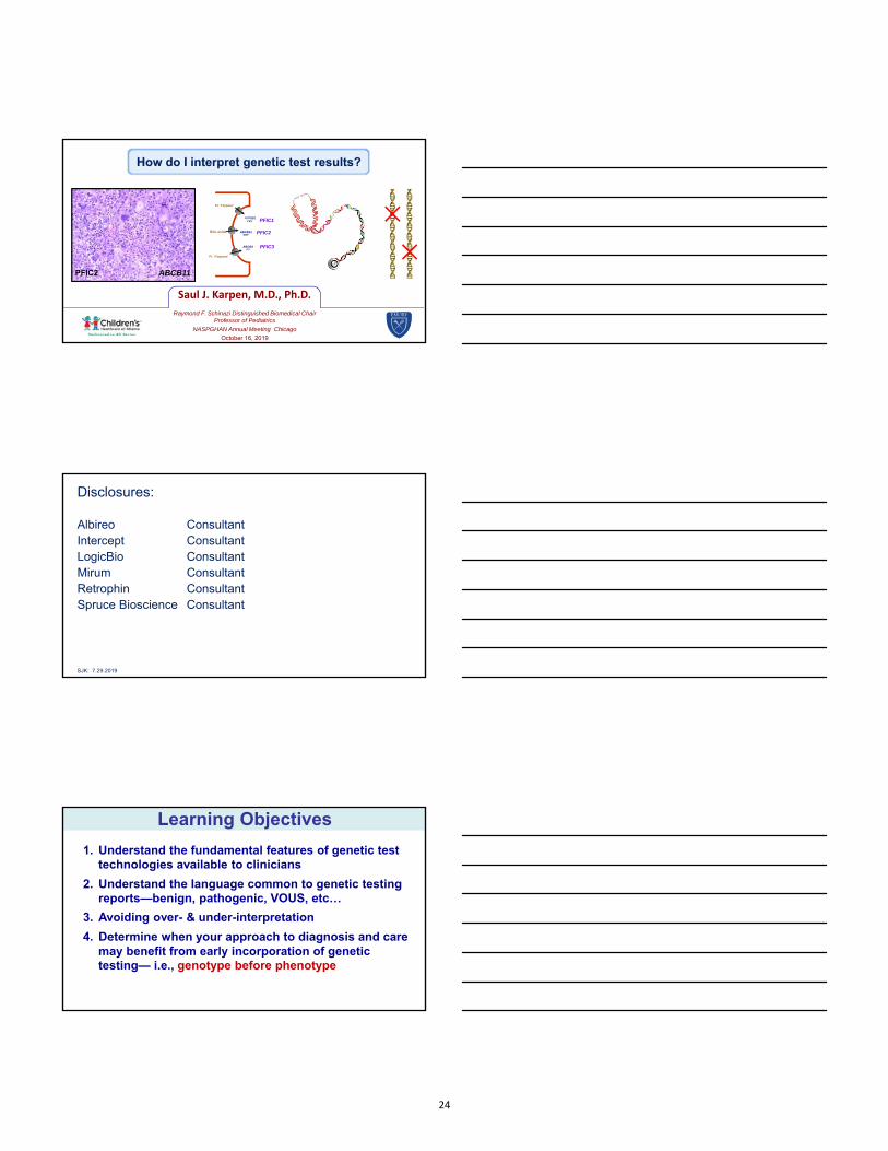
Saul J. Karpen, M.D., Ph.D.
Raymond F. Schinazi Distinguished Biomedical ChairProfessor of Pediatrics
NASPGHAN Annual Meeting Chicago
October 16, 2019
How do I interpret genetic test results?How do I interpret genetic test results?
ATP8B1FIC1
ABCB11BSEP
ABCB4MDR3
Bile acids
PL ‘Flippase’
PL ‘Floppase’
PFIC2
PFIC3
PFIC1
PFIC2 ABCB11
Disclosures:
Albireo ConsultantIntercept ConsultantLogicBio ConsultantMirum ConsultantRetrophin ConsultantSpruce Bioscience Consultant
SJK: 7.29.2019
1. Understand the fundamental features of genetic test technologies available to clinicians
2. Understand the language common to genetic testing reports—benign, pathogenic, VOUS, etc…
3. Avoiding over- & under-interpretation
4. Determine when your approach to diagnosis and care may benefit from early incorporation of genetic testing— i.e., genotype before phenotype
Learning Objectives
24
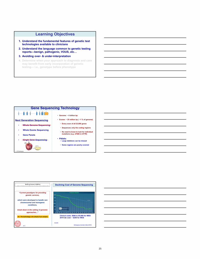
1. Understand the fundamental features of genetic test technologies available to clinicians
2. Understand the language common to genetic testing reports—benign, pathogenic, VOUS, etc…
3. Avoiding over- & under-interpretation
4. Determine when your approach to diagnosis and care may benefit from early incorporation of genetic testing— i.e., genotype before phenotype
Learning Objectives
Next Generation Sequencing
• Whole Genome Sequencing
• Whole Exome Sequencing
• Gene Panels
• Single Gene Sequencing
Gene Sequencing Technology
• Genome: ~ 6 billion bp
• Exome: ~ 25 million bp ( ~1 % of genome)
• Every exon of all 23,000 genes
• Sequences only the coding regions
• No need to know to look for individual mutations (e.g. F508 in CFTR)
• Pitfalls:• Large deletions can be missed
• Some regions are poorly covered
U. Of Washington
2012
“Current paradigms for providing
genetic services,
which were developed to handle rare
chromosomal and monogenic
conditions,
break down in the setting of genomic
approaches...”
Clinical costs: $500 to $5,000 for WES2019 lab cost: ~ $250 for WES
Declining Cost of Genome Sequencing
NHGRI
Schwarze Genetic Med 2018
Or: The technology is far ahead of our analysis
25
“Help” from the Report
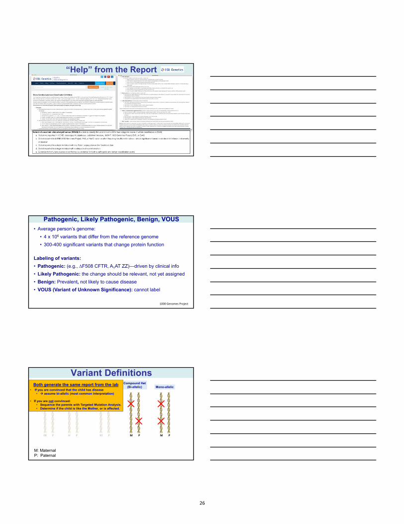
Pathogenic, Likely Pathogenic, Benign, VOUS
• Average person’s genome:
• 4 x 10⁶ variants that differ from the reference genome
• 300-400 significant variants that change protein function
Labeling of variants:
• Pathogenic: (e.g., F508 CFTR, A1AT ZZ)—driven by clinical info
• Likely Pathogenic: the change should be relevant, not yet assigned
• Benign: Prevalent, not likely to cause disease
• VOUS (Variant of Unknown Significance): cannot label
1000 Genomes Project
Variant DefinitionsHomozygous Heterozygous
Compound Het(Bi-allelic) Mono-allelic
“No VariantsDetected”
M P M PM P M PM P
Both generate the same report from the lab• If you are convinced that the child has disease
• assume bi-allelic (most common interpretation)
• If you are not convinced: • Sequence the parents with Targeted Mutation Analysis.• Determine if the child is like the Mother, or is affected.
M: MaternalP: Paternal
26
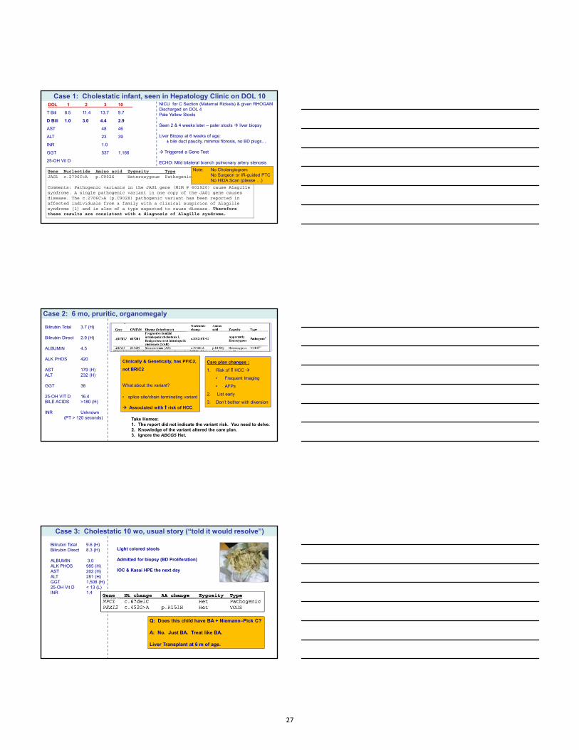
Gene Nucleotide Amino acid Zygosity Type JAG1 c.2706C>A p.C902X Heterozygous Pathogenic
Comments: Pathogenic variants in the JAG1 gene (MIM # 601920) cause Alagillesyndrome. A single pathogenic variant in one copy of the JAG1 gene causes disease. The c.2706C>A (p.C902X) pathogenic variant has been reported in affected individuals from a family with a clinical suspicion of Alagillesyndrome [1] and is also of a type expected to cause disease. Therefore these results are consistent with a diagnosis of Alagille syndrome.
DOL 1 2 3 10 28
T Bili 8.5 11.4 13.7 9.7 4.9
D Bili 1.0 3.0 4.4 2.9 3.2
AST 48 46 55
ALT 23 39 39
INR 1.0 0.9
GGT 537 1,166 244
25-OH Vit D 23
NICU for C Section (Maternal Rickets) & given RHOGAMDischarged on DOL 4Pale Yellow Stools
Seen 2 & 4 weeks later – paler stools liver biopsy
Liver Biopsy at 6 weeks of age:± bile duct paucity, minimal fibrosis, no BD plugs…
Triggered a Gene Test
ECHO: Mild bilateral branch pulmonary artery stenosis
Note: No CholangiogramNo Surgeon or IR-guided PTCNo HIDA Scan (please …)
Case 1: Cholestatic infant, seen in Hepatology Clinic on DOL 10
Bilirubin Total 3.7 (H)
Bilirubin Direct 2.9 (H)
ALBUMIN 4.5
ALK PHOS 420
AST 179 (H)ALT 232 (H)
GGT 38
25-OH VIT D 16.4BILE ACIDS >180 (H)
INR Unknown(PT > 120 seconds)
Case 2: 6 mo, pruritic, organomegaly
Clinically & Genetically, has PFIC2,
not BRIC2
What about the variant?
• splice site/chain terminating variant
Associated with ⬆ risk of HCC.
Care plan changes :
1. Risk of ⬆ HCC
• Frequent Imaging
• AFPs
2. List early
3. Don’t bother with diversion
Take Homes: 1. The report did not indicate the variant risk. You need to delve.2. Knowledge of the variant altered the care plan.3. Ignore the ABCG5 Het.
Bilirubin Total 9.6 (H)Bilirubin Direct 8.3 (H)
ALBUMIN 3.0ALK PHOS 985 (H)AST 202 (H)ALT 281 (H)GGT 1,508 (H)25-OH Vit D < 13 (L)INR 1.4
Light colored stools
Admitted for biopsy (BD Proliferation)
IOC & Kasai HPE the next day
Case 3: Cholestatic 10 wo, usual story (“told it would resolve”)
Q: Does this child have BA + Niemann–Pick C?
A: No. Just BA. Treat like BA.
Liver Transplant at 6 m of age.
27
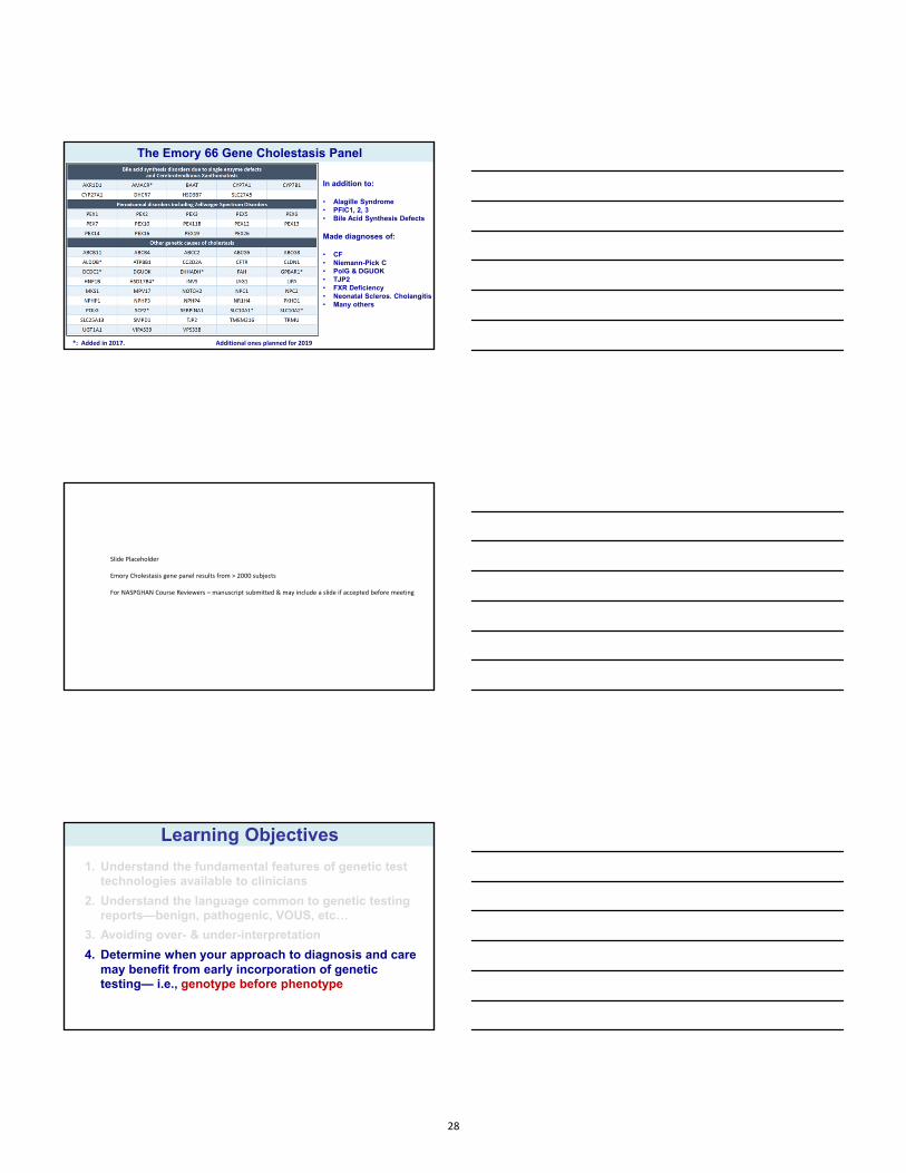
The Emory 66 Gene Cholestasis Panel
*: Added in 2017. Additional ones planned for 2019
In addition to:
• Alagille Syndrome• PFIC1, 2, 3• Bile Acid Synthesis Defects
Made diagnoses of:
• CF• Niemann-Pick C• PolG & DGUOK• TJP2• FXR Deficiency• Neonatal Scleros. Cholangitis• Many others
Slide Placeholder
Emory Cholestasis gene panel results from > 2000 subjects
For NASPGHAN Course Reviewers – manuscript submitted & may include a slide if accepted before meeting
1. Understand the fundamental features of genetic test technologies available to clinicians
2. Understand the language common to genetic testing reports—benign, pathogenic, VOUS, etc…
3. Avoiding over- & under-interpretation
4. Determine when your approach to diagnosis and care may benefit from early incorporation of genetic testing— i.e., genotype before phenotype
Learning Objectives
28
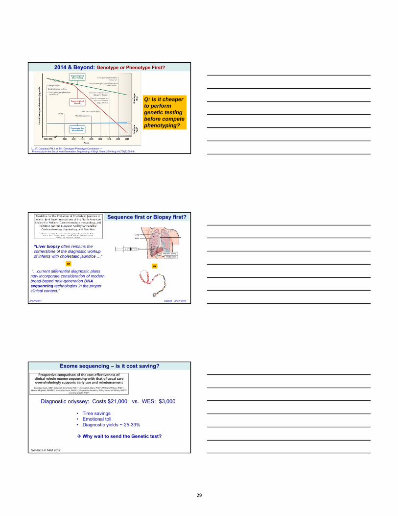
2014 & Beyond: Genotype or Phenotype First?
Lu JT, Campeau PM, Lee BH. Genotype–Phenotype Correlation —Promiscuity in the Era of Next-Generation Sequencing. N Engl J Med. 2014 Aug 14;371(7):593–6.
Q: Is it cheaper to perform genetic testing before compete phenotyping?
JPGN 2017
“…current differential diagnostic plans now incorporate consideration of modern broad-based next-generation DNA sequencing technologies in the proper clinical context.”
“Liver biopsy often remains the cornerstone of the diagnostic workup of infants with cholestatic jaundice …”
Dezsőfi JPGN 2014
oror
Sequence first or Biopsy first?
Exome sequencing – is it cost saving?
Genetics in Med 2017
Diagnostic odyssey: Costs $21,000 vs. WES: $3,000
• Time savings• Emotional toll• Diagnostic yields ~ 25-33%
Why wait to send the Genetic test?
29
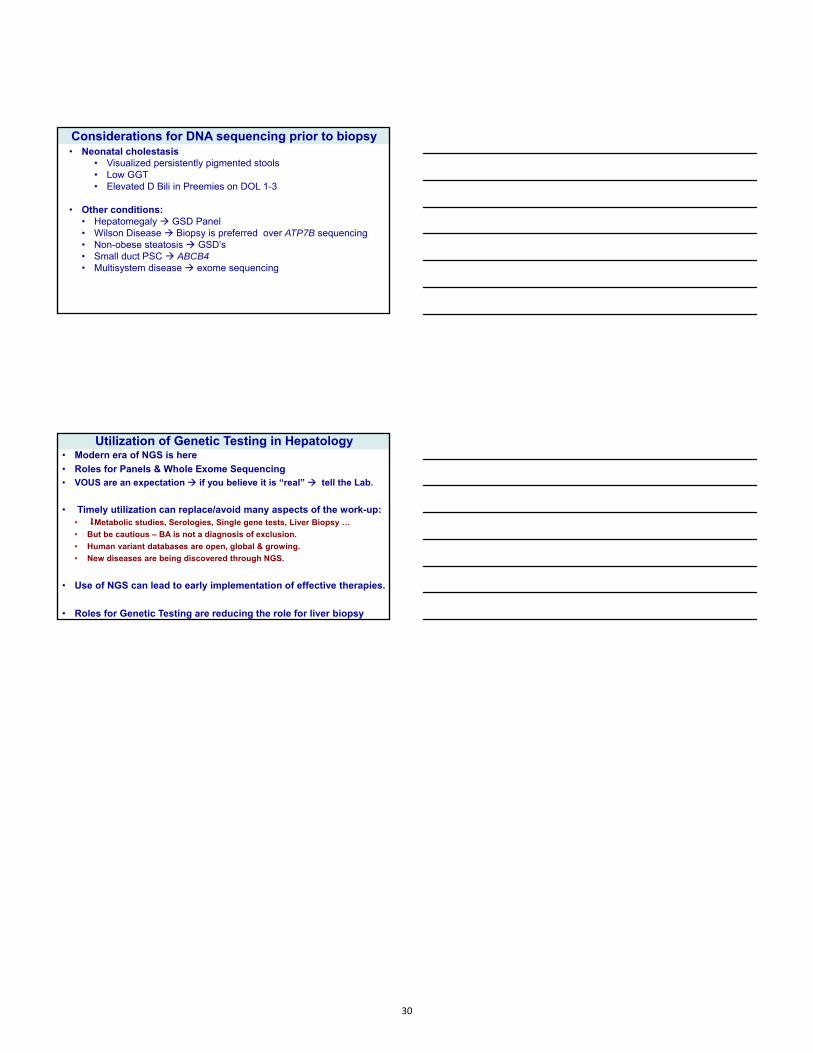
Considerations for DNA sequencing prior to biopsy • Neonatal cholestasis
• Visualized persistently pigmented stools• Low GGT• Elevated D Bili in Preemies on DOL 1-3
• Other conditions:• Hepatomegaly GSD Panel• Wilson Disease Biopsy is preferred over ATP7B sequencing• Non-obese steatosis GSD’s • Small duct PSC ABCB4• Multisystem disease exome sequencing
Utilization of Genetic Testing in Hepatology• Modern era of NGS is here
• Roles for Panels & Whole Exome Sequencing• VOUS are an expectation if you believe it is “real” tell the Lab.
• Timely utilization can replace/avoid many aspects of the work-up:• ⬇Metabolic studies, Serologies, Single gene tests, Liver Biopsy …
• But be cautious – BA is not a diagnosis of exclusion.
• Human variant databases are open, global & growing.
• New diseases are being discovered through NGS.
• Use of NGS can lead to early implementation of effective therapies.
• Roles for Genetic Testing are reducing the role for liver biopsy
30
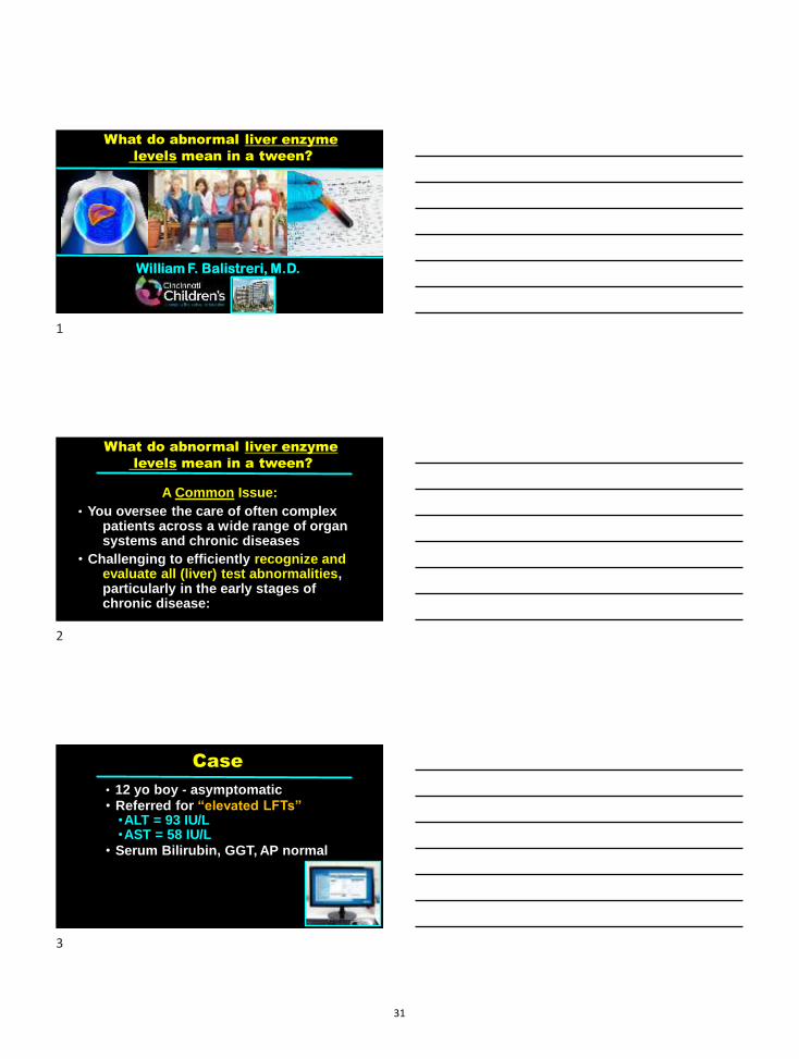
What do abnormal liver enzyme
levels mean in a tween?
William F. Balistreri, M.D.
A Common Issue:• You oversee the care of often complex
patients across a wide range of organsystems and chronic diseases
• Challenging to efficiently recognize andevaluate all (liver) test abnormalities,particularly in the early stages ofchronic disease:
What do abnormal liver enzyme
levels mean in a tween?
Case
• 12 yo boy - asymptomatic• Referred for “elevated LFTs”•ALT = 93 IU/L•AST = 58 IU/L
• Serum Bilirubin, GGT, AP normal
1
2
3
31
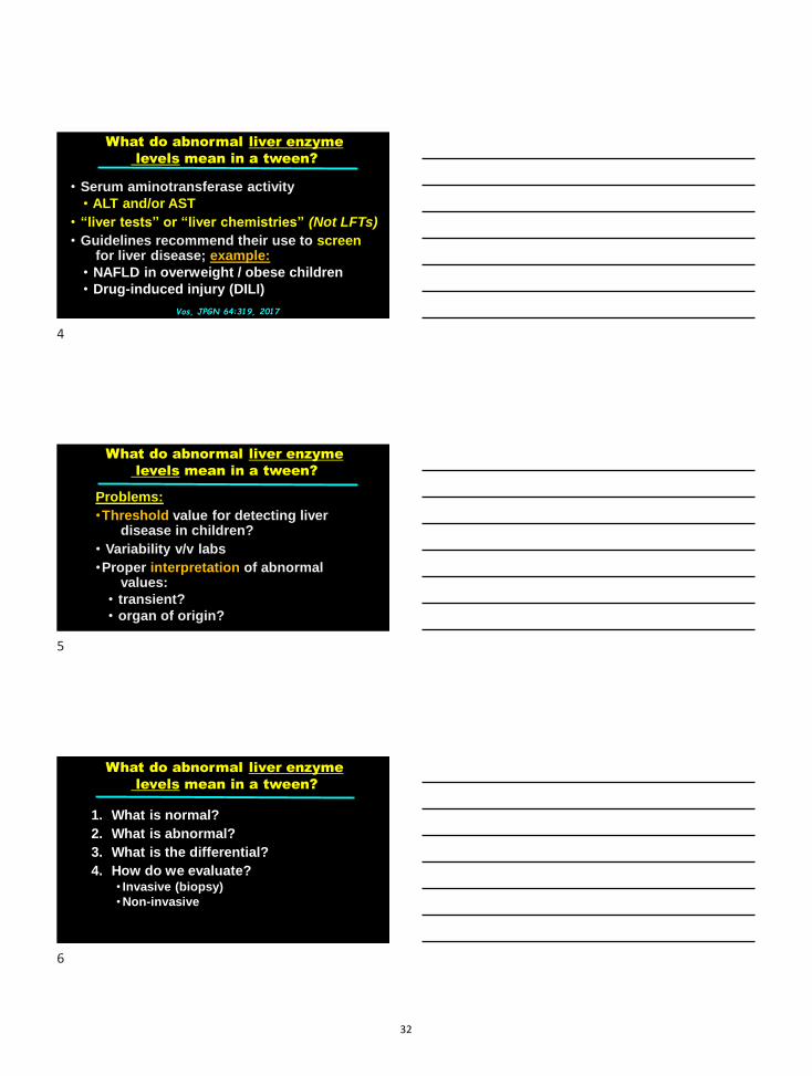
• Serum aminotransferase activity• ALT and/or AST
• “liver tests” or “liver chemistries” (Not LFTs)
• Guidelines recommend their use to screenfor liver disease; example:
• NAFLD in overweight / obese children• Drug-induced injury (DILI)
What do abnormal liver enzyme
levels mean in a tween?
Vos, JPGN 64:319, 2017
Problems:•Threshold value for detecting liver
disease in children?• Variability v/v labs•Proper interpretation of abnormal
values:• transient?• organ of origin?
What do abnormal liver enzyme
levels mean in a tween?
1. What is normal?2. What is abnormal?3. What is the differential?4. How do we evaluate?
• Invasive (biopsy)•Non-invasive
What do abnormal liver enzyme
levels mean in a tween?
4
5
6
32
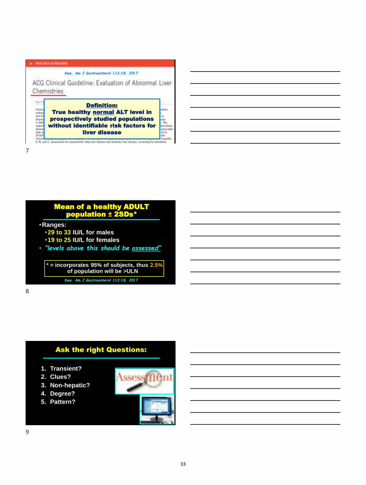
Kwo, Am J Gastroenterol 112:18, 2017
Definition:
True healthy normal ALT level in
prospectively studied populations
without identifiable risk factors for
liver disease
Mean of a healthy ADULTpopulation ± 2SDs*
•Ranges:•29 to 33 IU/L for males•19 to 25 IU/L for females
• “levels above this should be assessed”
* = incorporates 95% of subjects, thus 2.5%of population will be >ULN
Kwo, Am J Gastroenterol 112:18, 2017
1. Transient?2. Clues?3. Non-hepatic?4. Degree?5. Pattern?
Ask the right Questions:
7
8
9
33
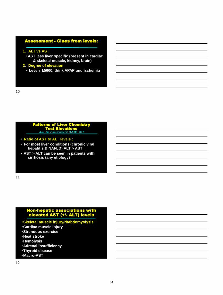
1. ALT vs AST•AST less liver specific (present in cardiac
& skeletal muscle, kidney, brain)2. Degree of elevation• Levels ≥5000, think APAP and ischemia
Assessment - Clues from levels:
• Ratio of AST to ALT levels :• For most liver conditions (chronic viral
hepatitis & NAFLD) ALT > AST• AST > ALT can be seen in patients with
cirrhosis (any etiology)
Kwo, Am J Gastroenterol 112:18, 2017
Patterns of Liver Chemistry Test Elevations
Non-hepatic associations with elevated AST (+/- ALT) levels
•Skeletal muscle injury/rhabdomyolysis•Cardiac muscle injury•Strenuous exercise•Heat stroke•Hemolysis•Adrenal insufficiency•Thyroid disease•Macro-AST
10
11
12
34
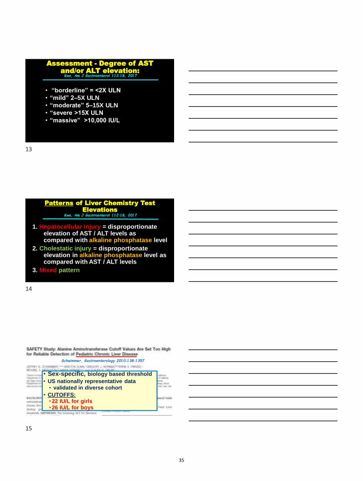
• “borderline” = <2X ULN
• “mild” 2–5X ULN• “moderate” 5–15X ULN• “severe >15X ULN
• “massive” >10,000 IU/L
Kwo, Am J Gastroenterol 112:18, 2017
Assessment - Degree of AST and/or ALT elevation:
Patterns of Liver Chemistry Test Elevations
1. Hepatocellular injury = disproportionateelevation of AST / ALT levels ascompared with alkaline phosphatase level
2. Cholestatic injury = disproportionateelevation in alkaline phosphatase level ascompared with AST / ALT levels
3. Mixed pattern
Kwo, Am J Gastroenterol 112:18, 2017
Schwimmer, Gastroenterology 2010;138:1357
• Sex-specific, biology based threshold• US nationally representative data• validated in diverse cohort
• CUTOFFS:•22 IU/L for girls•26 IU/L for boys
Schwimmer, Gastroenterology 2010;138:1357
13
14
15
35
Colantonio, Clin Chem 58:854, 2012
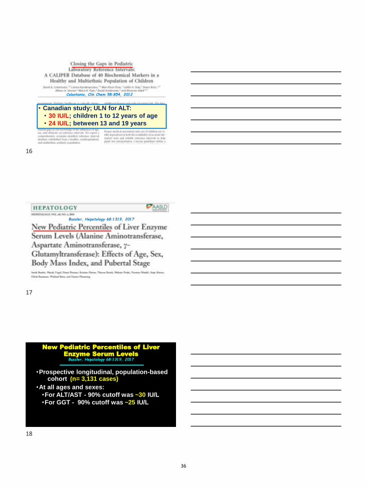
• Canadian study; ULN for ALT:• 30 IU/L; children 1 to 12 years of age• 24 IU/L; between 13 and 19 years
Bussler, Hepatology 68:1319, 2017
New Pediatric Percentiles of Liver Enzyme Serum Levels
Bussler, Hepatology 68:1319, 2017
•Prospective longitudinal, population-basedcohort (n= 3,131 cases)
•At all ages and sexes:•For ALT/AST - 90% cutoff was ~30 IU/L•For GGT - 90% cutoff was ~25 IU/L
16
17
18
36
Bus
sler,
Hepa
tology
68:1
319,
2017
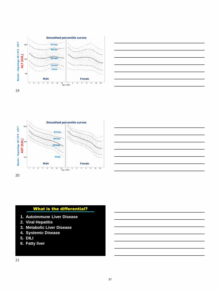
Male Female
ALT
(IU
/L)
97%ile
90%ile
50%ile
10%ile
3%ile
Smoothed percentile curves Smoothed percentile curves
Bus
sler,
Hepa
tology
68:1
319,
2017
AS
T (
IU/L
)
Male Female
3%ile
50%ile
90%ile
97%ile
Smoothed percentile curves
What is the differential?
1. Autoimmune Liver Disease2. Viral Hepatitis3. Metabolic Liver Disease4. Systemic Disease5. DILI6. Fatty liver
19
20
21
37
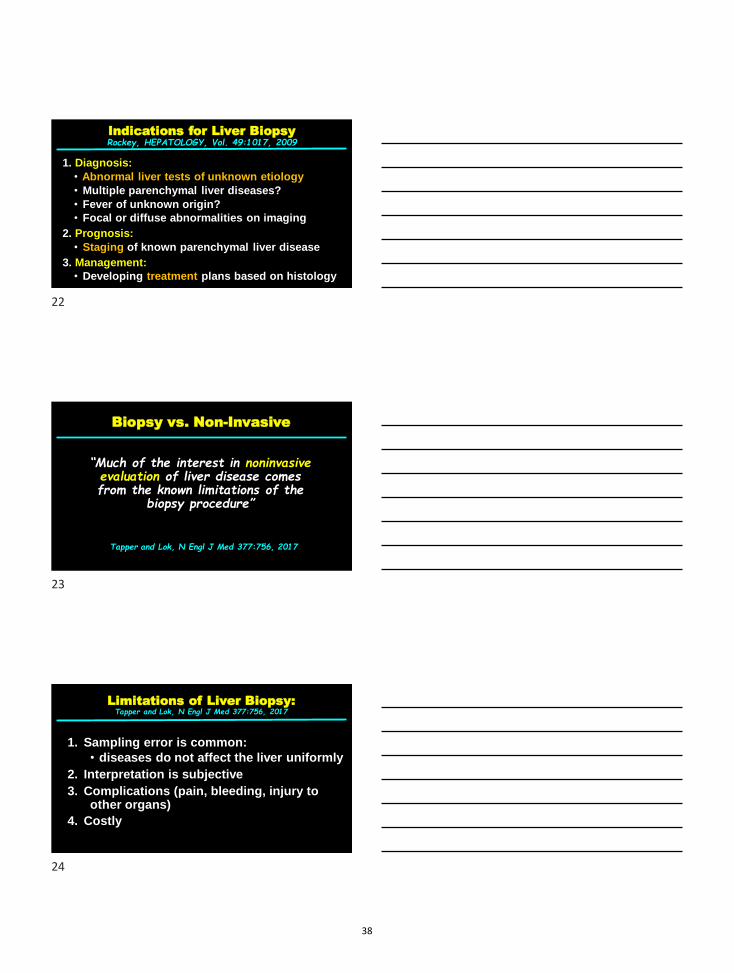
Indications for Liver BiopsyRockey, HEPATOLOGY, Vol. 49:1017, 2009
1. Diagnosis:• Abnormal liver tests of unknown etiology• Multiple parenchymal liver diseases?• Fever of unknown origin?• Focal or diffuse abnormalities on imaging
2. Prognosis:• Staging of known parenchymal liver disease
3. Management:• Developing treatment plans based on histology
“Much of the interest in noninvasive evaluation of liver disease comes from the known limitations of the
biopsy procedure”
Biopsy vs. Non-Invasive
Tapper and Lok, N Engl J Med 377:756, 2017
Limitations of Liver Biopsy:Tapper and Lok, N Engl J Med 377:756, 2017
1. Sampling error is common:• diseases do not affect the liver uniformly
2. Interpretation is subjective3. Complications (pain, bleeding, injury to
other organs)4. Costly
22
23
24
38
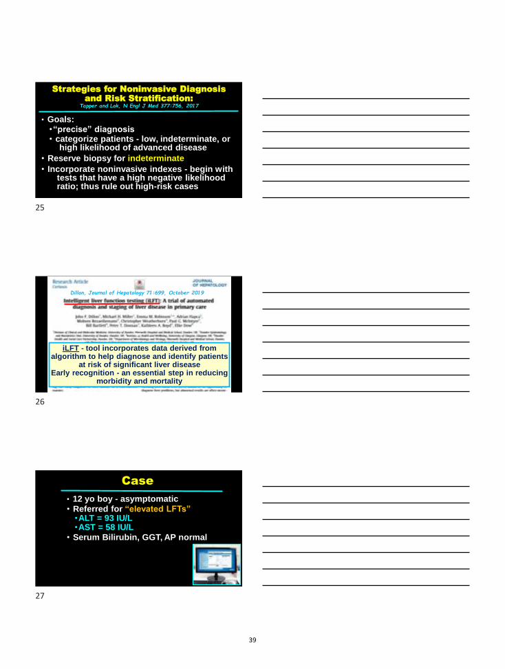
Strategies for Noninvasive Diagnosis and Risk Stratification:
Tapper and Lok, N Engl J Med 377:756, 2017
• Goals:•“precise” diagnosis• categorize patients - low, indeterminate, or
high likelihood of advanced disease• Reserve biopsy for indeterminate• Incorporate noninvasive indexes - begin with
tests that have a high negative likelihoodratio; thus rule out high-risk cases
Dillon, Journal of Hepatology 71:699, October 2019iLFT - tool incorporates data derived fromalgorithm to help diagnose and identify patients
at risk of significant liver diseaseEarly recognition - an essential step in reducing
morbidity and mortality
IU/L
Dillon, Journal of Hepatology 71:699, October 2019
Case
• 12 yo boy - asymptomatic• Referred for “elevated LFTs”•ALT = 93 IU/L•AST = 58 IU/L
• Serum Bilirubin, GGT, AP normal
25
26
27
39
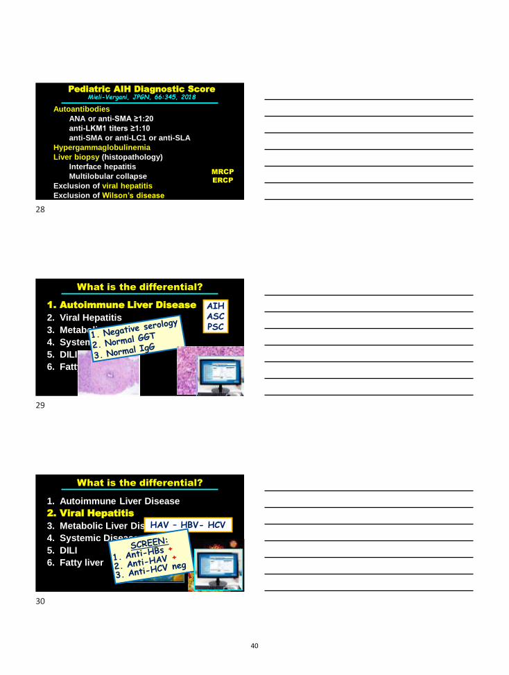
Pediatric AIH Diagnostic ScoreMieli-Vergani, JPGN, 66:345, 2018
AutoantibodiesANA or anti-SMA ≥1:20
anti-LKM1 titers ≥1:10
anti-SMA or anti-LC1 or anti-SLAHypergammaglobulinemiaLiver biopsy (histopathology)
Interface hepatitisMultilobular collapse
Exclusion of viral hepatitisExclusion of Wilson’s disease
MRCP
ERCP
What is the differential?
1. Autoimmune Liver Disease
2. Viral Hepatitis3. Metabolic Liver Disease4. Systemic Disease5. DILI6. Fatty liver
AIHASCPSC
What is the differential?
1. Autoimmune Liver Disease2. Viral Hepatitis
3. Metabolic Liver Disease4. Systemic Disease5. DILI6. Fatty liver
HAV – HBV- HCV
28
29
30
40
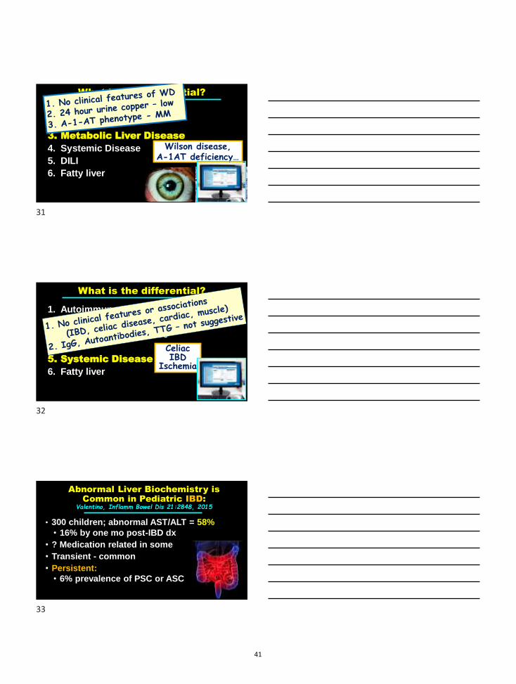
What is the differential?
1. Autoimmune Liver Disease2. Viral Hepatitis3. Metabolic Liver Disease
4. Systemic Disease5. DILI6. Fatty liver
Wilson disease, A-1AT deficiency…
What is the differential?
1. Autoimmune Liver Disease2. Viral Hepatitis3. Metabolic Liver Disease4. DILI5. Systemic Disease
6. Fatty liver
CeliacIBD
Ischemia
Abnormal Liver Biochemistry is Common in Pediatric IBD:
Valentino, Inflamm Bowel Dis 21:2848, 2015
• 300 children; abnormal AST/ALT = 58%• 16% by one mo post-IBD dx
• ? Medication related in some• Transient - common• Persistent:• 6% prevalence of PSC or ASC
31
32
33
41
Prevalence and causes of abnormal liver tests in patients with Celiac Disease
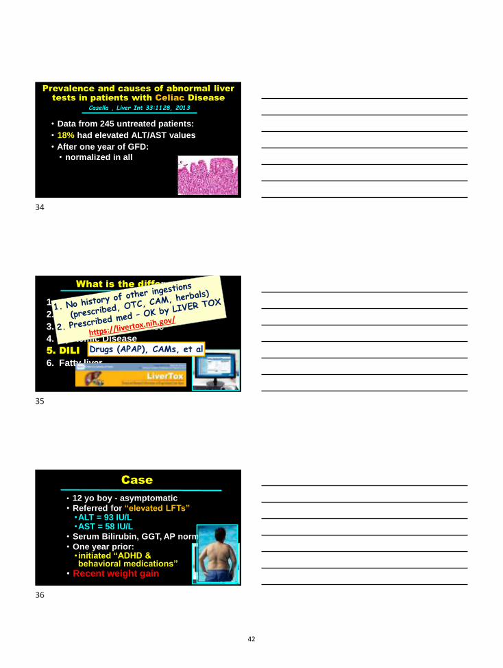
Casella , Liver Int 33:1128, 2013
• Data from 245 untreated patients:• 18% had elevated ALT/AST values• After one year of GFD:• normalized in all
What is the differential?
1. Autoimmune Liver Disease2. Viral Hepatitis3. Metabolic Liver Disease4. Systemic Disease5. DILI
6. Fatty liver
Drugs (APAP), CAMs, et al
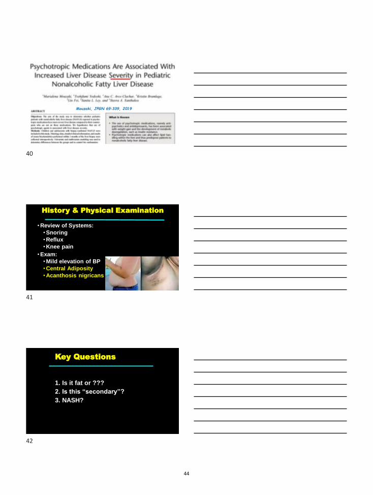
Case
• 12 yo boy - asymptomatic• Referred for “elevated LFTs”•ALT = 93 IU/L•AST = 58 IU/L
• Serum Bilirubin, GGT, AP normal• One year prior:• initiated “ADHD &behavioral medications”
• Recent weight gain
34
35
36
42
139# (6/9/19)
87# (6/9/18)
Weight
Case
initiated “behavioral meds”
BMI 35 kg/m2(140% of 95th percentile)
Nicol, JAMA Psychiatry. 75:788, 2018
De Hert & Detraux, JAMA Psychiatry 75:771, 2018
37
38
39
43
Mouzaki, JPGN 69:339, 2019
History & Physical Examination
•Review of Systems:•Snoring•Reflux•Knee pain
•Exam:•Mild elevation of BP•Central Adiposity•Acanthosis nigricans
Key Questions
1. Is it fat or ???2. Is this “secondary”?
3. NASH?
40
41
42
44
Causes of Secondary Steatosis
Macrovesicular steatosis:• Medications: MTX, steroids• Mauriac• Wilson’s disease• CF• Lipodystrophy• Starvation• TPN• A-beta-LP• XS alcohol consumption• “Syndromic”
Microvesicular steatosis:• Reye’s Syndrome
• Meds (valproate)• Acute fatty liver of pregnancy• HELLP• Inborn errors of metabolism:•LAL-D•CESD•Wolman disease•LCAT deficiency
Causes of Secondary Steatosis
Chalasani, Hepatology 67:328, 2018
43
44
45
45
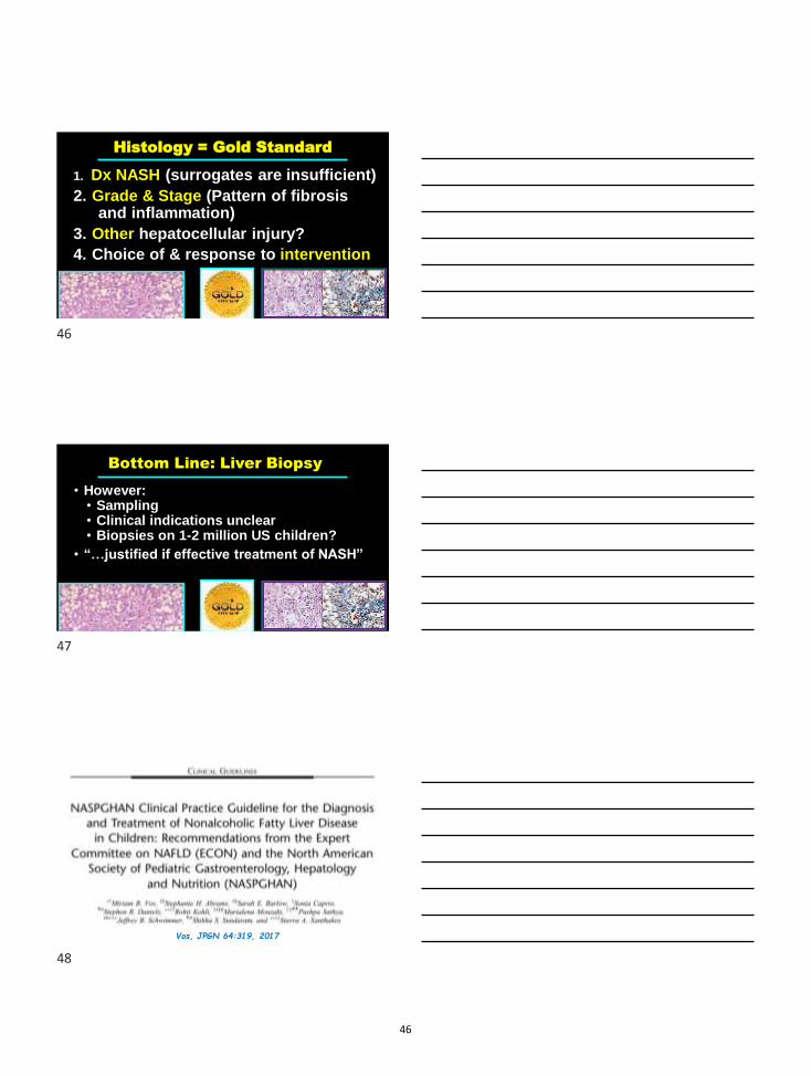
Histology = Gold Standard
1. Dx NASH (surrogates are insufficient)2. Grade & Stage (Pattern of fibrosis
and inflammation)3. Other hepatocellular injury?4. Choice of & response to intervention
Bottom Line: Liver Biopsy
• However:• Sampling• Clinical indications unclear• Biopsies on 1-2 million US children?
• “…justified if effective treatment of NASH”
Vos, JPGN 64:319, 2017
46
47
48
46
Consider biopsy for the assessment of NAFLD in children:
Those with an increased risk of NASHand/or advanced fibrosis:• Splenomegaly• Higher ALT levels (>80 U/l)• AST/ALT >1• Panhypopituitarism• Type 2 diabetes
Vos, JPGN 64:319, 2017
The “ideal test”:
1. Simple, easy to use, validated(age-specific), cost-effective
2. Accurate:- Dx of NASH- Staging of fibrosis- Risk stratification- Monitoring response to
interventions
3. Predicts progression?
Non-invasive Diagnosis and Monitoring of NASH
1. Markers of Injury (ALT, AST)
•Presence, degree, pattern nonspecific•Poor correlation with histology
Limitations of AST and ALT:
49
50
51
47
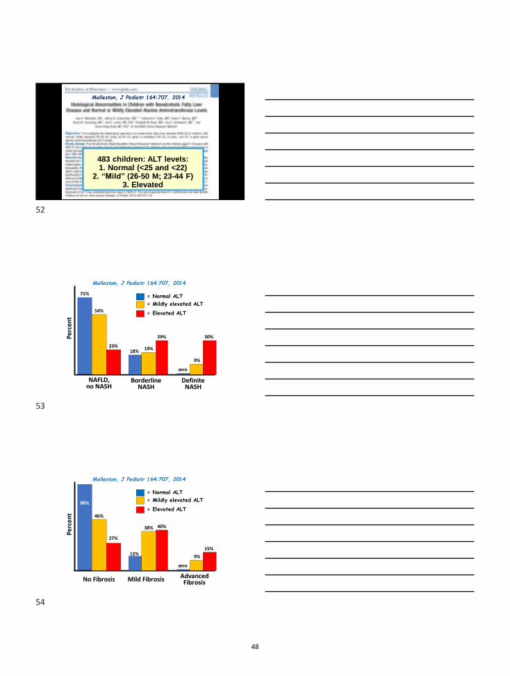
Molleston, J Pediatr 164:707, 2014
483 children: ALT levels:1. Normal (<25 and <22)
2. “Mild” (26-50 M; 23-44 F)3. Elevated
Molleston, J Pediatr 164:707, 2014
71%
NAFLD,no NASH
BorderlineNASH
DefiniteNASH
Pe
rce
nt
= Normal ALT
= Mildly elevated ALT
= Elevated ALT54%
23%18%
19%
29% 30%
9%
zero
Molleston, J Pediatr 164:707, 2014
88%
No Fibrosis Mild Fibrosis Advanced Fibrosis
Per
cen
t
= Normal ALT
= Mildly elevated ALT
= Elevated ALT46%
27%
12%
38% 40%
15%
9%
zero
52
53
54
48
Non-invasive Diagnosis and Monitoring of NASH
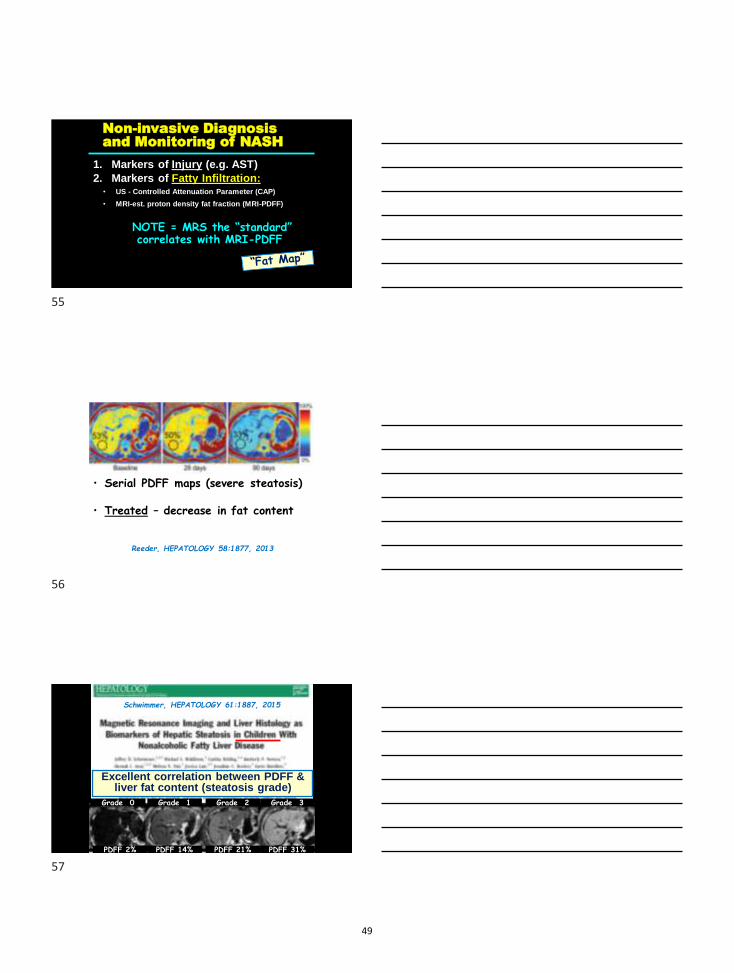
1. Markers of Injury (e.g. AST)2. Markers of Fatty Infiltration:
• US - Controlled Attenuation Parameter (CAP)
• MRI-est. proton density fat fraction (MRI-PDFF)
NOTE = MRS the “standard”correlates with MRI-PDFF
• Serial PDFF maps (severe steatosis)
• Treated – decrease in fat content
Reeder, HEPATOLOGY 58:1877, 2013
Excellent correlation between PDFF &liver fat content (steatosis grade)
Grade 0 Grade 1 Grade 2 Grade 3
PDFF 2% PDFF 14% PDFF 21% PDFF 31%
Schwimmer, HEPATOLOGY 61:1887, 2015
55
56
57
49
Middleton, HEPATOLOGY 67:858, 2018
“MRI-estimated PDFF has high diagnostic accuracy to
classify and predict histological steatosis grade”
Follow-Up Study
Dillman, AJR. Jan 2018
Dillman,AJR
210:1, 2018
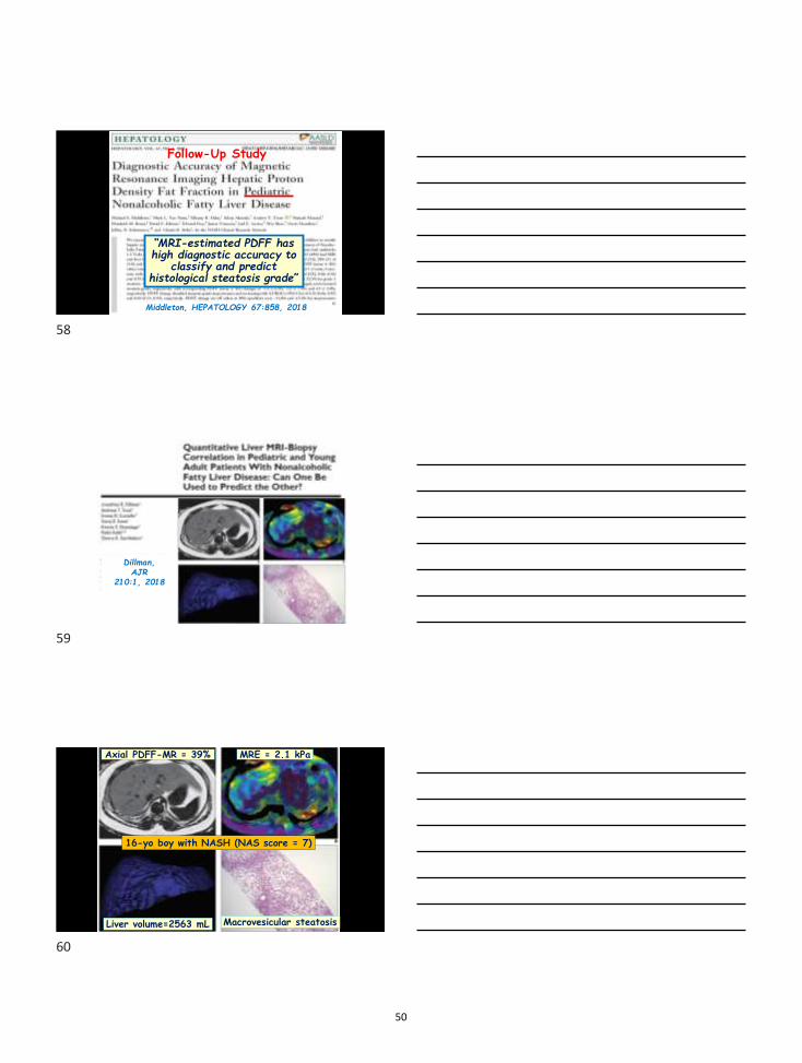
16-yo boy with NASH (NAS score = 7)
Axial PDFF-MR = 39% MRE = 2.1 kPa
Liver volume=2563 mL Macrovesicular steatosis
58
59
60
50
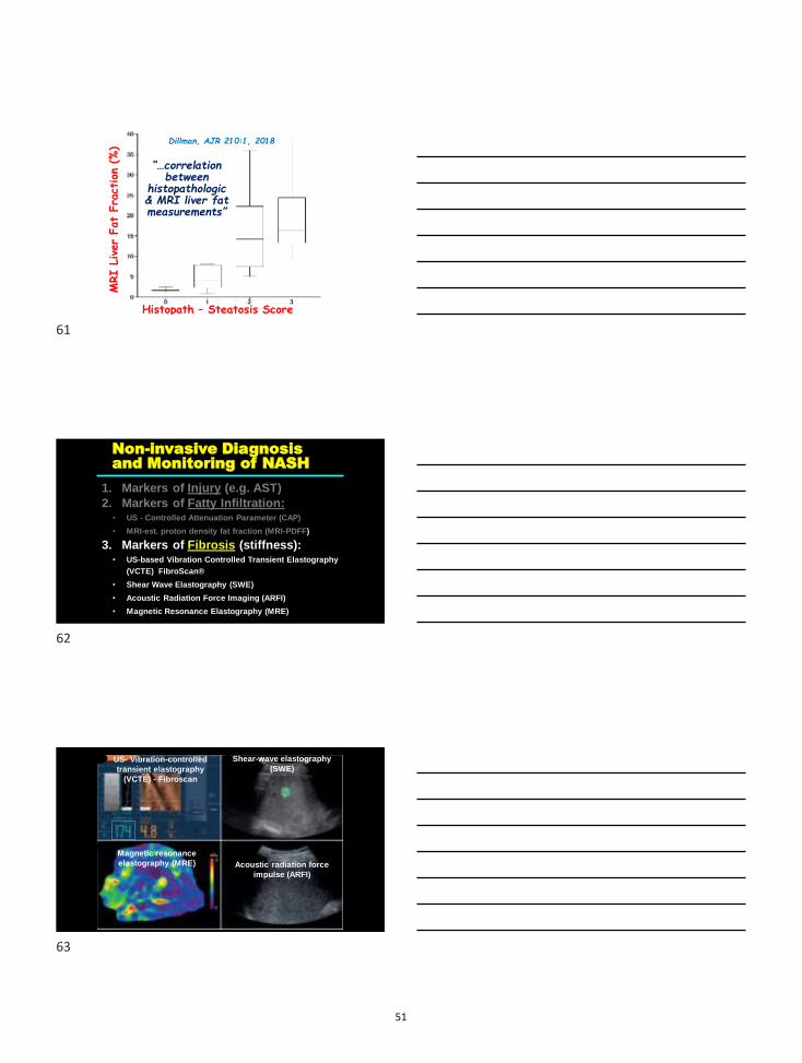
Dillman, AJR 210:1, 2018
MRI L
iver
Fat
Fra
ction
(%)
Histopath – Steatosis Score
“…correlation between
histopathologic & MRI liver fat measurements”
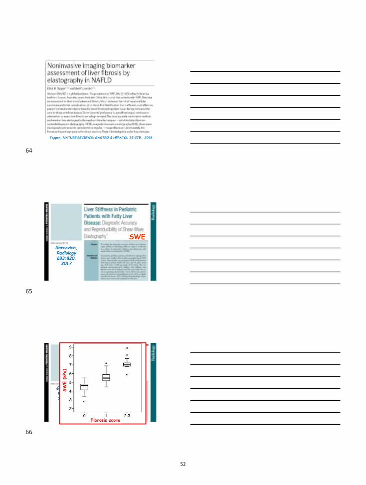
Non-invasive Diagnosis and Monitoring of NASH
1. Markers of Injury (e.g. AST)2. Markers of Fatty Infiltration:
• US - Controlled Attenuation Parameter (CAP)
• MRI-est. proton density fat fraction (MRI-PDFF)
3. Markers of Fibrosis (stiffness):• US-based Vibration Controlled Transient Elastography
(VCTE) FibroScan®
• Shear Wave Elastography (SWE)
• Acoustic Radiation Force Imaging (ARFI)
• Magnetic Resonance Elastography (MRE)
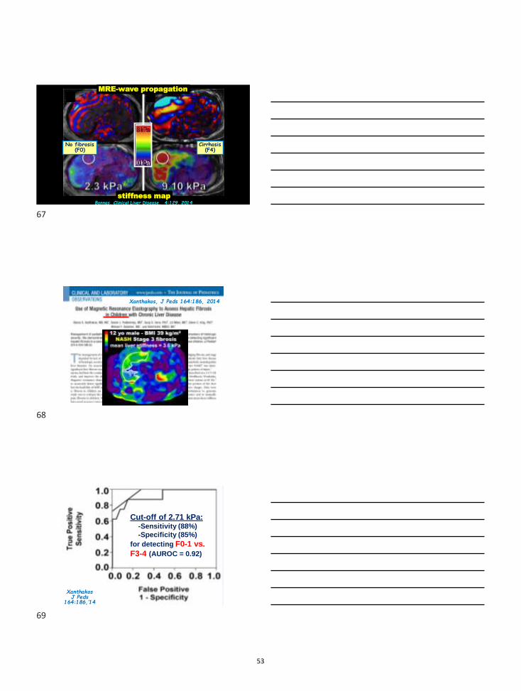
US- Vibration-controlled transient elastography
(VCTE) - Fibroscan
Shear-wave elastography(SWE)
Magnetic resonanceelastography (MRE) Acoustic radiation force
impulse (ARFI)
61
62
63
51
Tapper, NATURE REVIEWS; GASTRO & HEPATOL 15:275, 2018
Garcovich,Radiology283:820,
2017
SWE
Garcovich,Radiology283:820,
2017
SW
E (kPa)
Fibrosis score
64
65
66
52
Bannas, Clinical Liver Disease, 4:129, 2014
No fibrosis(F0)
Cirrhosis(F4)
MRE-wave propagation
stiffness map
Xanthakos, J Peds 164:186, 2014
Cut-off of 2.71 kPa:-Sensitivity (88%)-Specificity (85%)
for detecting F0-1 vs.F3-4 (AUROC = 0.92)
XanthakosJ Peds
164:186,‘14
67
68
69
53
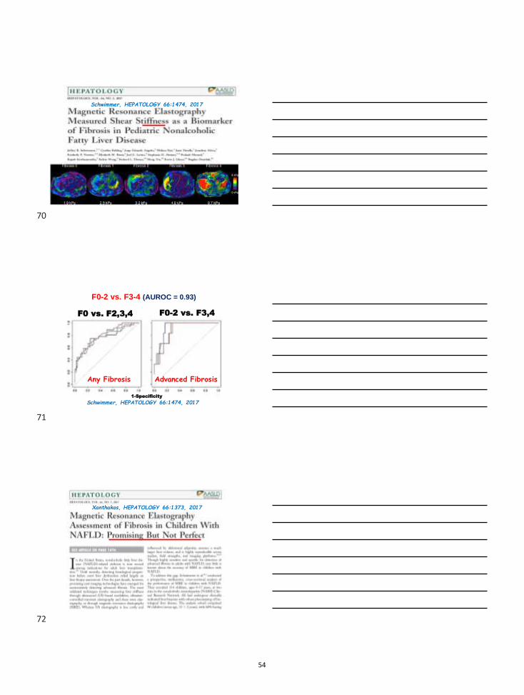
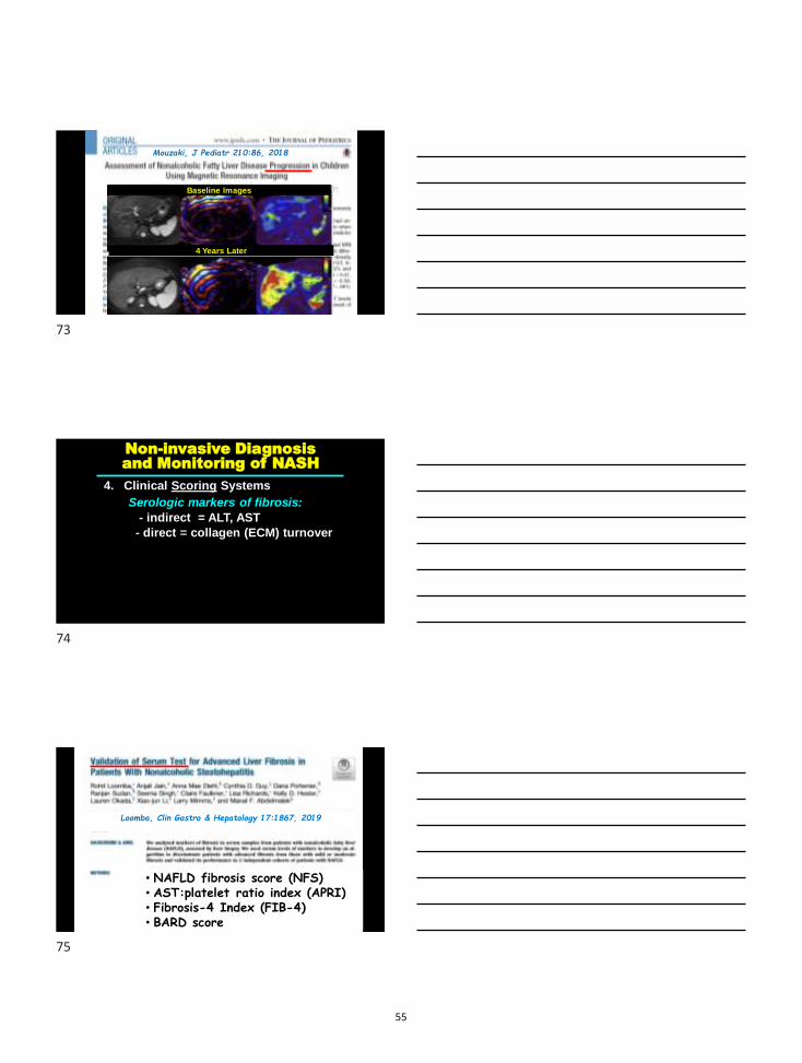
Schwimmer, HEPATOLOGY 66:1474, 2017
Schwimmer, HEPATOLOGY 66:1474, 2017
F0 vs. F2,3,4 F0-2 vs. F3,4
1-Specificity
F0-2 vs. F3-4 (AUROC = 0.93)
Any Fibrosis Advanced Fibrosis
Xanthakos, HEPATOLOGY 66:1373, 2017
70
71
72
54
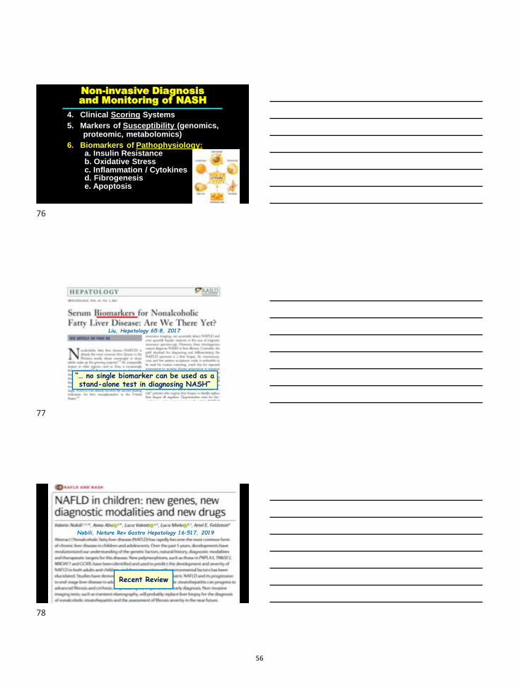
Mouzaki, J Pediatr 210:86, 2018
Baseline Images
4 Years Later
Non-invasive Diagnosis and Monitoring of NASH
4. Clinical Scoring SystemsSerologic markers of fibrosis:
- indirect = ALT, AST- direct = collagen (ECM) turnover
Loomba, Clin Gastro & Hepatology 17:1867, 2019
• NAFLD fibrosis score (NFS)• AST:platelet ratio index (APRI)• Fibrosis-4 Index (FIB-4)• BARD score
73
74
75
55
Non-invasive Diagnosis and Monitoring of NASH
4. Clinical Scoring Systems5. Markers of Susceptibility (genomics,
proteomic, metabolomics)6. Biomarkers of Pathophysiology:
a. Insulin Resistanceb. Oxidative Stressc. Inflammation / Cytokinesd. Fibrogenesise. Apoptosis
“… no single biomarker can be used as a stand-alone test in diagnosing NASH”
Liu, Hepatology 65:8, 2017
Nobili, Nature Rev Gastro Hepatology 16:517, 2019
Recent Review
76
77
78
56
Bottom Line:
• In most “tweens” with elevated liverenzymes… chronic liver disease canbe diagnosed and staged based on:
•Clinical history and examination•Routinely available serum-based and
radiologic studies:•multiple non-invasive methods toassess for significant fibrosis
Our Approach to this Patient
Steatohepatitis Center
CCHMC Liver Clinic:Initial Visit
•CBC, CMP, cbili, GGT, PT/INR•Fasting lipids, insulin; HbA1c•Auto-antibody panel•A1AT phenotype•HBsAg and anti-HBs; Anti-HCV•U/S (if not done within 12 mos)•TSH, fT4; TTG, CK, Aldolase•Nutrition consult
79
80
81
57
28
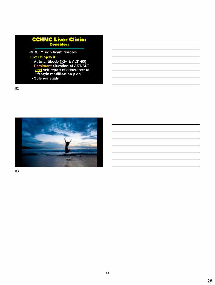
CCHMC Liver Clinic:Consider:
•MRE: ? significant fibrosis•Liver biopsy if:
- Auto-antibody (>2+ & ALT>50)- Persistent elevation of AST/ALT
and self report of adherence tolifestyle modification plan
- Splenomegaly
82
83
58
INDIANA UNIVERSITY SCHOOL OF MEDICINE
NASPGHAN Single Topic Symposium, October 16, 2019
What Do I Do with this Abnormal Radiology Finding?
Chronic Liver Disease Management for the Gastroenterologist
Jean Pappas Molleston, MD
Professor of Clinical Pediatrics
Division Chief, Pediatric Gastroenterology, Hepatology, and Nutrition
Riley Hospital for Children/Indiana University School of Medicine
INDIANA UNIVERSITY SCHOOL OF MEDICINE
Disclosures
Research funding from Mirum,Abbvie, Gillead unrelated to this talk
Research funding from CFfoundation, also unrelated
I am NOT a radiologist!
D Boll RadioGraphics 2009B Yeh RadioGraphics 2009
INDIANA UNIVERSITY SCHOOL OF MEDICINE
Objectives
Outline the differential diagnosis and evaluation of focal liverlesions.
Recognize congenital and acquired vascular abnormalities ofthe liver.
Identify appropriate imaging approaches to suspected biliary tract disease.
Differentiate various parenchymal liver abnormalities.
59
INDIANA UNIVERSITY SCHOOL OF MEDICINE
Focal Liver Lesions
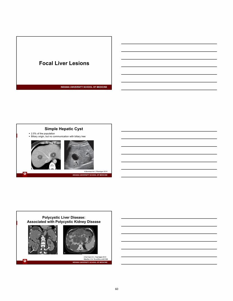
INDIANA UNIVERSITY SCHOOL OF MEDICINE
Simple Hepatic Cyst 2.5% of the population Biliary origin, but no communication with biliary tree
A Bornhani Am J Roentgen 2014
INDIANA UNIVERSITY SCHOOL OF MEDICINE
Polycystic Liver Disease: Associated with Polycystic Kidney Disease
60
INDIANA UNIVERSITY SCHOOL OF MEDICINE
A Borhani Am J Roentgen 2014
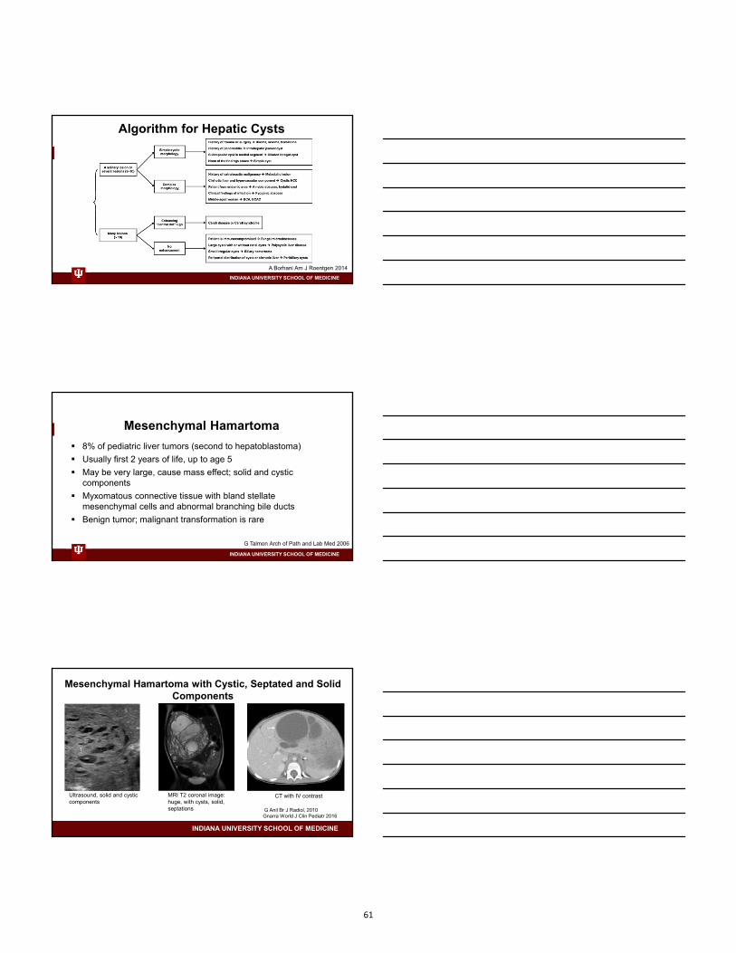
Algorithm for Hepatic Cysts
INDIANA UNIVERSITY SCHOOL OF MEDICINE
Mesenchymal Hamartoma
8% of pediatric liver tumors (second to hepatoblastoma)
Usually first 2 years of life, up to age 5
May be very large, cause mass effect; solid and cystic components
Myxomatous connective tissue with bland stellate mesenchymal cells and abnormal branching bile ducts
Benign tumor; malignant transformation is rare
G Talmon Arch of Path and Lab Med 2006
INDIANA UNIVERSITY SCHOOL OF MEDICINE
Ultrasound, solid and cystic components
MRI T2 coronal image: huge, with cysts, solid, septations
Mesenchymal Hamartoma with Cystic, Septated and Solid Components
G Anil Br J Radiol, 2010Gnarra World J Clin Pediatr 2016
CT with IV contrast
61
INDIANA UNIVERSITY SCHOOL OF MEDICINE
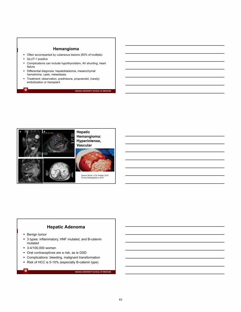
Hemangioma Often accompanied by cutaneous lesions (60% of multiple)
GLUT-1 positive
Complications can include hypothyroidism, AV shunting, heart failure
Differential diagnosis: hepatoblastoma, mesenchymal hamartoma, cysts, metastases
Treatment: observation, prednisone, propranolol, (rarely) embolization or transplant
Gnarra World J Clin Pediatr 2016Chung Radiographics 2010
Hepatic Hemangioma: Hyperintense, Vascular
INDIANA UNIVERSITY SCHOOL OF MEDICINE
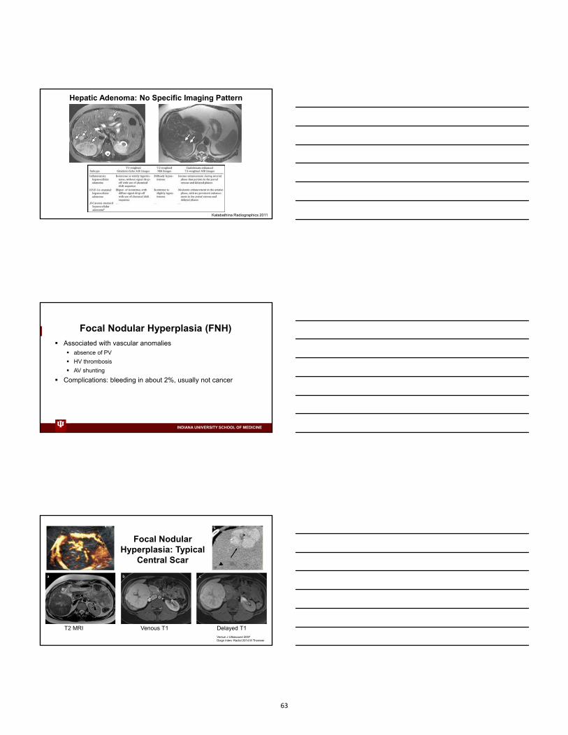
Hepatic Adenoma
Benign tumor
3 types: inflammatory, HNF mutated, and B-catenin mutated
3-4/100,000 women
Oral contraceptives are a risk, as is GSD
Complications: bleeding, malignant transformation
Risk of HCC is 5-10% (especially B-catenin type)
62
Katabathina Radiographics 2011
Hepatic Adenoma: No Specific Imaging Pattern
INDIANA UNIVERSITY SCHOOL OF MEDICINE
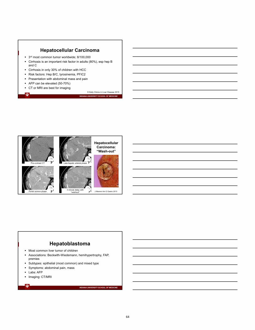
Focal Nodular Hyperplasia (FNH)
Associated with vascular anomalies absence of PV
HV thrombosis
AV shunting
Complications: bleeding in about 2%, usually not cancer
T2 MRIVenturi J Ultrasound 2007Diagn Interv Radiol 2014 M Thomeer
Focal Nodular Hyperplasia: Typical
Central Scar
Venous T1 Delayed T1
63
INDIANA UNIVERSITY SCHOOL OF MEDICINE
Hepatocellular Carcinoma 3rd most common tumor worldwide; 8/100,000
Cirrhosis is an important risk factor in adults (80%), esp hep B and C
Cirrhosis in only 30% of children with HCC
Risk factors: Hep B/C, tyrosinemia, PFIC2
Presentation with abdominal mass and pain
AFP can be elevated (50-70%)
CT or MRI are best for imagingD Kelly Clinics in Liver Disease 2015
Pre-contrast CT Late-hepatic arterial phase
Portal venous phase3 minute delay with
“washout” J Marrero Am G Gastro 2013
HepatocellularCarcinoma: “Wash-out”
INDIANA UNIVERSITY SCHOOL OF MEDICINE
Hepatoblastoma Most common liver tumor of children
Associations: Beckwith-Wiedemann, hemihypertrophy, FAP, premies
Subtypes: epithelial (most common) and mixed type
Symptoms: abdominal pain, mass
Labs: AFP
Imaging: CT/MRI
64
INDIANA UNIVERSITY SCHOOL OF MEDICINE
Radiopaedia.org
HepatoblastomaHeterogeneous, well defined, necrosis/hemorrhage, calcification
INDIANA UNIVERSITY SCHOOL OF MEDICINE
Vascular Lesions
INDIANA UNIVERSITY SCHOOL OF MEDICINE
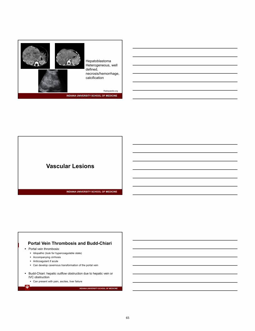
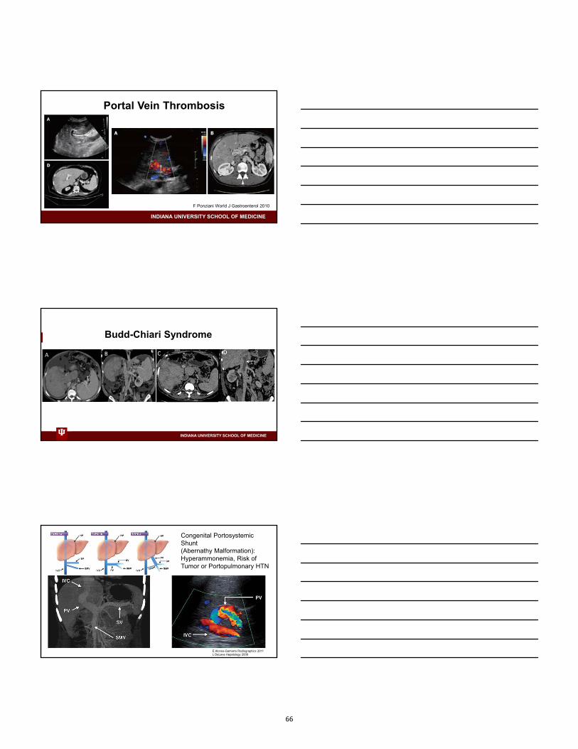
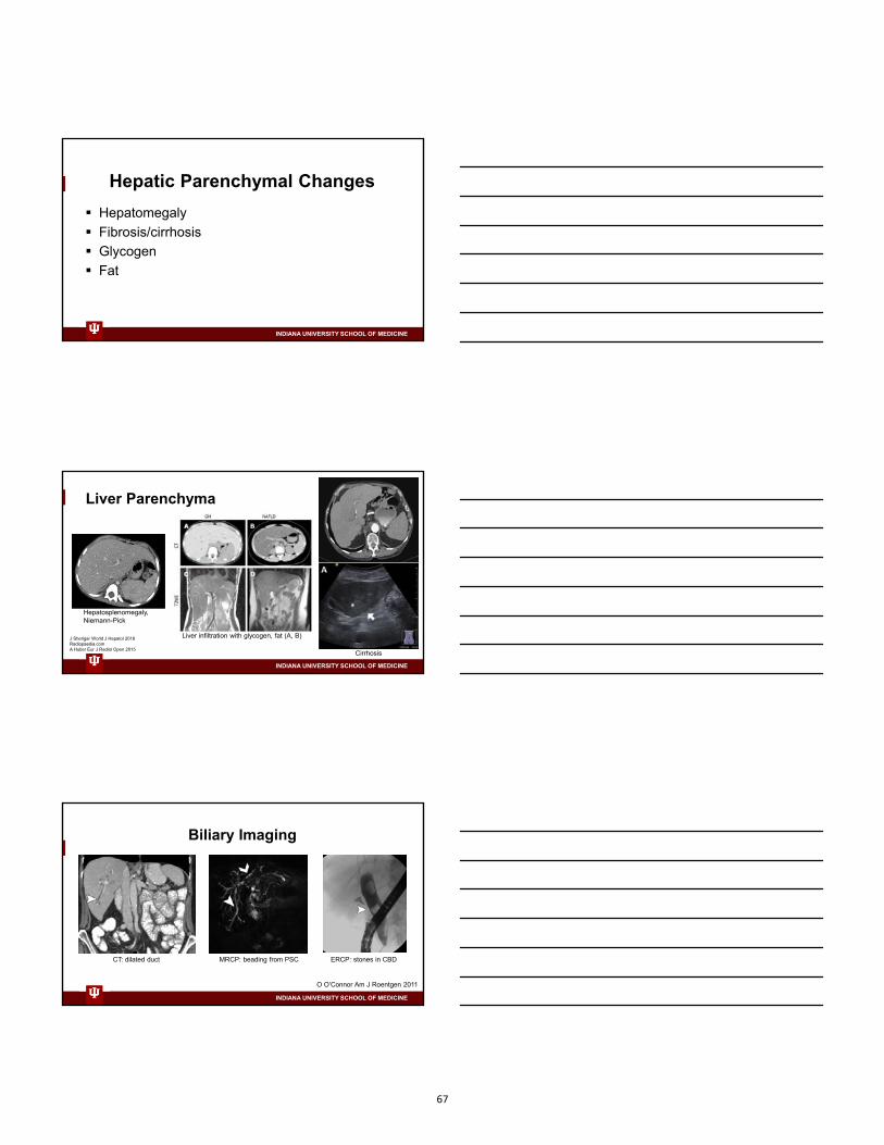
Portal Vein Thrombosis and Budd-Chiari Portal vein thrombosis:
Idiopathic (look for hypercoagulable state)
Accompanying cirrhosis
Anticoagulant if acute
Can develop cavernous transformation of the portal vein
Budd-Chiari: hepatic outflow obstruction due to hepatic vein or IVC obstruction Can present with pain, ascites, liver failure
65
INDIANA UNIVERSITY SCHOOL OF MEDICINE
F Ponziani World J Gastroenterol 2010
Portal Vein Thrombosis
INDIANA UNIVERSITY SCHOOL OF MEDICINE
Budd-Chiari Syndrome
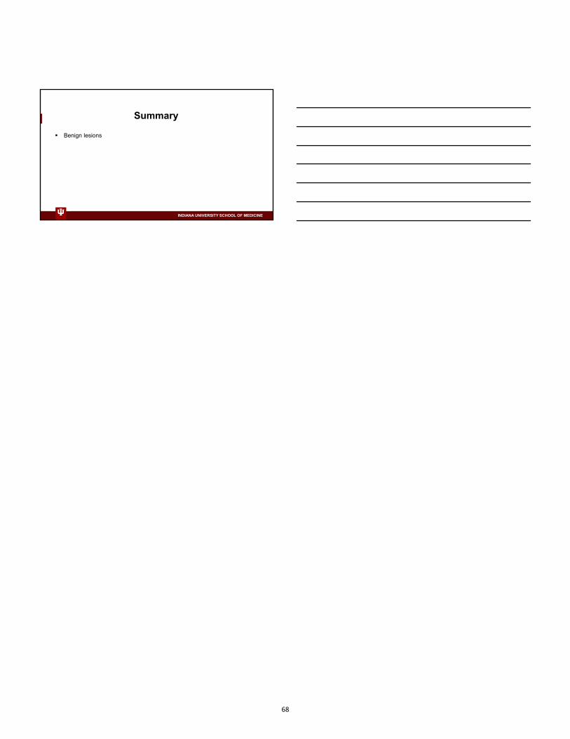
E Alonso-Gamarra Radiographics 2011L DeLeve Hepatology 2009
Congenital Portosystemic Shunt (Abernathy Malformation):Hyperammonemia, Risk of Tumor or Portopulmonary HTN
66
INDIANA UNIVERSITY SCHOOL OF MEDICINE
Hepatic Parenchymal Changes
Hepatomegaly
Fibrosis/cirrhosis
Glycogen
Fat
INDIANA UNIVERSITY SCHOOL OF MEDICINE
Hepatosplenomegaly,Niemann-Pick
Liver infiltration with glycogen, fat (A, B)J Sherigar World J Hepatol 2018Radiopaedia.comA Huber Eur J Radiol Open 2015
Cirrhosis
Liver Parenchyma
INDIANA UNIVERSITY SCHOOL OF MEDICINE
Biliary Imaging
O O’Connor Am J Roentgen 2011
CT: dilated duct MRCP: beading from PSC ERCP: stones in CBD
67
INDIANA UNIVERSITY SCHOOL OF MEDICINE
Summary
Benign lesions
68
Recognition and Stabilization of the Pediatric Patient with Acute Liver Failure
Robert H. Squires, MDProfessor of PediatricsUniversity of Pittsburgh
Disclosures for this Presentation
In the past 12 months, I have had the following relevant financial relationships with the following manufacturer(s) of any commercial product(s) and/or provider(s) of commercial services discussed in this CME activity:
• Up-to-Date: Royalty for chapter contributions on Acute Liver Failure
Objectives
• Recognize features needed to establish a clinical diagnosis of acute liver failure
• Identify and initiate management of multi-system complications associated with acute liver failure.
• Appreciate age-specific differences in the etiology of acute liver failure
• Know when to make early contact with and/or transfer to a pediatric liver transplant center.
69
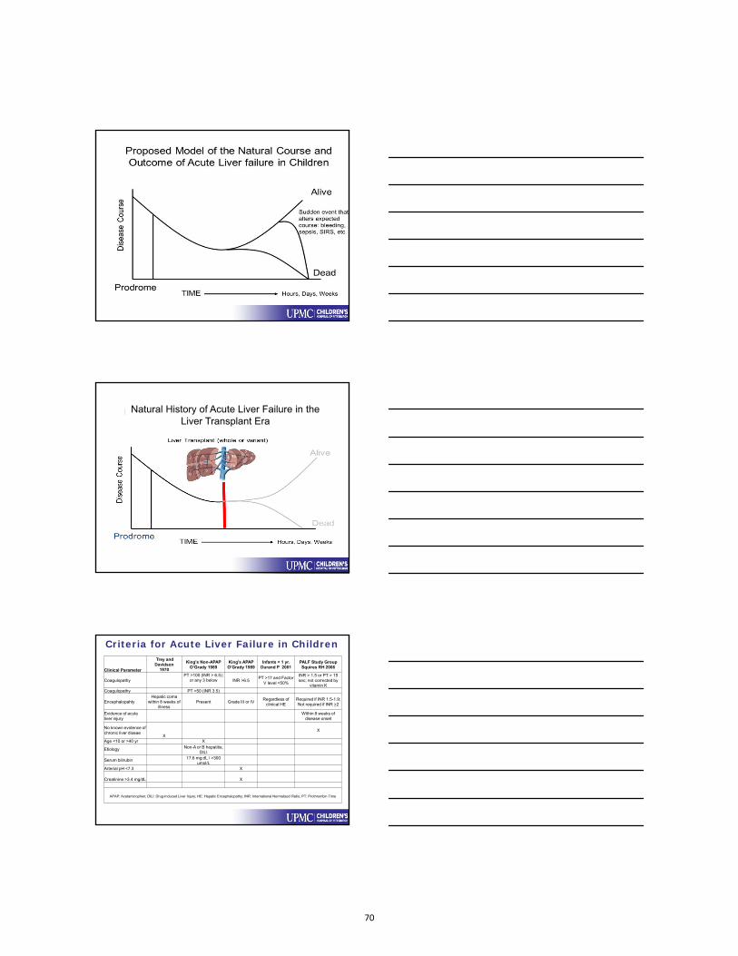
Natural History of Acute Liver Failure in the Liver Transplant Era
Clinical Parameter
Trey and Davidson
1970
King's Non-APAP O’Grady 1989
King's APAPO’Grady 1989
Infants < 1 yr.Durand P 2001
PALF Study GroupSquires RH 2006
CoagulopathyPT >100 (INR > 6.5);
or any 3 below INR >6.5PT >17 and Factor
V level <50%
INR > 1.5 or PT > 15 sec; not corrected by
vitamin KCoagulopathy PT >50 (INR 3.5)
EncephalopahtyHepatic coma
within 8 weeks of illness
Present Grade III or IVRegardless of
clinical HERequired if INR 1.5-1.9; Not required if INR >2
Evidence of acute liver injury
Within 8 weeks of disease onset
No known evidence of chronic liver diseae
XX
Age <10 or >40 yr X
Etiology Non-A or B hepatitis; DILI
Serum bilirubin 17.6 mg;dL / >300 umol/L
Arterial pH <7.3 X
Creatinine >3.4 mg/dL X
APAP: Acetaminophen; DILI: Drug-Induced Liver Injury; HE: Hepatic Encephalopathy; INR: International Normalized Ratio; PT: Prothrombin Time
Criteria for Acute Liver Failure in Children
70
Characterization of PALF
• PALFSG Consensus entry criteria for the PALF study in children
• No evidence of chronic liver disease• Evidence of acute liver injury• Coagulopathy unresponsive to Vitamin K
• PT > 15 sec. or INR > 1.5 with clinical encephalopathy• PT > 20 sec. or INR > 2.0 with/without clinical encephalopathy
• Identifies a spectrum that includes severe liver injury to fulminant liver failure
• Meeting these criteria should prompt at least contact with a liver transplant center.
Biochemical Evidence of Acute Liver Injury in Children
• Suggested parameters indicating evidence of liver injury• AST >100 IU/L or,• ALT >100 IU/L or,• Total bilirubin > 5mg/dL or,• Direct or conjugated bilirubin >2.0 mg/dL.
• PT/INR should always be obtained with evidence of acute liver injury to assess liver function.
• Acetaminophen toxicity: normal or near normal bilirubin with very high aminotransaminase levels
• Gestational Alloimmune Liver Disease, Tyrosinemia, Galactosemia: normal or near normal aminotransferase levels with high total and direct bilirubin
• Note serum alkaline phosphatase is NOT a lab test traditionally used to determine acute liver injury in children
• These cutoff values were not part of the PALF study criteria.
Effect of Vitamin K Administration on Rate of Warfarin Reversal
Polito NB, et.al. Transfusion. 2019
71
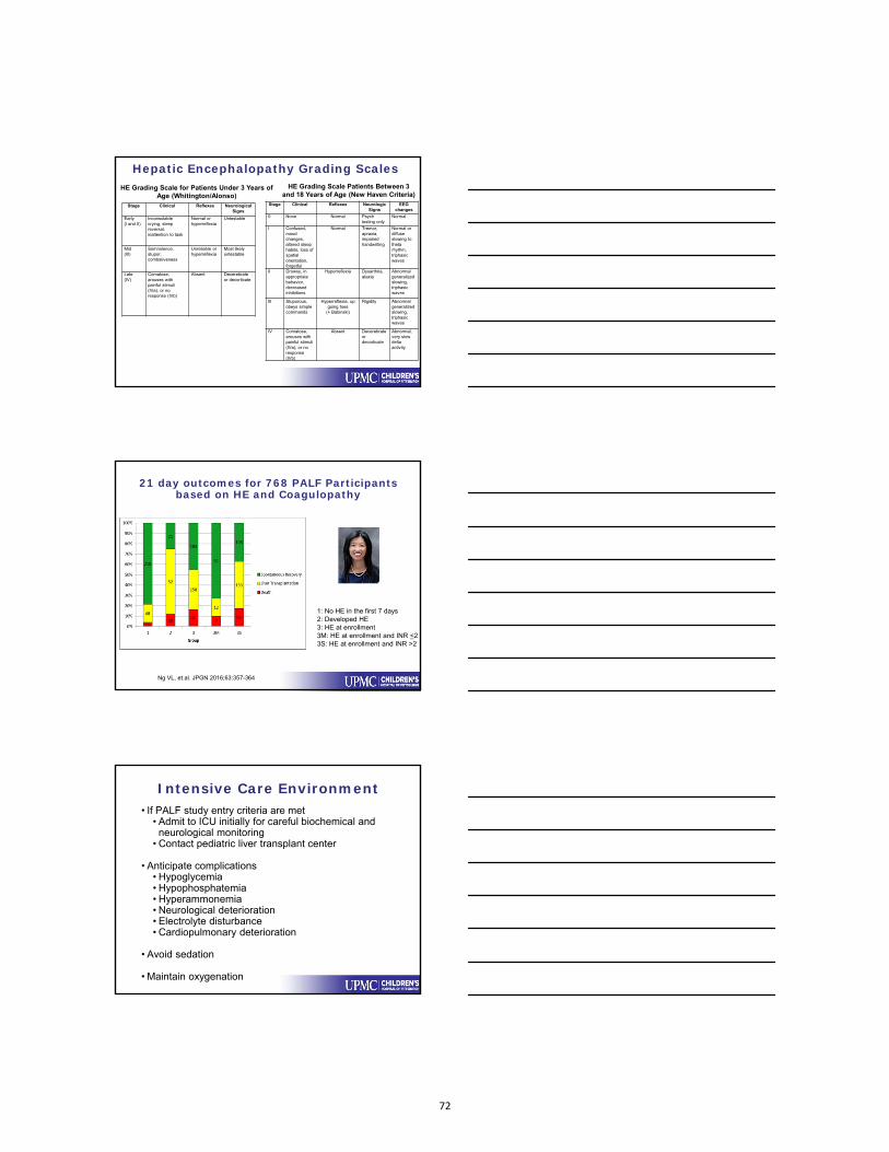
Stage Clinical Reflexes Neurologic Signs
EEG changes
0 None Normal Psych testing only
Normal
I Confused, mood changes, altered sleep habits, loss of spatial orientation, forgetful
Normal Tremor, apraxia, impaired handwriting
Normal or diffuse slowing to theta rhythm, triphasic waves
II Drowsy, in appropriate behavior, decreased inhibitions
Hyperreflexia Dysarthria, ataxia
Abnormal generalized slowing, triphasic waves
III Stuporous, obeys simple commands
Hyperreflexia, up going toes
(+ Babinski)
Rigidity Abnormal generalized slowing, triphasic waves
IV Comatose, arouses with painful stimuli (IVa), or no response (IVb)
Absent Decerebrate or decorticate
Abnormal, very slow delta activity
HE Grading Scale Patients Between 3 and 18 Years of Age (New Haven Criteria)
Stage Clinical Reflexes Neurological Signs
Early(I and II)
Inconsolable crying, sleep reversal, inattention to task
Normal or hyperreflexia
Untestable
Mid (III)
Somnolence, stupor, combativeness
Unreliable or hyperreflexia
Most likely untestable
Late (IV)
Comatose, arouses with painful stimuli (IVa), or no response (IVb)
Absent Decerebrate or decorticate
HE Grading Scale for Patients Under 3 Years of Age (Whitington/Alonso)
Hepatic Encephalopathy Grading Scales
21 day outcomes for 768 PALF Participants based on HE and Coagulopathy
1: No HE in the first 7 days2: Developed HE3: HE at enrollment3M: HE at enrollment and INR <23S: HE at enrollment and INR >2
Ng VL, et.al. JPGN 2016;63:357-364
Intensive Care Environment• If PALF study entry criteria are met
• Admit to ICU initially for careful biochemical and neurological monitoring
• Contact pediatric liver transplant center
• Anticipate complications• Hypoglycemia• Hypophosphatemia• Hyperammonemia• Neurological deterioration• Electrolyte disturbance• Cardiopulmonary deterioration
• Avoid sedation
• Maintain oxygenation
72
Hematologic Support
• Administer vitamin K parenterally only once
• In the setting of ALF, prolonged INR is a good measure of liver dysfunction, but is not a measure of bleeding risk
• Avoid giving FFP or cryoprecipitate just to correct the INR
• Consider giving blood products if• An invasive procedure is planned• Significant hemorrhage• INR is very high (e.g, >5-6) after parenteral vitamin K
Minimal Effects of Acute Liver Injury/Acute Liver Failure on Hemostasis as Assessed by
Thromboelastography (TEG)
• INR is a measure of liver function and not bleeding risk
• 51 patients
• Mean INR 3.4 + 1.7 (range 1.5‐9.6)
• Mean TEG parameters were normal
• Factor V and VII (pro‐coagulants) were decreased
• Proportional decreases in anti‐coagulant proteins
• Inversely proportional to elevated Factor VIII levels
• Most patients with ALI/ALF maintain normal hemostasis due to:
• Increased clot strength
• Increases Factor VIII and Von Willebrand levels
• Commensurate decline in pro‐ and anti‐coagulant proteins
Stravitz RT, et.al., Journal of Hepatology 2012;56:129-36
Metabolic Support
• Maintain glucose between 90-110 mg/dL• Dextrose containing fluids, may require D10 or D12.5
• Maintain phosphorus >3.5 mmol/L
• Monitor electrolyte and renal function
• Careful assessment of fluid status• Fluid resuscitation if needed• Fluid restriction (90-95% of maintenance) preferred• Avoid unnecessary use of blood products
73
Renal Support
• Renal injury at presentation• Shock / hypovolemia• Toxic injury (e.g. Acetaminophen or Mushroom toxicity)
• Careful monitoring of I & O
• Avoid over-zealous diuresis• Precipitate hepatorenal syndrome• Worsen encephalopathy
• Prepare for dialysis / hemofiltration (transplant center only)
Neurological Support
• Patient should be in a liver transplant center if there is any evidence of encephalopathy
• Minimize stimulation
• If the patient is confused or combative, protect from injury
• Elevate the head of the bed
• Lactulose for hyperammonemia
• Careful fluid management
• CT / continuous EEG
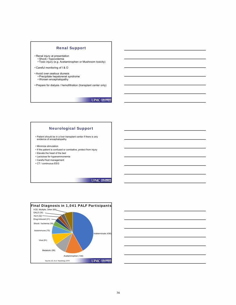
Final Diagnosis in 1,041 PALF Participants
Metabolic (99)
Viral (91)
Autoimmune (70)
VOD, Multiple, Other (69)
Acetaminophen (144)
Shock / Ischemia (39)
Indeterminate (436)
Drug-Induced (31)
HLH (32)
GALD (30)
Squires JE, et.al. Hepatology 2019
74
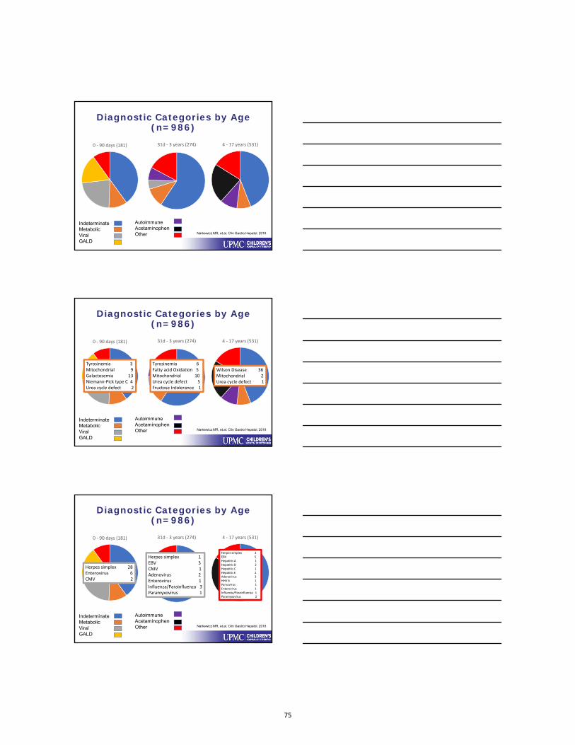
Diagnostic Categories by Age (n=986)
0 ‐ 90 days (181) 31d ‐ 3 years (274) 4 ‐ 17 years (531)
Indeterminate MetabolicViralGALD
AutoimmuneAcetaminophenOther Narkewicz MR, et.al. Clin Gastro Hepatol. 2018
Diagnostic Categories by Age (n=986)
0 ‐ 90 days (181) 31d ‐ 3 years (274) 4 ‐ 17 years (531)
Indeterminate MetabolicViralGALD
AutoimmuneAcetaminophenOther Narkewicz MR, et.al. Clin Gastro Hepatol. 2018
Tyrosinemia 3Mitochondrial 9Galactosemia 13Niemann‐Pick type C 4Urea cycle defect 2
Tyrosinemia 6Fatty acid Oxidation 5Mitochondrial 10Urea cycle defect 5Fructose Intolerance 1
Wilson Disease 36Mitochondrial 2Urea cycle defect 1
Diagnostic Categories by Age (n=986)
0 ‐ 90 days (181) 31d ‐ 3 years (274) 4 ‐ 17 years (531)
Indeterminate MetabolicViralGALD
AutoimmuneAcetaminophenOther Narkewicz MR, et.al. Clin Gastro Hepatol. 2018
Herpes simplex 28Enterovirus 6CMV 2
Herpes simplex 1EBV 3CMV 1Adenovirus 2Enterovirus 1Influenza/Parainfluenza 3Paramyxovirus 1
Herpes simplex 2EBV 5Hepatitis A 1Hepatitis B 2Hepatitis C 1Hepatitis E 2Adenovirus 3HHV 6 1Parvovirus 1Enterovirus 1Influenza/Parainfluenza 1Paramyxovirus 2
75
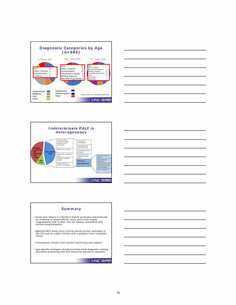
Diagnostic Categories by Age (n=986)
0 ‐ 90 days (181) 31d ‐ 3 years (274) 4 ‐ 17 years (531)
Indeterminate MetabolicViralGALD
AutoimmuneAcetaminophenOther
Narkewicz MR, et.al. Clin Gastro Hepatol. 2018
HLH 6Shock / Ischemia 6Acetaminophen 1Leukemia 1
HLH 10Shock / Ischemia 8Acetaminophen 12Venoocclusive disease 6Multiple diagnoses 5Non‐APAP Drug‐induced 2
HLH 9Shock / Ischemia 17Venoocclusive disease 6Multiple diagnoses 13Non‐APAP Drug‐induced 27Viral 27Mushroom 5Budd‐Chiari 3
Indeterminate PALF is Heterogeneous
Unrecognized acetaminophen
Unrecognized drug‐induced liver injury
Unrecognized or novel viral agents
Unrecognized or novelmetabolic or genetic defects
Immune dysregulation
Autoimmunemarker positive
Natural Killer cell dysfunction
Gestational alloimmune liver disease (neonatal hemochromatosis)
Other mechanisms
MetabolicDrug‐inducedViralAutoimmuneOther(44%)
Diagnostic testing interrupted by death, transplant, clinicalimprovement
Diagnosis not considered
Blood volume and logistical issues
Variability in diagnostic approach
Complete diagnostic evaluation was negative
Indeterminate(42%)
APAP(14%)
Summary
• Acute liver failure is a dynamic clinical syndrome characterized by evidence of hepatocellular injury and a liver-based coagulopathy that is often, but not always, associated with clinical encephalopathy.
• Meeting PALF study entry criteria should prompt admission to the ICU and an urgent contact with a pediatric liver transplant center.
• Immediately initiate multi-system monitoring and support
• Age-specific etiologies should prioritize initial diagnostic testing; all infants presenting with ALF should be started on acyclovir.
76
Future Direction
• Automate age-specific diagnostic testing.
• Develop effective liver support systems.
• Auxiliary or hepatocyte transplantation to bridge to recovery of the native liver.
• Improve clinical and in silico models that predict outcome and inform liver transplant decisions.
Take Home Message for Patients Meeting PALF Study Entry Criteria
• Admit all patients to the ICU.
• Immediately contact a pediatric liver transplant center.
• Maintain glucose and phosphorous levels; carefully monitor neurological status; avoid overuse of blood products.
• Initiate diagnostic testing, prioritizing potentially treatable conditions; acyclovir should be given to all infants and also be considered in adolescents.
Board Questions for MOC• offer MOC Part II credits as part of the symposium. To this end, please provide 2-3 board style multiple choice questions based on slide content to be submitted along with your slides. These will be used for MOC part II credit for the membership.
•
77

Disclosures
• Advisory boards: Roche, Alexion, Kadmon
• Grant Support: Gilead, Abbvie, Merck, Alexion
• Discussing UNAPPROVED therapy
• Advisory boards: Roche, Alexion, Kadmon
• Grant Support: Gilead, Abbvie, Merck, Alexion
• Discussing UNAPPROVED therapy
What Would Hamlet Do: To Rx or Not to Rx?Objectives
What Would Hamlet Do: To Rx or Not to Rx?Objectives
Virus-DiseaseVirus-Disease
Treatment CandidatesTreatment Candidates
MedicationsMedications
78
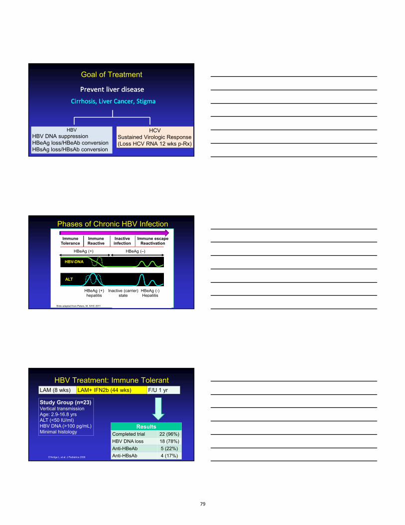
Goal of Treatment
Prevent liver diseasePrevent liver disease
HBVHBV DNA suppressionHBeAg loss/HBeAb conversionHBsAg loss/HBsAb conversion
HCVSustained Virologic Response(Loss HCV RNA 12 wks p-Rx)
Cirrhosis, Liver Cancer, StigmaCirrhosis, Liver Cancer, Stigma
Immune escapeReactivation
HBeAg (+) HBeAg (–)
HBV-DNA
Inactive (carrier)state
HBeAg (-)Hepatitis
HBeAg (+) hepatitis
ImmuneTolerance
ImmuneReactive
Inactiveinfection
Phases of Chronic HBV Infection
Slide adapted from Peters, M. IVHC 2011
ALT
HBsAg (-)
HBV Treatment: Immune Tolerant LAM (8 wks) LAM+ IFN2b (44 wks) F/U 1 yr
Study Group (n=23)Vertical transmission Age: 2.9-16.8 yrsALT (<50 IU/ml)HBV DNA (>100 pg/mL)Minimal histology
ResultsCompleted trial 22 (96%)
HBV DNA loss 18 (78%)
Anti-HBeAb 5 (22%)
Anti-HBsAb 4 (17%)D'Antiga L, et al. J Pediatrics 2006
79
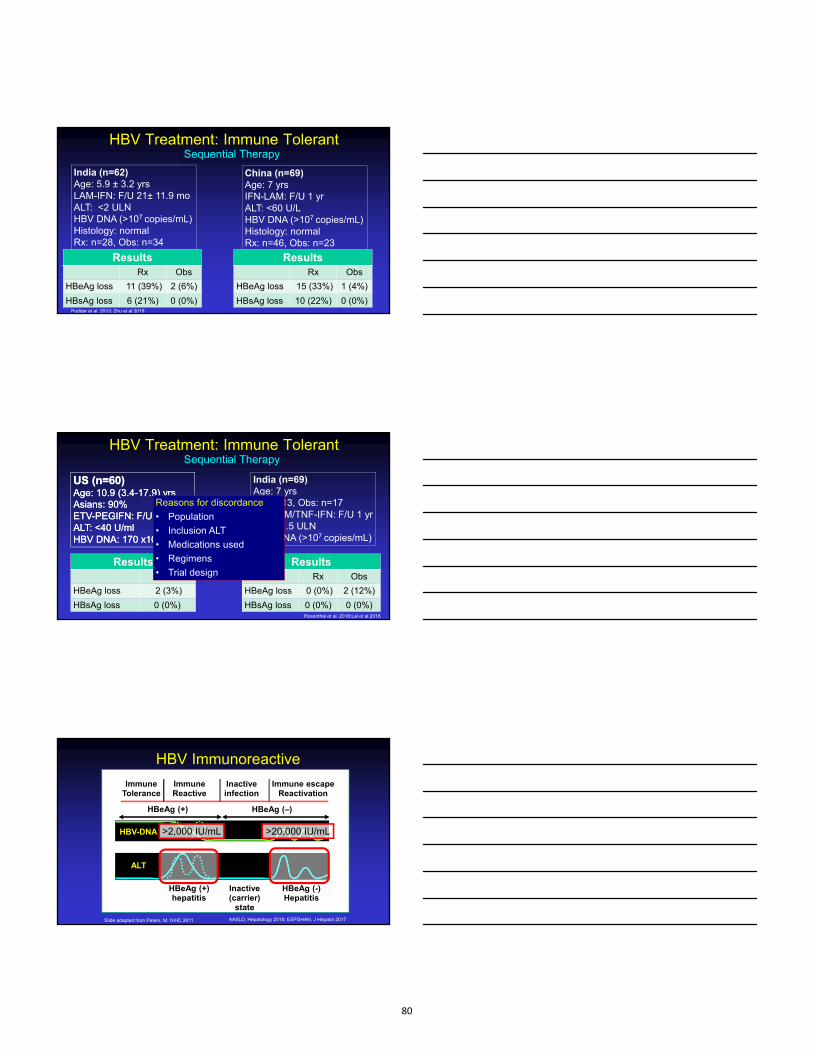
HBV Treatment: Immune Tolerant Sequential Therapy
India (n=62)Age: 5.9 ± 3.2 yrsLAM-IFN: F/U 21± 11.9 mo ALT: <2 ULNHBV DNA (>107 copies/mL)Histology: normalRx: n=28, Obs: n=34
Poddar et al. 2013; Zhu et al 2018
China (n=69)Age: 7 yrsIFN-LAM: F/U 1 yr ALT: <60 U/LHBV DNA (>107 copies/mL)Histology: normalRx: n=46, Obs: n=23
ResultsRx Obs
HBeAg loss 15 (33%) 1 (4%)
HBsAg loss 10 (22%) 0 (0%)
ResultsRx Obs
HBeAg loss 11 (39%) 2 (6%)
HBsAg loss 6 (21%) 0 (0%)
HBV Treatment: Immune Tolerant Sequential Therapy
Rosenthal et al. 2018;Lal et al 2018
India (n=69)Age: 7 yrsRx: n=13, Obs: n=17IFN-LAM/TNF-IFN: F/U 1 yr ALT: <1.5 ULNHBV DNA (>107 copies/mL)
ResultsRx Obs
HBeAg loss 0 (0%) 2 (12%)
HBsAg loss 0 (0%) 0 (0%)
ResultsRx
HBeAg loss 2 (3%)
HBsAg loss 0 (0%)
US (n=60)Age: 10.9 (3.4-17.9) yrsAsians: 90%ETV-PEGIFN: F/U 48 wksALT: <40 U/mlHBV DNA: 170 x106 U/mL
US (n=60)Age: 10.9 (3.4-17.9) yrsAsians: 90%ETV-PEGIFN: F/U 48 wksALT: <40 U/mlHBV DNA: 170 x106 U/mL
Reasons for discordance
• Population
• Inclusion ALT
• Medications used
• Regimens
• Trial design
Immune escapeReactivation
HBeAg (+) HBeAg (–)
HBV-DNA
Inactive (carrier)
state
HBeAg (-)Hepatitis
HBeAg (+) hepatitis
ImmuneTolerance
ImmuneReactive
Inactiveinfection
HBV Immunoreactive
Slide adapted from Peters, M. IVHC 2011
ALT
>
>2,000 IU/mL >20,000 IU/mL
AASLD, Hepatology 2018; ESPGHAN, J Hepatol 2017
80
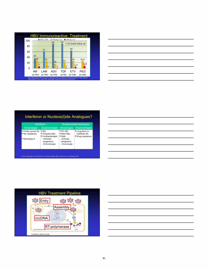
HBV Immunoreactive: Treatment
INF LAM ADV TDF ETV PEG(n=251) (n=191) (n=115) (n=52) (n=120) (n=101)
>12 month follow-up
Vajro 1996; Barbera 1994; Utili 1991; Ruiz-Moreno 1991, Ruiz-Moreno 1990; Sokal 1998; Lai 1991; Gregorio 1996; Murray K, 2012; Jonas 2002; Jonas 2008; Jonas 2015;Jonas 2016: Wirth, 2018
Interferon or Nucleos(t)ide Analogues?
Interferon Nucleos(t)ide Analogues
Advantages Disadvantages Advantages Disadvantages Finite course RxNo resistance
Genotype A
SQ Frequent AEsContraindicated
-cirrhosis-pregnancy -immunosupp
PO QDRare AEsSafe
-cirrhosis-pregnancy- imunosupp
Long-term or indefinite RxDrug resistance
Lok AS. Hepatology. 2010; Buster EH, et al. Gastroenterology. 2008; Lange CM, et al. Hepatology. 2009.
HBV Treatment Pipeline
cccDNA
Covalently closed circular
Entry
www.intmedpress.com
cccDNA
Assembly
RT-polymerase
81
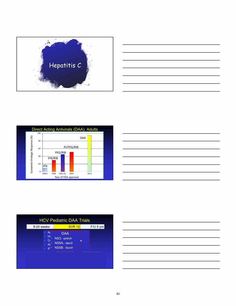
Hepatitis C
Direct Acting Antivirals (DAA): Adults
0
20
40
60
80
100
Sustained
Virologic Response (%)
1990’s 1998 2001‐02 2004 2014
IFN
IFN/RIB
PEG/RIB
DAA
Year of FDA approval
PI/PEG/RIB
3-6 y3-6 yPK (n=10)PK (n=10)
ExtendedExtended
12-17 yPK (n=10)PK (n=10)
ExtendedExtended
6-11 y6-11 yPK (n=10)PK (n=10)
ExtendedExtended
HCV Pediatric DAA Trials8-24 weeks SVR 12 F/U 5 yrs
• Age: >3 years
• HCV RNA > 1,000 IU/mL
• Compensated liver disease
• ALT: Normal or Abnormal
• IFN Naïve-experienced
DAA• NS3: -previr
• NS5A: -asvir
• NS5B: -buvir
82
98 98 100 100
0
20
40
60
80
100
SV
R12
, %
GlecaprevirPibrentasvir
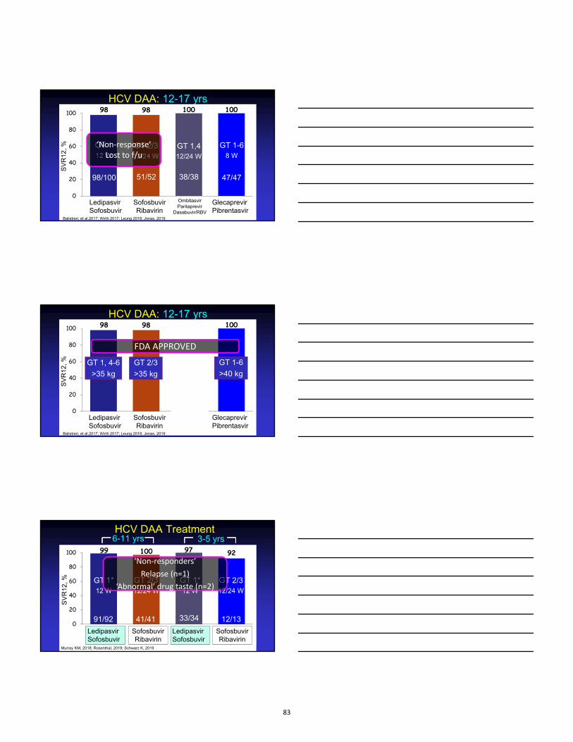
HCV DAA: 12-17 yrs
LedipasvirSofosbuvir
GT 1-68 W
47/47
GT 112 W
98/100
GT 2/312/24 W
51/52
GT 1,412/24 W
38/38
98/100SofosbuvirRibavirin
OmbitasvirParitaprevir
Dasabuvir/RBVBalistreri, et al.2017; Wirth 2017; Leung 2018; Jonas, 2019
‘Non‐response’ Lost to f/u
98 98 100 100
0
20
40
60
80
100
SV
R12
, %
GlecaprevirPibrentasvir
HCV DAA: 12-17 yrs
LedipasvirSofosbuvir
SofosbuvirRibavirin
OmbitasvirParitaprevir
Dasabuvir/RBVBalistreri, et al.2017; Wirth 2017; Leung 2018; Jonas, 2019
GT 1, 4-6
>35 kg
GT 2/3
>35 kg
GT 1-6
>40 kg
FDA APPROVED
99 97100 92
0
20
40
60
80
100
SV
R12
, %
HCV DAA Treatment
12/1391/92 41/41 33/34
3-5 yrs6-11 yrs
LedipasvirSofosbuvir
SofosbuvirRibavirin
Murray KM, 2018; Rosenthal, 2019; Schwarz K, 2019
LedipasvirSofosbuvir
SofosbuvirRibavirin
GT 2/312/24 W
GT 2/312/24 W
GT 1*12 W
GT 1*12 W
‘Non‐responders’
Relapse (n=1)
‘Abnormal’ drug taste (n=2)
83
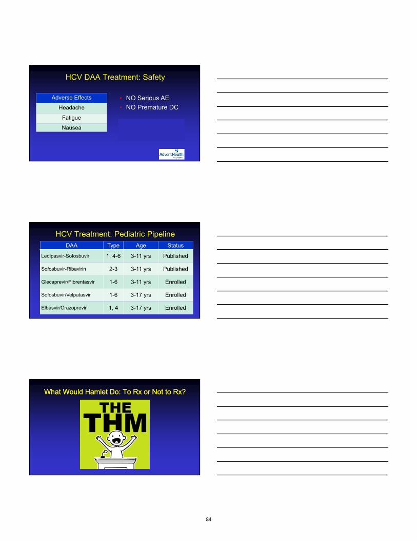
HCV DAA Treatment: Safety
• NO Serious AE
• NO Premature DC
• ‘Bad Taste’
• HBV Reactivation
Adverse Effects
Headache
Fatigue
Nausea
HCV Treatment: Pediatric PipelineDAA Type Age Status
Ledipasvir-Sofosbuvir 1, 4-6 3-11 yrs Published
Sofosbuvir-Ribavirin 2-3 3-11 yrs Published
Glecaprevir/Pibrentasvir 1-6 3-11 yrs Enrolled
Sofosbuvir/Velpatasvir 1-6 3-17 yrs Enrolled
Elbasvir/Grazoprevir 1, 4 3-17 yrs Enrolled
What Would Hamlet Do: To Rx or Not to Rx?What Would Hamlet Do: To Rx or Not to Rx?
84
What Would Hamlet Do: To Rx or Not to Rx?What Would Hamlet Do: To Rx or Not to Rx?
HBV
ImmunotolerantNormal ALT
High HBV DNA
MonitorALT, HBeAg, HBV DNA
ImmunoreactiveElevated ALTHBV DNA
TreatExclude other causesEntecavir, Tenofovir
Pegylated IFN
www.clinicaloptions.com/HepBConsult
What Would Hamlet Do: To Rx or Not to Rx?What Would Hamlet Do: To Rx or Not to Rx?
HCV
>12 yrs Ledipasvir‐SOF (1, 4‐6)Ledipasvir‐RBV (2,3)Glecaprevir‐PIB (GT 1‐6)
3‐12 yrsPeg‐IFN‐RBV
DEFER…
<3 yrsMonitor
What Would Hamlet Do: To Rx or Not to Rx?What Would Hamlet Do: To Rx or Not to Rx?
Regino P. Gonzalez-Peralta, MD
Pediatric GI, Hepatology and Liver Transplant
85
ARE THERE ANY MEDICAL THERAPIES FOR NASH?Single Topic Symposium, NASPGHAN 2019
Marialena Mouzaki, MD MSc
Cincinnati Children’s Hospital Medical Center
Disclosures
• In the past 12 months, I have had no relevant financial relationships with the manufacturer(s) of any commercial product(s) and/or provider(s) of commercial services discussed in this CME activity.
Learning Objectives
• Present the gaps in the therapeutic armamentarium for pediatric NASH
• Evaluate the preliminary data on the efficacy of novel medications currently being investigated for the treatment of NASH
86
NAFLD: “a growing problem”
Roberts E. J Hepatology 2007Anderson et al. PLoS One 2015;Thiagarajan et al. J Clin Experiment Hepatol 2019
2.30%
12.50%
36.10%
0%
10%
20%
30%
40%
Normal weight Overweight Obese
NAFLD PrevalenceGeneral Population Studies
Pathogenesis and treatment targets
Sanyal A. Nat Rev Gastroenterol Hep 2019
Recap of NAFLD histology scoring
NAFLD Activity Score (NAS; 0‐8)
Steatosis: 0‐3
Lobular inflammation: 0‐3
Ballooning: 0‐2
Fibrosis stage
Stage 0: absent
Stage 1‐3: mild‐severe
Stage 4: cirrhosis
Kleiner et al. Hepatol 2005
87
Medical therapies investigated to date
Successful
• Weight loss• Diet and exercise
• Exact amount is not clear
• Vitamin E• 800 IU daily
• NASH resolution in 58% on vitE vs. 28% on placebo
Not successful
• Metformin• 1000 mg daily
• Cysteamine bitartrate• 300‐450 mg BID
Vos et al. J Pediatr Gastroenterol Nutr 2017;Lavine et al. JAMA 2011;Schwimmer et al. Gastroenterol 2016
NASPGHAN guidelines
• Lifestyle changes• Limit sugar‐sweetened beverages
• Well‐balanced diet
• Moderate vigorous exercise
• Limiting screen time <2h per day
• Medications• No medications or supplements are currently recommended
• Surgical options• Medical weight loss surgery may be considered in select cases
Vos et al. J Pediatr Gastroenterol Nutr 2017
Novel treatment approaches
Konerman et al. J Hepatol 2018
88
Obeticholic acid leads to improved histology
• FXR agonist• Improves insulin sensitivity
• Decreases hepatic steatosis and inflammation
• FLINT trial: RCT 25 mg OCA vs. placebo x72 weeks
• Primary outcome: NAS by 2 points without worsening of fibrosis
22%
13%
35%
19%
OCA (n=110) Placebo (n=109)
Resolution of NASH Improvement of fibrosis
Neuschwander‐Tetriet al. Lancet 2014
REGENERATE –Obeticholic acid, phase III
• 25 mg vs. 10 mg vs. placebo x 48 months – target ~2,400 pt
• 2 primary endpoints:• Fibrosis improvement ≥1 stage without NASH worsening
• NASH resolution without fibrosis worsening
• Results of interim analysis at 18 months:
Younossi et al. Int Liv Congress Austria 2019
NGM282 ameliorates fat fraction in patients with NASH
Primary outcome: ≥5% reduction in PDFF
• Phase IIa RCT, 12 weeks • Humanized FGF19 analogue
Harrison et al. Lancet 2018;Patel et al. Therap Adv Gastroenterol 2015
≥30% in PDFF associated with ≥2 NAS in prior studies
89
NGM282 also associated with LDL‐C increase in patients with NASH
• Increase predominantly in large, buoyant LDL‐C particles
• Thought to be less atherogenic
Cholesterol
Bile acids
CYP7A1
NGM282
• Open label trial of NGM282 (lower doses (0.3mg, 1 mg, 3 mg) and rosuvastatin (up to 40 mg) x12 weeks for NASHRelative change
in PDFF: 34%Relative change in PDFF: 57%
Relative change in PDFF: 67%
Rinella et al. J Hepatol 2019
Novel treatment approaches
Konerman et al. J Hepatol 2018
Elafibranor (dual PPAR‐a/δ agonist)
• Fatty acid oxidation
• Fatty acid transport (TG, HDL)
• Anti‐inflammatory effectsPPARαPPARα
• Fatty acid oxidation
• Fatty acid transport (HDL)
• Improve glucose homeostasis
• Anti‐inflammatory activities
PPARδPPARδ
Ratziu et al. Gastroenterol 2016;Konerman et al. J Hepatol 2018
90
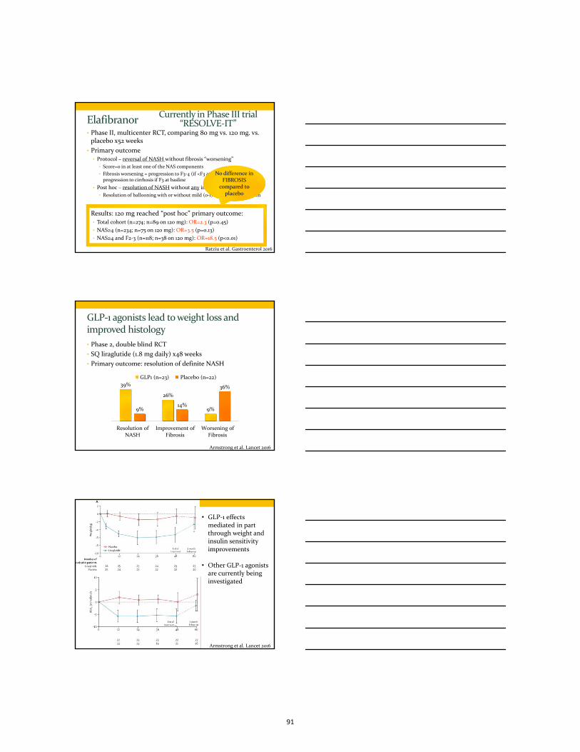
Elafibranor• Phase II, multicenter RCT, comparing 80 mg vs. 120 mg. vs. placebo x52 weeks
• Primary outcome• Protocol – reversal of NASH without fibrosis “worsening”
• Score=0 in at least one of the NAS components
• Fibrosis worsening = progression to F3‐4 (if <F3 at baseline), or progression to cirrhosis if F3 at basline
• Post hoc – resolution of NASH without any increase in fibrosis stage
• Resolution of ballooning with or without mild (0‐1) lobular inflammation
• Results: 120 mg reached “post hoc” primary outcome:• Total cohort (n=274; n=89 on 120 mg): OR=2.3 (p=0.45)
• NAS≥4 (n=234; n=75 on 120 mg): OR=3.5 (p=0.13)
• NAS≥4 and F2‐3 (n=118; n=38 on 120 mg): OR=18.5 (p<0.01)
Ratziu et al. Gastroenterol 2016
No difference in No difference in FIBROSIS
compared to placebo
Currently in Phase III trial “RESOLVE‐IT”
GLP‐1 agonists lead to weight loss and improved histology
• Phase 2, double blind RCT
• SQ liraglutide (1.8 mg daily) x48 weeks
• Primary outcome: resolution of definite NASH
Armstrong et al. Lancet 2016
39%
26%
9%9%14%
36%
Resolution ofNASH
Improvement ofFibrosis
Worsening ofFibrosis
GLP1 (n=23) Placebo (n=22)
Armstrong et al. Lancet 2016
• GLP‐1 effects mediated in part through weight and insulin sensitivity improvements
• Other GLP‐1 agonists are currently being investigated
91
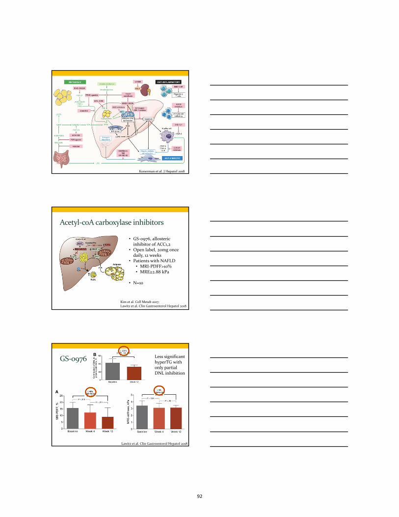
Novel treatment approaches
Konerman et al. J Hepatol 2018
Acetyl‐coA carboxylase inhibitors
Kim et al. Cell Metab 2017;Lawitz et al. Clin Gastroenterol Hepatol 2018
• GS‐0976, allosteric inhibitor of ACC1,2
• Open label, 20mg once daily, 12 weeks
• Patients with NAFLD• MRI‐PDFF>10%• MRE≥2.88 kPa
• N=10
Less significant hyperTG with only partial DNL inhibition
Lawitz et al. Clin Gastroenterol Hepatol 2018
GS‐0976
92
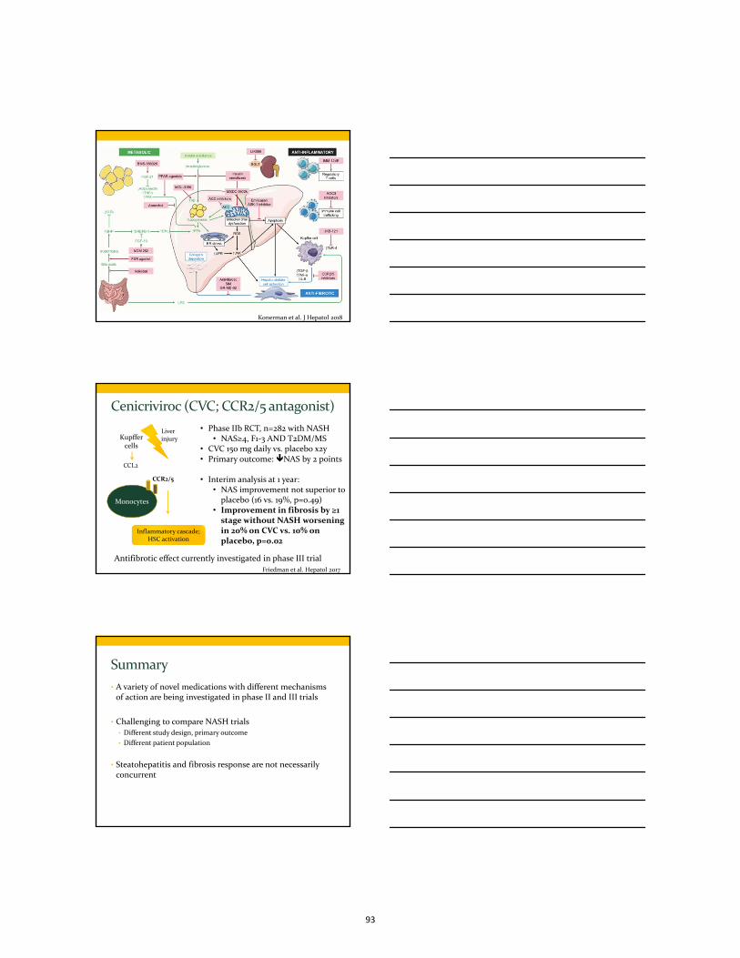
Novel treatment approaches
Konerman et al. J Hepatol 2018
Cenicriviroc (CVC; CCR2/5 antagonist)
Liver injuryΚupffer
cells
CCL2
Monocytes
CCR2/5
Inflammatory cascade; HSC activation
• Phase IIb RCT, n=282 with NASH• NAS≥4, F1‐3 AND T2DM/MS
• CVC 150 mg daily vs. placebo x2y• Primary outcome: NAS by 2 points
• Interim analysis at 1 year:• NAS improvement not superior to placebo (16 vs. 19%, p=0.49)
• Improvement in fibrosis by ≥1 stage without NASH worsening in 20% on CVC vs. 10% on placebo, p=0.02
Friedman et al. Hepatol 2017
Antifibrotic effect currently investigated in phase III trial
Summary
• A variety of novel medications with different mechanisms of action are being investigated in phase II and III trials
• Challenging to compare NASH trials• Different study design, primary outcome
• Different patient population
• Steatohepatitis and fibrosis response are not necessarily concurrent
93
Future Directions
• Address the methodological challenges:
• Population
• Take into account the phenotypic variability of NAFLD
• Thoughtful consideration of who actually needs treatment
• Outcome
• Study the impact of new agents on NASH and fibrosis independently
• Optimize histology surrogates to be used as outcomes
• Study combinations of agents
Take Home Messages
1. No medication is currently approved for the treatment of NAFLD
2. There is a plethora of new agents currently being investigated for the treatment of NAFLD/NASH
3. Careful selection of the patients who need treatment
4. Effective long‐term treatment approaches will likely include a combination of agents
Thank you
94
Other meds trialed
• Recombinant, pegylated FGF21 analogue BMS‐986026
Ratziu et al. Gastroenterol 2016
Elafibranor ‐ additional
Obeticholic acid also causes dyslipidemia
Neuschwander‐Tetriet al. Lancet 2014
95
Saghieri et al. Front Endocrinol 2018
96
When there is good function but the flow is all wrong; approach to non‐cirrhotic
portal hypertensionEvelyn Hsu, MD
Seattle Children’s Hospital
Learning Objectives
• Know the key aspects of diagnostic evaluation of children with portal hypertension
• Understand the terminology and management of children with non‐cirrhotic portal hypertension
Outline
Approach to portal hypertension in a child
Definitions
Epidemiology
Natural History
Management
Future Directions
Extrahepatic portal venous obstruction
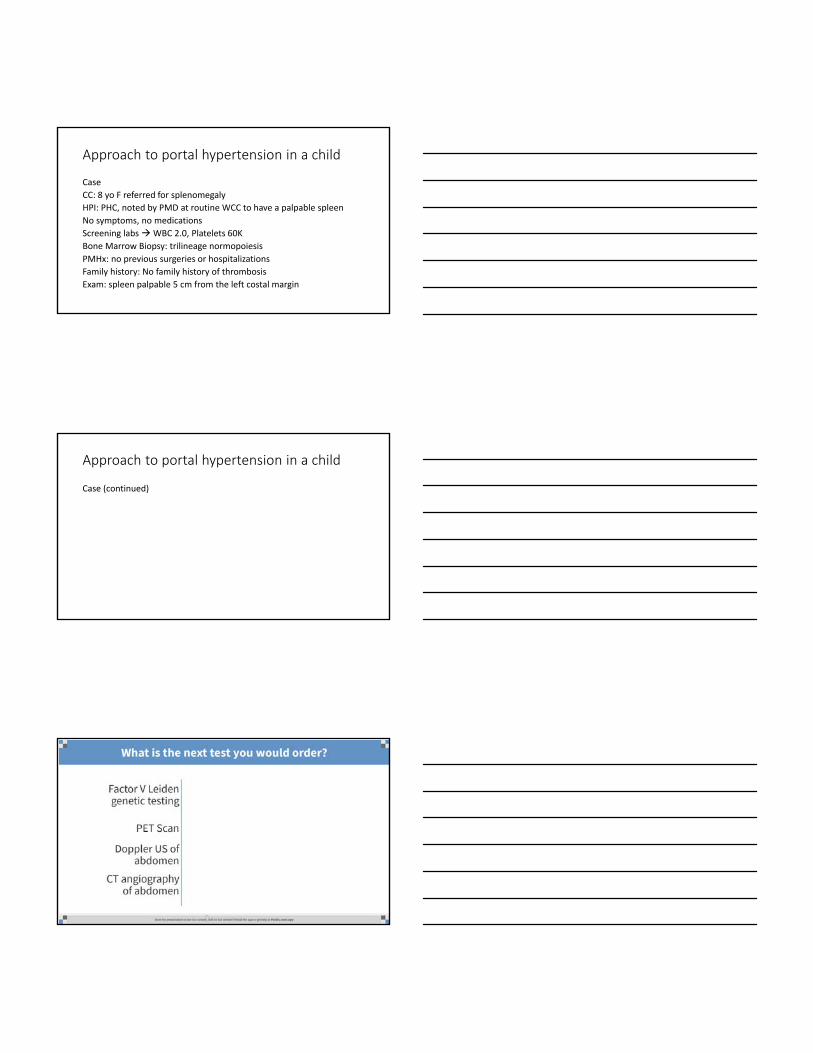
Approach to portal hypertension in a child
Case
CC: 8 yo F referred for splenomegaly
HPI: PHC, noted by PMD at routine WCC to have a palpable spleen
No symptoms, no medications
Screening labs WBC 2.0, Platelets 60K
Bone Marrow Biopsy: trilineage normopoiesis
PMHx: no previous surgeries or hospitalizations
Family history: No family history of thrombosis
Exam: spleen palpable 5 cm from the left costal margin
Approach to portal hypertension in a child
Case (continued)
Imaging modalities
• Doppler US
• CT angiography
Biopsy
• Liver biopsy performed, under IR
• Hepatic Wedge Pressure Gradient ~5 mm Hg
• Results show normal liver histology
Now what? What do we call this?
• 2011 Recommendations:
• Proposal that Idiopathic non‐cirrhotic portal hypertension be viewed as a distinct single entity with various pathological aspects
• Idiopathic non‐cirrhotic portal hypertension• Portal hypertension• Absence of cirrhosis as documented histologically in an appropriate liver specimen
• Absence of obstruction of the extra hepatic portal vein or hepatic vein outflow obstruction
• Absence of sarcoidosis, schistosomiasis, congenital hepatic fibrosis, or other causes of cirrhosis
Schouten JNL, et al. Hepatology. 2011 Sep 2;54(3):1071–81.
Problems with 2011 recommendations
• Liver lesions observed can be seen in the absence of portal hypertension
• Portal vein thrombosis is a frequent complication during disease course
• Requires exclusion of other causes of liver disease
De Gottardi A, et al. Lancet Gastroenterol Hepatol. 2019 May;4(5):399–411.
De Gottardi A, et al. Lancet Gastroenterol Hepatol. 2019 May;4(5):399–411.
New recommendations as defined by VALDIG
• Porto‐sinusoidal vascular disease• Histological alterations associated with porto‐sinusoidal vascular disease with portal hypertension encompass several elementary features that can be observed in isolated or combined fashion
1) Obliterative portal venopathy
2) Nodular regenerative hyperplasia
3) Incomplete septa cirrhosis
• These criteria are specific enough to be regarded as diagnostic for porto‐sinusoidal vascular disease, even in the absence of any clinical, imaging, or laboratory features of portal hypertension
De Gottardi A, et al. Lancet Gastroenterol Hepatol. 2019 May;4(5):399–411.
Obliterative portal venopathy and hepatoportal sclerosis• Affects primarily small and medium branches of the portal vein, with the key feature being phlebosclerosis
• Increased amounts of portal connective tissues around the vessels with irregular wall thickening and eccentric narrowing of the vessel lumen, possibly up to complete occlusion and absence of the vein
De Gottardi A, et al. Lancet Gastroenterol Hepatol. 2019 May;4(5):399–411.
Nodular regenerative hyperplasia
• Transformation of normal hepatic parenchyma into small nodules, mostly 1‐3mm, macroscopically paler than normal parenchyma
De Gottardi A, et al. Lancet Gastroenterol Hepatol. 2019 May;4(5):399–411.
Incomplete septal fibrosis
• Presence of incomplete, thin, perforated, or blind‐ended septa, and intermittently delimited rudimentary nodules
De Gottardi A, et al. Lancet Gastroenterol Hepatol. 2019 May;4(5):399–411.
Natural history
• Natural history is unknown
• Some patients may develop portal hypertension
• No recognized treatment exists, screening for signs of portal hypertension can be offered
• No data to recommend screening methodology or interval
• Cohort studies show good long‐term outcomes following a strategy based upon current management guidelines for cirrhosis‐ beta blockers, endoscopic band ligation
• TIPS have been used for variceal bleeding/refractory ascites
• Splenectomy or partial splenic embolization can increase platelet count, but benefits have not been demonstrated to outweigh risks
• Liver transplant data is scarce‐ survival is favorable, there is no defined risk of recurrence but some cases have been reported
Epidemiology
• Terminologies vary worldwide
• Developing countries have higher occurrences that have improved over time, favoring an association with intra‐abdominal infection
• 15‐34% of cases in India, 3‐7% in Europe, US, Canada
Pathogenesis
• Unknown‐ but relies on the development of vascular changes within the liver
• Associated conditions:• Immunity
• Blood diseases/prothrombotic conditions
• Infections
• Congenital or familial defects
• Drug exposure
De Gottardi A, et al. Lancet Gastroenterol Hepatol. 2019 May;4(5):399–411.
Baveno VI recommendations“Idiopathic portal hypertension, non‐cirrhotic portal fibrosis, and non‐cirrhotic portal hypertension indicate the same clinical entity”
Diagnosis requires exclusion of cirrhosis and other causes of non‐cirrhotic portal hypertension
Liver biopsy is mandatory and HVPG is recommended for diagnosis
Immunologic diseases and pro‐thrombotic disorders should be screened
Insufficient data for what therapy should be preferred for portal hypertension prophylaxis; management according to cirrhosis guidelines recommended
These patients should be screened for development of portal vein thrombosis‐ every 6 month doppler ultrasounds
Anticoagulation should be started in those who develop PVT
Extrahepatic portal vein obstruction
Evaluation, management, presentation
Natural history
• Extrahepatic portal vein obstruction is defined by obstruction of extrahepatic portal vein with or without involvement of the intrahepatic portal branches
• Referred with varied manifestations: • GI hemorrhage from variceal bleeding
• Splenomegaly, hypersplenism, less commonly with ascites
• Cavernous transformation: effort to bypass the thrombus and replace physiological portal venous flow or establish a spontaneous portosystemic shut
Imaging recommendations
• Doppler US
• CT angiography
• Established protocol for management (Alberti, et al JPBJ 2013 57(5) 619‐626)
Conservative management
• Medical therapy• Beta‐blockers
• anticoagulation
• Endoscopic therapy• Sclerotherapy
• Endoscopic variceal band ligation
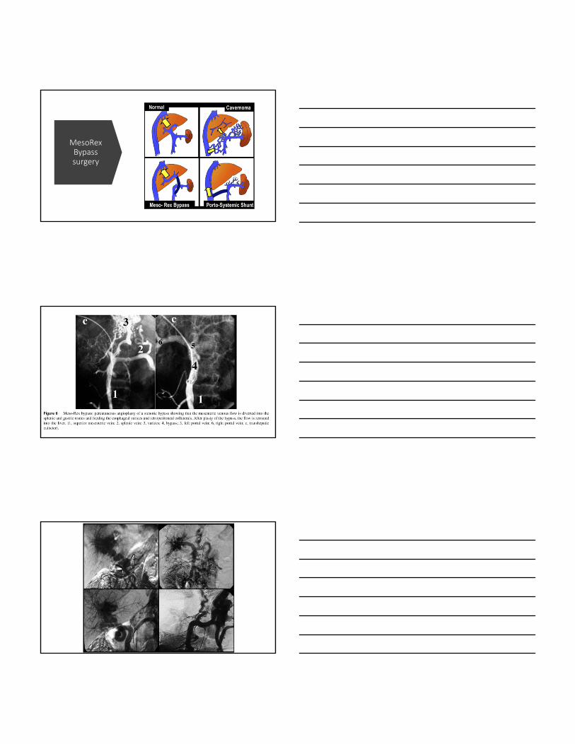
MesoRex Bypass surgery
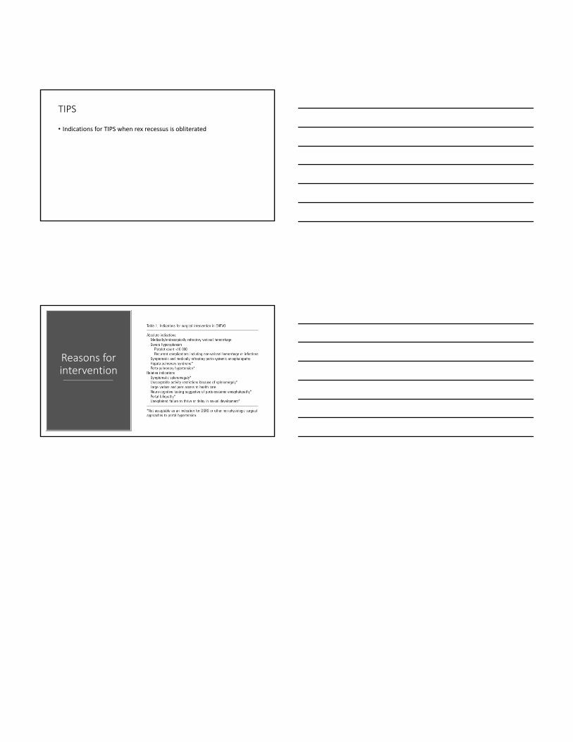
TIPS
• Indications for TIPS when rex recessus is obliterated
Reasons for intervention
What do I do now…. The management of portal hypertensive complications: varices, ascites, and encephalopathy
René Romero, M.D.Professor of Pediatrics
Clinical Director, Pediatric Hepatology
Medical Director, Pediatric Liver Transplant Program
Children’s Healthcare of Atlanta
Emory University School of Medicine
2
Disclosures
• In the past 12 months, I have had no relevant financial relationships with the manufacturer(s) of any commercial product(s) and/or provider(s) of commercial services discussed in this CME activity.
• I do not intend to discuss an unapproved or investigative use of a commercial product or device in my presentation.
Objectives
• Understand the appropriate medical management (pharmacologic and endoscopic) of acute variceal hemorrhage.
• Understand dosing and monitoring of diuretics in the management of ascites and the appropriate use of paracentesis.
• Approaches to the recognition and management of encephalopathy in pediatric chronic liver disease.
3
97
• Management of complications of portal hypertension in children is challenging
– Lack of rigorous clinical studies that inform practice
– Nature of disease leading to portal hypertension dissimilar to adults
– Rarity of the condition
– Limitations of equipment and techniques to study the condition in patient populations at risk
– Consequences of complications may be different than adults
• The many controversies surrounding these subjects will not be settled in this presentation 4
Portal Hypertension in Children
Alcoholic liver disease
Biliary Atresia
Ling et al. JPGN. 2011; 52: 254‐261Shneider et al. Hepatology. 2016; 63 (4): 1368‐1380.Grammatikopoulos et al. Arch Dis Child. 2018; 103: 186‐191.http://www.clevelandclinicmeded.com/medicalpubs/diseasemanagement/hepatology/alcoholic‐liver‐disease/https://childrennetwork.org/For‐Physicians/Digital‐Liver‐Histopathology‐Library
What do I do…. Why is it important to knowwhich child is at risk for bleeding from varices?
5
“This study makes the argument that if the risk of death from the first variceal bleed is presumed to be low, the need for primary prophylaxis of the first AVB in children would depend on the extent of the associated morbidity.” Bass JPGN 2018;67(3):312‐3
• How do we identify children who are at risk of developing and bleedingfrom esophageal varices in childhood?
• Gold standard in adults would be HVWPG– Not practical
• Clinical findings frequently used– not sensitive or specific enough
for to predict bleeders
• Imaging or Noninvasive measures of hepatic fibrosis– not sensitive or specific enough
to predict bleeders
• Scoring systems based on combination of labs and clinical features– May be the future
6
What do I do…. Which child is at risk for bleeding from varices?
Shneider et al. JPGN 2012; 55: 567‐573.Sutton et al. JPGN 2018; 66:559‐569.
98
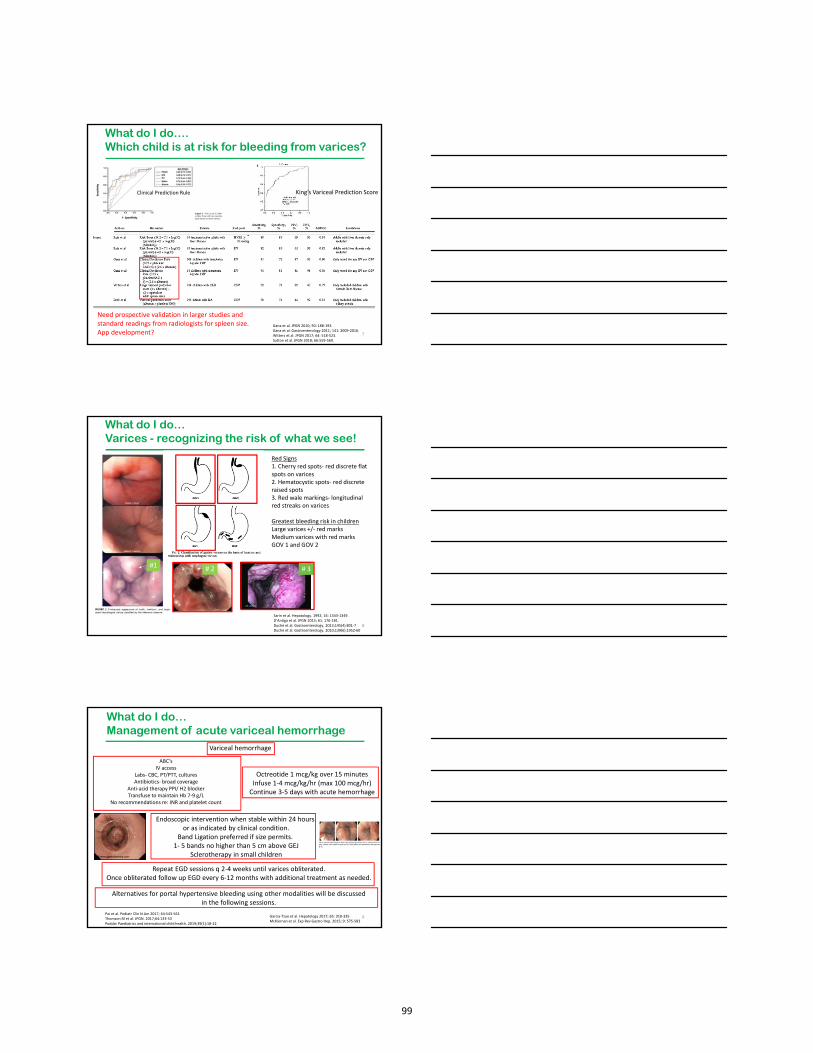
What do I do…. Which child is at risk for bleeding from varices?
7
Need prospective validation in larger studies and standard readings from radiologists for spleen size. App development?
Clinical Prediction Rule King’s Variceal Prediction Score
Gana et al. JPGN 2010; 50: 188‐193.Gana et al. Gastroenterology 2011; 141: 2009‐2016.Witters et al. JPGN 2017; 64: 518‐523.Sutton et al. JPGN 2018; 66:559‐569.
What do I do…Varices - recognizing the risk of what we see!
8
Red Signs1. Cherry red spots‐ red discrete flat spots on varices2. Hematocystic spots‐ red discrete raised spots3. Red wale markings‐ longitudinal red streaks on varices
Greatest bleeding risk in childrenLarge varices +/‐ red marksMedium varices with red marksGOV 1 and GOV 2
# 3# 2#1
Sarin et al. Hepatology. 1992; 16: 1343‐1349.D’Antiga et al. JPGN 2015; 61: 176‐181.Duché et al. Gastroenterology. 2013;145(4):801‐7Duché et al. Gastroenterology. 2010;139(6):1952‐60
What do I do…Management of acute variceal hemorrhage
9
Variceal hemorrhage
Octreotide 1 mcg/kg over 15 minutesInfuse 1‐4 mcg/kg/hr (max 100 mcg/hr)Continue 3‐5 days with acute hemorrhage
Endoscopic intervention when stable within 24 hours.or as indicated by clinical condition.
Band Ligation preferred if size permits. 1‐ 5 bands no higher than 5 cm above GEJ
Sclerotherapy in small children
Repeat EGD sessions q 2‐4 weeks until varices obliterated.Once obliterated follow up EGD every 6‐12 months with additional treatment as needed.
Alternatives for portal hypertensive bleeding using other modalities will be discussedin the following sessions.
ABC’sIV access
Labs‐ CBC, PT/PTT, culturesAntibiotics‐ broad coverage
Anti‐acid therapy PPI/ H2 blockerTransfuse to maintain Hb 7‐9 g/L
No recommendations re: INR and platelet count
Garcia‐Tsao et al. Hepatology 2017; 65: 310‐335McKiernan et al. Exp Rev Gastro Hep. 2015; 9: 575‐583
Pai et al. Pediatr Clin N Am 2017; 64:543‐561Thomson M et al. JPGN. 2017;64:133‐53Poddar Paediatrics and international child health. 2019;39(1):18‐22
99
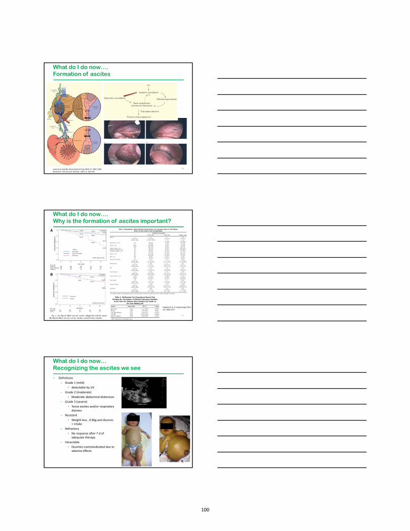
What do I do now….Formation of ascites
10Lane et al. Exp Rev Gastroenterol Hep 2015; 9: 1281‐1292Romero R. Clinical Liver Disease. 2013; 2: 210‐214
What do I do now….Why is the formation of ascites important?
11
Pugliese R et al. Hepatology 2014; 59: 1964‐1971
• Definitions
– Grade 1 (mild)
• detectable by US
– Grade 2 (moderate)
• Moderate abdominal distension
– Grade 3 (severe)
• Tense ascites and/or respiratory distress
– Resistant
• Weight loss , 0.8kg and diuresis < intake
– Refractory
• No response after 7 d of adequate therapy
– Intractable
• Diuretics contraindicated due to adverse effects
12
What do I do now…Recognizing the ascites we see
100
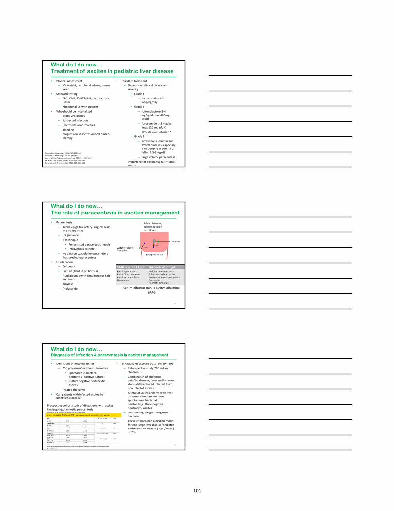
• Physical Assessment
– VS, weight, peripheral edema, neuro exam
• Standard testing
– CBC, CMP, PT/PTT/INR, UA, Ucr, Una, Uosm
– Abdominal US with Doppler
• Who should be hospitalized
– Grade 2/3 ascites
– Suspected infection
– Electrolyte abnormalities
– Bleeding
– Progression of ascites on oral diuretic therapy
• Standard treatment
– Depends on clinical picture and severity
• Grade 1
– Na restriction 1‐2 meq/kg/day
• Grade 2
– Spironolactone 2‐4 mg/kg/d (max 400mg adult)
– Furosemide 1‐ 3 mg/kg (max 120 mg adult)
– 25% albumin infusion?
• Grade 3
– Intravenous albumin and IV/oral diuretics especially with peripheral edema or Salb < 2.5‐3.0 g/dL
– Large volume paracentesis
– Importance of optimizing nutritional status
13
What do I do now…Treatment of ascites in pediatric liver disease
Runyon BA. Hepatology. 2009;49(6):2087‐107Runyon BA. Hepatology. 2013;57(4):1651‐3Lane et al. Exp Rev Gastroenterol Hep 2015; 9: 1281‐1292Bes et al. Arch Argent Pediatr 2017; 115: 385‐390Bes et al. Arch Argent Pediatr 2017; 115: 505‐ 511
• Paracentesis
– Avoid epigastric artery, surgical scars and visible veins
– US guidance
– Z‐technique
• Fenestrated paracentesis needle
• Intravenous catheter
– No data on coagulation parameters that preclude paracentesis
• Fluid analysis
– Cell count
– Culture (10ml in BC bottles)
– Fluid albumin with simultaneous Salbfor SAAG
– Amylase
– Triglyceride
14
What do I do now…The role of paracentesis in ascites management
Adult distances, approx. location in children
Serum albumin minus ascites albumin=SAAG
• Definitions of infected ascites
– 250 polys/mm3 without alternative
• Spontaneous bacterial peritonitis (positive culture)
• Culture negative neutrocyticascites
– Treated the same
• Can patients with infected ascites be identified clinically?
• Srivastava et al. JPGN 2017; 64: 194‐199
– Retrospective study 262 Indian children
– Combination of abdominal pain/tenderness, fever and/or loose stools differentiated infected from non infected ascites
– A total of 28.6% children with liver disease‐related ascites have spontaneous bacterial peritonitis/culture‐negative neutrocytic ascites
– commonly grew gram‐negative bacteria
– These children had a median model for end‐stage liver disease/pediatric endstage liver disease [PELD/MELD] of 23)
15
What do I do now…Diagnosis of infection & paracentesis in ascites management
Ghobrail et al. PloS one. 2018;13(10):e0203808
Prospective cohort study of 66 patients with ascitesUndergoing diagnostic paracentesis
Fever, elevated ANC and CRP was associated with infected ascites
101
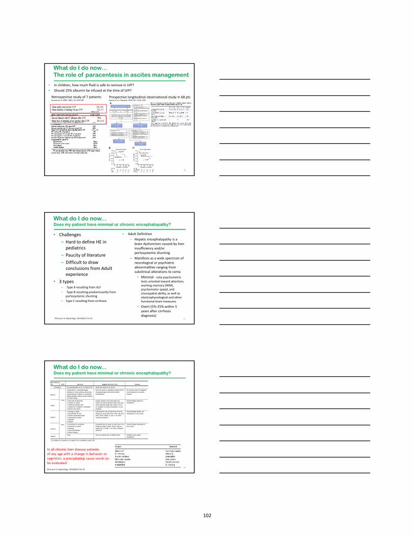
What do I do now…The role of paracentesis in ascites management
• In children, how much fluid is safe to remove in LVP?
• Should 25% albumin be infused at the time of LVP?
16
Kramer et al. JPGN. 2001; 33: 245‐249
Retrospective study of 7 patientsSarma et al. J Hepatol. 2015; 63: 1126‐1132
Prospective longitudinal observational study in 68 pts
• Challenges
– Hard to define HE in pediatrics
– Paucity of literature
– Difficult to draw conclusions from Adult experience
• 3 types– Type A resulting from ALF
– Type B resulting predominantly from portosystemic shunting
– Type C resulting from cirrhosis
• Adult Definition
– Hepatic encephalopathy is a brain dysfunction caused by liver insufficiency and/or portosystemic shunting
– Manifests as a wide spectrum of neurological or psychiatric abnormalities ranging from subclinical alterations to coma
• Minimal ‐ only psychometric tests oriented toward attention, working memory (WM), psychomotor speed, and visuospatial ability, as well as electrophysiological and other functional brain measures
• Overt (5%‐25% within 5 years after cirrhosis diagnosis)
17
What do I do now...Does my patient have minimal or chronic encephalopathy?
Vilstrup et al. Hepatology. 2014;60(2):715‐35
What do I do now...Does my patient have minimal or chronic encephalopathy?
18
In all chronic liver disease patientsof any age with a change in behavior or cognition, a precipitating cause needs to be evaluated
Vilstrup et al. Hepatology. 2014;60(2):715‐35
102
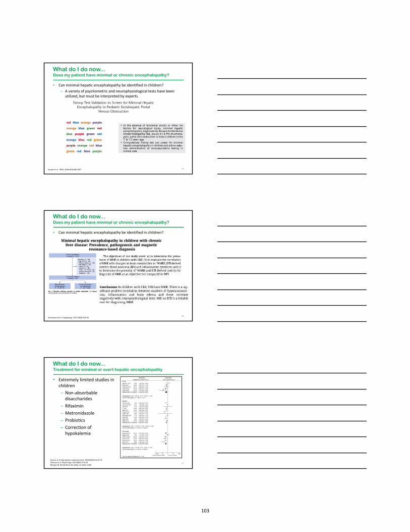
What do I do now...Does my patient have minimal or chronic encephalopathy?
• Can minimal hepatic encephalopathy be identified in children?
– A variety of psychometric and neurophysiological tests have been utilized, but must be interpreted by experts
19Suresh et al. JPGN. 2018;66(5):802‐807
What do I do now...Does my patient have minimal or chronic encephalopathy?
• Can minimal hepatic encephalopathy be identified in children?
20Srivastava et al. J Hepatology. 2017;66(3):528‐36
• Extremely limited studies in children
– Non‐absorbable disaccharides
– Rifaximin
– Metronidazole
– Probiotics
– Correction of hypokalemia
21
What do I do now...Treatment for minimal or overt hepatic encephalopathy
Ayra et al. Postgraduate medical journal. 2010;86(1011):34‐41Vilstrup et al. Hepatology. 2014;60(2):715‐35Morgan M. Metab Brain Dis.2016; 31:1361–1364
103
What do I do now…. The management of portal hypertensive complications: varices, ascites, and encephalopathy
• Conclusions– The management of portal hypertensive complications in children is
challenging due to its rarity and scientific evidence of best practices.
– Secondary prophylaxis of variceal bleeding with endoscopic therapy is appropriate and should be initiated as soon as is clinically feasible.
– The development of ascites can be managed with diuretics but is clearly an indicator of liver disease progression.
– The detection and treatment of all forms of hepatic encephalopathy in children remains problematic and requires specialized testing in the earliest stages.
– Pediatric Gastroenterologists should utilize frequent consultation with Pediatric Liver Centers for the management of patients with portal hypertensive complications
22
104
Pediatric Liver Transplant: Indications, Timing and Options
Shikha S. Sundaram, MD, MSCI, FAASLD
Medical Director, Pediatric Liver Transplantation
Children’s Hospital Colorado
Disclosures
• None
Objectives
• Understand indications/contraindications to liver transplant
• Understand when to refer a patient for transplant evaluation
• Understand the transplant evaluation process
• Understand how to help your patient choose a transplant program
• Understand PELD/MELD scores and limitations
117
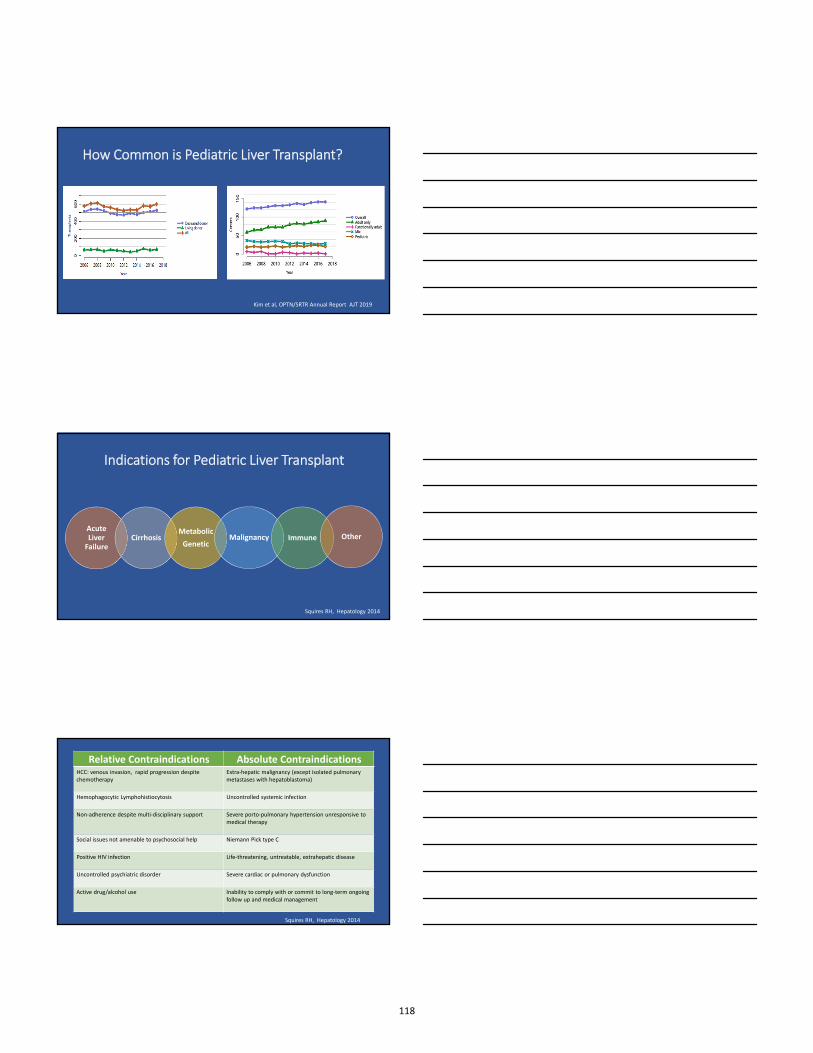
How Common is Pediatric Liver Transplant?
Kim et al, OPTN/SRTR Annual Report AJT 2019
Indications for Pediatric Liver Transplant
Acute Liver Failure
CirrhosisMetabolic
GeneticMalignancy Immune Other
Squires RH, Hepatology 2014
Relative Contraindications Absolute ContraindicationsHCC: venous invasion, rapid progression despite chemotherapy
Extra‐hepatic malignancy (except isolated pulmonary metastases with hepatoblastoma)
Hemophagocytic Lymphohistiocytosis Uncontrolled systemic infection
Non‐adherence despite multi‐disciplinary support Severe porto‐pulmonary hypertension unresponsive to medical therapy
Social issues not amenable to psychosocial help Niemann Pick type C
Positive HIV infection Life‐threatening, untreatable, extrahepatic disease
Uncontrolled psychiatric disorder Severe cardiac or pulmonary dysfunction
Active drug/alcohol use Inability to comply with or commit to long‐term ongoing follow up and medical management
Squires RH, Hepatology 2014
118
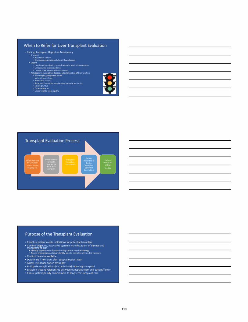
When to Refer for Liver Transplant Evaluation
• Timing: Emergent, Urgent or Anticipatory• Emergent
• Acute Liver Failure• Acute decompensation of chronic liver disease
• Urgent• Liver based metabolic crises refractory to medical management• Unresectable hepatoblastoma• Unresectable hepatocellular carcinoma
• Anticipatory: chronic liver disease and deterioration of liver function• Poor weight gain/growth failure• Variceal hemorrhage• Intractable ascites• Recurrent cholangitis, spontaneous bacterial peritonitis• Severe pruritus• Encephalopathy• Uncorrectable coagulopathy
Transplant Evaluation Process
Patient Referred for Evaluation
‐ Gather records, imaging, etc.
Permission to Evaluate granted by insurance company
Transplant Evaluation Performed
Patient Presented to
Center Transplant Selection Committee
Patient Transplant Listing
Yes/No
Purpose of the Transplant Evaluation
• Establish patient meets indications for potential transplant• Confirm diagnosis, associated systemic manifestations of disease and management plan
• Identify opportunities for maximizing current medical therapy• Assess immunization status, identify plan to complete all needed vaccines
• Confirm finances available• Determine if non‐transplant surgical options exist• Assess live donor option feasibility• Anticipate complications (and solutions) following transplant• Establish trusting relationship between transplant team and patient/family• Ensure patient/family commitment to long term transplant care
119
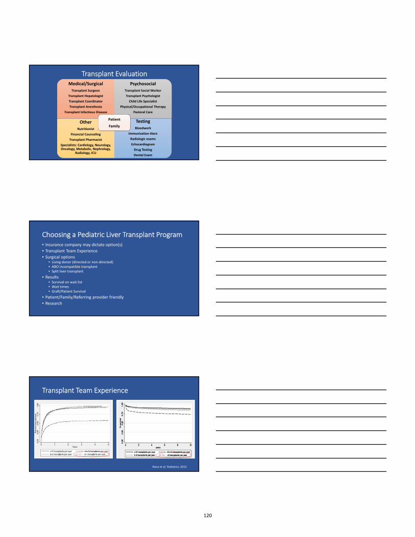
Transplant EvaluationMedical/Surgical
Transplant Surgeon
Transplant Hepatologist
Transplant Coordinator
Transplant Anesthesia
Transplant Infectious Disease
Psychosocial
Transplant Social Worker
Transplant Psychologist
Child Life Specialist
Physical/Occupational Therapy
Pastoral Care
Other
Nutritionist
Financial Counseling
Transplant Pharmacist
Specialists: Cardiology, Neurology, Oncology, Metabolic, Nephrology,
Radiology, ICU
Testing
Bloodwork
Immunization titers
Radiologic exams
Echocardiogram
Drug Testing
Dental Exam
Patient
Family
Choosing a Pediatric Liver Transplant Program• Insurance company may dictate option(s)
• Transplant Team Experience
• Surgical options• Living donor (directed or non‐directed)• ABO incompatible transplant• Split liver transplant
• Results• Survival on wait list• Wait times• Graft/Patient Survival
• Patient/Family/Referring provider friendly
• Research
Transplant Team Experience
Rana et al. Pediatrics 2015
120
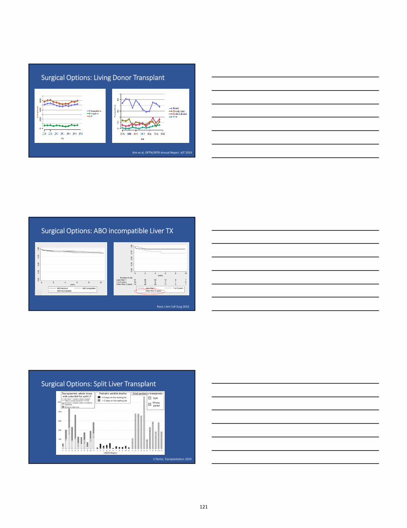
Surgical Options: Living Donor Transplant
Kim et al, OPTN/SRTR Annual Report AJT 2019
Surgical Options: ABO incompatible Liver TX
Rana J Am Coll Surg 2016
Surgical Options: Split Liver Transplant
E Perito, Transplantation 2019
121
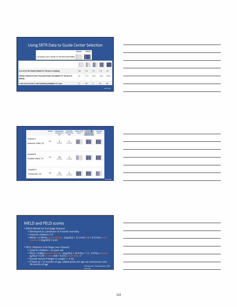
Using SRTR Data to Guide Center Selection
unos.org
Hospital A
Awesome Valley, NY
Hospital B
Paradise Island, TX
Hospital C
Fantasyville, CA
unos.org
MELD and PELD scores• MELD (Model for End Stage Disease)
• Developed as a predictor of 3 month mortality• Used for children ≥ 12• MELD = 3.78×ln[serum bilirubin (mg/dL)] + 11.2×ln[INR] + 9.57×ln[serum creatinine (mg/dL)] + 6.43
• PELD (Pediatric End‐Stage Liver Disease) • Used for children < 12 years old• PELD = 4.80[Ln serum bilirubin (mg/dL)] + 18.57[Ln INR] ‐ 6.87[Ln albumin(g/dL)] + 4.36(<1 year old) + 6.67(growth failure)
• Growth failure if height or weight < ‐2 SD• If listed at < 12 months of age, added points for age are maintained until 24 months of age
S McDiarmid, Transplantation 2002unos.org
122
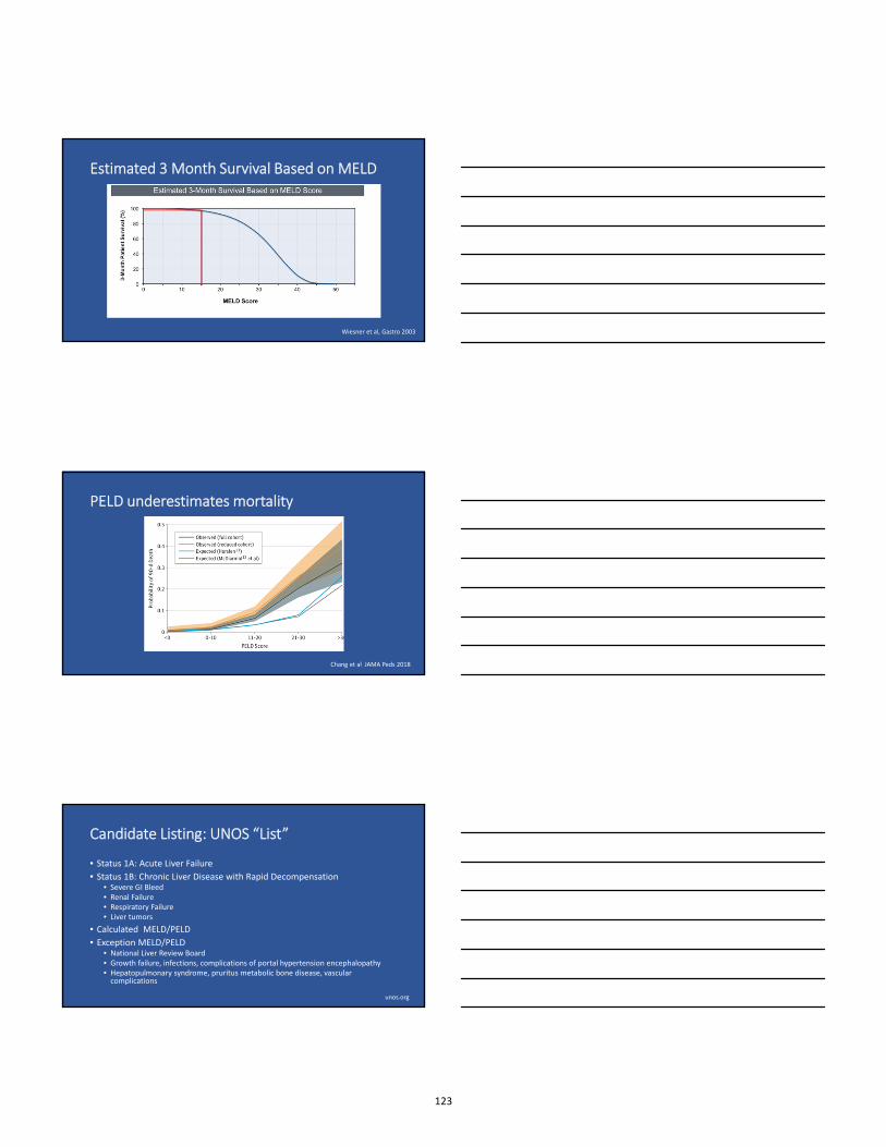
Estimated 3 Month Survival Based on MELD
Wiesner et al, Gastro 2003
PELD underestimates mortality
Chang et al JAMA Peds 2018
Candidate Listing: UNOS “List”
• Status 1A: Acute Liver Failure• Status 1B: Chronic Liver Disease with Rapid Decompensation
• Severe GI Bleed• Renal Failure• Respiratory Failure• Liver tumors
• Calculated MELD/PELD
• Exception MELD/PELD• National Liver Review Board• Growth failure, infections, complications of portal hypertension encephalopathy• Hepatopulmonary syndrome, pruritus metabolic bone disease, vascular complications
unos.org
123
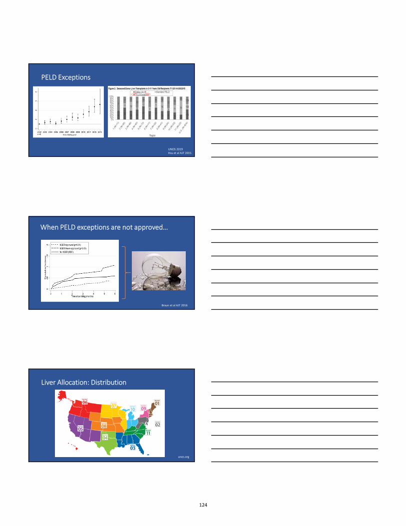
PELD Exceptions
UNOS 2019Hsu et al AJT 2015
When PELD exceptions are not approved…
Braun et al AJT 2016
Liver Allocation: Distribution
unos.org
124
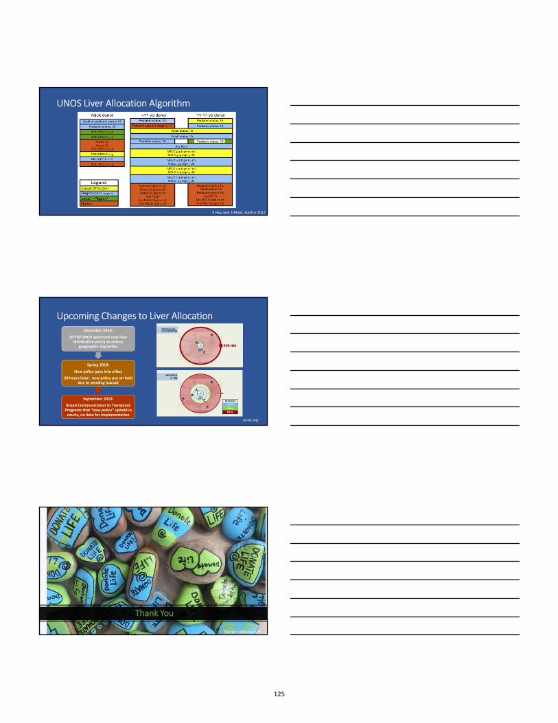
UNOS Liver Allocation Algorithm
E Hsu and S Rhee, Gastro 2017
Upcoming Changes to Liver AllocationDecember 2018:
OPTN/UNOS approved new liver distribution policy to reduce
geographic disparities
Spring 2019:
New policy goes into effect
24 hours later: new policy put on hold due to pending lawsuit
September 2019:
Broad Communication to Transplant Programs that “new policy” upheld in courts, no date for implementation
unos.org
Thank You
Twitter: @DonateLife
125
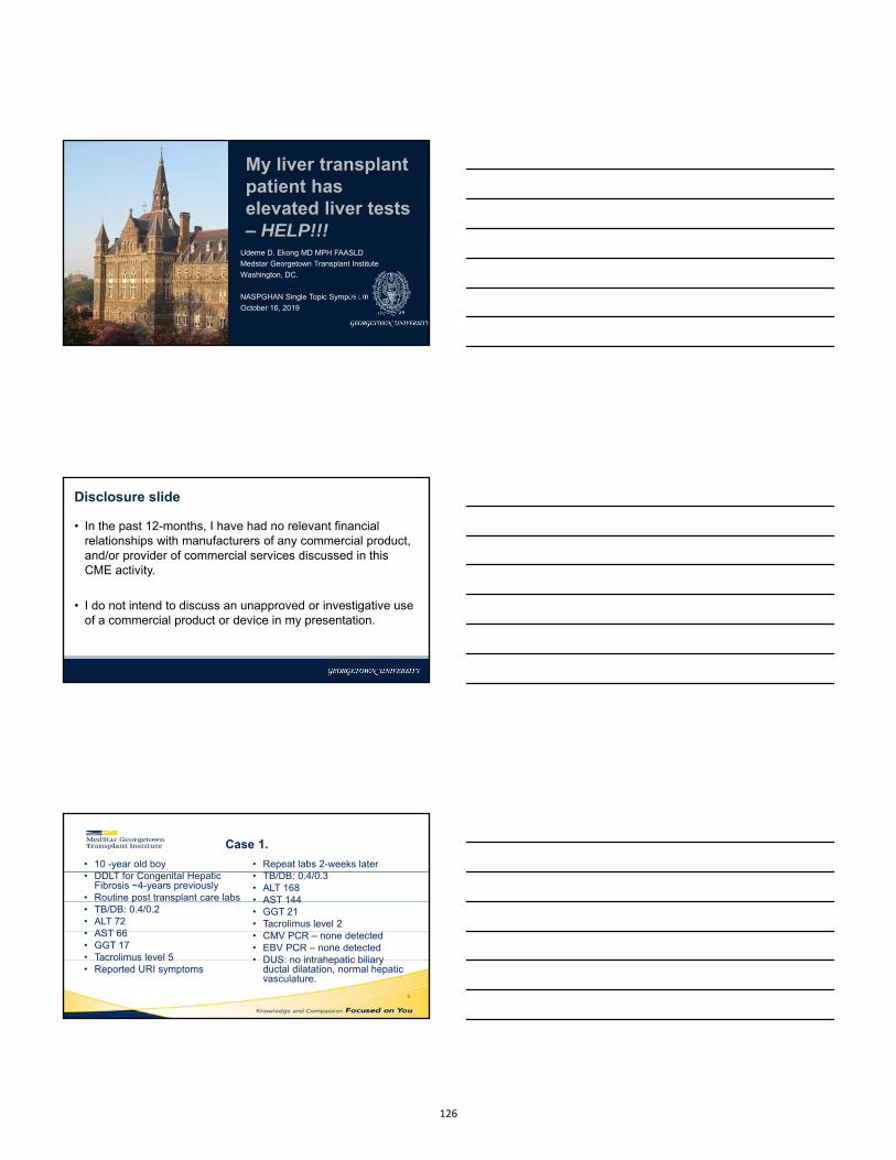
My liver transplant patient has elevated liver tests – HELP!!!
Udeme D. Ekong MD MPH FAASLD
Medstar Georgetown Transplant Institute
Washington, DC.
NASPGHAN Single Topic Symposium
October 16, 2019
Disclosure slide
• In the past 12-months, I have had no relevant financial relationships with manufacturers of any commercial product, and/or provider of commercial services discussed in this CME activity.
• I do not intend to discuss an unapproved or investigative use of a commercial product or device in my presentation.
Case 1.
• 10 -year old boy• DDLT for Congenital Hepatic
Fibrosis ~4-years previously• Routine post transplant care labs• TB/DB: 0.4/0.2• ALT 72• AST 66• GGT 17• Tacrolimus level 5• Reported URI symptoms
• Repeat labs 2-weeks later• TB/DB: 0.4/0.3• ALT 168• AST 144• GGT 21• Tacrolimus level 2• CMV PCR – none detected• EBV PCR – none detected• DUS: no intrahepatic biliary
ductal dilatation, normal hepatic vasculature.
3
126
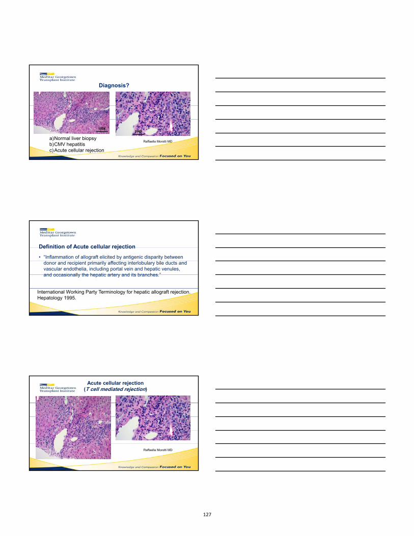
Diagnosis?
a)Normal liver biopsyb)CMV hepatitisc)Acute cellular rejection
Raffaella Morotti MD
100X200X
Definition of Acute cellular rejection
• “Inflammation of allograft elicited by antigenic disparity between donor and recipient primarily affecting interlobulary bile ducts and vascular endothelia, including portal vein and hepatic venules, and occasionally the hepatic artery and its branches.”
International Working Party Terminology for hepatic allograft rejection. Hepatology 1995.
Acute cellular rejection (T cell mediated rejection)
Raffaella Morotti MD
127
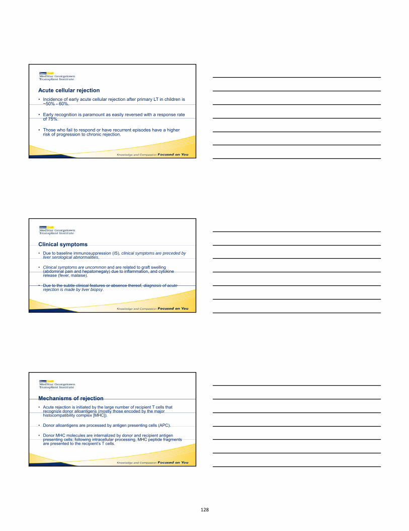
Acute cellular rejection
• Incidence of early acute cellular rejection after primary LT in children is ~50% - 60%.
• Early recognition is paramount as easily reversed with a response rate of 75%.
• Those who fail to respond or have recurrent episodes have a higher risk of progression to chronic rejection.
Clinical symptoms
• Due to baseline immunosuppression (IS), clinical symptoms are preceded by liver serological abnormalities.
• Clinical symptoms are uncommon and are related to graft swelling (abdominal pain and hepatomegaly) due to inflammation, and cytokine release (fever, malaise).
• Due to the subtle clinical features or absence thereof, diagnosis of acute rejection is made by liver biopsy.
Mechanisms of rejection
• Acute rejection is initiated by the large number of recipient T cells that recognize donor alloantigens (mostly those encoded by the major histocompatibility complex [MHC]).
• Donor alloantigens are processed by antigen presenting cells (APC).
• Donor MHC molecules are internalized by donor and recipient antigen presenting cells; following intracellular processing, MHC peptide fragments are presented to the recipient’s T cells.
128
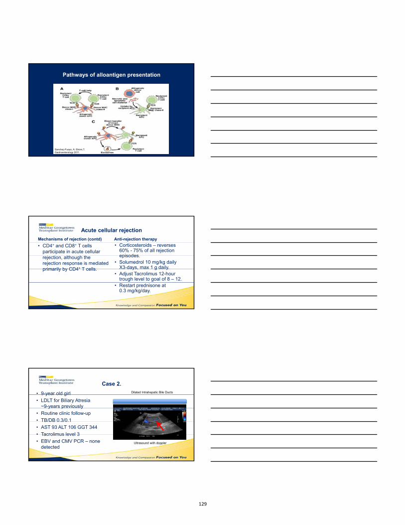
Pathways of alloantigen presentation
Sanchez-Fueyo, A; Strom,T.Gastroenterology 2011.
Acute cellular rejection
Mechanisms of rejection (contd) Anti-rejection therapy
• CD4+ and CD8+ T cells participate in acute cellular rejection, although the rejection response is mediated primarily by CD4+ T cells.
• Corticosteroids – reverses 60% - 75% of all rejection episodes.
• Solumedrol 10 mg/kg daily X3-days, max 1 g daily.
• Adjust Tacrolimus 12-hour trough level to goal of 8 – 12.
• Restart prednisone at 0.3 mg/kg/day.
Case 2.
• 9-year old girl
• LDLT for Biliary Atresia ~9-years previously
• Routine clinic follow-up
• TB/DB 0.3/0.1
• AST 93 ALT 106 GGT 344
• Tacrolimus level 3
• EBV and CMV PCR – none detected
Dilated Intrahepatic Bile Ducts
Ultrasound with doppler
129
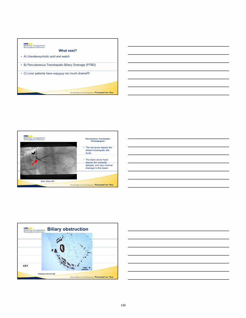
What next?
• A) Ursodeoxycholic acid and watch
• B) Percutaneous Transhepatic Biliary Drainage (PTBD)
• C) Liver patients have wayyyyy too much drama!!!!
Percutaneous TranshepaticCholangiogram
• The red arrow depicts the dilated intrahepatic bile ducts.
• The black arrow head depicts the markedly delayed, and very minimal drainage to the bowel.
Brian Dillon MD
Biliary obstruction
CK7
Raffaella Morotti MD
100X
130
Biliary complications following pediatric liver transplantation
• Biliary complications after LT remain frequent despite improvements/innovations in surgical techniques.
• These complications occasionally lead to graft failure or even death.
• The reported incidence of biliary complications after LDLT is 10% - 35% in pediatric recipients.
• Suggested risk factors for biliary complications include:– Hepatic arterial thrombosis– Acute cellular rejection– Prolonged cold ischemia time– Older age of donor– LDLT– ABO-incompatible LT
• 2 major therapeutic options for post-transplant biliary strictures:
– Surgical – biliary reconstruction (Roux-en-Y hepaticojejunostomy)
– Nonsurgical – PTBD
• In cases of post-transplant biliary strictures in duct-to-duct biliary reconstruction, conventional endoscopic intervention is the first-line Rx
Biliary obstruction following pediatric liver transplantation
• In biliary reconstruction at pediatric LT, Roux-en-Y hepaticojejunostomy is a standard procedure because the most common original disease is biliary atresia.
• In cases of post-transplant biliary strictures in hepaticojejunal biliary reconstruction, conventional endoscopy cannot approach the hepaticojejunalanastomotic site*
• Therefore PTBD is the first-line Rx for pediatric post-transplant biliary strictures.
Biliary obstruction following pediatric liver transplantation
131
Biliary obstruction following pediatric liver transplantationSanada et al. Clinical Transplantation 2019.
• Retrospective analysis of their experience on long-term outcome of PTBD for post-transplant biliary strictures.
• Success rate of PTBD – 90.6%
• 15-year primary patency rate of PTBD –52.6%
• The recurrence rate of biliary strictures after PTBD - 18.8%
Outcomes of PTBD for pediatric post-transplant biliary strictures
Case 3.• TB/DB: 0.6/0.1• ALT: 127• AST: 140• GGT: 255
• EBV PCR – none detected• CMV PCR – none detected
• DUS: no intrahepatic biliary ductal dilatation, patent hepatic vasculature.
• 16-year old F• DDLT 15 ½-years previously for
ESLD secondary to Biliary Atresia• Routine clinic visit• IS: Last four Tacrolimus levels: 9, 2,
8, 9
• PE: no peripheral adenopathy• Tonsils 2+ bilaterally, no exudates• BP 50% for age, height, and gender• Liver not palpable or percussed
beneath SCM, span 6 cm. No splenomegaly, no ascites.
132
Lymphoplasmacytic portal inflammation extending into the lobule (arrows) and interface hepatitis.
Additional labs obtained:
• ANA 1:320
• IgG elevated
• Anti-smooth muscle Ab, anti-LKM Ab, SLA all normal.
Raffaella Morotti MDH&E
200X
Clinical parameters used for diagnosing dnAIH(Kerkar, Vergani. J. Autoimmunity 2018).
Reports of dnAIH in pediatrics from around the world
Kerkar, Vergani. J Autoimmunity 2018.
133
De novo Autoimmune hepatitis (dnAIH)• Characterized by chronic liver damage with interface hepatitis, high
transaminase levels, high IgG levels and positive autoantibodies.
• Occurs in LT recipients transplanted for non-autoimmune liver disorders.
• The possibility that an antibody directed to a drug-metabolizing enzyme expressed at high levels within the liver and kidneys – namely glutathione-S-transferase T1 (GSTT1)is instrumental to the development of dnAIH has been suggested.
Immune pathogenesis of dnAIH
Regulatory T cells (Tregs) of these patientsare dysfunctional; and produce the pro-inflammatory cytokines – IFN-γ & IL-17A
dnAIH
normalLT
patient
Arterbery, Ekong et al. J. Immunology 2016.
• Monocytes/Macrophages seem to be key in promoting an inflammatory milieu that subsequently leads to the observed functional impairment of Tregs.
Immune pathogenesis of dnAIH
134
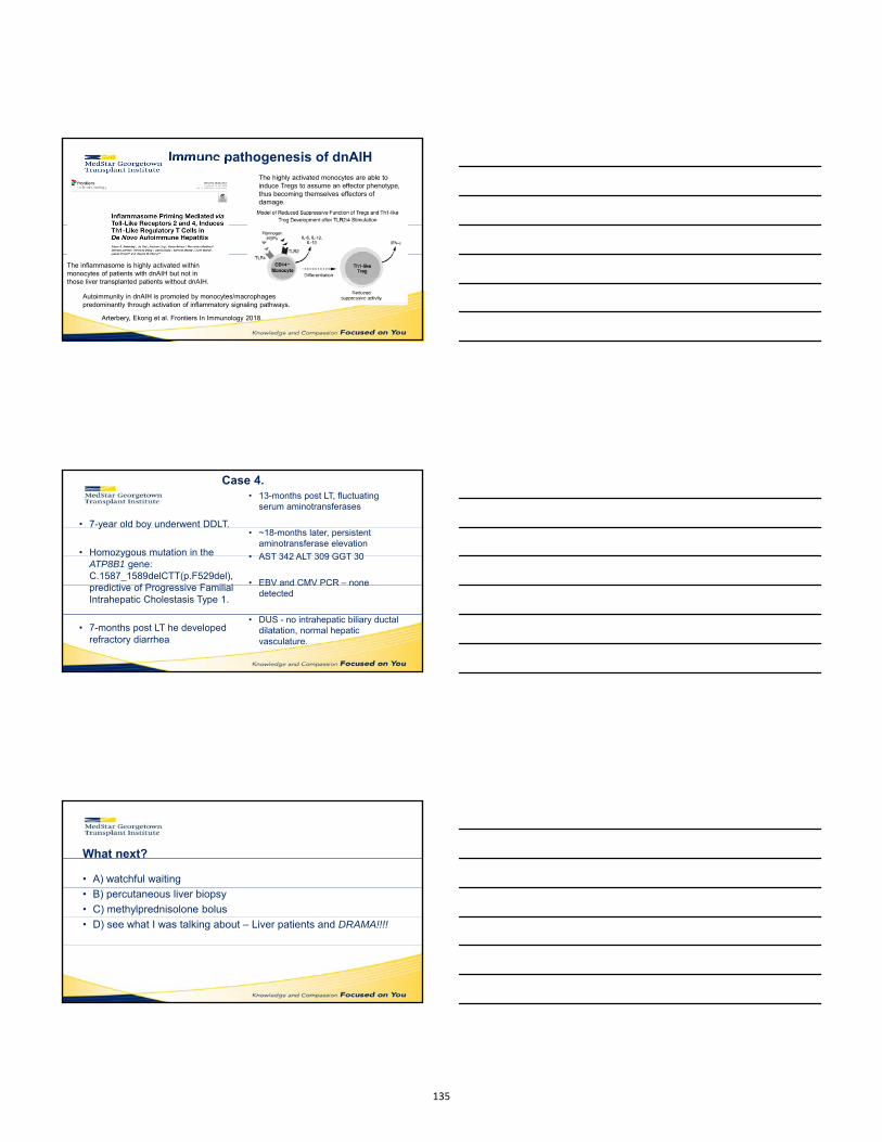
Immune pathogenesis of dnAIHThe highly activated monocytes are able to induce Tregs to assume an effector phenotype, thus becoming themselves effectors of damage.
Autoimmunity in dnAIH is promoted by monocytes/macrophages predominantly through activation of inflammatory signaling pathways.
Arterbery, Ekong et al. Frontiers In Immunology 2018.
The inflammasome is highly activated withinmonocytes of patients with dnAIH but not inthose liver transplanted patients without dnAIH.
Case 4.• 13-months post LT, fluctuating
serum aminotransferases
• ~18-months later, persistent aminotransferase elevation
• AST 342 ALT 309 GGT 30
• EBV and CMV PCR – none detected
• DUS - no intrahepatic biliary ductal dilatation, normal hepatic vasculature.
• 7-year old boy underwent DDLT.
• Homozygous mutation in the ATP8B1 gene: C.1587_1589delCTT(p.F529del), predictive of Progressive Familial Intrahepatic Cholestasis Type 1.
• 7-months post LT he developed refractory diarrhea
What next?
• A) watchful waiting
• B) percutaneous liver biopsy
• C) methylprednisolone bolus
• D) see what I was talking about – Liver patients and DRAMA!!!!
135
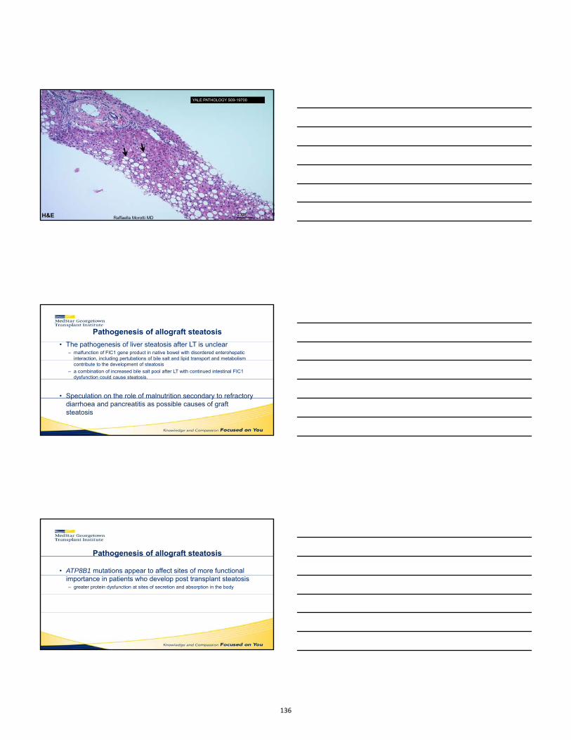
YALE PATHOLOGY S09-19700
H&E 100XRaffaella Morotti MD
Pathogenesis of allograft steatosis
• The pathogenesis of liver steatosis after LT is unclear– malfunction of FIC1 gene product in native bowel with disordered enterohepatic
interaction, including pertubations of bile salt and lipid transport and metabolism contribute to the development of steatosis
– a combination of increased bile salt pool after LT with continued intestinal FIC1 dysfunction could cause steatosis.
• Speculation on the role of malnutrition secondary to refractory diarrhoea and pancreatitis as possible causes of graft steatosis
Pathogenesis of allograft steatosis
• ATP8B1 mutations appear to affect sites of more functional importance in patients who develop post transplant steatosis– greater protein dysfunction at sites of secretion and absorption in the body
136
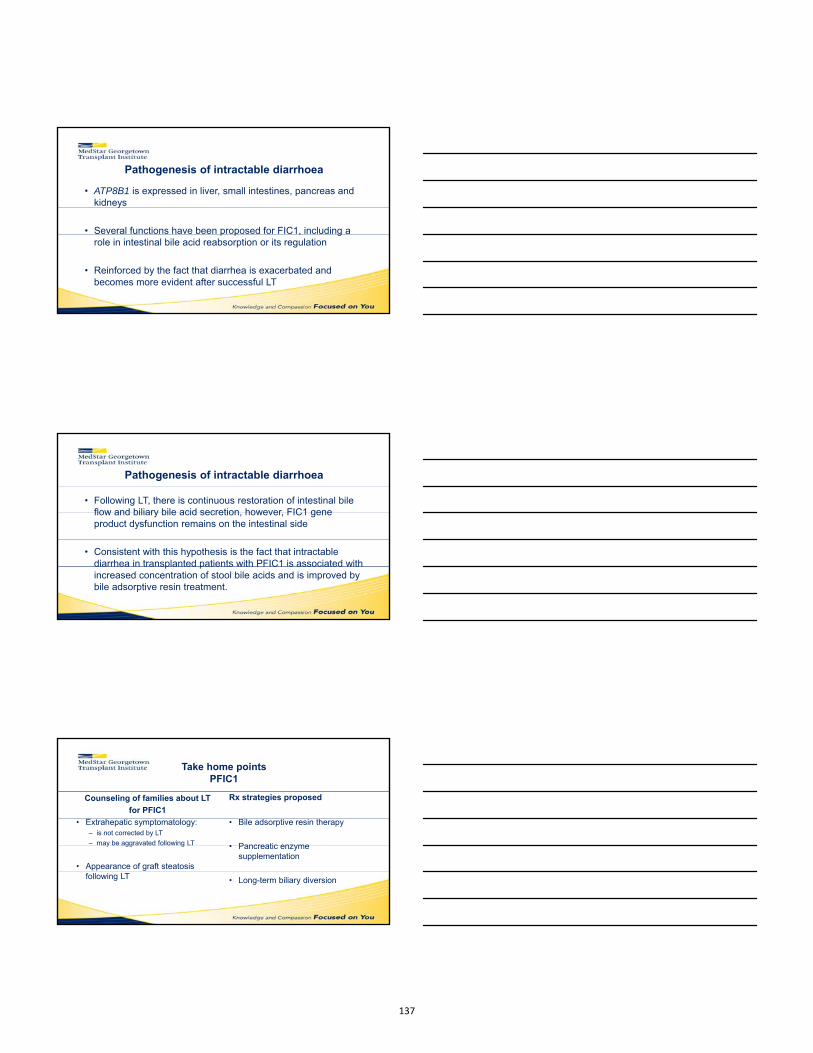
Pathogenesis of intractable diarrhoea
• ATP8B1 is expressed in liver, small intestines, pancreas and kidneys
• Several functions have been proposed for FIC1, including a role in intestinal bile acid reabsorption or its regulation
• Reinforced by the fact that diarrhea is exacerbated and becomes more evident after successful LT
Pathogenesis of intractable diarrhoea
• Following LT, there is continuous restoration of intestinal bile flow and biliary bile acid secretion, however, FIC1 gene product dysfunction remains on the intestinal side
• Consistent with this hypothesis is the fact that intractable diarrhea in transplanted patients with PFIC1 is associated with increased concentration of stool bile acids and is improved by bile adsorptive resin treatment.
Take home pointsPFIC1
Counseling of families about LT
for PFIC1
• Extrahepatic symptomatology: – is not corrected by LT
– may be aggravated following LT
• Appearance of graft steatosisfollowing LT
Rx strategies proposed
• Bile adsorptive resin therapy
• Pancreatic enzyme supplementation
• Long-term biliary diversion
137
What is a “normal” childhood after liver transplantation
Estella M. Alonso, M.D.Siragusa Transplant CenterAnn and Robert H. Lurie Children’ HospitalFeinberg School of MedicineNorthwestern University, Chicago, IL
Disclosures
• In the past 12 months, I have had no relevant financial relationships with the manufacturer(s) of any commercial product(s) and/or provider(s) of commercial services discussed in this CME activity.
Objectives
• Identify the common physical and psychosocial challenges children experience after LT
• Review risk factors for lower than expected physical function and school performance after LT
• Discuss screening strategies to implement in a post‐transplant ambulatory setting to identify high risk patients
138
ExpectationsWhat is a “normal” life?
• Normal physical health• No signs of chronic liver disease and no chronic or life‐threatening infections• No chronic complications of immunosuppression
• Normal growth and lack of physical limitations
• Normal energy level
• Ability to attend school regularly and participate in age‐appropriate activities
• Average or above average school performance
• Average psychosocial health and family function
• Ability to become an independent adult
Timeline of EventsWhat is the toll?• Hospital Days• Ambulance Ride• ED visits• Ambulatory Visits• Lab Draws• Imaging• Days of Physical Therapy• Missed School Days
Physical Function at One Year Post‐LT
Liver Trans 2016;22:495‐504
Predictors of Lower Physical Function• Primary disease• Height Z score<‐1.64 • ≥4 days of hospitalization • Not listed as Status 1
• Lower physical function scores compared to matched health group (p<0.01)
• 35.0% with physical function score >1 SD below the healthy mean
• Physical function scores higher in survivors with optimal health (p<0.01)
139
Common Medical Complications
• Rejection• 40‐60% of recipients with at least one episode in the first year• 2‐3% per year after the first year
• Consider non‐adherence
• Vascular stenosis or thrombosis < 10%• Hepatic artery
• Progressive scarring versus immune phenomenon • Portal Vein
• External compression versus scarring
• Biliary strictures up to 30%• Any interval from transplant• Anastomotic versus intra‐hepatic• Re‐admission/ Long‐term drain placement
Problematic Post‐Transplant Infections
• More severe community acquired viral infections• Norovirus• Adenovirus
• Chronic upper respiratory infections • Otitis Media
• EBV driven tonsillar hypertrophy• Opportunistic infection rare, but real• Bacteremia/Sepsis
• Invasive Pneumococcal Disease• Enteric bacteria associated with biliary obstruction• Ischemic abscess
Transpl Infect Dis 2016;18:333‐40
What to do about a fever?
Post‐transplantation period
Febrile hospitalizations Bacterial infectionn (%), of febrile hospitalizations
<1 month 3 1 (33)
1 month–1 year 39 13 (33)
1–5 years 58 38 (66)
>5 years 33 21 (64)
Number and timing of bacterial infections in children post liver transplantation hospitalized for fever
140
Post‐transplantation period
Febrile hospitalizations n
Bacterial infection n (%), of
febrile hospitalizations
<1 month 3 1 (33)
1 month–1 year 39 13 (33)
1–5 years 58 38 (66)
>5 years 33 21 (64)
Risk of Blood Stream Infection
• 29/340 (9%) LT recipients > 6 Months
• 42 organisms • 9 GPC• 33 GNR
• Intraabdominal infection most frequent source (47%) • 39% episodes with no apparent focus.
• Risk factors in multivariate analysis• Operative time > 12 hours (odds ratio [OR] = 3.55; P = 0.04) • Biliary stenosis (OR = 4.60; P = 0.006)
Pediatr Infect Dis J 2018;37:263‐8
School Readiness
• Language delay (>1 SD below norms) in infants awaiting LT• 40% with receptive language delay• 67% with expressive language delay
• Diagnosis of ADHD approximately double
• 15% sensorineural hearing loss after transplant
• 25% LT recipients have mild to moderate developmental delay at age 5‐7 yrs
• Cognitive and math deficits tend to persist up to age 9• Reading may improve • Predictors of lower FSIQ include single parent household (P < .002), parent education (P < .01), weight z‐score at liver transplantation (P < .03), and transfusion volume during liver transplantation (P < .0001)
J Pediatr 2010;156:936‐40 Pediatr Transplantation 2003: 7: 265–269J Pediatr 2014;165:65‐72
141
Distribution of IQ and Achievement Scores Following LT
Sorensen LG et al, Am J Transplant 2011;11:303‐11
Cognitive Delay
Special Education• 36% at Time 1 • 45% at Time 2
J Pediatr 2014;165:65‐72
Physical Growth
• 60% of children malnourished at transplant
• Usually achieve nutritional rehabilitation within 12‐24 months
• Complete catch‐up for weight within 12‐24 months
• Catch‐up linear growth not achieved until after 12‐24 months
• Sub‐optimal linear growth continues into long‐term follow‐up
• Pre‐transplant nutritional status
• Post‐transplant complications and medications
142
Distribution of Patients Below 10th and 5th Percentiles by Post Transplant Follow-up Month
54.4
44.1
33.829.3 27.8 28.7
33.2
24.119.9 20.0
22.1
46.0
0
10
20
30
40
50
60
0(N=1143)
12(N=911)
24(N=696)
36(N=564)
48(N=435)
60(N=321)
Post Transplant Follow-up Month
Pe
rce
nt
Height < 10th Percentile
Height < 5th Percentile
AJT 2009; 9:1389‐97
Factor Comparison Reference OR 95% CI p-value
Primary diseaseoverall p=0.0146
Other CholestaticALFMetabolic DiseaseOther
Biliary atresia 1.401.104.402.16
0.64, 3.040.38, 3.171.83,10.590.95, 4.91
0.40280.85560.00090.0675
Prednisone use up to 24 monthsoverall p=0.0046
6-17.9 months18+ months
<6 months1.423.02
0.70, 2.901.39, 6.55
0.33500.0053
Weight z score at transplant
continuous 0.80 0.65, 0.99 0.0385
Height z score at transplant
continuous 0.62 0.51, 0.77 <0.0001
Total Bilirubin at transplant
continuous 1.05 1.02, 1.07 0.0010
GGTP at 12 months
continuous 1.0021.000, 1.004
0.0328
Multivariate Analysis for Growth Impairment at 2 Years
AJT 2009; 9:1389‐97
J Pediatr 2103; 163:1354–60
Linear Growth in 353 Post‐Pubertal Children
143
Obesity
JPGN 2012;55:657‐62
60
65
70
75
80
85
90
Total Physical Psychosocial
Liver JRA Type 1 DiabetesCancer in Remission Cardiac Renal TransplantRenal Dialysis IBD
PedsQL™ Scores for LT Compared to6 Chronic Disease Groups
**
****
* < 0.001, ** <0.01, § <0.05Hochberg adjusted significance levels
**§
Pediatr Transpl 2011: 15: 245–253
Absent > 10 days/year
0%10%20%30%40%50%60%
Age (p=0.001)
6-11 yr 12-14 yr15-18 yr
0%10%20%30%40%50%60%
Interval (p<0.001)
0-18 mth 19-36 mth> 36 mth
Liver Transpl 2010;16:1041‐48
144
PedsQL 4.0 Parent Proxy‐Report
Scales
LT Sample(Mean±SD)
n=869
Healthy Children(Mean±SD)
n=3869
Effect Size
Total Score 77.26±17.58*** 84.12±13.74 0.47
Physical Health 79.33±22.07*** 86.87±17.05 0.42
Psychosocial Health 75.72±17.33*** 82.53±14.09 0.46
Emotional Functioning 73.27±19.28*** 81.02±15.93 0.47
Social Functioning 78.99±20.63*** 85.93±16.98 0.39
School Functioning 67.42±22.43*** 79.34±18.37 0.62
*** p<0.001
30.6% scored > 1 SD below mean for healthy sample
J Pediatr 2010;156:270‐6
PedsQL 4.0 Child Self‐Report
ScalesLT Sample(Mean±SD)
n=363
Healthy Children(Mean±SD) n=1844
Effect Size
Total Score 77.21±14.28*** 83.68±12.26 0.51
Physical Health 82.29±15.62*** 88.07±12.30 0.45
Psychosocial Health 74.51±15.83*** 81.33±14.04 0.48
Emotional Functioning 74.00±19.90*** 78.56±18.29 0.25
Social Functioning 80.95±19.09*** 85.50±16.93 0.26
School Functioning 68.53±18.56*** 79.83±16.33 0.68
*** p<0.001
31.1% scored > 1 SD below mean for healthy sample
School Functioning Scale Components
Parent Report LTn=746
Mean (±SD)
Healthyn=3215
Mean (±SD)
EffectSize
School Functioning 67.4±22.4 79.3±18.4 -0.62*
Cognitive 67.2±27.1 74.1±24.4 -0.28*
Missing School 67.7±23.8 84.0±17.8 -0.85*
Sub‐analysis of School Function
* p < 0.001
145
PedsQLTMMultidimensional Fatigue Scale
iWITH Unpublished Data
iWITH Baseline Healthy Controls
Mean SD n Mean SD Adjusted Significance Level
Effect Size
Child Report n=81Total Fatigue 74.9 17.75 157 82.19 12.27 0.0012 0.51
General Fatigue 80.2 18.89 157 86.36 13.11 0.0144 0.40
Sleep/Rest Fatigue 71.3 20.42 157 77.44 15.41 0.04 0.36
Cognitive Fatigue 73.4 22.15 157 82.78 16.26 0.0012 0.51
Parent Proxy‐Report n=87Total Fatigue 78.0 16.11 157 87.24 10.91 0.0004 0.71
General Fatigue 80.1 16.28 157 88.40 11.67 0.0004 0.62
Sleep/Rest Fatigue 81.4 16.57 157 86.70 12.63 0.022 0.38
Cognitive Fatigue 72.4 24.00 157 86.62 16.36 0.0004 0.74
Health‐Related Quality of Life and Cognitive Functioning in Liver Transplant Recipients During a 10‐year Time SpanObjectives • Determine if early developmental assessment at time of school entry predicts cognitive function in adolescence
• Determine if HRQOL changes as a function of time since transplant in long‐term survivors
Methods• Longitudinal neurocognitive assessment cohort study age 5‐6, 7‐8 and in adolescence (age 12 up to 18)
• Adolescent assessment; online surveys regarding perceived cognitive functioning and HRQOL
Conclusions• HRQOL does not appear to improve in adolescence. Over half of adolescent LT recipients appear to be at risk for lower cognitive and school function
Main Findings
0%
10%
20%
30%
40%
50%
60%
70%
HRQOL School Functioning Cognitive Functioning
Age 5‐6 Age 12‐18
Percentage of Participants Reporting Poor Functioning (1 SD below the healthy population mean)*
*On parent‐report PedsQLTM 4.0 Generic Core Scale Total Score, School Functioning Scale, and PedsQLTM Cognitive Functioning Scale
AASLD 2018
Is early childhood assessment predictive of functioning in adolescence?
• Early childhood PedsQLTM (Parent Report) showed excellent predictive value for poor functioning in adolescence
• BRIEF scores at T1 predict PedsQLTM Cognitive Function scores at T3 • BRIEF Metacognition Index (p = 0.0298)
• BRIEF Working Memory (p <0.0001)
Poor Function at T1 (n)
Poor Function at T1 and T3 (n) Positive Predictive Value
PedsQLTM 4.0 Total Score 19 14 74%
PedsQLTM School Functioning 21 18 86%
PedsQLTM Cognitive Functioning 31 22 71%
146
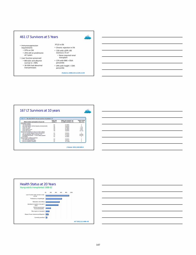
461 LT Survivors at 5 Years
• Immunosuppression requirements
• 97% on CNI
• 25% still on prednisone at 5 years
• Liver function preserved
• Bilirubin and albumin normal in > 90%
• 30‐50% had abnormal transaminases
PTLD in 6%
• Chronic rejection in 5%
• 13% with cGFR <90 ml/min/1.73 m2
• None required renal transplant
• 12% with BMI > 95th percentile
• 29% with height < 10th percentile
Pediatrics 2008;122:e1128‐e1135
167 LT Survivors at 10 years
J Pediatr 2012;160:820‐6
Health Status at 20 YearsYoung adults transplanted 1988‐92
0% 20% 40% 60% 80% 100%
Liver Function tests normal or nearnormal
Followed by a hepatologist
Medication side effects
Admitted to hospital in the past 3years
Receive governmentinsurance/disability
May require re transplant
Biopsy Proven Autoimmune/Rejection
Currently jaundiced
AJT 2012;12:1486–95
147
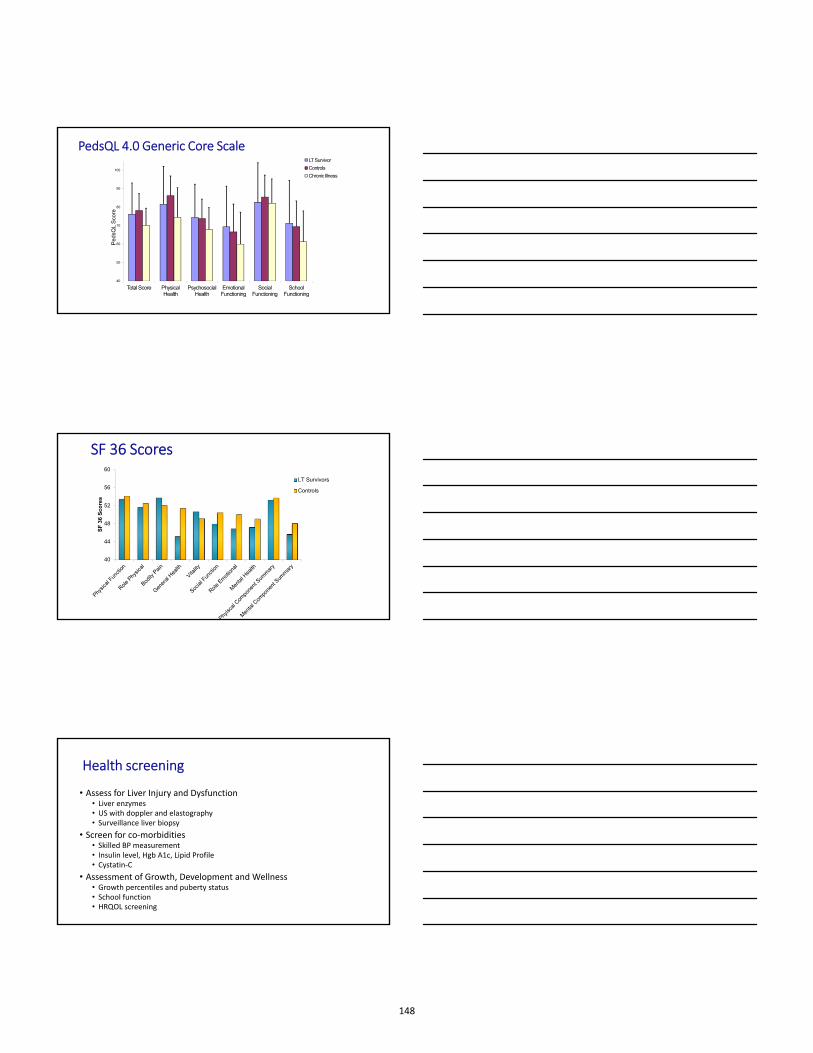
PedsQL 4.0 Generic Core Scale
40
50
60
70
80
90
100
Total Score Physical Health
Psychosocial Health
Emotional Functioning
Social Functioning
School Functioning
Ped
sQL
Sco
re
LT Survivor
Controls
Chronic Illness
SF 36 Scores
40
44
48
52
56
60
SF
36
Sco
res
LT Survivors
Controls
Health screening
• Assess for Liver Injury and Dysfunction• Liver enzymes• US with doppler and elastography• Surveillance liver biopsy
• Screen for co‐morbidities• Skilled BP measurement• Insulin level, Hgb A1c, Lipid Profile• Cystatin‐C
• Assessment of Growth, Development and Wellness• Growth percentiles and puberty status• School function• HRQOL screening
148
Summary
• Medical complications following Pediatric LT are not uncommon
• Interval from transplant impacts type of problems
• Functional outcomes are good, but lower than healthy peers• Especially in the area of school function
• Missed school days are important• LT survivors may have more fatigue
• Cognitive function maybe impaired• Recovery versus adaptation in early adulthood
149