Chronic Liver Disease - cmetracker.net antitrypsin deficiency ... Case #1 A 65 year old ... Chronic...
8
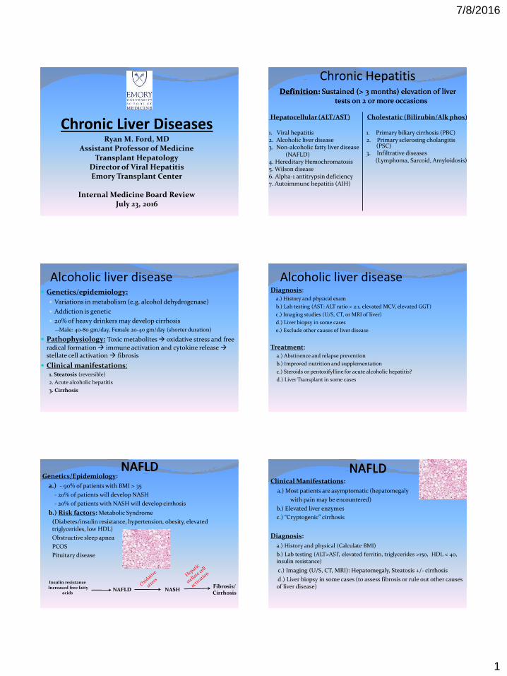
7/8/2016 1 Ryan M. Ford, MD Assistant Professor of Medicine Transplant Hepatology Director of Viral Hepatitis Emory Transplant Center Internal Medicine Board Review July 23, 2016 Chronic Hepatitis Hepatocellular (ALT/AST) 1. Viral hepatitis 2. Alcoholic liver disease 3. Non-alcoholic fatty liver disease (NAFLD) 4. Hereditary Hemochromatosis 5. Wilson disease 6. Alpha-1 antitrypsin deficiency 7. Autoimmune hepatitis (AIH) Cholestatic (Bilirubin/Alk phos) 1. Primary biliary cirrhosis (PBC) 2. Primary sclerosing cholangitis (PSC) 3. Infiltrative diseases (Lymphoma, Sarcoid, Amyloidosis) Definition: Sustained (> 3 months) elevation of liver tests on 2 or more occasions Alcoholic liver disease Genetics/epidemiology: Variations in metabolism (e.g. alcohol dehydrogenase) Addiction is genetic 20% of heavy drinkers may develop cirrhosis --Male: 40-80 gm/day, Female 20-40 gm/day (shorter duration) Pathophysiology: Toxic metabolites oxidative stress and free radical formation immune activation and cytokine release stellate cell activation fibrosis Clinical manifestations: 1. Steatosis (reversible) 2. Acute alcoholic hepatitis 3. Cirrhosis Diagnosis: a.) History and physical exam b.) Lab testing (AST: ALT ratio > 2:1, elevated MCV, elevated GGT) c.) Imaging studies (U/S, CT, or MRI of liver) d.) Liver biopsy in some cases e.) Exclude other causes of liver disease Treatment: a.) Abstinence and relapse prevention b.) Improved nutrition and supplementation c.) Steroids or pentoxifylline for acute alcoholic hepatitis? d.) Liver Transplant in some cases Alcoholic liver disease NAFLD Genetics/Epidemiology: a.) - 90% of patients with BMI > 35 - 20% of patients will develop NASH - 20% of patients with NASH will develop cirrhosis b.) Risk factors: Metabolic Syndrome (Diabetes/insulin resistance, hypertension, obesity, elevated triglycerides, low HDL) Obstructive sleep apnea PCOS Pituitary disease Insulin resistance Increased free fatty acids NAFLD NASH Fibrosis/ Cirrhosis NAFLD Clinical Manifestations: a.) Most patients are asymptomatic (hepatomegaly with pain may be encountered) b.) Elevated liver enzymes c.) “Cryptogenic” cirrhosis Diagnosis: a.) History and physical (Calculate BMI) b.) Lab testing (ALT>AST, elevated ferritin, triglycerides >150, HDL < 40, insulin resistance) c.) Imaging (U/S, CT, MRI): Hepatomegaly, Steatosis +/- cirrhosis d.) Liver biopsy in some cases (to assess fibrosis or rule out other causes of liver disease)
Transcript of Chronic Liver Disease - cmetracker.net antitrypsin deficiency ... Case #1 A 65 year old ... Chronic...
7/8/2016
1
Ryan M. Ford, MD Assistant Professor of Medicine
Transplant Hepatology Director of Viral Hepatitis Emory Transplant Center
Internal Medicine Board Review
July 23, 2016
Chronic Hepatitis
Hepatocellular (ALT/AST) 1. Viral hepatitis 2. Alcoholic liver disease 3. Non-alcoholic fatty liver disease (NAFLD) 4. Hereditary Hemochromatosis 5. Wilson disease 6. Alpha-1 antitrypsin deficiency 7. Autoimmune hepatitis (AIH)
Cholestatic (Bilirubin/Alk phos)
1. Primary biliary cirrhosis (PBC) 2. Primary sclerosing cholangitis
(PSC) 3. Infiltrative diseases (Lymphoma, Sarcoid, Amyloidosis)
Definition: Sustained (> 3 months) elevation of liver tests on 2 or more occasions
Alcoholic liver disease Genetics/epidemiology:
Variations in metabolism (e.g. alcohol dehydrogenase)
Addiction is genetic
20% of heavy drinkers may develop cirrhosis
--Male: 40-80 gm/day, Female 20-40 gm/day (shorter duration)
Pathophysiology: Toxic metabolites oxidative stress and free radical formation immune activation and cytokine release stellate cell activation fibrosis
Clinical manifestations: 1. Steatosis (reversible)
2. Acute alcoholic hepatitis
3. Cirrhosis
Diagnosis: a.) History and physical exam
b.) Lab testing (AST: ALT ratio > 2:1, elevated MCV, elevated GGT)
c.) Imaging studies (U/S, CT, or MRI of liver)
d.) Liver biopsy in some cases
e.) Exclude other causes of liver disease
Treatment: a.) Abstinence and relapse prevention
b.) Improved nutrition and supplementation
c.) Steroids or pentoxifylline for acute alcoholic hepatitis?
d.) Liver Transplant in some cases
Alcoholic liver disease
NAFLD Genetics/Epidemiology:
a.) - 90% of patients with BMI > 35
- 20% of patients will develop NASH
- 20% of patients with NASH will develop cirrhosis
b.) Risk factors: Metabolic Syndrome
(Diabetes/insulin resistance, hypertension, obesity, elevated triglycerides, low HDL)
Obstructive sleep apnea
PCOS
Pituitary disease
Insulin resistance Increased free fatty
acids NAFLD NASH
Fibrosis/Cirrhosis
NAFLD Clinical Manifestations:
a.) Most patients are asymptomatic (hepatomegaly
with pain may be encountered)
b.) Elevated liver enzymes
c.) “Cryptogenic” cirrhosis
Diagnosis:
a.) History and physical (Calculate BMI)
b.) Lab testing (ALT>AST, elevated ferritin, triglycerides >150, HDL < 40, insulin resistance)
c.) Imaging (U/S, CT, MRI): Hepatomegaly, Steatosis +/- cirrhosis
d.) Liver biopsy in some cases (to assess fibrosis or rule out other causes of liver disease)
7/8/2016
2
NAFLD Treatment: a) Lifestyle Change with a goal of at least 10% body weight reduction b) Control of metabolic syndrome risk factors (HTN, DM, dyslipidemia) Statins Metformin Pioglitazone Fenofibrates c.) Surgery: Gastric bypass surgery, gastric banding, gastric sleeve d.)Anti-inflammatories (Vitamin E, coffee, other antioxidants?) e.) Liver transplant: NASH cirrhosis is becoming a very common indication for
liver transplant in the U.S.
Hereditary Hemochromatosis Genetics/epidemiology:
Autosomal recessive (~1/250 Caucasians)
Low penetrance of clinical disease
HFE gene is most common mutation
C282Y homozygote is most commonly seen with clinical disease
Pathophysiology: Unregulated, increased uptake of dietary iron without negative feedback loop
Results in iron overload with organ deposition and hepatic fibrosis
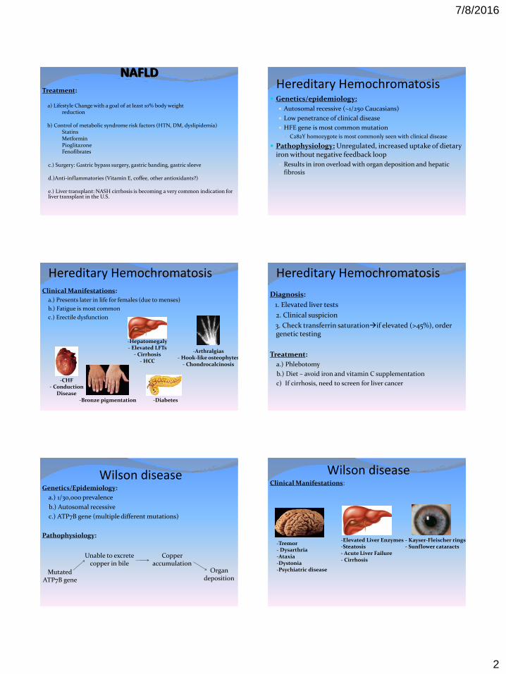
Clinical Manifestations:
a.) Presents later in life for females (due to menses)
b.) Fatigue is most common
c.) Erectile dysfunction
-Hepatomegaly - Elevated LFTs
- Cirrhosis - HCC
-CHF - Conduction
Disease -Bronze pigmentation
-Arthralgias - Hook-like osteophytes
- Chondrocalcinosis
-Diabetes
Hereditary Hemochromatosis
Diagnosis:
1. Elevated liver tests
2. Clinical suspicion
3. Check transferrin saturationif elevated (>45%), order genetic testing
Treatment:
a.) Phlebotomy
b.) Diet – avoid iron and vitamin C supplementation
c) If cirrhosis, need to screen for liver cancer
Hereditary Hemochromatosis
Genetics/Epidemiology:
a.) 1/30,000 prevalence
b.) Autosomal recessive
c.) ATP7B gene (multiple different mutations)
Pathophysiology:
Mutated ATP7B gene
Unable to excrete copper in bile
Copper accumulation
Organ deposition
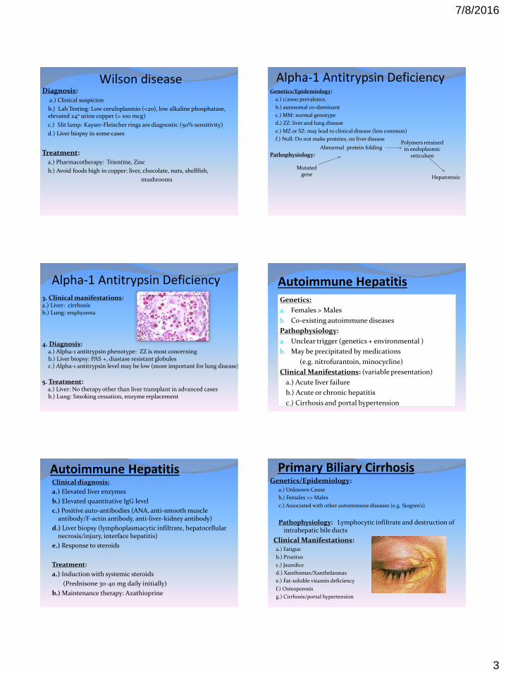
Wilson disease Clinical Manifestations:
-Elevated Liver Enzymes -Steatosis - Acute Liver Failure - Cirrhosis
-Tremor - Dysarthria -Ataxia -Dystonia -Psychiatric disease
- Kayser-Fleischer rings - Sunflower cataracts
Wilson disease
7/8/2016
3
Diagnosis:
a.) Clinical suspicion
b.) Lab Testing: Low ceruloplasmin (<20), low alkaline phosphatase, elevated 240 urine copper (> 100 mcg)
c.) Slit lamp: Kayser-Fleischer rings are diagnostic (50% sensitivity)
d.) Liver biopsy in some cases
Treatment: a.) Pharmacotherapy: Trientine, Zinc
b.) Avoid foods high in copper: liver, chocolate, nuts, shellfish,
mushrooms
Wilson disease Genetics/Epidemiology:
a.) 1/2000 prevalence,
b.) autosomal co-dominant
c.) MM: normal genotype
d.) ZZ: liver and lung disease
e.) MZ or SZ: may lead to clinical disease (less common)
f.) Null: Do not make proteins, no liver disease
Pathophysiology:
Mutated gene
Abnormal protein folding Polymers retained
in endoplasmic reticulum
Hepatotoxic
Alpha-1 Antitrypsin Deficiency
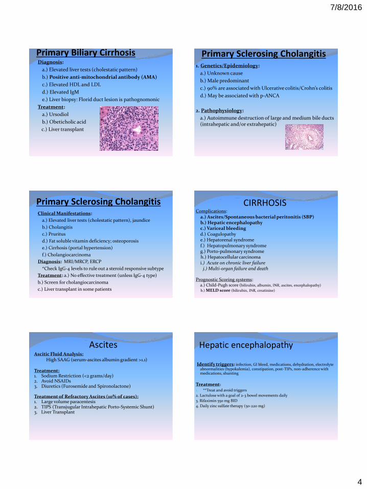
3. Clinical manifestations: a.) Liver: cirrhosis b.) Lung: emphysema
4. Diagnosis: a.) Alpha-1 antitrypsin phenotype: ZZ is most concerning b.) Liver biopsy: PAS +, diastase resistant globules c.) Alpha-1 antitrypsin level may be low (more important for lung disease)
5. Treatment: a.) Liver: No therapy other than liver transplant in advanced cases b.) Lung: Smoking cessation, enzyme replacement
Alpha-1 Antitrypsin Deficiency Autoimmune Hepatitis Genetics:
a. Females > Males
b. Co-existing autoimmune diseases
Pathophysiology:
a. Unclear trigger (genetics + environmental )
b. May be precipitated by medications
(e.g. nitrofurantoin, minocycline)
Clinical Manifestations: (variable presentation)
a.) Acute liver failure
b.) Acute or chronic hepatitis
c.) Cirrhosis and portal hypertension
Autoimmune Hepatitis Clinical diagnosis:
a.) Elevated liver enzymes
b.) Elevated quantitative IgG level
c.) Positive auto-antibodies (ANA, anti-smooth muscle antibody/F-actin antibody, anti-liver-kidney antibody)
d.) Liver biopsy (lymphoplasmacytic infiltrate, hepatocellular necrosis/injury, interface hepatitis)
e.) Response to steroids
Treatment:
a.) Induction with systemic steroids
(Prednisone 30-40 mg daily initially)
b.) Maintenance therapy: Azathioprine
Primary Biliary Cirrhosis Genetics/Epidemiology:
a.) Unknown Cause
b.) Females >> Males
c.) Associated with other autoimmune diseases (e.g. Sjogren’s)
Pathophysiology: Lymphocytic infiltrate and destruction of intrahepatic bile ducts
Clinical Manifestations: a.) Fatigue
b.) Pruritus
c.) Jaundice
d.) Xanthomas/Xanthelasmas
e.) Fat-soluble vitamin deficiency
f.) Osteoporosis
g.) Cirrhosis/portal hypertension
7/8/2016
4
Primary Biliary Cirrhosis Diagnosis:
a.) Elevated liver tests (cholestatic pattern)
b.) Positive anti-mitochondrial antibody (AMA)
c.) Elevated HDL and LDL
d.) Elevated IgM
e.) Liver biopsy: Florid duct lesion is pathognomonic
Treatment:
a.) Ursodiol
b.) Obeticholic acid
c.) Liver transplant
Primary Sclerosing Cholangitis 1. Genetics/Epidemiology:
a.) Unknown cause
b.) Male predominant
c.) 90% are associated with Ulcerative colitis/Crohn’s colitis
d.) May be associated with p-ANCA
2. Pathophysiology:
a.) Autoimmune destruction of large and medium bile ducts (intrahepatic and/or extrahepatic)
Primary Sclerosing Cholangitis Clinical Manifestations:
a.) Elevated liver tests (cholestatic pattern), jaundice
b.) Cholangitis
c.) Pruritus
d.) Fat soluble vitamin deficiency; osteoporosis
e.) Cirrhosis (portal hypertension)
f.) Cholangiocarcinoma
Diagnosis: MRI/MRCP, ERCP
*Check IgG-4 levels to rule out a steroid responsive subtype
Treatment: a.) No effective treatment (unless IgG-4 type)
b.) Screen for cholangiocarcinoma
c.) Liver transplant in some patients
Complications: a.) Ascites/Spontaneous bacterial peritonitis (SBP) b.) Hepatic encephalopathy c.) Variceal bleeding d.) Coagulopathy e.) Hepatorenal syndrome f.) Hepatopulmonary syndrome g.) Porto-pulmonary syndrome h.) Hepatocellular carcinoma i.) Acute on chronic liver failure j.) Multi-organ failure and death
Prognostic Scoring systems: a.) Child-Pugh score (bilirubin, albumin, INR, ascites, encephalopathy)
b.) MELD score (bilirubin, INR, creatinine)
CIRRHOSIS
Ascitic Fluid Analysis: High SAAG (serum-ascites albumin gradient >1.1) Treatment: 1. Sodium Restriction (<2 grams/day) 2. Avoid NSAIDs 3. Diuretics (Furosemide and Spironolactone) Treatment of Refractory Ascites (10% of cases): 1. Large volume paracentesis 2. TIPS (Transjugular Intrahepatic Porto-Systemic Shunt) 3. Liver Transplant
Ascites
Identify triggers: infection, GI bleed, medications, dehydration, electrolyte abnormalities (hypokalemia), constipation, post-TIPs, non-adherence with medications, shunting
Treatment:
1. **Treat and avoid triggers
2. Lactulose with a goal of 2-3 bowel movements daily
3. Rifaximin 550 mg BID
4. Daily zinc sulfate therapy (50-220 mg)
Hepatic encephalopathy
7/8/2016
5
Risks for bleeding
- Higher portal pressure
- Varix size
-High MELD score
- Child’s class B/C
- Prior variceal bleed
Esophageal varices Red wale signs Variceal banding
Esophageal varices Screening recommendations:
EGD at time of diagnosis of cirrhosis
EGD every 1-3 years based on history and risk
Treatment/prophylaxis:
1. Non-selective beta blockers (Nadolol, Propranolol) with goal heart rate near 55-60 as long as blood pressure will tolerate
2. Variceal band ligation
Esophageal varices
Hepatocellular Carcinoma *HCC is fastest-growing cause of cancer-death in USA
Often asymptomatic until late stage
Screening:
Ultrasound, CT, MRI q 6 months
serum AFP
Who to screen?
Any patient with cirrhosis
Patients with chronic hepatitis B virus
Treatment?
Resection, loco-regional therapy, liver transplant
Hepatitis B--Global 400 million carriers in the world
#1 cause of cirrhosis in the world
#1 cause of HCC in the world
Highly endemic in Southeast Asia and Africa: 60-80% lifetime risk of infection
--Perinatal or horizontal spread as children
Hepatitis B in U.S. 1.25 million carriers in U.S.
Most carriers are immigrants or first generation Americans
7/8/2016
6
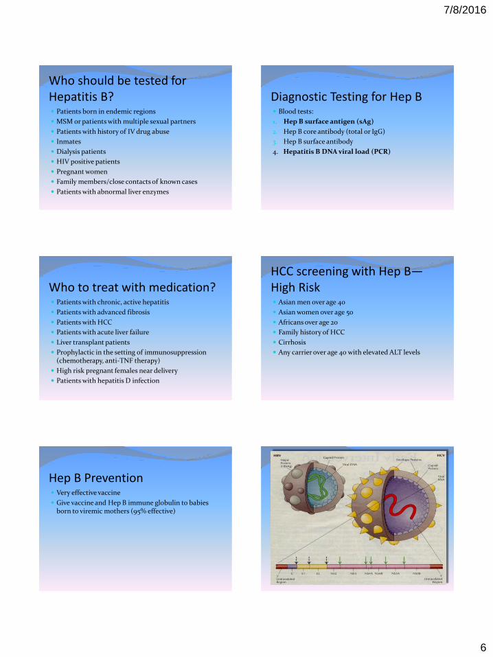
Who should be tested for Hepatitis B? Patients born in endemic regions
MSM or patients with multiple sexual partners
Patients with history of IV drug abuse
Inmates
Dialysis patients
HIV positive patients
Pregnant women
Family members/close contacts of known cases
Patients with abnormal liver enzymes
Diagnostic Testing for Hep B Blood tests:
1. Hep B surface antigen (sAg)
2. Hep B core antibody (total or IgG)
3. Hep B surface antibody
4. Hepatitis B DNA viral load (PCR)
Who to treat with medication? Patients with chronic, active hepatitis
Patients with advanced fibrosis
Patients with HCC
Patients with acute liver failure
Liver transplant patients
Prophylactic in the setting of immunosuppression (chemotherapy, anti-TNF therapy)
High risk pregnant females near delivery
Patients with hepatitis D infection
HCC screening with Hep B—High Risk Asian men over age 40
Asian women over age 50
Africans over age 20
Family history of HCC
Cirrhosis
Any carrier over age 40 with elevated ALT levels
Hep B Prevention Very effective vaccine
Give vaccine and Hep B immune globulin to babies born to viremic mothers (95% effective)
7/8/2016
7
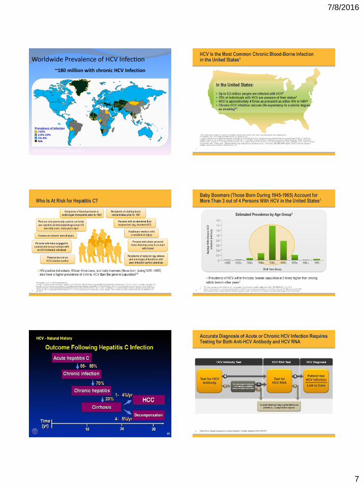
Worldwide Prevalence of HCV Infection
>10% 2.5%-10%
1%-5% N/A
Prevalence of Infection
~180 million with chronic HCV Infection
http://www.who.int/csr/disease/hepatitis/Hepc.pdf. 2
4 6
HCV - Natural History
41
Outcome Following Hepatitis C Infection
9
7/8/2016
8
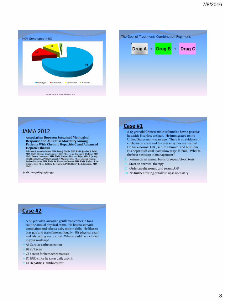
HCV Genotypes in US
Germer JJ, et al. J Clin Microbiol. 2011
73
13
12 2
Genotype 1 Genotype 2 Genotype 3 All Others
The Goal of Treatment: Combination Regimens
Drug A Drug B Drug C + +
JAMA 2012 Association Between Sustained Virological
Response and All-Cause Mortality Among Patients With Chronic Hepatitis C and Advanced Hepatic Fibrosis
Adriaan J. van der Meer, MD; Bart J. Veldt, MD, PhD; Jordan J. Feld, MD, PhD; Heiner Wedemeyer, MD, PhD; Jean-François Dufour, MD, PhD; Frank Lammert, MD, PhD; Andres Duarte-Rojo, MD; E. Jenny Heathcote, MD, PhD; Michael P. Manns, MD, PhD; Lorenz Kuske; Stefan Zeuzem, MD, PhD; W. Peter Hofmann, MD, PhD; Robert J. de Knegt, MD, PhD; Bettina E. Hansen, PhD; Harry L. A. Janssen, MD, PhD
JAMA. 2012;308(24):2584-2593
Case #1 A 65 year old Chinese male is found to have a positive
hepatitis B surface antigen. He immigrated to the United States many years ago. There is no evidence of cirrhosis on exam and his liver enzymes are normal. He has a normal CBC, serum albumin, and bilirubin. His hepatitis B viral load is low at 150 IU/mL. What is the best next step in management?
A) Return on an annual basis for repeat blood tests
B) Start on antiviral therapy
C) Order an ultrasound and serum AFP
D) No further testing or follow-up is necessary
Case #2
A 66 year old Caucasian gentleman comes in for a routine annual physical exam. He has no somatic complaints and takes a baby aspirin daily. He likes to play golf and travel internationally. His physical exam and lab testing are normal. What should be included in your work-up?
A) Cardiac catheterization
B) PET scan
C) Screen for hemochromatosis
D) EGD since he takes daily aspirin
E) Hepatitis C antibody test