2016 INS Standards Update - Infusion Nurses of Oregon · Session Objective •Describe the...
46
2016 INS Standards Update Lisa A. Gorski MS, HHCNS-BC, CRNI ® , FAAN
Transcript of 2016 INS Standards Update - Infusion Nurses of Oregon · Session Objective •Describe the...

2016 INS
Standards
Update
Lisa A. Gorski MS, HHCNS-BC, CRNI®,
FAAN

Disclosures
• Speakers Bureau - BD Medical,
Access Scientific, Genentech
• Advisory Board Member -- ivWatch

Session Objective
• Describe the methodology for revising
the 2016 Infusion Therapy Standards of
Practice.
• Identify new standards and major
changes in existing standards.
• Discuss findings from the INS Vesicant
Task Force.
.

International
Opportunities to
Present the INS
Standards

Acknowledgement:
2016 Standards Committee
• Chair - Lisa Gorski, MS, RN, HHCNS-BC, CRNI®,
FAAN
• Lynn Hadaway, Med, RN-BC, CRNI®
• Mary E. Hagle, PhD, RN-BC, FAAN
• Mary McGoldrick, MS, RN, CRNI®
• Marsha Orr, MS, RN
• Darcy Doellman, MSN, RN, CRNI®, VA-BC
CITATION FOR STANDARDS:
Gorski, LA, Hadaway, L, Hagle, M, McGoldrick, M, Orr, M, Doellman, D.
Infusion therapy standards of practice. J Infus Nurs 2016: 39 (suppl 1): S1-
S159.

INS Mission & History of INS
Standards
• Mission
• Develop and disseminate standards of practice
• 1980, 1990, 1998, 2000, 2006, 2011, 2016
• Provide professional development opportunities and education
• Advance best practice through synthesis and research
• Support professional certification
• Advocate for the public

Foreword• “Whether the purpose lies in informing patient care, legal
proceedings, or personal edification and growth, no
document is more versatile, time-tested, or valuable in
the field of infusion practice.”
• “As a physician researcher dedicated to improving the
safety of patients who require vascular access and
infusion-based therapies, the Standards has informed
the work that I do, the questions I ask, and the clinical
care I provide. Quite simply put, there is nothing else like
it.”
Vineet Chopra, MD, MSC
Ann Arbor VA Medical Center &
University of Michigan Health System

Additional Resource
• Policies and Procedures for
Infusion Therapy
• Translate the Standards into
clinical procedures

From the “Infusion Nursing Standards
of Practice” to the “Infusion Therapy
Standards of Practice”
• ‘Infusion therapy does not “belong” to one group of clinicians, but it is the responsibility of any clinician who is involved in the practice.’
• Standard 3. Scope of Practice • Clear definition of roles, responsibility, and accountability
for each clinician in accordance with regulations, organizational policy
• Collaboration among the healthcare team
• Address nurses, unlicensed assistive personnel, radiologic/respiratory technician/technologist/therapist, paramedics

2016 Table of Contents
64 Standards – divided into 9 sections
1: Infusion Therapy Practice
2: Patient & Clinician Safety
3: Infection Prevention & Control
4: Infusion Equipment
5: Vascular Access Device (VAD) Selection & Placement
6: VAD Management
7: VAD-Related Complications
8: Other Infusion Devices
9: Infusion Therapies

Appendices
• Infusion Team Definition
• New illustrations
• Glossary greatly expanded
• Definitional information from previous
document moved to glossary
Crosswalk
document is
available on
the INS Web
site at the INS
Learning
Center
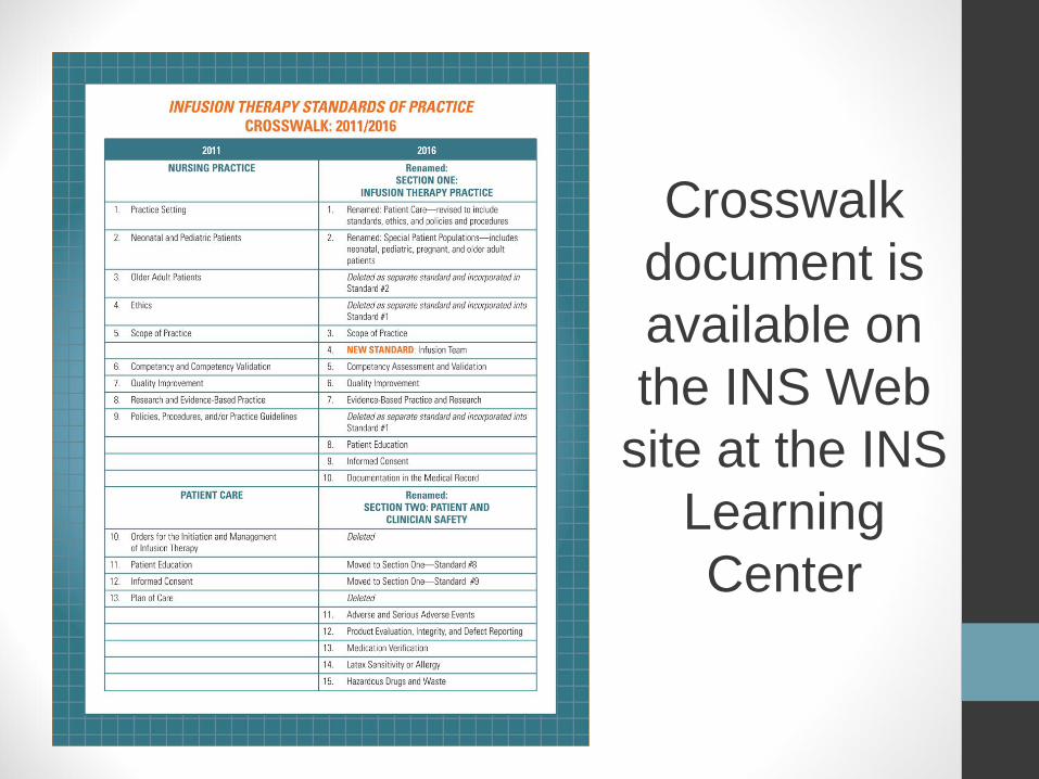
Format of the Standards
– Not changed but less
repetition
• Standards and Practice Criteria
• Standards• Expectations of practice applicable to infusion
therapy in all settings
• Not rated
“Section”
Standards
• Apply to all Standards in the section – ↓ repetition
• Section Standards included in Sections 4-9
• Example Section 7: VAD-Related Complications • “To ensure patient safety, the clinician is competent to recognize s/s
of VAD-related complications during insertion, management, and
removal, and appropriately intervene.”
• “Prevention, assessment and management …are addressed in
organizational policies, procedures, and/or practice guidelines”
Gorski et al, JIN, p. S95.

Format of the Standards
Practice Criteria• Provide specific guidance in the implementation of
the corresponding Standard
• Each Practice Criterion is supported by evidence
• Each Criterion is rated as reflecting the strength of the body of evidence
• Reflects the body of evidence available and retrievable at the time of review
• Rating Scale: I, IA/P, II-V, Regulatory
• References for each Practice Criterion are listed


Evidence -- The Evolving
Science of Infusion
Therapy
• INS Standards 2011
• Level I (highest) evidence – 3.8% of rankings
• Level V evidence – 67%
• INS Standards 2016
• Level I (highest) evidence – 5.8% of rankings
• Level V evidence – 46%
• 350 more references

A brief look at some
new and changed 2016
Standards
NOTE: Information provided includes selected excerpts of Standards and Practice Criteria, not the complete content from any Standard.

Standard 4.
Infusion Team• Scope of service to meet patient and organization needs
• VAD insertion and management assigned to individuals/teams
with infusion therapy education, training, and validated
competency
• Peripheral catheter insertion by team = increased insertion
success, decreased hospital acquired BSI, local infection,
occlusions, and accidental removals
• Team managing VADs decrease BSI and related costs, phlebitis
and infiltration, and increase patient satisfaction
• More studies needed to expand and increase level of
evidence ranking

Standard 5. Competency Assessment &
Validation
•Standard
• As a method of public protection to ensure patient safety, the clinician is competent in the safe delivery of infusion therapy and VAD insertion and/or management within his/her scope of practice.
• Clinician responsible and accountable for attaining and maintaining competence
• Competency assessment and validation is performed initially and on an ongoing basis
Gorski, Hadaway, Hagle et al (2016) Infusion therapy standards of practice. JIN 39 (1, suppl), S18.

Standard 5. Competency
Assessment & Validation
• Practice Criteria
• Competence goes “beyond psychomotor skills to include application of knowledge, critical thinking skills, decision making” (IV)
• Use a combination of different measurement techniques such as self-assessment (promote self-efficacy, confidence), written tests (knowledge), clinical scenarios (critical thinking), psychomotor skills (simulation, observation in work environment preferred for invasive infusion procedures) (IV)
• Qualifications for role of competency assessor --the person validating the specific skill should be competent with the skill. (IV)
Gorski, Hadaway, Hagle et al (2016) Infusion therapy standards of practice. JIN 39 (1, suppl), S18-19.

Standard 22. Vascular
Visualization
• Visible light devices for transillumination
• Cold light source needed
• Near-infrared light devices
• More informed decisions about peripheral veins,
bifurcation, tortuosity
• Ultrasound
• Peripheral sites – requires longer catheters
• Addresses CVADs
• Dynamic or “real-time” use is recommended
• Sterile TSM dressing (peripheral sites), sheath cover and gel
Standard 23. CVAD
Tip Location• Standard
• Tip location of a CVAD is determined radiographically or by
other imaging technologies prior to initiation of infusion
therapy or when clinical signs and symptoms suggest tip
malposition
• The CVAD tip location with the greatest safety profile in
adults and children is the cavoatrial junction (CAJ)
• Practice Criteria
• ECG methods – “real time” identification of placement (II)
• Chest radiograph required in absence of technology used
during the procedure (II)
• Position tip in lower superior vena cava at or near CAJ (II)
• Lower body insertion sites – inferior vena cava above level
of diaphragm (IV)Gorski, Hadaway, Hagle et al (2016) Infusion therapy standards of practice. JIN 39 (1, suppl), S46-48.

Standard 26. VAD Planning
– A critically important
Standard Standards:
• “.. appropriate VAD is selected to accommodate the patient’s
vascular access needs based on the prescribed therapy or
treatment regimen; anticipated duration of therapy; vascular
characteristics; and patient’s age, comorbidities, history of infusion
therapy, preference for VAD location, and ability and resources
available to care for the device”
• VAD selection occurs as a collaborative process among the
interprofessional team, the patient, and the patient’s caregivers
• Fewest number of lumens, least invasive, smallest outer diameter

VAD Planning translated into
the Policy/Procedure Manual
• “..overarching goal …choose the least invasive VAD that has the greatest likelihood of reaching end of the planned infusion therapy with the fewest number of replacements and the lowest rate of complications.”
• “Selection of the most appropriate VAD, and site of placement, are critical decisions that impact the clinical outcome as well as the patient experience and satisfaction with care.”
• “..a complex decision that requires critical thinking and analysis; the decision is generally not based on a single factor, such as the drug or solution category of vesicant or irritant.”
Gorski, Hadaway, Hagle et al
(2016) Infusion therapy
policies & procedures, 5th ed.
Norwood, MA: INS, p. 42.

pH limitations removed from 2016
Standards
• After performing a literature review, unable to
substantiate 2011 Standards recommendation that
“therapies not appropriate for short peripheral
catheters/midlines include infusates with a pH of < 5
or > 9”
Gorski, Hagle, Bierman (2015) Intermittently delivered IV medication and pH: Re-evaluating the
evidence. JIN 38 (1), 27-46.
Infusion Nurses Society (2011) Infusion nursing standards of practice. JIN 34(1S), S1- S110.

New Work from the INS Vesicant
Task Force: An Evidence-Based
List of Noncytoxic Vesicants
• “The first step in reducing the risk of extravasation is to identify and recognize drugs and solutions that are associated with tissue damage when the solution escapes from the vascular pathway.”
Lisa A. Gorski (Task Force Chair), Marc Stranz, Lynda Cook, James Joseph, Kathy Kokotis, Pam Sabatino-Holmes, Lori Van Gosen

INS Vesicant Task Force:
Noncytoxic Vesicants
RED LIST : Well-recognized
vesicants with multiple citations and reports of tissue damage upon extravasation
Calcium chloride
Calcium gluconate
Contrast media - nonionic
Dextrose Concentration ≥12.5%
Dobutamine
Dopamine
Epinephrine
Norepinephrine
Parenteral nutrition solutions exceeding
900mOsm/L
Phenylephrine
Phenytoin
Promethazine
Sodium bicarbonate
Sodium chloride ≥3%
Vasopressin

INS Vesicant Task Force:
Noncytoxic Vesicants• YELLOW LIST:
Vesicants
associated with
fewer published
reports of
extravasation;
published drug
information and
infusate
characteristics
indicate caution
and potential for
tissue damage.
Acyclovir
Amiodarone
Arginine
Dextrose Concentration ≥10%-12.5%
Mannitol ≥20%
Nafcillin
Pentamidine
Pentobarbital Sodium
Phenobarbital Sodium
Potassium ≥60mEq/L
Vancomycin Hydrochloride
Standard 27. Site
Selection • Use site most likely to last the full length of the prescribed
therapy, using the forearm to increase dwell time, decrease
pain during dwell time, promote self-care, and prevent
accidental removal and occlusions. Consider veins found on
the dorsal and ventral surfaces of the upper extremities,
including the metacarpal, cephalic, basilic, and median veins.
(IV)
• Do not use lower extremity veins (adults)
• Discuss arm preference for VAD site selection, including a
recommendation to use sites in the nondominant arm. (V)
• Areas to avoid: inner aspect of wrist, areas of flexion/pain and
sites distal to these areas, compromised veins, areas of previous
infiltration or extravasation
• Ultrasound for short peripheral catheter placement in adult and
pediatric patients with difficult venous access and/or after failed
venipuncture attempts

Standard 33. Vascular Access
Site Preparation & Device
Placement• Make no more than 2 attempts at short
peripheral catheter placement per clinician;
limit total attempts to no more than 4.
• “Multiple unsuccessful attempts cause patient
pain, delay treatment, limit future vascular
access, increase cost, and increase risk for
complications. Patients with difficult vascular
access require a careful assessment of VAD
needs and collaboration with the health care
team to discuss appropriate options.”

Standard 33. Vascular Access Site
Preparation and Device Placement
• Short peripheral catheters
• Committee Consensus: Consider
increased attention to aseptic
technique, including strict attention
to skin antisepsis and use of sterile
gloves….lack of evidence comparing
BSI rates with or without use of
sterile gloves, longer dwell times
have raised concerns regarding risk
for BSI ..furthermore contamination
of nonsterile gloves is documented”

34. Needleless Connectors
• Practice Criteria
• Need for NC between the VAD hub and administration set used
for continuous infusions is unknown. Primary purpose of NCs is
eliminating needles and risk for needlestick injury with
intermittent infusions
• NCs are potential sites for intraluminal microbial contamination
– need for careful adherence to infection prevention practices
• Vigorous mechanical scrub prior to each access using 70%
alcohol, iodophors, >0.5% chlorhexidine in alcohol
• Duration of scrub time dependent on design of NC and
properties of disinfectant - range 5-60 seconds – more
research needed
Gorski et al, JIN, p. S68-70.

Standard 34. Needleless
Connectors
• Passive disinfection cap recommendations
• Committee Consensus: After removal, multiple
accesses of the VAD may be required to administer
a medication (eg, flush syringes and administration
sets) and require additional disinfection before each
entry. Scrubbing time, technique, and agents for
disinfection of the needleless connector between
subsequent connections is unknown due to a lack of
research. Consider using a vigorous 5- to 15-
second scrub time with each subsequent entry
into the VAD, depending upon the needleless
connector design.
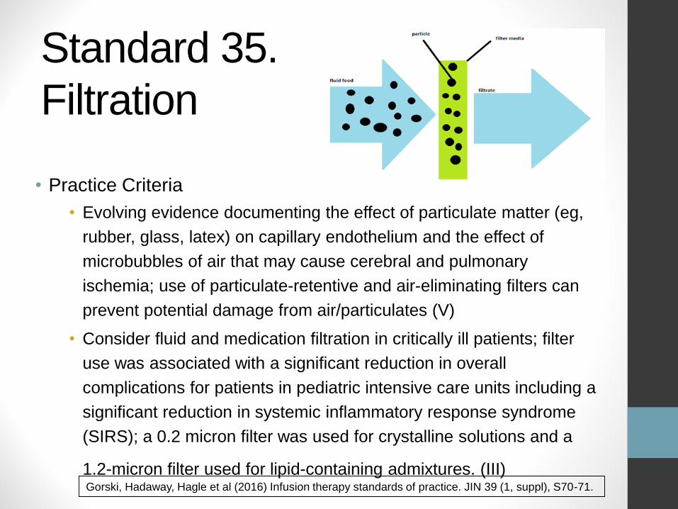
Standard 35.
Filtration
• Practice Criteria
• Evolving evidence documenting the effect of particulate matter (eg,
rubber, glass, latex) on capillary endothelium and the effect of
microbubbles of air that may cause cerebral and pulmonary
ischemia; use of particulate-retentive and air-eliminating filters can
prevent potential damage from air/particulates (V)
• Consider fluid and medication filtration in critically ill patients; filter
use was associated with a significant reduction in overall
complications for patients in pediatric intensive care units including a
significant reduction in systemic inflammatory response syndrome
(SIRS); a 0.2 micron filter was used for crystalline solutions and a
1.2-micron filter used for lipid-containing admixtures. (III)Gorski, Hadaway, Hagle et al (2016) Infusion therapy standards of practice. JIN 39 (1, suppl), S70-71.

Standard 37. VAD
Stabilization
• Stabilize and secure vascular access devices (VADs) to
prevent VAD complications and unintentional loss of
access.
• Practice Criteria
• Consider use of an engineered stabilization device (ESD)
• Glossary Term: Engineered Stabilization Device. A device
or system placed subcutaneously or topically; specifically
designed and engineered to control movement at the catheter
hub.
• Movement at the catheter hub increases the risk for phlebitis,
infiltration, and risk for accidental dislodgement
• Tape and sutures are not an effective alternative to an ESD

What Not to Do• Practice Criteria
• Do not use rolled bandages, with or without elastic properties, to secure any type of VAD
• They do not adequately secure the VAD, can obscure signs and symptoms of complications, and can impair circulation or the flow of infusion.
• Presence of skin disorders that contradict the use of medical adhesives (ie, pediatric epidermolysis bullosa; toxic epidermal necrolysis) may necessitate the use of tubular gauze mesh rather than adhesive engineered stabilization devices (ESD).

Standard 40. Flushing and
Locking• Flush with normal saline, aspirate for blood return, clear
medication from lumen, prevent contact between
incompatible solutions
• Minimum volume = twice internal volume of catheter system
• Larger volume may be needed
• Lock solutions
• Peripheral catheters – normal saline in adults; heparin 0.5 to 10
units per mL OR normal saline for neonates and pediatrics
• Midline catheters – insufficient evidence for recommendation
• CVADs – heparin 10 units per mL OR normal saline
• Volume = internal volume of catheter system plus 20%

Standard 41. VAD Assessment,
Care, Dressing Changes
• PRACTICE CRITERIA:
• Assess VAD catheter-sin junctions site and surrounding area
for redness, tenderness, swelling, and drainage by visual
inspection and palpation through the intact dressing and
through patient reports about any discomfort including pain,
paresthesias, numbness, or tingling.
• CVAD and midline catheter sites at least once per day.
• Routinely assess short peripheral catheter sites
• At least every 4 hours
• Every 1 to 2 hours for patients who are critically ill/sedated or
have cognitive deficits
• Hourly for neonatal/pediatric patients
• Vesicant infusions – more often

Standard 41. VAD Assessment,
Care, Dressing Changes
• Use of chlorhexidine dressings• Use chlorhexidine-impregnated dressings over CVADs to
reduce infection risk when the extraluminal route is the primary
source of infection. Even when organizations show a low
baseline central line-associated bloodstream infection
(CLABSI) rate, further reduction in CLABSI rate has been
demonstrated with use of chlorhexidine-impregnated
dressings.
• Consider use of chlorhexidine dressings with peripheral arterial
catheters.
• Use with caution – premature neonates, those with fragile skin
or complex skin pathologies due to risk for contact dermatitis
and pressure necrosis

Standard 44. VAD Removal
• Peripheral and nontunneled CVAD need assessed
daily
• Removed for unresolved complications,
discontinuation of infusion therapy, no longer
necessary for plan of care
• Peripheral catheter – remove if not used for 24
hours
• List of clinical indications for removal of
peripheral and midline catheters

Standard 44. VAD Removal
--Focus on Short Peripheral
Catheters
• Critical issues to consider when changing policy from timed catheter removal (i.e. every 96 hours) to clinically indicated short peripheral catheter removal
• Appropriate site selection
• Appropriate device selection
• Attention to conditions under which catheter placed ---ASEPTIC technique is critical
• Stabilization
• Maintenance of intact dressing

Standard 47.
Nerve Injuries
• Standard
• During peripheral venipuncture and catheter
dwell time, reports of paresthesia-type pain
require immediate removal of the VAD.
• During the insertion or dwell of CVADs,
clinicians will maintain a high index of
suspicion for nerve injuries when the patient
complains of respiratory difficulty or unusual
presentations of pain or discomfort.

Standard 47. Nerve Injuries
• Practice Criteria
• Selecting specific peripheral venous and arterial puncture
sites for the purpose of avoiding nerves is not possible;
however, common sites have a greater risk of nerve injury.
Venipuncture sites with the greatest risk include:
• Distal sensory branches of the radial and ulnar nerves for
sites in the dorsal hand.
• Superficial radial nerve at the cephalic vein of the radial wrist.
• Median nerve on the volar aspects of the wrist.
• Median and anterior interosseous nerve at or above the
antecubital fossa.
• Lateral and medial antebrachial nerves for the antecubital
fossa.
• Brachial plexus nerve for subclavian and jugular sites.
Gorski et al, JIN, p. S102-104.
Standard 47. Nerve Injuries
• Practice Criteria
• Review the patient’s medication list for systemic
anticoagulant medication(s) prior to making a puncture in
a vein or artery.
• Do not use subcutaneous probing techniques or multiple
passes of the needle or catheter when performing any
puncture procedure as this increases risk of nerve
damage. (V)
Gorski et al, JIN, p. S102-104.


![Infusion Nursing Standards of Practice - INCATIVincativ.es/documentos/guias/INS_Standards_of_Practice_2011[1].pdf · Infusion Nursing Standards of Practice Reviewers Jeanette Adams,](https://static.fdocuments.net/doc/165x107/5d05992288c99375438c30ce/infusion-nursing-standards-of-practice-1pdf-infusion-nursing-standards-of.jpg)