2015 Reuss et al, Molecular Neurobiology
17
Neuroglobin Expression in the Mammalian Auditory System Stefan Reuss & Ovidiu Banica & Mirra Elgurt & Stephanie Mitz & Ursula Disque-Kaiser & Randolf Riemann & Marco Hill & Dawn V. Jaquish & Fred J. Koehrn & Thorsten Burmester & Thomas Hankeln & Nigel K. Woolf Received: 23 September 2014 /Accepted: 29 December 2014 # Springer Science+Business Media New York 2015 Abstract The energy-yielding pathways that provide the large amounts of metabolic energy required by inner ear sen- sorineural cells are poorly understood. Neuroglobin (Ngb) is a neuron-specific hemoprotein of the globin family, which is suggested to be involved in oxidative energy metabolism. Here, we present quantitative real-time reverse transcription PCR, in situ hybridization, immunohistochemical, and West- ern blot evidence that neuroglobin is highly expressed in the mouse and rat cochlea. For primary cochlea neurons, Ngb expression is limited to the subpopulation of type I spiral ganglion cells, those which innervate inner hair cells, while the subpopulation of type II spiral ganglion cells which inner- vate the outer hair cells do not express Ngb. We further inves- tigated Ngb distribution in rat, mouse, and human auditory brainstem centers, and found that the cochlear nuclei and su- perior olivary complex (SOC) also express considerable amounts of Ngb. Notably, the majority of olivocochlear neu- rons, those which provide efferent innervation of outer hair cells as identified by neuronal tract tracing, were Ngb-immu- noreactive. We also observed that neuroglobin in the SOC frequently co-localized with neuronal nitric oxide synthase, the enzyme responsible for nitric oxide production. Our find- ings suggest that neuroglobin is well positioned to play an important physiologic role in the oxygen homeostasis of the peripheral and central auditory nervous system, and provides the first evidence that Ngb signal differentiates the central projections of the inner and outer hair cells. Keywords Neuroglobin . Cochlea . Hair cell . Spiral ganglion . Mice . Hearing . Auditory Abbreviations pb Base pairs DPO Dorsal paraolivary nucleus FG Fluoro-Gold GAPDH Glyceraldehyde-3-phosphate dehydrogenase Electronic supplementary material The online version of this article (doi:10.1007/s12035-014-9082-1) contains supplementary material, which is available to authorized users. S. Reuss (*) Department of Nuclear Medicine, University Medical Center, Johannes Gutenberg-University, Langenbeckstr. 1, Bld. 210, 55101 Mainz, Germany e-mail: [email protected] O. Banica : M. Elgurt : S. Mitz Department of Anatomy and Cell Biology, University Medical Center, Johannes Gutenberg-University, 55099 Mainz, Germany U. Disque-Kaiser Department of Radiation Oncology, University Medical Center, Johannes Gutenberg-University, 55101 Mainz, Germany R. Riemann Department of ENT, Elbe-Kliniken, 21682 Stade, Germany M. Hill : T. Hankeln Institute of Molecular Genetics, Johannes Gutenberg-University, 55099 Mainz, Germany D. V. Jaquish : F. J. Koehrn : N. K. Woolf Department of Surgery, University of California at San Diego, San Diego, CA 92093-0604, USA D. V. Jaquish : F. J. Koehrn : N. K. Woolf Veterans Affairs Research Service, VA San Diego Healthcare System, San Diego, CA 92093-0604, USA T. Burmester Institute of Zoology and Zoological Museum, University of Hamburg, Hamburg, Germany Mol Neurobiol DOI 10.1007/s12035-014-9082-1
-
Upload
nigel-woolf -
Category
Documents
-
view
95 -
download
1
Transcript of 2015 Reuss et al, Molecular Neurobiology
Neuroglobin Expression in the Mammalian Auditory System
Stefan Reuss & Ovidiu Banica & Mirra Elgurt &Stephanie Mitz & Ursula Disque-Kaiser &
Randolf Riemann & Marco Hill & Dawn V. Jaquish &
Fred J. Koehrn & Thorsten Burmester & Thomas Hankeln &
Nigel K. Woolf
Received: 23 September 2014 /Accepted: 29 December 2014# Springer Science+Business Media New York 2015
Abstract The energy-yielding pathways that provide thelarge amounts of metabolic energy required by inner ear sen-sorineural cells are poorly understood. Neuroglobin (Ngb) is aneuron-specific hemoprotein of the globin family, which issuggested to be involved in oxidative energy metabolism.Here, we present quantitative real-time reverse transcriptionPCR, in situ hybridization, immunohistochemical, and West-ern blot evidence that neuroglobin is highly expressed in themouse and rat cochlea. For primary cochlea neurons, Ngbexpression is limited to the subpopulation of type I spiralganglion cells, those which innervate inner hair cells, whilethe subpopulation of type II spiral ganglion cells which inner-vate the outer hair cells do not express Ngb. We further inves-tigated Ngb distribution in rat, mouse, and human auditorybrainstem centers, and found that the cochlear nuclei and su-perior olivary complex (SOC) also express considerableamounts of Ngb. Notably, the majority of olivocochlear neu-rons, those which provide efferent innervation of outer hair
cells as identified by neuronal tract tracing, were Ngb-immu-noreactive. We also observed that neuroglobin in the SOCfrequently co-localized with neuronal nitric oxide synthase,the enzyme responsible for nitric oxide production. Our find-ings suggest that neuroglobin is well positioned to play animportant physiologic role in the oxygen homeostasis of theperipheral and central auditory nervous system, and providesthe first evidence that Ngb signal differentiates the centralprojections of the inner and outer hair cells.
Keywords Neuroglobin . Cochlea . Hair cell .
Spiral ganglion .Mice . Hearing . Auditory
Abbreviationspb Base pairsDPO Dorsal paraolivary nucleusFG Fluoro-GoldGAPDH Glyceraldehyde-3-phosphate dehydrogenase
Electronic supplementary material The online version of this article(doi:10.1007/s12035-014-9082-1) contains supplementary material,which is available to authorized users.
S. Reuss (*)Department of Nuclear Medicine, University Medical Center,Johannes Gutenberg-University, Langenbeckstr. 1, Bld. 210,55101 Mainz, Germanye-mail: [email protected]
O. Banica :M. Elgurt : S. MitzDepartment of Anatomy and Cell Biology, University MedicalCenter, Johannes Gutenberg-University, 55099 Mainz, Germany
U. Disque-KaiserDepartment of Radiation Oncology, University Medical Center,Johannes Gutenberg-University, 55101 Mainz, Germany
R. RiemannDepartment of ENT, Elbe-Kliniken, 21682 Stade, Germany
M. Hill : T. HankelnInstitute of Molecular Genetics, Johannes Gutenberg-University,55099 Mainz, Germany
D. V. Jaquish : F. J. Koehrn :N. K. WoolfDepartment of Surgery, University of California at San Diego, SanDiego, CA 92093-0604, USA
D. V. Jaquish : F. J. Koehrn :N. K. WoolfVeterans Affairs Research Service, VA San Diego HealthcareSystem, San Diego, CA 92093-0604, USA
T. BurmesterInstitute of Zoology and Zoological Museum, University ofHamburg, Hamburg, Germany
Mol NeurobiolDOI 10.1007/s12035-014-9082-1
IHC Inner hair cellsISH In situ hybridizationLOC/MOC
Lateral/medial olivocochlear neurons
LSO Lateral superior oliveMNTB Medial nucleus of the trapezoid bodyMSO Medial superior oliveNgb NeuroglobinnNOS Neuronal nitric oxide synthaseNO Nitric oxideOHC Outer hair cellsOCN Olivocochlear neuronsPBS Phosphate-buffered 0.9 % salineRPO Rostral paraolivary nucleusrt-RT-PCR
Real-time reverse transcription polymerase chain-reaction
RT Room temperatureSgn Spiral ganglion neuronsSOC Superior olivary complexSPO Superior paraolivary nucleusVNTB Ventral nucleus of the trapezoid bodyVPO Ventral paraolivary nucleus
Introduction
The globin family is comprised of small porphyrin-containingproteins, which can reversibly bind O2 by means of an iron(Fe2+) ion of the heme prosthetic group [1, 2]. Besides thebetter known examples of hemoglobin (Hb) and myoglobin(Mb), the vertebrate globin family includes six additionalmembers: neuroglobin (Ngb) [3], cytoglobin (Cygb) [4–6],globin E (GbE) [7], globin X (GbX) [8], globin Y (GbY)[9], and androglobin (Adgb) [10]. The vertebrate globins havea complex evolutionary history and may serve different func-tions which are, however, often only poorly defined [11].While Hb, Mb, Ngb, Cygb, and Adgb are present in mostjawed vertebrates [10, 12], GbE, GbX, and GbY appear tohave been lost in placental mammals and other vertebrate taxa[11].
Ngb has been identified in a wide range of vertebrates, andits structure has been highly conserved (e.g., there is 94 %amino acid sequence identity between human and murineNgb) [13]. Ngb is mainly expressed in neurons of the centraland peripheral nervous systems and in endocrine tissues [3,14–24]. Ngb has also been found in the retinae of man, mouse,and guinea pig [25–28]. Within the retina, Ngb is expressedprimarily at sites of high oxygen consumption: the inner seg-ments of photoreceptor cells, the inner and outer nuclearlayers, and the retinal ganglion cell layer [25, 26]. There isconvincing evidence that in retina and brain Ngb is associatedwith mitochondria and thus with oxidative metabolism [25,
26, 18]. Moreover, across vertebrates total Ngb levels in brainseem to be positively correlated with species’ tolerance forhypoxia [29, 24, 20].
Experimental investigations have demonstrated that Ngbserves to protect cultured cerebral neurons from hypoxiain vitro [30]. Ngb may also help the brain in vivo to resistneuronal injury during experimental stroke [31], althoughstudies with knockout mice rather contradict this conclusion[32]. In summary, while the exact physiological role of Ngb isstill a matter of debate [33, 34], it is likely that Ngb contributesto local O2 supply or, alternatively, provides protection fromreactive oxygen species (ROS).
The anatomical structure of the mammalian inner earmakes the neurosensory hair cells and vestibulocochlear nerveneurons uniquely vulnerable to hypoxia/ischemia [35]. Al-though the anatomy of the cochlea has been well describedand considerable progress has been made in characterizing thestructural and physiological properties of the inner ear (cf.[36–40], at present, we still know relatively little about thespecific cellular mechanisms by which the inner ear satisfiesits normal metabolic energy demands and defends itselfagainst pathologic conditions. Early studies of oxygen con-sumption in the cochlea have suggested that metabolic rateswere higher in external wall structures (e.g., stria vascularis)than in sensorineural cells (reviewed in [41]). Subsequent in-vestigations utilizing the 2-deoxyglucose (2-DG) radiotracertechnique determined that acoustic stimulation alters cochlearmetabolism: in silence the rate of 2-DG incorporation in thecochlea was highest in external wall structures (i.e., striavascularis, spiral ligament, and spiral prominence), and rela-tively low in neurosensory cells (i.e., inner and outer hair cells,and vestibulocochlear (VIIIth cranial) nerve ganglia), whilenoise exposure significantly increased the rate of 2-DG uptakein the same cochlear external wall structures and auditoryneurosensory cells [42–44].
The metabolic energy required for neurosensory cell func-tion in the cochlea is provided primarily by adenosine triphos-phate (ATP). ATP levels have been measured in cultured co-chlear hair cells [41] and CNS neurons [45], and these levelswere found to be on the same order of magnitude as freeze-dried outer hair cell preparations [46]. In addition, it has beendemonstrated that the induction of cochlear pathology (e.g.,hypoxia/ischemia) significantly reduces the ATP content ofthe organ of Corti in vivo [47]. However, questions remainabout the relative contributions of the glycolytic and oxidativepathways to the synthesis and maintenance of ATP levels inthe inner ear. Glycolysis results in the formation of lactate, butsince attempts to measure lactate in the cochlea have failed[48], it has long been assumed that low-level aerobic metab-olism is the primary source of cochlear ATP. On the otherhand, there is evidence that outer hair cells deprived of glucoseex vivo generate ATP, which suggests that these sensory cellsmay also possess unidentified intracellular metabolic
Mol Neurobiol
substrates that can augment oxidative phosphorylation underhypoxic conditions [41].
Oxygen required for aerobic glucose metabolism is sup-plied to the inner ear by the Hb of the blood. While the spiral(auditory) and Scarpa’s (vestibular) ganglia of thevestibulocochlear nerve are well vascularized by radiatingcapillary arcades, the neuroepithelia of the auditory (i.e., organof Corti), and vestibular (i.e., maculae of the utricle and sac-cule, and cristae ampullares of the semicircular canals) sys-tems do not receive any direct blood supply [49]. Consequent-ly, oxygen must diffuse relatively large distances to reach theauditory and vestibular sensory hair cells. Notably, the vascu-lar anatomy of the inner ear is homologous to that of the eye,since the retina similarly lacks any direct blood supply to itssensory cells (i.e., photoreceptors) [50]. Because the inner earand eye share similar challenges in providing an adequatesupply of oxygen to their neurosensory cells during periodsof oxidative stress, we hypothesized that the auditory andvestibular systems may have evolved similar cellular mecha-nisms to fuel their oxidative metabolic demands as those pres-ent in the retina.
Besides one report that Ngb is expressed by spiral ganglionneurons [51], detailed information about the distribution ofNgb in mammalian peripheral and central auditory system islacking. We, therefore, studied the cochlea of rats and mice,and the auditory brainstem of rats, mice, and man, using Ngbimmunohistochemistry, in situ hybridization, Western blot-ting, quantitative rt-RT-PCR, and neuronal tract tracing tocharacterize Ngb-expressing anatomical regions and neuronalcell types. We describe here that Ngb is highly expressed in asubpopulation of primary afferent neurons in thevestibulocochlear nerve but not in auditory sensory hair cells.Ngb is also present in the auditory brainstem, in particular inthe superior olivary complex, and in a functionally importantsubpopulation, the olivocochlear neurons.
Materials and Methods
Animals
The experiments were conducted on (1) 6–10-week-old-maleC.B-17 mice (Taconic Farms, Germantown, NY, USA)housed under constant conditions (light:dark 12:12 h, temper-ature 22+2 °C) with food and water ad libitum at the VA SanDiego Healthcare System (VASDHS) Veterinary MedicalUnit. Experimental protocols were approved by the VASDHSInstitutional Animal Care and Use Committee, and conformedto the PHS BGuide for the Care and Use of Laboratory Ani-mals,^ (2) male Balb/C-mice, aged 2 months, and (3) 9–12-week-old-male Sprague-Dawley rats, held under constantconditions (see group 1) in the animal facility unit of theDepartment of Anatomy and Cell Biology, Johannes
Gutenberg-University, Mainz, Germany. Animals of groups2 and 3 were bred in-house. Experimental protocols (animalhousing and neuronal tracing) were approved by the localAdministration District Official Committee and were in accor-dance with the published European Health Guidelines. Allefforts were made to minimize the number of animals andtheir suffering.
Neuroglobin Immunohistochemistry
Mouse cochleae and brains were dissected from animals of theC.B-17 strain that were killed by cervical dislocation. Thecochleae were then immersion-fixed in 5 % paraformaldehydein phosphate-buffered 0.9 % saline (PBS) for 2 h at 4 °C, andthen decalcified in 10 % EDTA at 4 °C. Decalcified cochleaewere subsequently immersed in 30 % sucrose until they sank,embedded in OCT, frozen, sectioned at 10 μm, collected ontoBioBond-coated (Ted Pella, Redding, CA). Fisher SuperFrostslides (Fisher Scientific, Tustin, CA), air and vacuum dried,and then stored until needed at −70 °C. Two different Ngbantibodies directed against a synthetic peptide which coversthe conserved amino acid positions 55–70 of mammalian Ngb(H2N-CLSSPEFLDHIRKVML-CONH2) [30, 25] were inde-pendently raised in rabbits. One was produced by StrategicBiosolutions Inc. (San Diego, CA), the other was producedby Eurogentec (Köln, Germany, see [25]). Antibodies wereaffinity-purified using a SulfoLink kit (Pierce Biochemicals,Rockford, IL) as described by the manufacturer.
Mouse sections on glass slides were washed in PBS-0.1 %Triton X-100, treated with blocking solution (10 % normaldonkey serum+5 % BSA in PBS-0.1 % Triton X-100), incu-bated overnight at RT with Ngb antibody (1:200; StrategicBiosolutions) and mouse monoclonal anti-peripherin interme-diate filament protein antibody (1:25; Chemicon, Temecula,CA) diluted in blocking solution (Mouse-On-Mouse PK-2200Kit, Vector Laboratories, Burlingame, CA) overnight at 4 °C.Slides were washed and incubated with secondary donkeyanti-rabbit antibody conjugated to Alexa 594 (1:300) and don-key anti-mouse antibody conjugated to Alexa 488 (1:300;Molecular Probes, Eugene, OR) in PBS for 1 h in the dark.Slides were washed and stained with bisbenzimide (nuclearstain, Sigma, St. Louis, MO), washed again, and coverslippedwith Prolong Mounting Media (Molecular Probes, Eugene,OR). Negative control experiments which substituted normalrabbit and mouse IgG for the primary antibodies resulted inthe complete absence of staining.
Rats were killed by ether overdose at the middle of the lightperiod and immediately perfused transcardially with PBS towhich 15,000 IU heparin/L were added, at RT, followed by anice-cold paraformaldehyde-lysine-periodate solution (PLP;[52]. The right atrium was opened to enable venous outflow.Cochleae were decalcified as described above, sectioned at30-μm thickness and mounted on gelatinized glass slides.
Mol Neurobiol
Sections were then incubated overnight at RT with Ngb anti-body (1:500; Eurogentec), to which 1% normal donkey serumand 0.1% Triton-X 100 were added. After three rinses in PBS,the reaction was visualized using Cy3 conjugated to an F(ab)2fragment of a donkey anti-rabbit IgG (1:200 in PBS; JacksonImmuno-Research, West Grove, PA). Sections were dried,cleared in xylene, and covered.
For brainstem immunohistochemistry, PLP-perfused ratand mouse brains were removed, postfixed in PLP for 1 h,and stored overnight at 4 °C in phosphate-buffered 30 % su-crose for cryoprotection. They were then sectioned serially at40-μm thickness on a freezing microtome in the frontal plane.Sections were collected in PBS and, for immunohistochemis-try, incubated free-floating overnight at RT in Ngb antibody(1:500, Eurogentec), and processed further as described abovefor the rat cochlea.
RNA Extraction
Total RNA was extracted from frozen C.B-17 mouse wholebrains and cochleae using TRizol reagent (Invitrogen, Carls-bad, CA), and from Balb/C mouse whole brains using theRNeasy kit (Qiagen, Hilden, Germany), and subsequentlytreated with DNase I Amplification Grade (Invitrogen) ac-cording to the manufacturer’s protocols.
DNA Synthesis and Cloning of Ngb-cDNA for ISH
Ngb cDNA was generated using the SuperScript One-StepRT-PCRwith PlatinumTaq (Invitrogen) according to the man-ufacturer’s protocol. In brief, 500 ng of DNase I-treated totalRNAwas added to 1× reaction mix buffer (containing 0.2 mMof each dNTP and 1.2 mM MgSO4), 1 μl RT/Platinum TaqMix and 0.2 μM of sense and antisense Ngb primers: senseprimer 5′-GTTGACTGCACCCACGCCT-3′; antisense prim-er 5′-GCACCACAGCTCCGTAGAGT-3′ (GenBank acces-sion number NM-022414), with the total volume being50 μl. The reaction was then given into a Deltacycler II(Ericomp Inc., San Diego, CA) and cycled at 50 °C for30 min then at 94 °C for 2 min, followed by 40 cycles of94 °C for 30 s, 65 °C for 30 s, and 72 °C for 1 min, with afinal annealing extension at 72 °C for 5 min. A 487 bp Ngbfragment was generated and then cloned using the TOPODualVector Cloning System (Invitrogen). The fragment was thensequenced by the UCSD/VMRF CFAR Genomics Core Fa-cility for verification.
Neuroglobin In Situ Hybridization
For the detection of Ngb-mRNA in the mouse cochlea, the487 bp Ngb cDNAwas cut with EcoRVand PstI to generate a308 bp fragment (290 bp of it being Ngb) which was thensubcloned into the EcoRV, PstI sites of the pSP72 vector
(Promega, Madison, WI). Sense and antisense Ngb probeswere generated by linearizing the subclone with HindIII orEcoRV, respectively, followed by labeling using the DIGRNA labeling kit (Roche, Indianapolis, IN) according to themanufacturer’s protocol. Paraffin-embedded sections weredeparaffinized in CitriSolve (Fisher Scientific, Waltham,MA), hydrated in graded alcohols, and rinsed 2× in 1×PBSfor 5 min prior to prehybridization. For prehybridization, thesections were placed in 0.2 N HCl at RT for 20 min, dipped indH2O, put in 2× standard saline citrate (SSC) at 70 °C for30 min, dipped in dH2O, digested in 20 mM Tris pH 7.4,2 mM CaCl2+4 μg/ml proteinase K at 37 °C for 30 min,and digestion terminated with 0.2 % glycine in 1×PBS for30 s, followed by 2× 5-min washes in 1×PBS, 3 min in0.1 M triethanolamine (TEA) pH 8.0, followed by an acetyla-tion step of 1:400 acetic anhydride in TEA for 10 min. Slideswere then passed 2× in 2× SSC for 2 min, dehydrated ingraded alcohols, and air-dried.
The hybridization buffer contained 50 % deionized form-amide, 10 % dextran sulfate, 0.3 M NaCl, 10 mMTris pH 7.6,5 mMEDTA pH7.6, 1XDenhardt’s solution, 100μg/ml yeasttRNA and 250 μg/ml sheared herring sperm DNA. Hybridi-zation was conducted overnight at 42 °C with 0.5 μg/μl ofprobe per slide. Washes consisted of 2× 15 min in 2×SSC+0.1 % Triton X-100 at 42 °C, 15 min in 0.2×SSC+0.1 %Triton X-100 at 42 °C, 15 min in 0.1×SSC+0.1 % TritonX-100 at 42 °C, and 2× 15 min in 1× washing buffer(100 mM Tris+150 mM NaCl pH 7.5)+0.5 % Triton X-100at RT. Blocking was with 2 % blocking buffer (Blocking Re-agent, Roche, Indianapolis, IN) in 1× washing buffer+0.5 %Triton X-100 at RT for 30 min. Slides were then incubatedwith a 1:400 dilution of anti-DIG antibody-AP (Roche) in 1 %blocking buffer (2 % blocking buffer diluted 1:1 with 1×washing buffer) at RT for 1.5 h. Slides were then washed 3×5 min at RT in 1× washing buffer+0.5 % Triton X-100,followed by an equilibration in 1X detection buffer(100 mM Tris+100 mM NaCl pH 9.5+10 mM MgCl2+240 μg/ml Levaminsol) at RT for 2 min. Development wascarried out overnight in the dark at RT using 1X detectionbuffer+0.33 mg/ml NBT+0.16 mg/ml BCIP. Developmentwas stopped by dipping the slides 2× in dH2O for 5 min.Sections were counterstained for 1 s in hematoxylin, andslides were coverslipped with crystal mount (Biomedia Corp.,Foster City, CA).
For the detection of Ngb-mRNA in the rat brainstem, PLP-perfused brains were postfixed in PLP for 1 h and storedovernight at 4 °C in phosphate-buffered 30 % sucrose forcryoprotection. They were then sectioned serially at 40-μmthickness on a freezing microtome in the frontal plane. Sec-tions were collected in PBS, mounted on silanized glass slidesand processed for in situ hybridization. The hybridization pro-cedures used on brainstem slices were described before [15]and resembled those used on cochlea sections (see above). In
Mol Neurobiol
brief, digoxigenin-labeled sense (negative control) and anti-sense RNA probes were in vitro transcribed by SP6 RNApolymerase (DIGRNALabeling Kit, Roche,Mannheim, Ger-many), using polymerase chain reaction (PCR)-generatedtemplates covering the 453-bp mouse Ngb coding region (ac-cession no. AJ245945). For template generation, a SP6 RNApolymerase promoter sequence was attached to the 5′ end ofthe sense or antisense PCR primers. For prehybridization,slices were incubated for 1 min with proteinase K (10 μg/ml)in PBS/Tween 20. Digestion was terminated with 0.2 % gly-cine in phosphate-buffered saline (PBS)/Tween 20 followedby 3×5 min in PBS/Tween 20 and 1×5 min in PBS. RNAprobes were diluted in 2× SSC/50 % formamide and sectionswere incubated with the probe at 42 °C overnight. Slides werewashed first in 2× SSC, followed by 0.1× SSC at 60 °C for20 min. Preparations were then treated with a mixture of RN-Ase A (25 μg/ml) for 30 min at 37 °C in a wet chamber, andthereafter washed in PBS/Tween 20. After blocking for15 min in 1 % blocking reagent (Roche) in 0.1 M Tris and0.15 M NaCl at pH 7.5, label was detected by alkalinephosphatase-coupled anti-digoxigenin antibodies (diluted1:100 in 1 % blocking reagent in 0.1 M Tris and 0.15MNaCl,pH 7.5); 30-min incubation at 37 °C, washing in PBS/Tween20 and NBT buffer (0.1 M Tris, 0.1 M NaCl, 0.05 M MgCl2-hexahydrate pH 9.5), and subsequent incubation with nitro-blue-tetrazolium/5-bromo-4-chloro-3-indolyl-phosphate sub-strate (1:50 diluted in NBT buffer; 30–45 min 37 °C in thedark). Substrate reaction was stopped by PBS washing. Sec-tions were dried, covered by PBS/glycerol (1:1) solution, andcoverslipped.
Quantitative Real-Time Reverse-Transcription PolymeraseChain-Reaction (rt-RT-PCR)
DNase I-treated total RNA (1 μg; see BRNA Extraction^) wasreverse transcribed using SuperScript II RNase H-ReverseTranscriptase (Invitrogen), according to the manufacturer’sprotocol. Reactions were primed using oligo(dT)12-18 primersand the total volume/reaction was 20 μl. Following comple-tion of the reaction, samples were brought to 10 ng/μl finalvolume with 5 μl being used for rt-RT-PCR.
Ngb-mRNA expression in C.B-17 mouse whole brains andcochleae was measured by quantitative rt-RT-PCR, based onTaqMan method, using the ABI PRISM 7700 Sequence De-tection System (Applied Biosystems, Foster City, CA).Primers and probe sequences for Ngb (GenBank accessionnumber AJ245945) were designed using Primer Express soft-ware (Applied Biosystems): forward primer 5′-TGCTGCCTCTCTTCCAGTACAA-3′; reverse primer 5′-GGAATTCTGGAGAGGAGAGACAGT-3′; and probe 5′-CCGCCAGTTCTCCAGCCCTGAG-3′. Quantitative rt-RT-PCR reactionswere performed by the UCSD/VMRF CFARGenomics Core.The total RNA was normalized by the amount of
glyceraldehyde-3-phosphate dehydrogenase (GAPDH) am-plified in each reaction. A GAPDH standard curve was pre-pared as the endogenous reference, and an Ngb standard curvewas constructed based on tenfold dilutions of the 487 bp NgbcDNA ranging from 1 ng to 10 μg. A total of 50 ng of the RTproduct was used for each sample reaction, with all reactionsrun in duplicate. Results were obtained by using the relativestandard curve analysis (Applied Biosystems User Bulletin#2). The amount of sample Ngb and GAPDH target was thendetermined from the appropriate standard curve. For eachsample the averaged Ngb target values were divided by theaveraged GAPDH target values to obtain the normalizedamount of Ngb mRNA target. The standard deviation for thenormalized amount of NgbmRNA target was calculated usingthe following formulae:
SD ¼ffiffiffiffiffiffiffiffiffiffiffiffiffiffiffiffiffiffiffiffiffiffiffiffiffiffiffiffiffiffiffiffiffiffiffiffiffiffiffi
CVNgb2 þ CVGAPDH
q
ð1Þ
where
CV ¼ s
x¼ standarddeviation
meanvalueð2Þ
One of the brain samples (brain #1) was arbitrarily chosenas the calibrator, and the average normalized Ngb sample val-ue was divided by the average normalized calibrator Ngb val-ue. Thus, the calibrator (i.e., brain #1) constituted the 1× sam-ple, and the relative amount of target Ngb mRNA in the dif-ferent samples was expressed as an n-fold modulation differ-ence relative to the calibrator.
For the quantification of Ngb-mRNA from specific brainregions, including the SOC and cochlear nucleus, we cut freshmouse brains in 1-mm thick coronal slices and microdissectedthe regions using neuropunch needles with defined inner di-ameter (Fine Science Tools, Heidelberg, Germany). Sampleswere processed as described above, with the following mod-ifications. For the TaqMan assay, we used the QuantiTectProbe PCR-Kit (Qiagen, Hilden, Germany) and the ABIPRISM 7000 Sequence Detection System (AppliedBiosystems) with the following primers for Ngb, designedusing IDTSciTools OAnalyzer 3.0 (Integrated DNATechnol-ogies, San Diego, CA).
Forward primer 5′-GAAGCATCGGGCAGTG-3; reverseprimer 5′-AGGCACCTCTCCAGCATGTAGAG-3; andprobe 6-FAM-5′-CTCAGCTCCTTCTCGACAGT-3-DarkQuencher. Acidic ribosomal protein (ARP) [forward primer5′-AGGGCGACCTGGAAGTCC-3; reverse primer 5′-GCATCTGCTTGGAGCCCA-3] was used for RNAnormalization.
Mol Neurobiol
The SPSS 12.0 program (SPSS, München, Germany) wasused for statistical analysis and to generate boxplots.
Olivocochlear Neuronal Tracing
These experiments were performed on five rats, which wereanesthetized with tribromoethanol (0.3 g/kg b.w., i.p.) andreceived pressure-injections of 200 nl of a 5 % Fluoro-Goldsolution (FG, Fluorochrome, Englewood, CO; dissolved indistilled water) into the left scala tympani via the round win-dow. The surgical approach was described previously [53,54]. After 5 days, rats were killed by ether overdose and per-fused with PLP as described above.
Double Immunofluorescence of Brainstem Sections
PLP-perfusion-fixed brains from Balb/C-mice werecryoprotected and sectioned as described above. Sectionswere incubated with Ngb-antibody (1:500, Eurogentec), visu-alized by Cy3 and, simultaneously, with sheep anti-neuronalnitric oxide synthase (nNOS, 1:500 in PBS; Abcam, Cam-bridge, England), visualized by anti-sheep IgG coupled toCy2 (1:200; Jackson, West Grove, PA), and treated furtheras described above. Control incubations showed that blockingthe antisera with the respective antigens as well as omission ofsingle or both primary and/or secondary antisera resulted inthe absence of respective staining, and that no cross-reactivitybetween primary and secondary antibodies was present.
Cell Counting
From each section, FG-labeled cells of the brainstem werequantified with regard to their location within the SOC. Neu-rons exhibiting immunoreactivity to the antibodies tested werecounted when immunoreactivity was clearly over the back-ground level. They were manually counted from the incubatedsections using an Olympus BX 51 research microscope (seeBImage Analysis^). Counts were corrected according to Ab-ercrombie [55] to prevent double counting of cells. Single-and double-labeled neurons were quantified separately andthe respective percentages were calculated for each nucleus.
Brainstem regions were identified using the stereotaxicbrain atlases for rat and mouse [56, 57], and the overviewsgiven by Schwartz [58], Warr [59], Kulesza et al. [60], andMalmiera and Merchan [61]. According to Warr [59] and Vet-ter and Mugnaini [62], FG-labeled neurons were divided intothree subgroups: lateral olivocochlear (LOC) neurons in thelateral superior olive (LSO), shell-neurons (surrounding theLSO, including periolivary regions), and medial OC (MOC)neurons in the ventral nucleus of the trapezoid body (VNTB)and rostral periolivary region (RPO).
Human Brainstem Immunohistochemistry
These studies were performed on sections from the humanbrainstem. Material was kindly provided by Dr. ThorstenFink, Department of Pathology, HSK Wiesbaden, Germany.Specimens were derived from a woman who died at the age of73 from peritonitis and septic shock, and a man died at the ageof 74 ears from pneumonia. Neither one suffered from anyknown neurological disease. Brainstems were taken postmor-tem, immersion-fixed, and stored in formalin at 4 °C.
For analysis, the brainstems were cut in half at the mediananterior fissure, and a section of 15-mm thickness was takenfrom the region cranial to the border between medullaoblongata and pons. This specimen was cryoprotected inphosphate-buffered sucrose, cut in 40-μm sections, and storedin 10 mM PBS at 4 °C.
Due to the high levels of autofluorescence found in theaged human brain specimens, and the unsatisfying results ofquenching procedures, we used a non-fluorescent detectionmethod. In brief, after incubation in primary Ngb-antibody(1:500, Eurogentec), sections were washed in PBS, incubatedfor 1.5 h at RT in a biotin-conjugated F(ab)2 fragment of adonkey anti-rabbit IgG (1:200 in PBS; Jackson Labs, WestGrove, PA), and processed for immunohistochemistry accord-ing to the avidin-biotin-peroxidase-complex method (ABCSet, Vector Labs, Burlingame, CA; [63], using alpha-naphthol as chromogen. Sections were mounted on gelatin-coated slides, dried, and coverslipped.
Image Analysis
Mouse cochlea immunohistochemistry and in situ hybridiza-tion slides were examined using a Zeiss Axioplan II micro-scope equipped with a KX85 CCD camera (Apogee Instru-ments, Tucson, AZ). Digital images were processed and opti-mized using either Image Pro (Media Cybernetics, SilverSprings, MD) or Adobe Photoshop (San Jose, CA). Quantifi-cation of somatic fluorescence was accomplished using onlythe linear range of the digital camera. Deconvolution imageswere collected using a DeltaVision Restoration microscopesystem (Applied Precision Inc., Issaquah, WA) equipped witha Sony Photometrics Coolsnap HQ charged-coupled device(CCD) camera system (10 MHz, 12 bit, 1392×1040; Sony,Burbank, CA) attached to an inverted, wide-field fluorescentNikon TE-200 microscope (Nikon Inc., Kanagawa, Japan).Optical sections were acquired using Nikon oil immersionobjectives in 0.2-μm steps in the z-axis using the attachedApplied Precision Inc. motorized stage. Images were saved,processed, and analyzed on SGI workstations (Oxygen & Oc-tane computers; Silicon Graphics, Mountain View, CA) usingthe DeltaVision SoftWorx software package (version 2.50).
Sections from rat cochlea, as well as from rat, mouse, andhuman brainstem (ISH, neuronal tracing, and double
Mol Neurobiol
immunofluorescence material) were analyzed using an Olym-pus BX51 research microscope equipped with anepifluorescence unit and filter sets for FG (excitation centerwavelength 360 nm, bandwidth 50 nm), Cy2 (480/40 nm) andCy3 (540/25 nm) as well as a digital color camera operatedusing the AnalySIS (Soft Imaging System, Münster, Germa-ny) image processing program. Images were merged, and con-trast and brightness adjusted using Adobe Photoshop (AdobeSystems Inc., San Francisco, CA).
Results
Experiments described here were conducted on mouse, rat,and human material.
Localization of Neuroglobin Protein
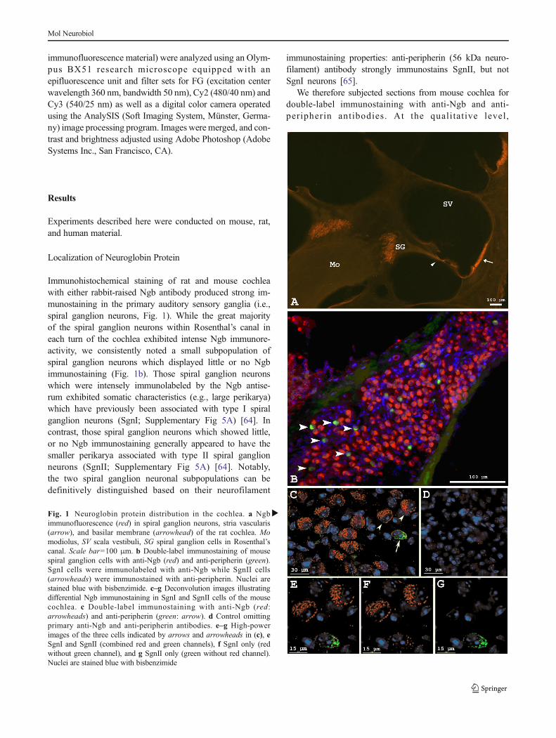
Immunohistochemical staining of rat and mouse cochleawith either rabbit-raised Ngb antibody produced strong im-munostaining in the primary auditory sensory ganglia (i.e.,spiral ganglion neurons, Fig. 1). While the great majorityof the spiral ganglion neurons within Rosenthal’s canal ineach turn of the cochlea exhibited intense Ngb immunore-activity, we consistently noted a small subpopulation ofspiral ganglion neurons which displayed little or no Ngbimmunostaining (Fig. 1b). Those spiral ganglion neuronswhich were intensely immunolabeled by the Ngb antise-rum exhibited somatic characteristics (e.g., large perikarya)which have previously been associated with type I spiralganglion neurons (SgnI; Supplementary Fig 5A) [64]. Incontrast, those spiral ganglion neurons which showed little,or no Ngb immunostaining generally appeared to have thesmaller perikarya associated with type II spiral ganglionneurons (SgnII; Supplementary Fig 5A) [64]. Notably,the two spiral ganglion neuronal subpopulations can bedefinitively distinguished based on their neurofilament
immunostaining properties: anti-peripherin (56 kDa neuro-filament) antibody strongly immunostains SgnII, but notSgnI neurons [65].
We therefore subjected sections from mouse cochlea fordouble-label immunostaining with anti-Ngb and anti-peripherin antibodies. At the quali tat ive level,
�Fig. 1 Neuroglobin protein distribution in the cochlea. a Ngbimmunofluorescence (red) in spiral ganglion neurons, stria vascularis(arrow), and basilar membrane (arrowhead) of the rat cochlea. Momodiolus, SV scala vestibuli, SG spiral ganglion cells in Rosenthal’scanal. Scale bar=100 μm. b Double-label immunostaining of mousespiral ganglion cells with anti-Ngb (red) and anti-peripherin (green).SgnI cells were immunolabeled with anti-Ngb while SgnII cells(arrowheads) were immunostained with anti-peripherin. Nuclei arestained blue with bisbenzimide. c–g Deconvolution images illustratingdifferential Ngb immunostaining in SgnI and SgnII cells of the mousecochlea. c Double-label immunostaining with anti-Ngb (red:arrowheads) and anti-peripherin (green: arrow). d Control omittingprimary anti-Ngb and anti-peripherin antibodies. e–g High-powerimages of the three cells indicated by arrows and arrowheads in (c), eSgnI and SgnII (combined red and green channels), f SgnI only (redwithout green channel), and g SgnII only (green without red channel).Nuclei are stained blue with bisbenzimide
Mol Neurobiol
immunolabeling revealed two distinct spiral ganglion neu-ronal subpopulations: SgnI (peripherin- negative) neuronsthat exhibited strong Ngb immunostaining, and SgnII(peripherin-positive) neurons which were not immuno-stained with Ngb (Fig. 1b). This distinct immunostainingpattern for SgnI and SgnII neurons was confirmed athigher resolution and magnification using DeltaVision Res-toration Microscopy System deconvolution images(Fig. 1c–g). Because a small number of SgnII neuronsexhibited Ngb immunofluorescence which appeared to ex-ceed background levels, a more quantitative analysis wasconducted (see supplemental material). We found that theaverage cross-sectional area of SgnI neurons was 15 %larger than SgnII neurons, and Ngb fluorescence moreintense in SgnI than in SgnII neurons (SupplementaryFigs. 3 and 4).
Specific Ngb immunofluorescence was also observed inthe stria vascularis (arrow in Fig. 1a) and basilar membrane(arrowhead in Fig. 1a) while no Ngb immunostaining wasobserved in hair cells, supporting cells, or Schwann cells sur-rounding SgnI neurons.
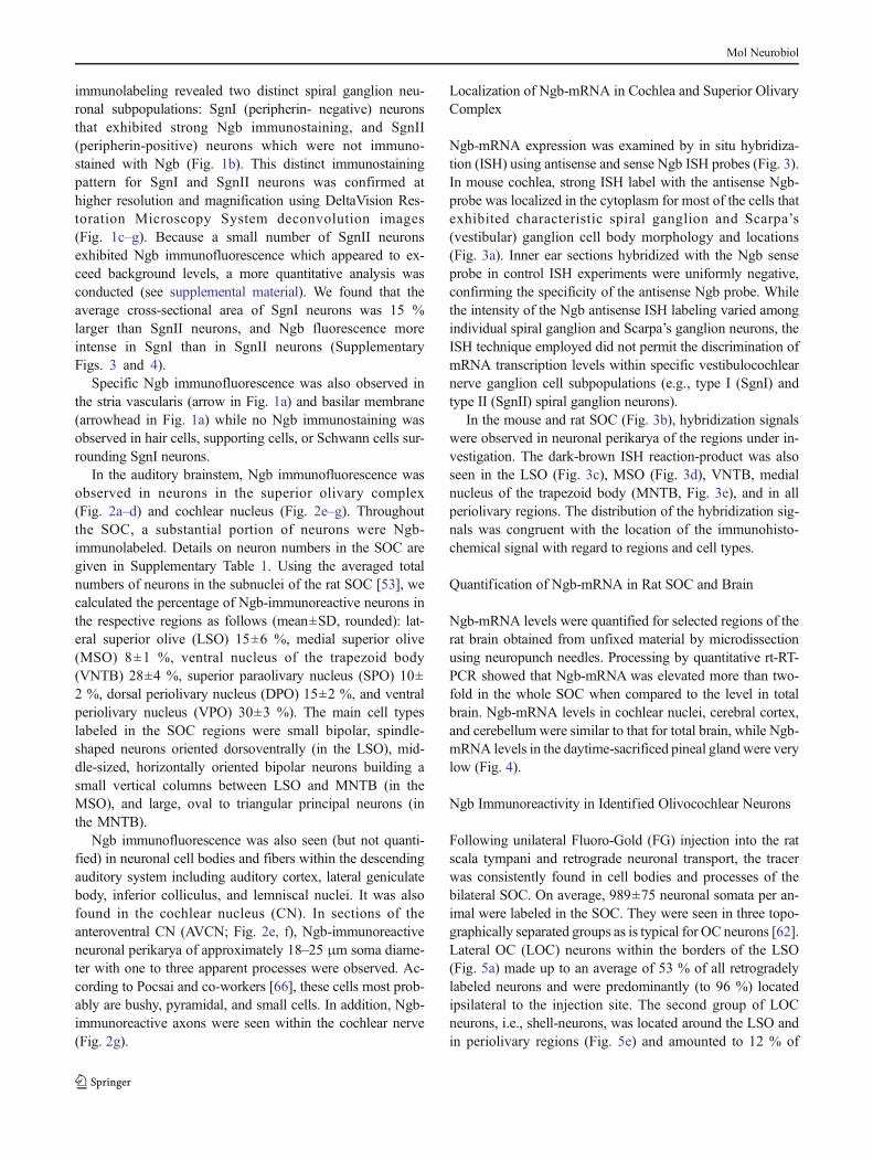
In the auditory brainstem, Ngb immunofluorescence wasobserved in neurons in the superior olivary complex(Fig. 2a–d) and cochlear nucleus (Fig. 2e–g). Throughoutthe SOC, a substantial portion of neurons were Ngb-immunolabeled. Details on neuron numbers in the SOC aregiven in Supplementary Table 1. Using the averaged totalnumbers of neurons in the subnuclei of the rat SOC [53], wecalculated the percentage of Ngb-immunoreactive neurons inthe respective regions as follows (mean±SD, rounded): lat-eral superior olive (LSO) 15±6 %, medial superior olive(MSO) 8±1 %, ventral nucleus of the trapezoid body(VNTB) 28±4 %, superior paraolivary nucleus (SPO) 10±2 %, dorsal periolivary nucleus (DPO) 15±2 %, and ventralperiolivary nucleus (VPO) 30±3 %). The main cell typeslabeled in the SOC regions were small bipolar, spindle-shaped neurons oriented dorsoventrally (in the LSO), mid-dle-sized, horizontally oriented bipolar neurons building asmall vertical columns between LSO and MNTB (in theMSO), and large, oval to triangular principal neurons (inthe MNTB).
Ngb immunofluorescence was also seen (but not quanti-fied) in neuronal cell bodies and fibers within the descendingauditory system including auditory cortex, lateral geniculatebody, inferior colliculus, and lemniscal nuclei. It was alsofound in the cochlear nucleus (CN). In sections of theanteroventral CN (AVCN; Fig. 2e, f), Ngb-immunoreactiveneuronal perikarya of approximately 18–25 μm soma diame-ter with one to three apparent processes were observed. Ac-cording to Pocsai and co-workers [66], these cells most prob-ably are bushy, pyramidal, and small cells. In addition, Ngb-immunoreactive axons were seen within the cochlear nerve(Fig. 2g).
Localization of Ngb-mRNA in Cochlea and Superior OlivaryComplex
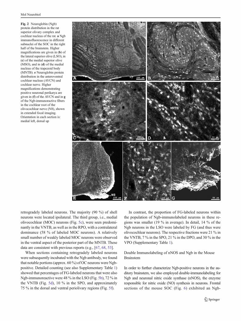
Ngb-mRNA expression was examined by in situ hybridiza-tion (ISH) using antisense and sense Ngb ISH probes (Fig. 3).In mouse cochlea, strong ISH label with the antisense Ngb-probe was localized in the cytoplasm for most of the cells thatexhibited characteristic spiral ganglion and Scarpa’s(vestibular) ganglion cell body morphology and locations(Fig. 3a). Inner ear sections hybridized with the Ngb senseprobe in control ISH experiments were uniformly negative,confirming the specificity of the antisense Ngb probe. Whilethe intensity of the Ngb antisense ISH labeling varied amongindividual spiral ganglion and Scarpa’s ganglion neurons, theISH technique employed did not permit the discrimination ofmRNA transcription levels within specific vestibulocochlearnerve ganglion cell subpopulations (e.g., type I (SgnI) andtype II (SgnII) spiral ganglion neurons).
In the mouse and rat SOC (Fig. 3b), hybridization signalswere observed in neuronal perikarya of the regions under in-vestigation. The dark-brown ISH reaction-product was alsoseen in the LSO (Fig. 3c), MSO (Fig. 3d), VNTB, medialnucleus of the trapezoid body (MNTB, Fig. 3e), and in allperiolivary regions. The distribution of the hybridization sig-nals was congruent with the location of the immunohisto-chemical signal with regard to regions and cell types.
Quantification of Ngb-mRNA in Rat SOC and Brain
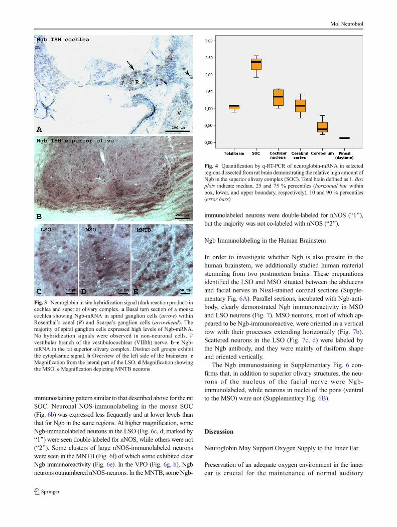
Ngb-mRNA levels were quantified for selected regions of therat brain obtained from unfixed material by microdissectionusing neuropunch needles. Processing by quantitative rt-RT-PCR showed that Ngb-mRNA was elevated more than two-fold in the whole SOC when compared to the level in totalbrain. Ngb-mRNA levels in cochlear nuclei, cerebral cortex,and cerebellum were similar to that for total brain, while Ngb-mRNA levels in the daytime-sacrificed pineal gland were verylow (Fig. 4).
Ngb Immunoreactivity in Identified Olivocochlear Neurons
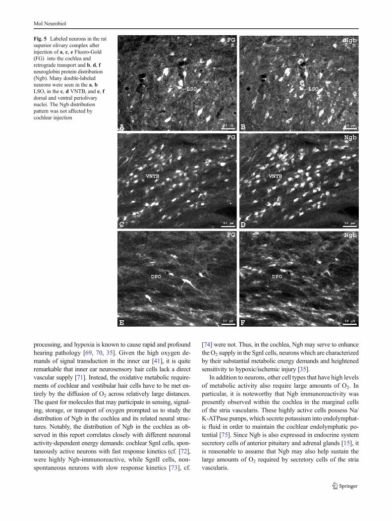
Following unilateral Fluoro-Gold (FG) injection into the ratscala tympani and retrograde neuronal transport, the tracerwas consistently found in cell bodies and processes of thebilateral SOC. On average, 989±75 neuronal somata per an-imal were labeled in the SOC. They were seen in three topo-graphically separated groups as is typical for OC neurons [62].Lateral OC (LOC) neurons within the borders of the LSO(Fig. 5a) made up to an average of 53 % of all retrogradelylabeled neurons and were predominantly (to 96 %) locatedipsilateral to the injection site. The second group of LOCneurons, i.e., shell-neurons, was located around the LSO andin periolivary regions (Fig. 5e) and amounted to 12 % of
Mol Neurobiol
retrogradely labeled neurons. The majority (90 %) of shellneurons were located ipsilateral. The third group, i.e., medialolivocochlear (MOC) neurons (Fig. 5c), were seen predomi-nantly in the VNTB, as well as in the RPO, with a contralateraldominance (58 % of labeled MOC neurons). A relativelysmall number of weakly labeled MOC neurons were observedin the ventral aspect of the posterior part of the MNTB. Thesedata are consistent with previous reports (e.g., [67, 68, 53].
When sections containing retrogradely labeled neuronswere subsequently incubated with the Ngb antibody, we foundthat notable portions (approx. 60%) of OC neurons were Ngb-positive. Detailed counting (see also Supplementary Table 1)showed that percentages of FG-labeled neurons that were alsoNgb-immunoreactive were 48% in the LSO (Fig. 5b), 72% inthe VNTB (Fig. 5d), 10 % in the SPO, and approximately75 % in the dorsal and ventral periolivary regions (Fig. 5f).
In contrast, the proportion of FG-labeled neurons withinthe population of Ngb-immunolabeled neurons in these re-gions was smaller (19 % in average). In detail, 14 % of theNgb neurons in the LSO were labeled by FG (and thus wereolivocochlear neurons). The respective fractions were 21 % inthe VNTB, 7 % in the SPO, 21% in the DPO, and 30 % in theVPO (Supplementary Table 1).
Double Immunolabeling of nNOS and Ngb in the MouseBrainstem
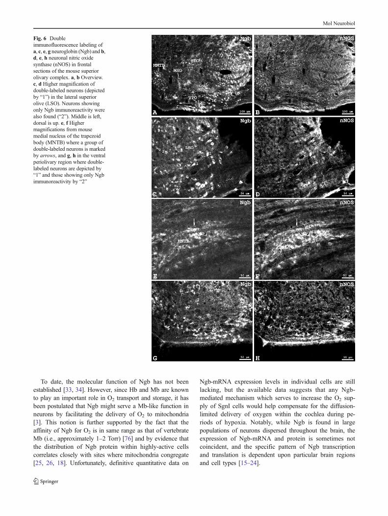
In order to further characterize Ngb-positive neurons in the au-ditory brainstem, we also employed double-immunolabeling forNgb and neuronal nitric oxide synthase (nNOS), the enzymeresponsible for nitric oxide (NO) synthesis in neurons. Frontalsections of the mouse SOC (Fig. 6) exhibited an Ngb-
Fig. 2 Neuroglobin (Ngb)protein distribution in the ratsuperior olivary complex andcochlear nucleus of the rat. a Ngbimmunofluorescence in differentsubnuclei of the SOC in the righthalf of the brainstem. Highermagnifications are given in (b) ofthe lateral superior olive (LSO), in(c) of the medial superior olive(MSO), and in (d) of the medialnucleus of the trapezoid body(MNTB). e Neuroglobin proteindistribution in the anteroventralcochlear nucleus (AVCN) andcochlear nerve. Highermagnifications demonstratingpositive neuronal perikarya aregiven in (f) of the AVCN and in gof the Ngb-immunoractive fibersin the cochlear root of theolivocochlear nerve (N8), shownin extended focal imaging.Orientation in each section is:medial left, dorsal up
Mol Neurobiol
immunostaining pattern similar to that described above for the ratSOC. Neuronal NOS-immunolabeling in the mouse SOC(Fig. 6b) was expressed less frequently and at lower levels thanthat for Ngb in the same regions. At higher magnification, someNgb-immunolabeled neurons in the LSO (Fig. 6c, d; marked byB1^) were seen double-labeled for nNOS, while others were not(B2^). Some clusters of large nNOS-immunolabeled neuronswere seen in the MNTB (Fig. 6f) of which some exhibited clearNgb immunoreactivity (Fig. 6e). In the VPO (Fig. 6g, h), Ngbneurons outnumbered nNOS-neurons. In theMNTB, someNgb-
immunolabeled neurons were double-labeled for nNOS (B1^),but the majority was not co-labeled with nNOS (B2^).
Ngb Immunolabeling in the Human Brainstem
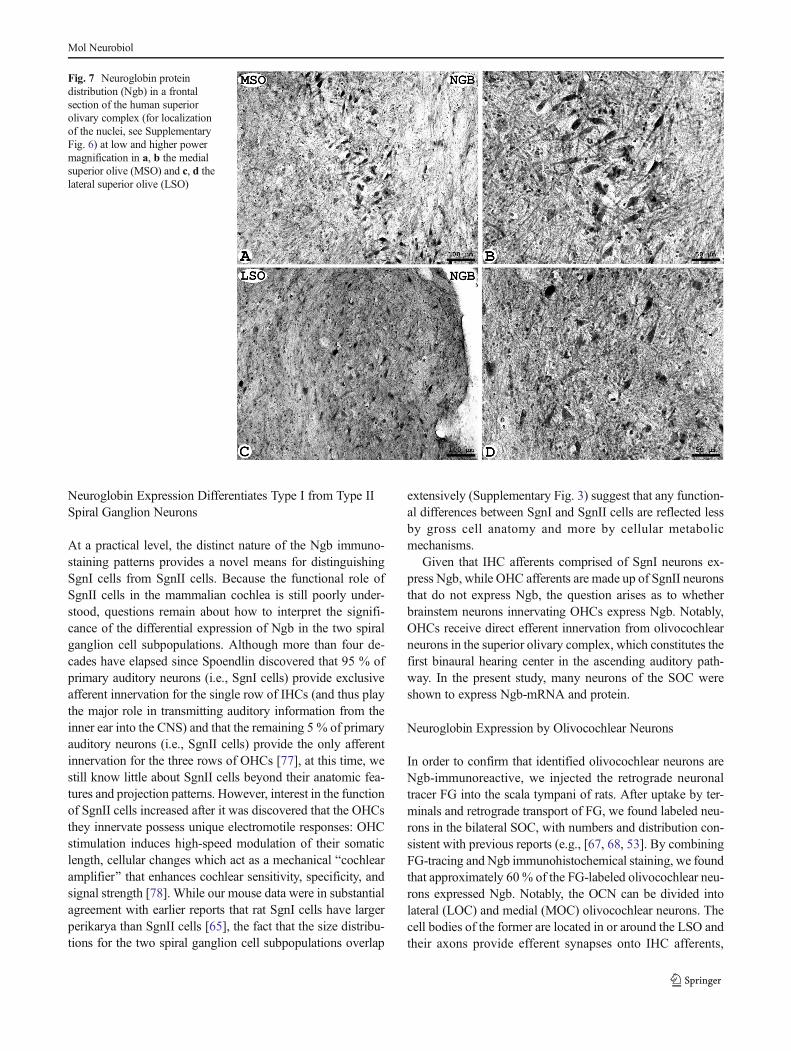
In order to investigate whether Ngb is also present in thehuman brainstem, we additionally studied human materialstemming from two postmortem brains. These preparationsidentified the LSO and MSO situated between the abducensand facial nerves in Nissl-stained coronal sections (Supple-mentary Fig. 6A). Parallel sections, incubated with Ngb-anti-body, clearly demonstrated Ngb immunoreactivity in MSOand LSO neurons (Fig. 7). MSO neurons, most of which ap-peared to be Ngb-immunoreactive, were oriented in a verticalrow with their processes extending horizontally (Fig. 7b).Scattered neurons in the LSO (Fig. 7c, d) were labeled bythe Ngb antibody, and they were mainly of fusiform shapeand oriented vertically.
The Ngb immunostaining in Supplementary Fig. 6 con-firms that, in addition to superior olivary structures, the neu-rons of the nucleus of the facial nerve were Ngb-immunolabeled, while neurons in nuclei of the pons (ventralto the MSO) were not (Supplementary Fig. 6B).
Discussion
Neuroglobin May Support Oxygen Supply to the Inner Ear
Preservation of an adequate oxygen environment in the innerear is crucial for the maintenance of normal auditory
Fig. 3 Neuroglobin in situ hybridization signal (dark reaction product) incochlea and superior olivary complex. a Basal turn section of a mousecochlea showing Ngb-mRNA in spiral ganglion cells (arrow) withinRosenthal’s canal (R) and Scarpa’s ganglion cells (arrowhead). Themajority of spiral ganglion cells expressed high levels of Ngb-mRNA.No hybridization signals were observed in non-neuronal cells. Vvestibular branch of the vestibulocochlear (VIIIth) nerve. b–e Ngb-mRNA in the rat superior olivary complex. Distinct cell groups exhibitthe cytoplasmic signal. b Overview of the left side of the brainstem. cMagnification from the lateral part of the LSO. dMagnification showingthe MSO. e Magnification depicting MNTB neurons
Fig. 4 Quantification by q-RT-PCR of neuroglobin-mRNA in selectedregions dissected from rat brain demonstrating the relative high amount ofNgb in the superior olivary complex (SOC). Total brain defined as 1. Boxplots indicate median, 25 and 75 % percentiles (horizontal bar withinbox, lower, and upper boundary, respectively), 10 and 90 % percentiles(error bars)
Mol Neurobiol
processing, and hypoxia is known to cause rapid and profoundhearing pathology [69, 70, 35]. Given the high oxygen de-mands of signal transduction in the inner ear [41], it is quiteremarkable that inner ear neurosensory hair cells lack a directvascular supply [71]. Instead, the oxidative metabolic require-ments of cochlear and vestibular hair cells have to be met en-tirely by the diffusion of O2 across relatively large distances.The quest for molecules that may participate in sensing, signal-ing, storage, or transport of oxygen prompted us to study thedistribution of Ngb in the cochlea and its related neural struc-tures. Notably, the distribution of Ngb in the cochlea as ob-served in this report correlates closely with different neuronalactivity-dependent energy demands: cochlear SgnI cells, spon-taneously active neurons with fast response kinetics (cf. [72],were highly Ngb-immunoreactive, while SgnII cells, non-spontaneous neurons with slow response kinetics [73], cf.
[74] were not. Thus, in the cochlea, Ngb may serve to enhancethe O2 supply in the SgnI cells, neurons which are characterizedby their substantial metabolic energy demands and heightenedsensitivity to hypoxic/ischemic injury [35].
In addition to neurons, other cell types that have high levelsof metabolic activity also require large amounts of O2. Inparticular, it is noteworthy that Ngb immunoreactivity waspresently observed within the cochlea in the marginal cellsof the stria vascularis. These highly active cells possess Na/K-ATPase pumps, which secrete potassium into endolymphat-ic fluid in order to maintain the cochlear endolymphatic po-tential [75]. Since Ngb is also expressed in endocrine systemsecretory cells of anterior pituitary and adrenal glands [15], itis reasonable to assume that Ngb may also help sustain thelarge amounts of O2 required by secretory cells of the striavascularis.
Fig. 5 Labeled neurons in the ratsuperior olivary complex afterinjection of a, c, e Fluoro-Gold(FG) into the cochlea andretrograde transport and b, d, fneuroglobin protein distribution(Ngb). Many double-labeledneurons were seen in the a, bLSO, in the c, d VNTB, and e, fdorsal and ventral periolivarynuclei. The Ngb distributionpattern was not affected bycochlear injection
Mol Neurobiol
To date, the molecular function of Ngb has not beenestablished [33, 34]. However, since Hb and Mb are knownto play an important role in O2 transport and storage, it hasbeen postulated that Ngb might serve a Mb-like function inneurons by facilitating the delivery of O2 to mitochondria[3]. This notion is further supported by the fact that theaffinity of Ngb for O2 is in same range as that of vertebrateMb (i.e., approximately 1–2 Torr) [76] and by evidence thatthe distribution of Ngb protein within highly-active cellscorrelates closely with sites where mitochondria congregate[25, 26, 18]. Unfortunately, definitive quantitative data on
Ngb-mRNA expression levels in individual cells are stilllacking, but the available data suggests that any Ngb-mediated mechanism which serves to increase the O2 sup-ply of SgnI cells would help compensate for the diffusion-limited delivery of oxygen within the cochlea during pe-riods of hypoxia. Notably, while Ngb is found in largepopulations of neurons dispersed throughout the brain, theexpression of Ngb-mRNA and protein is sometimes notcoincident, and the specific pattern of Ngb transcriptionand translation is dependent upon particular brain regionsand cell types [15–24].
Fig. 6 Doubleimmunofluorescence labeling ofa, c, e, g neuroglobin (Ngb) and b,d, e, h neuronal nitric oxidesynthase (nNOS) in frontalsections of the mouse superiorolivary complex. a, b Overview.c, d Higher magnification ofdouble-labeled neurons (depictedby “1”) in the lateral superiorolive (LSO). Neurons showingonly Ngb immunoreactivity werealso found (“2”). Middle is left,dorsal is up. e, f Highermagnifications from mousemedial nucleus of the trapezoidbody (MNTB) where a group ofdouble-labeled neurons is markedby arrows, and g, h in the ventralperiolivary region where double-labeled neurons are depicted by“1” and those showing only Ngbimmunoreactivity by “2”
Mol Neurobiol
Neuroglobin Expression Differentiates Type I from Type IISpiral Ganglion Neurons
At a practical level, the distinct nature of the Ngb immuno-staining patterns provides a novel means for distinguishingSgnI cells from SgnII cells. Because the functional role ofSgnII cells in the mammalian cochlea is still poorly under-stood, questions remain about how to interpret the signifi-cance of the differential expression of Ngb in the two spiralganglion cell subpopulations. Although more than four de-cades have elapsed since Spoendlin discovered that 95 % ofprimary auditory neurons (i.e., SgnI cells) provide exclusiveafferent innervation for the single row of IHCs (and thus playthe major role in transmitting auditory information from theinner ear into the CNS) and that the remaining 5 % of primaryauditory neurons (i.e., SgnII cells) provide the only afferentinnervation for the three rows of OHCs [77], at this time, westill know little about SgnII cells beyond their anatomic fea-tures and projection patterns. However, interest in the functionof SgnII cells increased after it was discovered that the OHCsthey innervate possess unique electromotile responses: OHCstimulation induces high-speed modulation of their somaticlength, cellular changes which act as a mechanical Bcochlearamplifier^ that enhances cochlear sensitivity, specificity, andsignal strength [78]. While our mouse data were in substantialagreement with earlier reports that rat SgnI cells have largerperikarya than SgnII cells [65], the fact that the size distribu-tions for the two spiral ganglion cell subpopulations overlap
extensively (Supplementary Fig. 3) suggest that any function-al differences between SgnI and SgnII cells are reflected lessby gross cell anatomy and more by cellular metabolicmechanisms.
Given that IHC afferents comprised of SgnI neurons ex-press Ngb, while OHC afferents are made up of SgnII neuronsthat do not express Ngb, the question arises as to whetherbrainstem neurons innervating OHCs express Ngb. Notably,OHCs receive direct efferent innervation from olivocochlearneurons in the superior olivary complex, which constitutes thefirst binaural hearing center in the ascending auditory path-way. In the present study, many neurons of the SOC wereshown to express Ngb-mRNA and protein.
Neuroglobin Expression by Olivocochlear Neurons
In order to confirm that identified olivocochlear neurons areNgb-immunoreactive, we injected the retrograde neuronaltracer FG into the scala tympani of rats. After uptake by ter-minals and retrograde transport of FG, we found labeled neu-rons in the bilateral SOC, with numbers and distribution con-sistent with previous reports (e.g., [67, 68, 53]. By combiningFG-tracing and Ngb immunohistochemical staining, we foundthat approximately 60% of the FG-labeled olivocochlear neu-rons expressed Ngb. Notably, the OCN can be divided intolateral (LOC) and medial (MOC) olivocochlear neurons. Thecell bodies of the former are located in or around the LSO andtheir axons provide efferent synapses onto IHC afferents,
Fig. 7 Neuroglobin proteindistribution (Ngb) in a frontalsection of the human superiorolivary complex (for localizationof the nuclei, see SupplementaryFig. 6) at low and higher powermagnification in a, b the medialsuperior olive (MSO) and c, d thelateral superior olive (LSO)
Mol Neurobiol
whereby they modulate the glutamatergic IHC-afferent termi-nal synapses, and thus serve to regulate SgnI activity in re-sponse to sound stimulation (cf. [79].
In contrast to LOCs, theMOC neuron cell bodies are locatedin the ventral nucleus of the trapezoid body (VNTB) and rostralperiolivary area (RPO), and their axons provide efferent synap-ses directly onto the OHCs [80, 62, 81–83], for review see [84].MOC activity has been shown to elicit outer hair cell mechan-ical effects, which modulate otoacoustic emissions and canprovide protection against acoustic injury (cf. [85, 86]. In thisstudy, we found that both LOC and MOC neurons expressNgb. Given the important role of the SOC in protecting haircells from sound-induced damage, it may be assumed that Ngbexpression in LOC andMOC neurons that have high dischargerates [87] and energy demands would help them physiological-ly during periods of noise-induced oxidative stress.
Possible Interaction Between Neuroglobin and Nitric Oxide
The specialized anatomy and physiology of the cochlea and itsrelated neural structures may provide unique insights into thepossible function of Ngb. For instance, it has been suggestedthat Ngb might scavenge nitric oxide (NO) [88, 89], which isproduced following the activation of nitric oxide synthase(NOS) during hypoxia, and other forms of environmentalstress [2].
The possibility of NO scavenging by Ngb is supported byearlier reports of neuronal NOS (nNOS) in the peripheral andcentral auditory system [90, 53, 91], and our evidence for theco-expression of nNOS [53] and Ngb (this report) in the ma-jority of spiral ganglion neurons (i.e., SgnI cells), and inmouse and rat SOC neurons (this report). In addition, thereis also an evidence of a substantial overlap in the constitutiveexpression patterns for Ngb [15] and nNOS [92] in non-auditory regions of the brain.
Finally, further evidence of possible interactions betweenNgb and NO in the central nervous system has previouslybeen provided by our group (cf. [34, 93], and by the observedco-expression of Ngb and nNOS in the rat hypothalamus [94].
Cellular Distribution and Localization of Neuroglobin
The present results demonstrate, by means of variousmethods, that neuroglobin is present in neuronal cells of thecochlea and the auditory brainstem. Notably, Ngbimmunolabeling was not observed in Schwann cells (i.e.,myelin-producing cells enveloping SgnI cells) or CNS astro-cytes. Ngb expression in CNS glial cells has only been ob-served in species which live in oxygen-challenged environ-ments such as the subterranean mole rat Spalax, as well asseals and whales [24, 18, 20].
The comparison between the immunohistochemical andISH-results revealed substantial agreement between the Ngb-
immunostaining and mRNA-expression patterns in the supe-rior olivary complex of the brainstem (this report) in spiralganglion neurons in the present and in an earlier study [51]as well as in Scarpa’s ganglion neurons (this report). However,the presence of Ngb protein in the absence of antisensemRNA-Ngb-hybridization in stria vascularis and the basilarmembrane suggests that Ngb either is transcribed in thesetissues at levels below the sensitivity of our ISH assays or itis transported to these sites after translation. In addition, therelatively weak Ngb protein bands from cochlea lysates (seeSupplementary Figs. 1 and 2) are probably due to the fact thatneurons constitute only a relatively small fraction of the totalcellular volume of the cochlea. Thus, it can be assumed thatneurons in the cochlea (and in brain regions with relativelyfew neuronal somata) express considerably more Ngb-mRNAand protein than would be expected from the estimates obtain-ed using the whole cochlea or brain tissue.
It should be noted that in the SOC 10–30 % of the neuronsare Ngb-positive. Nineteen percent of these Ngb-neuronswere olivocochlear neurons. It is currently unknown whetherNgb-positive SOC neurons possess a common feature such ashigh energy demand and/or whether they are anatomically orphysiologically connected, but it appears that the SOCwith itsvarious functions in the hearing processes has the unspecifiedadvantage of Ngb-producing neurons. Our quantitative rt-RT-PCR data determined that Ngb-mRNA levels were signifi-cantly higher in the SOC than those for total brain, whichfurther supports the hypothesis that Ngb may play a specialrole in the peripheral and central auditory nervous system.
In the cochlear nucleus (CN), Ngb-mRNA levels were inapproximately the same range as in total brain. Immunostain-ing with Ngb-antibody was confined to small and middle-sized somata scattered throughout the CN. Fibers of the co-chlear nerve (i.e., axons of spiral ganglion neurons) terminatein the CN, which serves as first relay center in the ascendingauditory pathway. From the variety of cell types previouslydescribed in rat CN, the observed cell distribution, soma size,and processes of Ngb-labeled neurons suggest that most werebushy cells, pyramidal cells, or small cells [66]. These types ofCN cells project centrally to the SOC, inferior colliculus (IC),and medial geniculate body (MGB) (cf. [61].
Ngb-immunoreactive neurons were observed in the IC,MGB, lemniscal nuclei, and auditory cortex as parts of thedescending auditory system, which, judging from cell shape,size, and distribution, project itself to the LSO, periolivaryregions, lateral lemniscus, and the IC. Although we did notquantify neuroglobin in these regions yet, it appears that theexpression levels did not exceed those in other brain regions.
Taken together, our cochlea and brainstem data show thatIHCs are contacted by Ngb-expressing SgnI afferent neurons,while OHCs are contacted by Ngb-expressing efferent neu-rons. Although we do not know yet what the local role of Ngbis, it is significant that both types of hair cell are contacted by
Mol Neurobiol
Ngb-expressing neurons. Thus, it is possible that Ngb mayplay an active role in the transport or storage of oxygen inauditory neurons, and that Ngb may also provide protectionfor the auditory nervous system under conditions when energydemands are high. In addition, Ngb may also act as signalmolecule in oxygen sensing. In any case, if Ngb-expressingafferent and efferent auditory system neurons do exert a directNgb-involving influence on cochlear hair cells, then the mech-anism would presumably involve Ngb-transport from neuro-nal soma to peripheral processes, in particular to the axonendings. The question as to whether Ngb is present in distalparts of neurons has not previously been discussed. Whilesome evidence of Ngb protein within axons can be found inthe figures from prior immunohistochemical studies of thedorsal root ganglion and in dorsal spinal cord [95] and aninvestigation of the central projections of the optic nerve[96], our present observations that Ngb-immunoreactivevestibulocochlear nerve axons project from the cochlea intothe brainstem cochlear nucleus (Fig. 2e, g) have confirmed forthe first time that Ngb can be transported within axonal pro-cesses over relatively long distances.
Finally, to extend our comparative understanding of Ngb ex-pression in the SOC, we included an investigation of brainstemslices from human postmortem brains. In frontal sections, thehuman SOC was located between abducens and facial nerves(see Supplementary Fig. 6), in agreement with the literature (cf.[97, 98]. Notably, within the human SOC, many MSO neurons,and scattered neurons in the LSO, were Ngb-immunoreactive,similar to the situation seen in both the mouse and rat. Otherneuronal groups at this brainstem level exhibited differentialNgb staining insofar as neurons of the facial nucleus were pos-itive while neurons of the nuclei of the pons were Ngb-negative.
Conclusions
Our study provides the first detailed analysis of Ngb expres-sion in the cochlea and superior olivary complex of the audi-tory brainstem. We demonstrate, by a variety of experimentalmethods, that Ngb is highly expressed in mouse and rat spiralganglion type I neurons, and in the SOC of rats, mice, andmen. Further studies, however, are required to elucidate thespecific role of Ngb within various sensory (e.g., visual, audi-tory, and vestibular) systems under normal and pathologicalconditions. Ngb has been shown to protect neurons fromhypoxic-ischemic injury [30, 31, 33, 34], which suggests thata better understanding of the role of Ngb in the inner ear andauditory brainstem may provide the basis for new clinicalapproaches to treat, prevent, and diagnose cochlear oxidativestress, a leading cause of sensorineural hearing loss (SNHL)for which, at present, no effective therapeutics exist.
Acknowledgments This work was supported by grants from the Na-tional Institutes of Health, National Institute for Deafness and Communi-cation Disorders grants DC00386 and DC02666, and the Research Ser-vice of the Department of Veterans Affairs (USA), from the Röttger-Stiftung and Hoffmann-Klose-Stiftung (Germany) and by the DeutscheForschungsgemeinschaft (Bu956/5 and Ha2103/3). We thank Dr.Thorsten Fink, Department of Pathology, HSK Wiesbaden, Germany,for providing the human brainstem specimens. The University of Califor-nia Cancer Center Biomedical Imaging Core is also thanked for theirimaging support. We thank Steve McMullen of the UCSD Cancer CenterDigital Imaging Shared Resource for his assistance in collecting thedeconvolution digital microscope images; Anuradha Desai of the Depart-ment of Surgery, University of California at San Diego and the VeteransAffairs Research Service, VA San Diego Healthcare System, CA, for hertechnical assistance with the Western blot assays; and Kunlin Jin andDavid A. Greenberg of Buck Institute for Age Research, Novato, CA,for their helpful discussions and comments.
Conflict of Interest The authors declare that they have no conflict ofinterest.
References
1. Weber RE, Vinogradov SN (2001) Nonvertebrate hemoglobins:functions and molecular adaptations. Physiol Rev 81(2):569–628
2. Wittenberg JB, Wittenberg BA (2003) Myoglobin functionreassessed. J Exp Biol 206(Pt 12):2011–2020
3. Burmester T, Weich B, Reinhardt S, Hankeln T (2000) A vertebrateglobin expressed in the brain. Nature 407:520–523
4. Trent JT, Hargrove MS (2002) A ubiquitously expressed humanhexacoordinate hemoglobin. J Biol Chem 277:19538–19545
5. Kawada N, Kristensen DB, Asahina K, Nakatani K, Minamiyama Y,Seki S, Yoshizato K (2001) Characterization of a stellate cellactivation-associated protein (STAP) with peroxidase activity foundin rat hepatic stellate cells. J Biol Chem 276:25318–25323
6. Burmester T, Ebner B, Weich B, Hankeln T (2002) Cytoglobin: anovel globin type ubiquitously expressed in vertebrate tissues. MolBiol Evol 19(4):416–421
7. Kugelstadt D, Haberkamp M, Hankeln T, Burmester T (2004)Neuroglobin, cytoglobin, and a novel, eye-specific globin fromchicken. Biochem Biophys Res Commun 325:719–725
8. Roesner A, Fuchs C, Hankeln T, Burmester T (2005) A globin geneof ancient evolutionary origin in lower vertebrates: evidence for twodistinct globin families in animals. Mol Biol Evol 22:12–20
9. Fuchs C, Burmester T, Hankeln T (2006) The amphibian globin generepertoire as revealed by the Xenopus genome. Cytogenet GenomeRes 112(3–4):296–306
10. Hoogewijs D, Ebner B, Germani F, Hoffmann FG, Fabrizius A,Moens L, Burmester T, Dewilde S, Storz JF, Vinogradov SN,Hankeln T (2012) Androglobin: a chimeric globin in metazoans thatis preferentially expressed in mammalian testes.Mol Biol Evol 29(4):1105–1114
11. Burmester T, Hankeln T (2014) Function and evolution of vertebrateglobins. Acta Physiol 211:501–514
12. Schwarze K, Burmester T (2013) Conservation of globin genes in theBliving fossil^ Latimeria chalumnae and reconstruction of the evolu-tion of the vertebrate globin family. Biochim Biophys Acta 1834(9):1801–1812
13. Burmester T, Haberkamp M, Mitz S, Roesner A, Schmidt M, EbnerB, Gerlach F, Fuchs C, Hankeln T (2004) Neuroglobin andcytoglobin: genes, proteins and evolution. IUBMB Life 56:703–707
14. Geuens E, Brouns I, Flamez D, DeWilde S, Timmermans JP, MoensL (2003) A globin in the nucleus! J Biol Chem 278:30417–30420
Mol Neurobiol
15. Reuss S, Saaler-Reinhardt S, Weich B, Wystub S, Reuss MH,Burmester T, Hankeln T (2002) Expression analysis of neuroglobinmRNA in rodent tissues. Neuroscience 115:645–656
16. Wystub S, Laufs T, Schmidt M, Burmester T, Maas U, Saaler-Reinhardt S, Hankeln T, Reuss S (2003) Localisation of neuroglobinprotein in the mouse brain. Neurosci Lett 346:114–116
17. Laufs TL, Wystub S, Reuss S, Burmester T, SaalerReinhardt S,Hankeln T (2004) Neuron-specific expression of neuroglobin inmammals. Neurosci Lett 362:83–86
18. Mitz SA, Reuss S, Folkow LP, Blix AS, Ramirez JM, Hankeln T,Burmester T (2009) When the brain goes diving: glial oxidative me-tabolism may confer hypoxia tolerance to the seal brain.Neuroscience 163:552–560
19. Hundahl CA, Allen GC, Nyengaard JR, Dewilde S, Carter BD,Kelsen J, Hay-Schmidt A (2008) Neuroglobin in the rat brain: local-ization. Neuroendocrinology 88:173–182
20. Schneuer M, Flachsbarth S, Czech-Damal NU, Folkow LP, SiebertU, Burmester T (2012) Neuroglobin of seals andwhales: evidence fora divergent role in the diving brain. Neuroscience 223:35–44
21. Mammen PPA, Shelton JM, Goetsch SC, Williams SC, RichardsonJA, GarryMG, Garry DJ (2002) Neuroglobin, a novel member of theglobin family, is expressed in focal regions of the brain. J HistochemCytochem 50:1591–1598
22. Hundahl CA, Allen GC, Hannibal J, Kjaer K, Rehfeld JF, Dewilde S,Nyengaard JR, Kelsen J, Hay-Schmidt A (2010) Anatomical charac-terization of cytoglobin and neuroglobin mRNA and protein expres-sion in the mouse brain. Brain Res 1331:58–73
23. Schmidt M, Laufs T, Reuss S, Hankeln T, Burmester T (2005)Divergent distribution of cytoglobin and neuroglobin in the murineeye. Neurosci Lett 374:207–211
24. Avivi A, Gerlach F, Joel A, Reuss S, Burmester T, Nevo E, Hankeln T(2010) Neuroglobin, cytoglobin, and myoglobin contribute to hyp-oxia adaptation of the subterranean mole rat Spalax. Proc Natl AcadSci U S A 107(50):21570–21575
25. Schmidt M, Giessl A, Laufs T, Hankeln T, Wolfrum U, Burmester T(2003) How does the eye breathe?—Evidence for neuroglobin-mediated oxygen supply in the mammalian retina. J Biol Chem278:1932–1935
26. Bentmann A, Schmidt M, Reuss S, Wolfrum U, Hankeln T,Burmester T (2005) Divergent distribution in vascular and avascularmammalian retinae links neuroglobin to cellular respiration. J BiolChem 280:20660–20665
27. Ostojic J, Sakaguchi DS, de Lathouder Y, Hargrove MS, Trent JT3rd, Kwon YH, Kardon RH, Kuehn MH, Betts DM, Grozdanic S(2006) Neuroglobin and cytoglobin: oxygen-binding proteins in ret-inal neurons. Invest Ophthalmol Vis Sci 47(3):1016–1023
28. Rajendram R, Rao NA (2007) Neuroglobin in normal retina andretina from eyes with advanced glaucoma. Br J Ophthalmol 91(5):663–666
29. Roesner A, Mitz SA, Hankeln T, Burmester T (2008) Globins andhypoxia adaptation in the goldfish, Carassius auratus. FEBS J275(14):3633–3643
30. Sun Y, Jin K, Mao XO, Zhu Y, Greenberg DA (2001) Neuroglobin isupregulated by and protects neurons from hypoxic-ischemic injury.Proc Natl Acad Sci U S A 98:15306–15311
31. Sun YJ, Jin KL, Peel A, Mao XO, Xie L, Greenberg DA (2003)Neuroglobin protects the brain from experimental stroke in vivo.Proc Natl Acad Sci U S A 100:3497–3500
32. Raida Z, Hundahl CA, Kelsen J, Nyengaard JR, Hay-Schmidt A(2012) Reduced infarct size in neuroglobin-null mice after experi-mental stroke in vivo. Exp Translat Stroke Med 4(1):15
33. Burmester T, Hankeln T (2009) What is the function of neuroglobin?J Exp Biol 212(Pt 10):1423–1428
34. Hankeln T, Ebner B, Fuchs C, Gerlach F, Haberkamp M, Laufs TL,Roesner A, Schmidt M, Weich B, Wystub S, Saaler-Reinhardt S,Reuss S, Bolognesi M, Sanctis DD, Marden MC, Kiger L, Moens
L, Dewilde S, Nevo E, Avivi A, Weber RE, Fago A, Burmester T(2005) Neuroglobin and cytoglobin in search of their role in thevertebrate globin family. J Inorg Biochem 99:110–119
35. Koga K, Hakuba N, Watanabe F, Shudou M, Nakagawa T, Gyo K(2003) Transient cochlear ischemia causes delayed cell death in theorgan of Corti: an experimental study in gerbils. J Comp Neurol 456:105–111
36. Brown MC, Santos-Sacchi J (2013) Audition. In: Squire L, Berg D,Bloom FE, du Lac S, Ghosh A (eds) Fundamental Neuroscience, 4thedn. Elsevier, Amsterdam, pp 553–576
37. Slepecky NB (1996) Structure of the mammalian cochlea. In: DallosP, Popper AN, Fay RR (eds) The Cochlea, vol 8, Springer Handbookof Auditory Research. Springer, New York, pp 44–129
38. Dallos P (1996) Overview: cochlear neurobiology. In: Dallos P,Popper AN, Fay RR (eds) The Cochlea, vol 8. Springer Handbookof Auditory Research. Springer, New York, pp 1–43
39. Møller A (2007) Hearing: anatomy, physiology, and disorders of theauditory system, 2nd edn. Elsevier, Amsterdam
40. Raphael Y, Altschuler RA (2003) Structure and innervation of thecochlea. Brain Res Bull 60:397–422
41. Puschner B, Schacht J (1997) Energy metabolism in cochlear outerhair cells in vitro. Hear Res 114(1–2):102–106
42. Ryan AF, Goodwin P, Woolf NK, Sharp F (1982) Auditory stimula-tion alters the pattern of 2-deoxyglucose uptake in the inner ear. BrainRes 234(2):213–225
43. Goodwin PC, Ryan AF, Sharp FR, Woolf NK, Davidson TM (1984)Cochlear deoxyglucose uptake: relationship to stimulus intensity.Hearing Res 15(3):215–224
44. Canlon B, Anniko M (1987) The postnatal development of stimulat-ed deoxyglucose uptake into the mouse cochlea and the inferiorcolliculus. Archives of oto-rhino-laryngology 244(5):273–277
45. Schousboe A, Booher J, Hertz L (1970) Content of ATP in cultivatedneurons and astrocytes exposed to balanced and potassium-rich me-dia. J Neurochem 17(10):1501–1504
46. Thalmann R, Thalmann I, Comegys TH (1972) Quantitative cyto-chemistry of the organ of Corti. Dissection, weight determination andanalysis of single outer hair cells. Laryngoscope 82(11):2059–2078
47. Thalmann R, Miyoshi T, Thalmann I (1972) The influence of ische-mia upon the energy reserves of inner ear tissues. Laryngoscope82(12):2249–2272
48. Scheibe F, Haupt H, Rothe E, Hache U (1981) On the glucose, py-ruvate, and lactate concentration of perilymph, blood, and cerebro-spinal fluid of unexposed and sound-exposed guinea pigs under ethylurethane anesthesia (author’s transl). Archives of oto-rhino-laryngology 233(1):89–97
49. Axelsson A (1988) Comparative anatomy of cochlear blood vessels.Am J Otolaryngol 9(6):278–290
50. Yu DY, Cringle SJ (2001) Oxygen distribution and consumptionwithin the retina in vascularised and avascular retinas and in animalmodels of retinal disease. Progr Retinal Eye Res 20(2):175–208
51. Lopez IA, Acuna D, Shahram Y, Mowlds D, Ngan AM,Rungvivatjarus T, Sharma Y, Edmond J (2010) Neuroglobin expres-sion in the cochlea of rat pups exposed to chronic very mild carbonmonoxide (25 ppm) in air during and after the prenatal period. BrainRes 1327:56–68
52. McLean IW, Nakane PK (1974) Periodate-lysine-paraformaldehydefixative. A new fixation for immunoelectron microscopy. JHistochem Cytochem 22:1077–1083
53. Riemann R, Reuss S (1999) Nitric oxide synthase in identifiedolivocochlear projection neurons in rat and guinea pig. Hear Res135:181–189
54. Reuss S, Disque-Kaiser U, Antoniou-Lipfert P, Najaf Gholi M,Riemann E, Riemann R (2009) Neurochemistry of olivocochlearneurons in the hamster. Anat Rec 292:461–471
55. Abercrombie M (1946) Estimation of nuclear population from mi-crotome sections. Anat Rec 94:239–247
Mol Neurobiol
56. Paxinos G, Franklin KBJ (2001) The mouse brain in stereotaxiccoordinates, 2nd edn. Academic, San Diego
57. Paxinos G, Watson C (2007) The rat brain in stereotaxic coordinates,4th edn. Academic, San Diego
58. Schwartz IR (1992) The superior olivary complex and lateral lemnis-cal nuclei. In: Webster DB, Popper AN, Fay RF (eds) TheMammalian Auditory Pathway: Neuroanatomy. Springer, NewYork, pp 117–167
59. Warr BW (1992) Organization of olivocochlear efferent systems inmammals. In: Webster DB, Popper AN, Fay RR (eds) The mamma-lian auditory pathway. Neuroanatomy Springer, New York, pp 410–448
60. Kulesza RJ, Vinuela A, Saldana E, Berrebi AS (2002) Unbiasedstereological estimates of neuron number in subcortical auditory nu-clei of the rat. Hear Res 168:12–24
61. MalmiercaMS, MerchanMA (2004) Auditory system. In: Paxinos G(ed) The rat nervous system, Thirdth edn. Elsevier Academic Press,Amsterdam, pp 997–1082
62. Vetter DE, Mugnaini E (1992) Distribution and dendritic features ofthree groups of rat olivocochlear neurons. A study with two retro-grade cholera toxin tracers. Anat Embryol 185:1–16
63. Hsu SM, Raine L, Fanger H (1981) Use of avidin-biotin-peroxidasecomplex (ABC) in immunoperoxidase techniques: a comparison be-tween ABC and unlabeled antibody (PAP) procedures. J HistochemCytochem 29:577–580
64. Berglund AM, Ryugo DK (1991) Neurofilament antibodies and spi-ral ganglion neurons of the mammalian cochlea. J Comp Neurol306(3):393–408
65. Hafidi A (1998) Peripherin-like immunoreactivity in type II spiralganglion cell body and projections. Brain Res 805:181–190
66. Pocsai K, Pal B, Pap P, Bakondi G, Kosztka L, Rusznak Z, Szucs G(2007) Rhodamine backfilling and confocal microscopy as a tool forthe unambiguous identification of neuronal cell types: a study of theneurones of the rat cochlear nucleus. Brain Res Bull 71(5):529–538
67. White JS, Warr WB (1983) The dual origins of the olivocochlearbundle in the albino rat. J Comp Neurol 219:203–214
68. Aschoff A, Ostwald J (1988) Distribution of cochlear efferents andolivo-collicular neurons in the brainstem of rat and guinea pig. Adouble labeling study with fluorescent tracers. Exp Brain Res 71:241–251
69. Sawada S, Mori N, Mount RJ, Harrison RV (2001) Differential vul-nerability of inner and outer hair cell systems to chronic mild hypoxiaand glutamate ototoxicity: insights into the cause of auditory neurop-athy. J Otolaryngol 30(2):106–114
70. Nakashima T, Naganawa S, Sone M, Tominaga M, Hayashi H,Yamamoto H, Liu XL, Nuttall AL (2003) Disorders of cochlearblood flow. Brain Res Rev 43:17–28
71. Axelsson A, Ryan A, Woolf N (1986) The early postnatal develop-ment of the cochlear vasculature in the gerbil. Acta Otolaryngol101(1–2):75–87
72. Taberner AM, Liberman MC (2005) Response properties of singleauditory nerve fibers in the mouse. J Neurophysiol 93(1):557–569
73. Robertson D (1984) Horseradish peroxidase injection of physiologi-cally characterized afferent and efferent neurones in the guinea pigspiral ganglion. Hear Res 15(2):113–121
74. Rusznak Z, Szucs G (2009) Spiral ganglion neurones: an overview ofmorphology, firing behaviour, ionic channels and function. PflugersArchiv: Eur J Physiol 457(6):1303–1325
75. Xia A, Kikuchi T, Hozawa K, Katori Y, Takasaka T (1999)Expression of connexin 26 and Na, K-ATPase in the developingmouse cochlear lateral wall: functional implications. Brain Res846:106–111
76. Dewilde S, Kiger L, Burmester T, Hankeln T, Baudin-Creuza V,Aerts T, Marden MC, Caubergs R, Moens L (2001) Biochemicalcharacterization and ligand binding properties of neuroglobin, a nov-el member of the globin family. J Biol Chem 276(42):38949–38955
77. Spoendlin H (1972) Innervation densities of the cochlea. ActaOtolaryngol 73(2):235–248
78. Brownell WE, Bader CR, Bertrand D, de Ribaupierre Y (1985)Evoked mechanical responses of isolated cochlear outer hair cells.Science 227(4683):194–196
79. Ruel J, Wang J, Rebillard G, Eybalin M, Lloyd R, Pujol R, Puel JL(2007) Physiology, pharmacology and plasticity at the inner hair cellsynaptic complex. Hear Res 227(1–2):19–27
80. Warr WB, Boche JB, Neely ST (1997) Efferent innervation of theinner hair cell region: origins and terminations of two lateralolivocochlear systems. Hear Res 108:89–111
81. HorvathM, Kraus KS, Illing RB (2000) Olivocochlear neurons send-ing axon collaterals into the ventral cochlear nucleus of the rat. JComp Neurol 422:95–105
82. Sanchez-Gonzalez MA, Warr WB, Lopez DE (2003) Anatomy ofolivocochlear neurons in the hamster studied with Fluoro-Gold.Hear Res 185:65–76
83. Hill JC, Prasher DK, Luxon LM (1997) Evidence for efferent effectson auditory afferent activity, and their functional relevance. ClinOtolaryngol Alied Sci 22(5):394–402
84. Guinan JJ Jr (2010) Cochlear efferent innervation and function. CurrOpin Otolaryngol Head Neck Surge 18(5):447–453
85. Wersinger E, Fuchs PA (2011)Modulation of hair cell efferents. HearRes 279(1–2):1–12
86. Guinan JJ Jr, Salt A, Cheatham MA (2012) Progress in cochlearphysiology after Bekesy. Hear Res 293(1–2):12–20
87. Liberman MC (1988) Response properties of cochlear efferent neu-rons: monaural vs. binaural stimulation and the effects of noise. JNeurophysiol 60(5):1779–1798
88. Brunori M, Giuffre A, Nienhaus K, Nienhaus GU, Scandurra FM,Vallone B (2005) Neuroglobin, nitric oxide, and oxygen: functionalpathways and conformational changes. Proc Natl Acad Sci U S A102:8483–8488
89. Brunori M, Vallone B (2007) Neuroglobin, seven years after. CellMol Life Sci 64:1259–1268
90. Fessenden JD, Altschuler RA, Seasholtz AF, Schacht J (1999) Nitricoxide/cyclic guanosine monophosphate pathway in the peripheraland central auditory system of the rat. J Comp Neurol 404:52–63
91. Zdanski CJ, Prazma J, Petrusz P, Grossman G, Raynor E, Smith TL,Pillsbury HC (1994) Nitric oxide synthase is an active enzyme in thespiral ganglion cells of the rat cochlea. Hear Res 79:39–47
92. Rodrigo J, Springall DR, Uttenthal O, Bentura ML, Abadia-MolinaF, Riveros-Moreno V, Martínez-Murillo R, Polak JM, Moncada S(1994) Localization of nitric oxide synthase in the adult rat brain.Philos Trans R Soc Lond B Biol Sci 345:175–221
93. Burmester T, Hankeln T (2008) Neuroglobin and other nerve hemo-globins. In: Bolognesi M, di Prisco G, Verde C (eds) DioxygenBinding and Sensing Proteins, vol 9. Protein Reviews. Springer,Milan, pp 211–222
94. Hundahl CA, Kelsen J, Dewilde S, Hay-Schmidt A (2008)Neuroglobin in the rat brain (II): co-localisation with neurotransmit-ters. Neuroendocrinology 88:183–198
95. Hankeln T, Wystub S, Laufs T, Schmidt M, Gerlach F, Saaler-Reinhardt S, Reuss S, Burmester T (2004) The cellular and subcel-lular localization of neuroglobin and cytoglobin—a clue to theirfunction? IUBMB Life 56(11–12):671–679
96. Lechauve C, Augustin S, Roussel D, Sahel JA, Corral-Debrinski M(2013) Neuroglobin involvement in visual pathways through the op-tic nerve. Biochim Biophys Acta 1834(9):1772–1778
97. Amunts K, Morosan P, Hilbig H, Zilles K (2012) Auditory system.In: Mai JK, Paxinos G (eds) The human nervous system, 3rd edn.Elsevier, Amsterdam, pp 1270–1300
98. Moore JK (2000) Organization of the human superior olivary com-plex. Microsc Res Tech 51:403–412
Mol Neurobiol