1.INTRODUCTION 9paduaresearch.cab.unipd.it/10063/1/Montagna_Aldo_Tesi.pdf · 1.3 Drosophila aa...
84
Transcript of 1.INTRODUCTION 9paduaresearch.cab.unipd.it/10063/1/Montagna_Aldo_Tesi.pdf · 1.3 Drosophila aa...


INDEX
1.INTRODUCTION ............................................................................................. 9
1.1 Mitochondrial network dynamics ............................................................. 11
Mechanisms of mitochondrial fusion ................................................................................. 11
Mechanisms of the mitochondrial fission ........................................................................... 13
Functions of mitochondrial dynamics ................................................................................. 15
1.2 Bioenergetics of mitochondria ................................................................ 18
Bioenergetic role of mitochondrial fusion .......................................................................... 21
Bioenergetic role of mitochondrial fission .......................................................................... 22
Adaptation of mitochondrial dynamics to bioenergetic conditions ................................... 23
1.2 Mitochondrial dynamics and disease ...................................................... 25
Autosomal Dominant Optic Atrophy (DOA) ........................................................................ 25
1.3 Drosophila as a model organism ............................................................ 28
Mitochondrial dynamics in Drosophila ............................................................................... 29
Drosophila as a model of primary defects of mitochondrial dynamincs: DOA and OPA1 .. 30
1.4 Genome Editing in Drosophila ................................................................ 33
CRISPR/Cas9 system ............................................................................................................ 33
Drosophila CRISPR system ................................................................................................... 35
Application of CRISPR/Cas9 system .................................................................................... 36
2.AIM OF WORK .............................................................................................. 39
3.METHODS .................................................................................................... 40
3.1 Molecular Biology ................................................................................... 40
Production of the chiRNAs .................................................................................................. 40
Production of the donor plasmids ...................................................................................... 40
Screening of adults for mutation ........................................................................................ 41
Transformation of chemiocompetent cells ......................................................................... 43
Drosophila genomic DNA extraction protocol .................................................................... 43
3.2 Drosophila genetics ................................................................................ 44
Embryo injection ................................................................................................................. 44
Generation of stable stocks ................................................................................................ 44
Drosophila strains used ....................................................................................................... 44

3.3 Microscopy ............................................................................................. 44
Immunohistochemistry ....................................................................................................... 44
Live imaging of mitochondrial network on muscles of Drosophila larvae .......................... 44
Mitochondria density analysis ............................................................................................ 45
Image analysis ..................................................................................................................... 45
3.4 Biochemical Assays ................................................................................ 45
Mitochondrial Respiration Assay ........................................................................................ 45
Activity of respiratory complexes ....................................................................................... 45
APPENDIX A: Stock and Solutions .................................................................. 48
APPENDIX B: Plasmids maps ......................................................................... 51
4. RESULTS ..................................................................................................... 52
4.1 Drosophila OPA1 MUTANTS: GENERATION AND
PHENOTYPIC CHARACTERIZATION ......................................................... 52
Generation of mutants........................................................................................................ 52
Phenotypic characterization of CRISPR/Cas9 dOPA1 mutants ........................................... 57
4.2 ANALYSIS OF MITOCHONDRIAL DYNAMICS ..................................... 59
Mitochondrial morphology and distribution in Drosophila larvae nerves ......................... 60
Mitochondrial morphology in Drosophila larvae muscles .................................................. 63
4.3 Analysis of the mitochondria functions .................................................... 65
Mitochondrial respiration in Drosophila larvae homogenate ............................................ 65
Analysis of the activity of respiratory complexes ............................................................... 66
5. DISCUSSION ............................................................................................... 68
6. Reference ..................................................................................................... 72

1
Summary Mitochondria are very dynamic organelles with a crucial role in life and death of
eukaryotic cells. These organelles regulate cellular energy generation, calcium
and redox homeostasis, and apoptosis. To perform the cellular functions
effectively, mitochondria continuously change their structure and morphology
through protein machineries controlling fission and fusion process
(mitochondrial dynamics). Strong evidence has emerged to implicate disturbed
mitochondrial fusion and fission as central pathological components
underpinning a number of childhood and adult-onset neurodegenerative
disorders. Several proteins that regulate the morphology of the mitochondrial
network have been identified, the most widely studied of which are Optic
Atrophy 1 (OPA1), Mitofusin1 and 2 (Mfn1 and 2) and Dynamin Related Protein
1 (DRP1).
OPA1 is a ubiquitously expressed dynamin-like GTPase in the inner
mitochondrial membrane. It plays important roles in mitochondrial fusion,
apoptosis, reactive oxygen species (ROS) and ATP production. Mutations of
OPA1 result in autosomal Dominant Optic Atrophy (DOA), a common hereditary
optic neuropathy characterized by retinal ganglion cell degeneration leading to
optic neuropathy, symmetrical central visual loss and dyschromatopsia. The
majority of OPA1 mutations result in premature termination codons, and the
resultant truncated mRNA species are highly unstable, being rapidly degraded
by protective surveillance mechanisms operating via nonsense-mediated
mRNA decay. Haploinsufficiency, therefore, is a major disease mechanism in
DOA, and the pathological consequences of a dramatic reduction in OPA1
protein levels is highlighted by those rare families who are heterozygous for
microdeletions spanning the entire OPA1 coding region. Progressive visual
failure remains the defining feature of DOA but, with greater availability of
genetic testing, a specific OPA1 mutation in exon 14 (c.1334G>A, p.Arg445His)
has been found to cause sensorineural deafness, ataxia, myopathy, peripheral
neuropathy, and classical chronic progressive external ophthalmoplegia. This
syndrome is called DOA plus.
2
The molecular mechanisms linking OPA1 mutations and DOA are not fully
understood. In this work a new model of OPA1-linked Dominant Optic Atrophy
was generated in Drosophila melanogaster, in order to use it for studying DOA
pathogenesis.
The Drosophila OPA1 gene (dOPA1) shares 51.2% similarity with its human
orthologue and the alignment of protein sequence of hOPA1 with dOPA1 shows
that the domains most subjected to pathogenic mutations are well conserved.
To address the pathophysiological mechanism of OPA1-linked DOA, we
generated two dOPA1 mutants: OPA1 R417H, a mutant that carries in
endogenous dOPA1 the mutation corresponding to R445H in humans; OPA1null
carrying a microdeletion leading to production of a inactive truncated protein of
482 amino acids.
To model these mutations we have used in vivo CRISPR/Cas9, a genome
engineering system that has revolutionized genetic analysis in many organisms.
For use in genome engineering the system requires two essential components:
gRNA and Cas9-endonuclease. The gRNA recognizes a 20-nt target sequence
next to a trinucleotide NGG protospacer adjacent motif (PAM) to direct Cas9-
dependent cleavage of both DNA strands within the target sequence. Several
groups have used the CRISPR/Cas9 system to induce targeted mutations in
Drosophila, but differ in their approach to supply the Cas9 protein and gRNA
components of the system. It has been demonstrated that two targeting gRNAs
can be used to generated a large defined deletions and the Cas9 catalyzed
gene replacement by homologous recombination.
The experimental design of my work requires the following steps: generation of
the gRNAs responsible for precisely targeting the genomic region where
recombination should take place; generation of the dsDNA templates containing
the desired genomic modifications to be introduced and homology arms for
accurate recombination; choice of a screening method.
The gRNAs guide the cut of the Cas9 on the genomic region of the dOPA1
gene through the target sequences and the PAM sites; the cut of genomic DNA
favors homologous recombination with the dOPA1 mutated fragment cloned
into dsDNA plasmid donor. The exogenous dOPA1 mutated gene in addition to
the pathological mutations carries a silent mutation that introduces a novel

3
BamHI restriction site necessary for screening the occurrence of homologous
recombination events by restriction digest.
The gRNAs and the dsDNA plasmids donors were microinjected in the embryo
of the Drosophila line expressing Cas9 protein in the ovary under control of
vasa regulatory sequences.
The mutants were screened for the presence of the mutation through PCR on
genomic DNA, restriction digest and sequencing of the OPA1 mutated
fragment. After having verified that the mutants had mutations of interest
without any other alterations, we described the phenotypic effects observed in
these mutants. We also analyzed the mitochondria in the nervous and muscular
systems using confocal microscopy and the mitochondrial functions through
biochemical assays.
Observations of the adults within the lines shows that both mutations in
heterozygosity do not cause any evident morphological alterations. However,
both mutations in homozygosity turned out to be lethal but differently R417H
homozygous mutants develop until the second instar larva stage whereas the
OPA1null homozygotes die earlier at the first instar larva stage.
We were more interested in studying the heterozygote dOPA1 mutants
because DOA is a dominant disease. The lifespan reduction of both dOPA1
mutants indicate that the heterozygous mutations of OPA1 is likely to cause
systemic consequences probably affecting multiple processes. Since OPA1 is
involved in mitochondrial dynamics we performed a series of experiments to
analyze mitochondrial morphology in the neuronal and muscular systems of
both dOPA1 mutants. Heterozygous dOPA1 mutants display defects of
mitochondria morphology in nerves and muscles in Drosophila third instar
larvae, mitochondria network shape is characterized by mild fragmentation and
clusterization. Mitochondria function was analyzed on homozygous and
heterozygous dOPA1 mutants. Mitochondrial respiration and the redox activity
of respiratory complexes was decreased in both mutants. Furthermore
heterozygous OPA1 R417H displayed more severe effects in some assays than
OPA1null heterozygotes. This suggested that R417H mutation could interfere
with the activity of the wild type copy of dOPA1 resulting in more severe
phenotypes than those caused by the presence of a single loss of function
allele.

4
In conclusion, we have produced a model to study the Dominant Optic Atrophy
which can be helpful to understand the pathogenesis of this disease caused by
different classes of mutations within the OPA1 gene.

5
Riassunto I mitocondri sono organelli dinamici fondamentali per la vita e la morte delle
cellule eucariotiche, svolgono diverse funzioni tra cui: produzione di ATP,
regolazione dell’omeostasi del Ca2+, produzione di ROS e regolazione
dell’apoptosi. I processi di fusione e fissione mitocondriale (dinamiche
mitocondriali) sono alla base del corretto funzionamento di questi organelli e
sono controllati da una serie di proteine che, di conseguenza, regolano forma e
struttura dei mitocondri.
Disturbi delle dinamiche mitocondriali sono alla base di diverse patologie
neurodegenerative che colpiscono bambini e giovani adulti. Le diverse proteine
che regolano la morfologia della network mitocondriale sono state individuate in
vari studi, le principali sono: Optic Atrophy 1 (OPA1), Mitofusin1 and 2 (Mfn1
and 2) and Dynamin Related Protein 1 (DRP1).
OPA1 è una proteina ubiquitaria della famiglia delle dianamine, con attività
GTPasica, situata sulla membrana interna dei mitocondri. Presenta un ruolo
fondamentale nel processo di fusione mitocondriale, apoptosi, produzione di
ROS e produzione di ATP. Mutazioni di OPA1 sono alla base dell’ Atrofia Ottica
Dominante (DOA), una comune neuropatia ottica ereditaria caratterizzata da
degenerazione delle cellule ganglionari della retina con conseguente
neuropatia, perdita della capacità visiva simmetrica centrale e discromatopsia.
La maggior parte delle mutazioni patologiche di OPA1 determinano la
formazione di codoni di stop, con conseguente produzione di forme troncate di
mRNA altamente instabili che vengono rapidamente degradate dai vari
meccanismi di controllo. L’aploinsufficienza è il principale meccanismo
patogenetico della DOA, le conseguenze di una drastica riduzione dei livelli di
OPA1 sono ben visibili in famiglie in cui sono state identificate microdelezioni in
eterozigosi, localizzate nella regione codificante del gene di OPA1. La perdita
progressiva della vista rimane la caratteristica principale della DOA ma, la
maggior disponibilità di test genetici, ha permesso di identificare una mutazione
specifica del gene OPA1 localizzata sull’esone 14 (c.1334G>A, p.Arg445His)
che causa DOA, associa a sordità neurosensoriale, atassia, miopatia,
neuropatia periferica e oftalmoplegia esterna progressiva cronica. Questa
sindrome è chiamata DOA plus.

6
I meccanismi molecolari alla base di DOA causata da mutazioni di OPA1 non
sono del tutto chiari. In questo lavoro abbiamo generato un nuovo modello di
Atrofia Ottica Dominante usando Drosophila melanogaster, al fine di utilizzarlo
per studiare e comprendere al meglio la patogenesi di questa malattia.
Il gene OPA1 di Drosophila (dOPA1) mostra il 51.2% di similarità con il gene
ortologo umano, l’allineamento della proteina umana con quella di Drosophila
mostra che i domini, su cui si localizzano la maggior parte delle mutazioni
patologiche, sono altamente conservati.
Per studiare il meccanismo patofisiologico della DOA dovuta a mutazioni di
OPA1, abbiamo generato due mutanti dOPA1: OPA1 R417H, un mutante che
porta la mutazione corrispondente alla mutazione umana OPA1 R445H; e il
mutante OPA1null in cui è stata inserita una microdelezione che determina la
produzione di una forma tronca inattiva di 482 aminoacidi.
Per inserire queste mutazione nel genoma di Drosophila abbiamo utilizzato il
sistema CRISPR/Cas9, un sistema che permette la modifica del DNA genomico
e che ha rivoluzionato le analisi genetiche in diversi organismi. Le componenti
fondamentali di questo sistema sono gRNA e endonucleasi Cas9. Il gRNA
riconosce una sequenza target di 20nt seguita da un sito PAM (protospacer
adjacent motif), costituito da tre nucleotidi NGG, e necessario per indirizzare il
taglio dei due filamenti del DNA genomico ad opera dell’endonucleasi Casλ.
Diversi gruppi di ricercatori hanno utilizzato il sistema CRISPR/Cas9 per
introdurre specifiche mutazioni nel genoma di Drosophila, mettendo a punto
vari metodi di somministrazione delle diverse componenti. È stato dimostrato
che un approccio metodologico efficace, è l’utilizzo di una coppia di gRNA che,
mediante il taglio del Ca9, determinano una larga delezione in una porzione
genomica ben definita e questo, in presenza di una dsDNA donatore, favorisce
la sostituzione genica mediante ricombinazione omologa.
Il disegno sperimentale del mio lavoro richiede i seguenti steps: generazione
dei gRNA in grado di indirizzare il taglio dell’endonucleasi sulla regione
genomica di interesse; generazione del dsDNA donatore contenete la porzione
genica con le mutazioni desiderate e due regioni di omologia limitrofe ad essa e
necessarie per la corretta ricombinazione e infine la scelta di un metodo di
screening.

7
I gRNA guidano il taglio del Cas9 sulla porzione genomica del gene dOPA1
grazie alla sequenza target di 20nt e ai siti PAM; il taglio del DNA genomico
favorisce la ricombinazione omologa con il frammento mutato di dOPA1 clonato
nel plasmide dsDNA donatore. Sul frammento genico esogeno, oltre alla
mutazione patologica di OPA1, è stata inserita una mutazione silente per
introdurre un sito di restrizione dell’enzima BamHI, necessario per lo screening
dei mutanti in cui è avvenuta la corretta ricombinazione.
I gRNAs e i plasmidi dsDNA donatori sono stati microiniettati in embrioni di una
linea di Drosophila in cui la proteina Cas9 è espressa nelle cellule germinali,
sotto il controllo del fattore di regolazione della trascrizione genica, vasa.
Lo screening per individuare i mutanti corretti è stato fatto mediante PCR sul
DNA genomico, taglio di restrizione e sequenziamento. Dopo aver verificato la
corretta ricombinazione omologa del frammento esogeno abbiamo eseguito
una caratterizzazione fenotipica dei mutanti. Mediante microscopia confocale,
abbiamo analizzato la morfologia dei mitocondri nel sistema nervoso e
muscolare; con dei saggi biochimici abbiamo poi testato la funzionalità
mitocondriale in larve mutanti.
Gli adulti eterozigoti per le mutazioni di dOPA1 non presentano evidenti
alterazioni morfologiche. Entrambe le mutazioni però, risultano letali in
omozigosi ma con delle differenze: la mutazione R417H risulta essere letale al
secondo stadio larvale mentre la completa assenza di OPA1, che si determina
nel mutante omozigote OPA1null, è letale al primo stadio larvale. Essendo DOA
una malattia genetica dominante, è importante studiare gli effetti di queste
mutazioni sui mutanti eterozigoti. Analizzando la durata media della vita dei
mutanti adulti eterozigoti, abbiamo osservato un importante riduzione di questo
parametro, simile per entrambe le mutazioni e collegato probabilmente agli
effetti sistemici che si hanno in seguito all’alterazione di vari processi cellulari
che coinvolgono OPA1 e i mitocondri. Dato che OPA1 è una proteina coinvolta
nella regolazione delle dinamiche mitocondriali, abbiamo messo a punto una
serie di esperimenti per analizzare la morfologia mitocondriale nel sistema
nervoso e muscolare dei mutanti dOPA1. In larve terzo stadio eterozigoti per
entrambe le mutazioni abbiamo osservato alterazioni della morfologia
mitocondriale in nervi e muscoli, il network mitocondriale è caratterizzato da
lieve frammentazione e presenza di cluster. La funzionalità mitocondriale è
8
stata analizzata nei mutanti dOPA1 eterozigoti ed omozigoti. Respirazione
mitocondriale e attività redox dei complessi della catena respiratoria risultano
ridotte in entrambi i mutanti. Inoltre, il mutante OPA1 R417H eterozigote risulta
avere deficit di funzionalità mitocondriale maggiore rispetto al mutante OPA1null
eterozigote. Questo suggerisce che la mutazione R417H possa interferire con
l’attività della copia wild type di dOPA1, determinando un fenotipo più grave
della perdita di un solo allele funzionante.
Per concludere, possiamo affermare di aver prodotto un modello di Atrofia
Ottica Dominate che potrebbe essere d’aiuto per lo studio della patogenesi di
questa malattia e per comprendere meglio come agiscono le diverse classi di
mutazioni sul gene OPA1.

9
1.INTRODUCTION Mitochondria are fundamental organelles in life and death of eukaryotic cells.
They are the main site of energy production and they have a central position in
the programmed cell death pathway. Moreover, they are involved in many
others processes, such as Ca2+ homeostasis, cellular differentiation, control of
cell cycle and growth, amplification of signaling cascades. Finally, mitochondria
are involved in several human diseases, including neurodegenerative disorders
and cancer, and may play a role in aging processes.
The structure of these organelles is very elaborate and supports their multiple
functions. In certain cell types they are organized in networks of interconnected
mitochondria. Similarly, the ultrastructure of mitochondria is extremely complex,
with the organelle bound by two distinct membrane: the outer membrane
(OMM) and the inner membrane (IMM).
Figure 1: Mitochondrial morphology. (A) Mitochondria are double membrane-bound organelles with
characteristic inner membrane folds, termed cristae. The schematic shows the structure of mitochondria. A
transmission electron microscopy image of mitochondria in ultrathin sections of human fibroblast cells is also shown. (B) In many cell types, mitochondria appear as long, tubular and sometimes branched
structures that spread throughout the entire cytoplasm. Mitochondria (green) were stained in human
osteosarcoma cells (U2OS) by indirect immunofluorescence using antibodies against the outer membrane
protein TOM20.Nuclei (blue) were stained with DAPI (4′,6-diamidino-2-phenylindole). Cells were analysed
by confocal microscopy.1

10
The IMM is organized in distinct compartments, the peripheral inner membrane
and the cristae that are separated from the peripheral inner membrane by
narrow tubular junction (Figure 1a). The cristae are key mitochondrial
structures: they are the site of oxidative phosphorylation where the complexes
of respiratory chain are localized. Furthermore live cell microscopy studies
showed that mitochondria are highly dynamic organelles that can build large
interconnected intracellular networks (Figure 1b). In many eukaryotic cell types,
mitochondria continuously move along cytoskeletal tracks and frequently fuse
and divide. These concerted activities control mitochondrial morphology and
intracellular distribution and determine their cell type-specific appearance1.
The morphology of the mitochondrial network is in a constant state of flux,
influenced by the delicate balance between opposing fusional and fissional
forces. The main players in this intricate and tightly coordinated process were
first identified in seminal experiments using yeast models. These mediators of
mitochondrial dynamics have been highly conserved throughout evolution,
which is in keeping with the critical regulatory roles of these proteins in both
simple and complex organisms. Pathogenic mutations have been identified in
several pro-fusion and pro-fission nuclear genes, with disease phenotypes
ranging from severe, early-onset and invariably lethal encephalomyopathies,
through isolated optic atrophy and peripheral neuropathy, to more-complex late-
onset multisystemic neuromuscular disorders. Mutations in the pro-fusion
genes optic atrophy 1 (OPA1) were initially reported in families with autosomal
dominant optic atrophy (DOA; OMIM #605290)2.

11
1.1 Mitochondrial network dynamics Research on mitochondrial fusion and fission (collectively termed mitochondrial
dynamics) gained much attention in recent years, as it is important for our
understanding of many biological processes, including the maintenance of
mitochondrial functions, apoptosis and ageing.
Mitochondria in cells of most tissues are tubular, but dynamic changes in
morphology are driven by fission, fusion and translocation (Figure 2)3. Figure 2: Mitochondrial fusion and fission dissected. (A) Fusion of mitochondria requires the sequential
interaction of outer and inner membranes. Fusion of the outer membranes of two adjacent mitochondria
requires low GTP levels, whereas the subsequent fusion of the inner membranes requires high GTP
levels. Two components of the mitochondrial fusion machinery are known in mammalian cells, the outer
membrane proteins mitofusins Mfn1 and Mfn2, which each have a cytosolic GTPase domain and two coiled-coil regions, and the intermembrane space proteins GTPase OPA1. (B) Models and molecules of
mitochondrial fission. Fission protein 1 (Fis1) is localized uniformly to the mitochondrial outer membrane,
whereas dynamin-related protein (Drp1) is localized to the cytosol and punctate spots on mitochondria.
Some of these spots are constriction sites that lead to mitochondrial fission. How Drp1 is recruited to
mitochondria is unclear.
Mechanisms of mitochondrial fusion Membrane fusion is a fundamental process in the life of eukaryotic cells. For
example, transport vesicles fuse with the organelles of the secretory pathway,
12
gametes fuse during fertilization, and enveloped viruses enter the host cytosol
by fusion with endosomal membranes. Mitochondrial fusion is a particularly
complex process, as mitochondria are double membrane-bound organelles and
must coordinate the fusion of four membranes.
The first known mediator of mitochondrial fusion was identified in 1997 by
molecular genetic analysis of the male sterile fuzzy onions (fzo) mutant in
Drosophila melanogaster4. Mitofusins in yeast and metazoa share a similar
domain structure. They are large GTPases that contain two transmembrane
regions in the mitochondrial outer membrane, with a short loop in the
intermembrane space and the major parts of the protein facing the cytosol5,6.
Yeast Fzo1 contains three predicted heptad repeat regions. Mammals have two
mitofusin isoforms, MFN1 and MFN2, both of which lack the most amino-
terminal heptad repeat.
Mgm1 is a dynamin-related large GTPase that is essential for inner membrane
fusion in yeast7. It has an N-terminal mitochondrial targeting sequence that is
cleaved by matrix-processing peptidase (MPP) following import. A large Mgm1
isoform contains an N-terminal transmembrane domain that anchors the protein
in the inner membrane, and its main part is located in the intermembrane
space. A fraction of Mgm1 molecules is processed further during import by the
rhomboid-related membrane protease Pcp1, generating a short isoform that
lacks the transmembrane anchor8. Both isoforms contain a GTPase domain, a
GTPase effector domain and several heptad repeats. The mammalian Mgm1
orthologue, optic atrophy protein 1 (OPA1), and related proteins in worms and
flies have also been shown to be required for mitochondrial fusion9,10. OPA1 is
present in eight isoforms that are generated by alternative splicing and
alternative processing at two cleavage sites that are located between the N-
terminal transmembrane domain and the first heptad repeat.
During fusion two mitochondria approaching each other are tethered in a
docking step, consistently the carboxy-terminal heptad repeats of mammalian
MFN1 have been shown to form an intermolecular antiparallel coiled coil that
may tether adjacent mitochondria prior to fusion11. Coiled coil formation by
mitofusins might then draw the membranes close together and initiate lipid
bilayer mixing, and the GTPase could provide biomechanical energy for outer
membrane fusion.

13
The first mechanistic insights into the role of Mgm1 in inner membrane fusion
came from the analysis of yeast mutant mitochondria in vitro. After the
completion of outer membrane fusion, Mgm1 is required in trans on both inner
membranes of the fusion partners. Certain mgm1 mutant alleles show a specific
defect in inner membrane tethering, whereas others are defective in inner
membrane fusion, suggesting that Mgm1 participates in both processes7.
Interestingly, studies using purified Mgm1 variants reconstituted with liposomes
showed that only the short Mgm1 isoform, which lacks the transmembrane
region, has GTPase activity, and that its GTPase is activated in heterotypic
complexes containing the membrane-bound long isoform. Thus, the long Mgm1
isoform is proposed to tether opposing inner membranes and harness GTPase
dependent conformational changes of the short isoform to initiate lipid bilayer
mixing of the inner membrane12.
Mechanisms of the mitochondrial fission Dynamin superfamily members are versatile large GTPases that mediate
various membrane remodeling processes in eukaryotic cells. Mgm1 and OPA1,
and in particular Fzo1 and mitofusins, are distantly related dynamin superfamily
members. Although these proteins function in membrane fusion, classical
dynamins are typically involved in membrane scission events in vesicle budding
pathways. Classical dynamins assemble into higher oligomeric structures that
form rings and spirals around membranes. These spirals are thought to sever
the enclosed membranes following GTP hydrolysis through the
mechanoenzymatic activity of dynamin1. Dynamin-related proteins have similar
roles in the division of membrane-bound organelles, including endosomes,
peroxisomes and mitochondria.
A dynamin-related protein, termed Dnm1 in yeast and dynamin-related protein
1 (DRP1) in mammals, is the master regulator of mitochondrial division in most
eukaryotic organisms. It is a soluble protein containing an N-terminal GTPase,
a middle domain and a C-terminal GTPase effector domain that is involved in
self-assembly. Cells lacking DRP1 contain highly interconnected mitochondrial
nets that are formed by ongoing fusion in the absence of fission activity13.
The function of the mitochondrial fission machinery is best understood in yeast.
Recruitment of Dnm1 from the cytosol and assembly in punctate structures on
the mitochondrial surface depends on two partner proteins, mitochondrial

14
fission-1 (Fis1) and mitochondrial division protein 1 (Mdv1)14. Fis1 is a small
tailanchored protein in the outer membrane. Its N-terminal domain faces the
cytosol, where it forms a six-helix bundle with tandem tetratricopeptide repeat
motifs (TPR) that provide an interface for interaction with the adaptor protein
Mdv115. Mdv1 contains an N-terminal extension for Fis1 binding, a heptad
repeat region mediating homo-oligomeric interactions and a C-terminal WD40
repeat domain that interacts with Dnm116.
Recent in vitro studies using purified proteins revealed mechanistic insights into
the role of Mdv1 as a dynamin effector that mediates the assembly of Dnm1
and suggests the following model of mitochondrial division in yeast. First, Fis1
recruits Mdv1 from the cytosol. Membrane-associated Mdv1 then nucleates the
assembly of Dnm1–GTP oligomers on the mitochondrial surface. Dnm1–GTP
oligomers proceed to form spirals that are eventually wrapped around the
organelle. Finally, Dnm1 spirals sever the mitochondrial membranes following
GTP hydrolysis in a manner that is probably similar to the action of classical
dynamins in vesicular budding pathways17,18.
In mammals FIS1 interacts with DRP1 and apparently has a similar role in
mitochondrial fission to its yeast counterpart, as FIS1 overexpression promotes
mitochondrial fragmentation and FIS1 depletion produces interconnected
mitochondrial nets19, 20. However, Mdv1 homologues have not been identified in
metazoans which indicates significant differences between the metazoan and
yeast mitochondrial division machineries. Furthermore, knockdown of human
FIS1 does not affect the distribution of DRP1 in mitochondria21, and deletion of
the two FIS1 homologous genes in Caenorhabditis elegans does not produce a
strong mitochondrial fission phenotype22, suggesting that additional pathways of
DRP1 recruitment exist in metazoans. One possible candidate for an alternative
fission factor is mitochondrial fission factor (MFF), a tail-anchored protein that is
conserved in metazoans but does not exist in yeast. MFF contains heptad
repeats and a C-terminal transmembrane domain that is embedded in the outer
membrane. Depletion of MFF attenuates mitochondrial division, both in
mammalian and D. melanogaster cells. Interestingly, MFF and FIS1 exist in
separate complexes, suggesting that they have different roles in mitochondrial
division23.
15
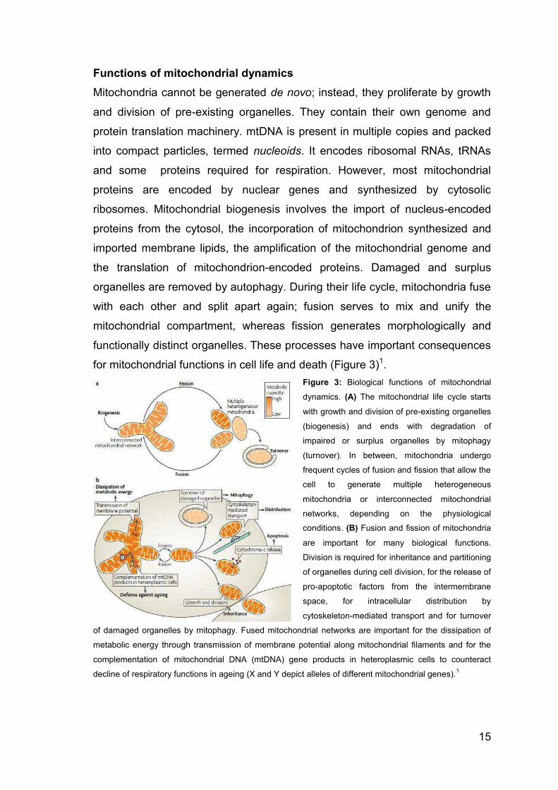
Functions of mitochondrial dynamics Mitochondria cannot be generated de novo; instead, they proliferate by growth
and division of pre-existing organelles. They contain their own genome and
protein translation machinery. mtDNA is present in multiple copies and packed
into compact particles, termed nucleoids. It encodes ribosomal RNAs, tRNAs
and some proteins required for respiration. However, most mitochondrial
proteins are encoded by nuclear genes and synthesized by cytosolic
ribosomes. Mitochondrial biogenesis involves the import of nucleus-encoded
proteins from the cytosol, the incorporation of mitochondrion synthesized and
imported membrane lipids, the amplification of the mitochondrial genome and
the translation of mitochondrion-encoded proteins. Damaged and surplus
organelles are removed by autophagy. During their life cycle, mitochondria fuse
with each other and split apart again; fusion serves to mix and unify the
mitochondrial compartment, whereas fission generates morphologically and
functionally distinct organelles. These processes have important consequences
for mitochondrial functions in cell life and death (Figure 3)1. Figure 3: Biological functions of mitochondrial
dynamics. (A) The mitochondrial life cycle starts
with growth and division of pre-existing organelles
(biogenesis) and ends with degradation of
impaired or surplus organelles by mitophagy
(turnover). In between, mitochondria undergo
frequent cycles of fusion and fission that allow the
cell to generate multiple heterogeneous
mitochondria or interconnected mitochondrial
networks, depending on the physiological conditions. (B) Fusion and fission of mitochondria
are important for many biological functions.
Division is required for inheritance and partitioning
of organelles during cell division, for the release of
pro-apoptotic factors from the intermembrane
space, for intracellular distribution by
cytoskeleton-mediated transport and for turnover
of damaged organelles by mitophagy. Fused mitochondrial networks are important for the dissipation of
metabolic energy through transmission of membrane potential along mitochondrial filaments and for the
complementation of mitochondrial DNA (mtDNA) gene products in heteroplasmic cells to counteract
decline of respiratory functions in ageing (X and Y depict alleles of different mitochondrial genes).1

16
Mitochondrial dynamics is essential in mammalian development. Mitochondrial
fusion is important for inheritance and maintenance of mtDNA. In yeast, fusion-
defective mutants rapidly lose their mitochondrial genome and consequently
show defects in respiration24. This is probably because fragmentation of
mitochondria produces multiple small organelles, most of which lack mtDNA, so
partitioning of these organelles to daughter cells produces a significant number
of progeny lacking mtDNA. As a result, mitochondrial genomes are lost from the
population after several generations.
Disruption of fusion in mammalian cells also leads to mitochondrial
heterogeneity and dysfunction, possibly as a consequence of nucleoid loss in
individual mitochondria25. Thus, it seems that fusion serves as a fundamental
mechanism to maintain a mitochondrial population with a full complement of
nucleus- and mitochondrion-encoded gene products. Although mitochondrial
fission inevitably generates organelles lacking nucleoids, fusion ensures that
the mitochondrial genome and gene products are replenished before
functionality is lost.
Cells defective in mitochondrial division contain highly interconnected net-like
mitochondria that typically accumulate in restricted areas, leaving large parts of
the cell devoid of mitochondria. Proper mitochondrial distribution depends on
division to split the mitochondrial network into transportable units. Obviously,
this is particularly important in large and extended cells, such as neurons.
Accordingly, DRP1 and OPA1 are crucial to establish proper mitochondrial
content and distribution in dendrites. This, in turn, is essential for the
maintenance of dendritic spines and synapses, which are neuronal structures
with a particularly high energy demand26,25.
In other cell types, fused mitochondrial networks act as electrically united
systems that transmit the membrane potential generated by the proton pumps
of the respiratory chain27. This mechanism was proposed to play an important
part in the dissipation of metabolic energy in muscle cells.
Hence, it seems that concerted activities of the mitochondrial fusion and fission
machineries shape the mitochondrial compartment and adapt it to the specific
requirements of the cells.
Furthermore research over the past decades leaves no doubt that mitochondria
have a crucial role in ageing. The mitochondrial theory of ageing postulates that

17
the respiratory chain produces reactive oxygen species (ROS) as byproducts of
oxidative phosphorylation. Because mitochondria are a major source for ROS,
mtDNA is particularly vulnerable to ROS-induced mutations and lesions. As a
result, gradual and progressive accumulation of mtDNA mutations leads to a
loss of functional respiratory chain complexes, resulting in a decline of
bioenergetic capacity and eventually age-associated pathologies and death28.
Mitochondrial dynamics was proposed to counteract this detrimental process
through two activities: rescue of non-functional organelles by fusion and
elimination of damaged organelles after fission. Irrespective of the level of
heteroplasmy, mitochondria showed a homogenous pattern of respiratory
activity at the cellular level as a result of fusion and inter mixing of mitochondrial
contents29. These suggests that fusion of mitochondria and complementation of
mitochondrial gene products are a defence mechanism against cellular ageing.
Autophagy is a process of self-degradation of cellular components that are
harmful or no longer required. damaged or surplus organelles or portions of
cytosol are sequestered by double-membrane autophagosomes that fuse with
lysosomes or vacuoles and are broken down by hydrolytic enzymes30. The
autophagic breakdown of mitochondria is termed mitophagy. It is tempting to
speculate that mitophagy constitutes a mechanism to remove dysfunctional
mitochondria from the cell and thereby prevent proliferation of mutated mtDNA.
Support for this hypothesis came recently from a seminal study that described
the behaviour of fluorescently labelled mitochondria in cultured mammalian
cells31. Mitochondrial division was found to frequently produce two uneven
daughter organelles, one with high membrane potential and one with decreased
membrane potential and reduced OPA1 levels. Intriguingly, mitochondria with
decreased membrane potential and reduced OPA1 levels are less likely to be
engaged in subsequent fusion events and, instead, are prone to removal by
mitophagy. Remarkably, inhibition of fission decreases mitophagy and results in
decline of respiratory capacity, whereas arrest of autophagy leads to the
accumulation of mitochondria with low membrane potential and low OPA1. on
the basis of these observations a hypothesis was proposed that integrates
mitochondrial dynamics and turnover in the mitochondrial life cycle.
Mitochondrial fission frequently generates solitary mitochondria that might
either maintain an intact membrane potential and re-fuse with the mitochondrial

18
network, or might be depolarized and depleted of OPA1, thereby preventing
further rounds of fusion. This enables subsequent elimination by mitophagy31.
Therefore, mitochondrial division may contribute to a quality control mechanism
that facilitates removal of damaged mitochondria from the cell.
At last mitochondrial fission is important for apoptosis. A key event in apoptosis
is mitochondrial outer membrane permeabilization, which releases cytochrome-
c and other pro-apoptotic factors from the intermembrane space into the cytosol
to trigger downstream cell death pathways32,33. Regulation of apoptosis
involves DRP1-dependent mitochondrial fragmentation in a wide range of
organisms, including yeast34, flies35, worms36 and mammals37.
Although many issues remain controversial, it seems that mitochondrial
fragmentation occurs early in the apoptotic pathway, just prior to or
simultaneously with outer membrane permeabilization and before effector
caspase activation. Further work is required to determine how the components
of mitochondrial fission and fusion actively participate in programmed cell
death.
1.2 Bioenergetics of mitochondria The cellular energy currency, ATP, can be produced in 3 ways: (a) by
anaerobic glycolysis (which takes place in the cytosol); when glucose is
converted to pyruvate only a small fraction of total free energy potentially
available for ATP synthesis is released with an overall net gain of 2 ATP
molecules; (b) by tricarboxylic acid (TCA), also known as citric acid or Krebs
cycle, which takes place in mitochondrial matrix and yields one ATP molecule
percycle, (c) by oxidative phosphorylation (OXPHOS), which takes place in the
IMM and allows~15 times more ATP to be made than that produced by
glycolysis. This is because mitochondria house the major enzymatic systems
used to complete the oxidation of sugars, fats and proteins that enter the Krebs
cycle after being converted to acetyl-CoA. Pyruvate produced by glycolysis
enters mitochondria, where pyruvate dehydrogenase catalyzes its conversion to
acetyl-CoA, also reducing NAD+ to NADH; fatty acids are converted to acetyl-
CoA by β oxidation; while various enzymes exist for the conversion of specific
amino acids in to pyruvate, acetyl-CoA or directly into specific citric acid cycle
intermediates. Once in the TCA cycle, CoA causes the acetyl moiety to react

19
with oxaloacetate to produce citrate. In a series of seven subsequent enzymatic
steps, citrate is oxidized back to oxaloacetate while giving off two molecules of
carbon in the form of CO2, three NADH and one of flavin adenine dinucleotide
FADH238. The latter two carry the free energy liberated from the Krebs cycle to
the mitochondrial electron transport chain made up of OXPHOS complexes I to
V (Figure 4).
Figure 4. Cellular respiration: the electron transport chain and ATP synthase. Complexes I-IV of electron transport chain (ETC) and the ATP synthase (Complex V) are embedded in the
IMM. The ETC substrates coming from Krebs cycle feed the electrons to complexes I and II, electrons are
then transferred along the chain due to the increasing redox potential of the OXPHOS enzymes. The flow
of electrons is accompanied by proton pumping from the matrix to theintermembrane space creating the
electrochemical proton gradient which then drives the synthesis of ATP. Complexes I to V can be inhibited
by rotenone, thenoyltrifluoroacetone (TTFA), antimycin A, cyanide and oligomycin, respectively.
NADH donates electrons to respiratory complex I, also called NADH
dehydrogenase, an L-shaped enzyme complex that contains a hydrophobic
domain embedded in the IMM and a hydrophilic arm which protrudes into the
mitochondrial matrix and contains the NADH binding site. The reduced cofactor
donates two electrons to a flavin mononucleotide prosthetic group contained in
the hydrophilic arm of complex I. These electrons are then passed down the
arm via a series of iron–sulphur clusters to the lipid soluble redox carrier
coenzyme Q (CoQ)38. The liberated energy is used to pump out four protons
(H+) from the matrix to the IMS against their concentration gradient. The other
cofactor formed in the citric acid cycle (FADH2) never leaves the complex, as
the dehydrogenase itself is a part of the electron transport chain. The enzyme,
also known as complex II, contains FAD as a prosthetic group alongside Fe–S
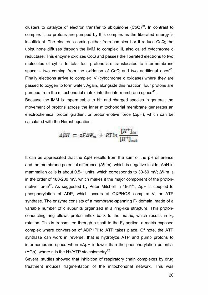
20
clusters to catalyze of electron transfer to ubiquinone (CoQ)39. In contrast to
complex I, no protons are pumped by this complex as the liberated energy is
insufficient. The electrons coming either from complex I or II reduce CoQ; the
ubiquinone diffuses through the IMM to complex III, also called cytochrome c
reductase. This enzyme oxidizes CoQ and passes the liberated electrons to two
molecules of cyt c. In total four protons are translocated to intermembrane
space – two coming from the oxidation of CoQ and two additional ones40.
Finally electrons arrive to complex IV (cytochrome c oxidase) where they are
passed to oxygen to form water. Again, alongside this reaction, four protons are
pumped from the mitochondrial matrix into the intermembrane space41.
Because the IMM is impermeable to H+ and charged species in general, the
movement of protons across the inner mitochondrial membrane generates an
electrochemical proton gradient or proton-motive force (ΔµH), which can be
calculated with the Nernst equation:
It can be appreciated that the ΔµH results from the sum of the pH difference
and the membrane potential difference (ΔΨm), which is negative inside. ΔpH in
mammalian cells is about 0.5-1 units, which corresponds to 30-60 mV; ΔΨm is
in the order of 180-200 mV, which makes it the major component of the proton-
motive force42. As suggested by Peter Mitchell in 196143, ΔµH is coupled to
phosphorylation of ADP, which occurs at OXPHOS complex V, or ATP
synthase. The enzyme consists of a membrane-spanning Fo domain, made of a
variable number of c subunits organized in a ring-like structure. This proton-
conducting ring allows proton influx back to the matrix, which results in Fo
rotation. This is transmitted through a shaft to the F1 portion, a matrix-exposed
complex where conversion of ADP+Pi to ATP takes place. Of note, the ATP
synthase can work in reverse, that is hydrolyze ATP and pump protons to
intermembrane space when nΔµH is lower than the phosphorylation potential
(ΔGp), where n is the H+/ATP stoichiometry42.
Several studies showed that inhibition of respiratory chain complexes by drug
treatment induces fragmentation of the mitochondrial network. This was
21
observed, for example, in HeLa cells44, 45, CV1-4A cells44, 46, mouse embryonic
fibroblasts46, human skin fibroblasts47, cultured cortical neurons47,MRC5
fibroblasts48, and other cell types49. In contrast, some cell types retain
filamentous mitochondria during respiratory chain inhibition, and the
phenotypes of respiratory-deficient cells lacking an intact mitochondrial genome
are ambiguous49. Mitochondria appear more elaborately interconnected and
ramified in HeLa cells when mitochondrial respiration is induced by growth in
galactose containing medium ( in comparison to glucose medium)50. However,
this effect was not observed in MRC5 fibroblasts50. In sum, the majority of the
available data point to a functional link between changes of energy metabolism
and adaptations of mitochondrial morphology in mammalian cells. It appears
that interconnected mitochondrial networks are frequently present in
metabolically and respiratory active cells, whereas small and fragmented
mitochondria are more prevalent in quiescent and respiratory inactive cells51.
Bioenergetic role of mitochondrial fusion Mitochondrial fusion allows efficient mixing of mitochondrial content, and it
generates extended mitochondrial networks. Both effects are advantageous
under conditions of high energy demand, and disruption of mitochondrial fusion
results in mitochondrial dysfunction and loss of respiratory capacity both in
yeast and in mammalian cells51-53.
Deletion of the FZO1 or MGM1 genes, encoding key components of the
mitochondrial fusion machinery, leads to rapid loss of the mitochondrial genome
in yeast24. As several respiratory chain subunits are encoded by the
mitochondrial DNA (mtDNA), it is difficult to determine whether loss of fusion
directly contributes to a decline of respiratory capacity, or whether respiratory
defects infusion-deficient yeast mutants are an indirect consequence of a defect
in mtDNA inheritance. Deletion of the DNM1 gene, encoding a key mediator of
mitochondrial fission, extends life span in yeast54. It is not exactly known
whether longevity is directly related to the highly fused, interconnected
mitochondrial network characteristic for fission-defective yeast mutants, or
whether it is linked to the inactivation of cell death pathways or other reasons.
Furthermore, deletion of the MGM1 gene reduces life span in yeast55,
suggesting that mitochondrial fusion is beneficial for cell physiology. However, it
remain sun known whether loss of mtDNA in mgm1 mutants has an impact on
22
life span, and whether there is a direct link between mitochondrial fusion activity
and respiratory capacity in yeast51.
Content mixing and complementation of gene products in fused mitochondria
were proposed to be crucial for maintenance of mitochondrial functions and
counteract cellular aging. During the process of aging, different mutations
accumulate in different mtDNA molecules. Thus, wild-type mtDNA coexists with
different mutant alleles or deletions, a state termed heteroplasmy. When
individual mitochondria have acquired mutations in different genes, each
mitochondrion will be respiratory deficient. However, when these mitochondria
fuse, each fusion partner contributes an intact allele, and complementation of
gene products restores respiratory activity56.
Several recent reports underscore the importance of mitochondrial fusion under
conditions of high energy demand in mammals. It was shown that some cell
stressors, including UV irradiation and several drugs that inhibit cytosolic
protein synthesis, can trigger increased mitochondrial fusion in mouse
embryonic fibroblasts, a process termed stress-induced mitochondrial
hyperfusion. Mitochondria elongate and form a mesh of highly interconnected
filaments in an Mfn1 and Opa1-dependent manner. Stress-induced
mitochondrial hyperfusion is accompanied by increased mitochondrial ATP
production. It is conceivable that fusion is necessary to optimize mitochondrial
function in order to allow the cell to cope with increased energy demand during
selective forms of stress57.
Bioenergetic role of mitochondrial fission Mitochondrial division serves a variety of different functions. These include
partitioning and inheritance of the organelles during cell division, release of
cytochrome c and other intermembrane space proteins during apoptosis, and
generation of transportable mitochondrial units for movement along the
cytoskeleton1. While these functions are not directly related to bioenergetics, it
was proposed that mitochondrial fission also serves to eliminate damaged
organelles from the mitochondrial network in order to allow their removal by
autophagy. This activity supposedly constitutes an organellar quality control
mechanism and contributes to maintenance of bioenergetic capacity51, 5831, 51,
5831, 31, 51, 58.
23
The observation of fluorescently labelled mitochondria in cultured mammalian
cells revealed that mitochondrial division frequently generates two uneven
daughter organelles, one with high membrane potential and another one with
decreased membrane potential. Strikingly, mitochondria with low membrane
potential were found to have reduced levels of the inner membrane fusion
factor Opa1, and thus are less likely to re-fuse with the mitochondrial network.
Instead, these dysfunctional mitochondria are removed from the cell by
autophagy31. Thus, mitochondrial fission followed by selective fusion provides a
mechanism to segregate damaged and dysfunctional mitochondria and permit
their degradation by autophagy. In the long term, this mechanism contributes to
the maintenance of a healthy mitochondrial population and maintenance of
bioenergetic capacity31.
Adaptation of mitochondrial dynamics to bioenergetic conditions What are the molecular processes that adapt the activities of the mitochondrial
fusion and fission machineries to the bioenergetic state of the cell? At least
three different, mutually non-exclusive mechanisms likely play important roles:
first, the activity of the mitochondrial fusion machinery might directly respond to
the bioenergetic state of mitochondria; second, several cellular signalling
pathways modulate the activity of fusion and fission proteins; and third, the
expression of key factors of mitochondrial dynamics is regulated at the
transcriptional level51.
Fusion and fission are antagonistic processes that predominate under different
conditions to adapt mitochondrial morphology and dynamics to the bioenergetic
requirements of the cell (Figure 5). Fused mitochondria are preferred when
optimal mitochondrial function is needed. Thus, fused mitochondrial networks
are frequently found in respiratory active cells. Apparently, mixing of the matrix
and the inner membrane allows the constituents of the respiratory machinery to
cooperate most efficiently. Furthermore, fusion engages the entire
mitochondrial compartment in respiration to maximize ATP synthesis. It is
conceivable that the sudden need for metabolic energy is the reason for the
formation of hyperfused mitochondrial networks that are formed upon exposure
of cells to stress, and that fusion optimizes mitochondrial function during
starvation. While fusion in stress-exposed or starving cells constitutes a short-
term adaptation to changing environmental conditions, it also plays a beneficial

24
role for maintenance of bioenergetic capacity in the long term. Upon aging,
fusion allows complementation of gene products and thus compensates for the
accumulation of mitochondrial mutations in heteroplasmic cells. Moreover,
fused mitochondrial networks contribute to the dissipation of energy in large
cells with a particularly high energy demand. In contrast, fragmented
mitochondria are frequently found in resting cells and might represent a
“default” morphological state when high respiratory activity is not required. The
activity of the mitochondrial fission machinery contributes to maintenance of
bioenergetic capacity as it allows the elimination of irreversibly damaged
mitochondria by autophagy. The activity of the key proteins of mitochondrial
dynamics is regulated at multiple levels, including transcription, post-
translational modification, and direct response to the bioenergetic state of
mitochondria51.
Figure 5. Model of adaptation of mitochondrial morphology to respiratory activity. Fragmented mitochondria constitute the preferred morphological state when respiratory activity is low.
Under respiratory conditions mitochondria undergo frequent cycles of fusion and fission to allow spreading
of metabolites and macromolecules throughout the entire compartment. At the same time, mitochondrial
fission is required for removal of damaged and inactive organelles by autophagy. When the bioenergetic
state becomes critical, for example under nutrient deprivation or exposure to certain forms of stress, highly
fused mitochondria are formed to optimize mitochondrial function51.
25
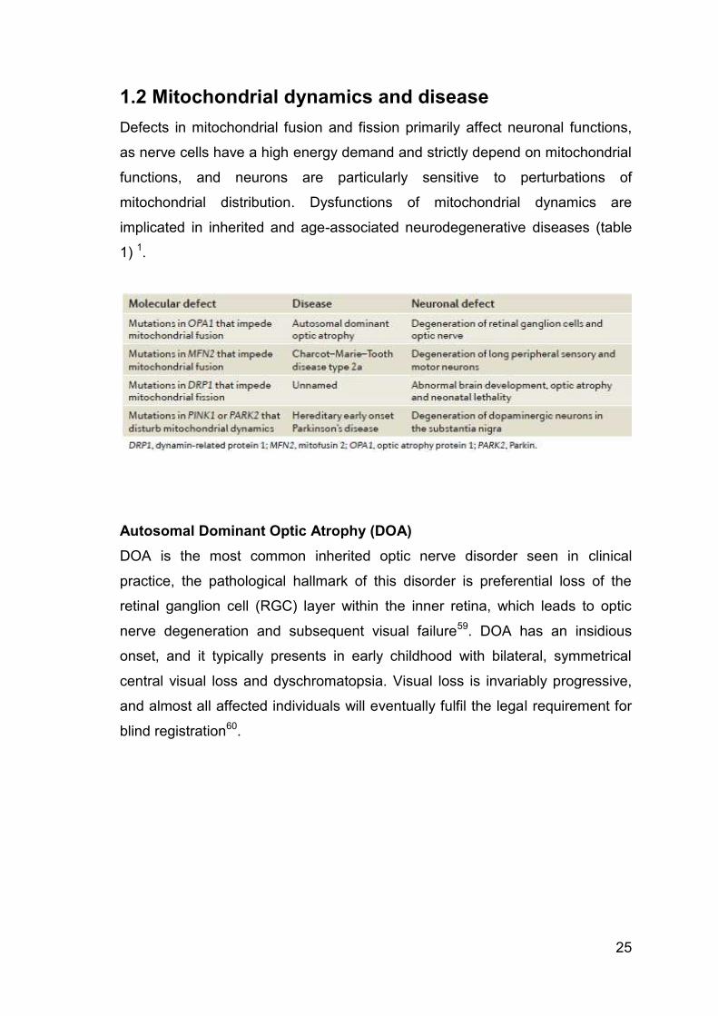
1.2 Mitochondrial dynamics and disease Defects in mitochondrial fusion and fission primarily affect neuronal functions,
as nerve cells have a high energy demand and strictly depend on mitochondrial
functions, and neurons are particularly sensitive to perturbations of
mitochondrial distribution. Dysfunctions of mitochondrial dynamics are
implicated in inherited and age-associated neurodegenerative diseases (table
1) 1.
Autosomal Dominant Optic Atrophy (DOA) DOA is the most common inherited optic nerve disorder seen in clinical
practice, the pathological hallmark of this disorder is preferential loss of the
retinal ganglion cell (RGC) layer within the inner retina, which leads to optic
nerve degeneration and subsequent visual failure59. DOA has an insidious
onset, and it typically presents in early childhood with bilateral, symmetrical
central visual loss and dyschromatopsia. Visual loss is invariably progressive,
and almost all affected individuals will eventually fulfil the legal requirement for
blind registration60.

26
The majority (50–65%) of families with DOA harbour pathogenic mutations
within the OPA1 gene, which consists of 30 coding exons spanning over 100 kb
of genomic DNA61. OPA1 codes for a 960-amino-acid, dynamin-related GTPase
that localizes to the inner mitochondrial membrane(Figure 6).
Figure 6: Domains and motifs identified in human OPA1. The GTPase domains are shown in blue with the
distinct G motifs shown in black bars (from G1 to G4). Coiled-coil regions (CC) and/or GED (GTPase
effector domain) are shown in yellow. The transmembrane domains (TM) are shown in red. The
mitochondrial import sequence (MIS) is shown in purple, with the cleavage site of mitochondrial
processing protease (MPP) in residue F88. The middle domain is colored in brown and the alternative
spliced region (Spl. Reg.) in black. The length and topology in amino acids for each region and domain are
shown. The primary sequence for each OPA1 splicing isoform is also depicted. The alternative splicing of
exons 4, 4b, and 5b generates eight isoforms of OPA1 with distinct numbers of total amino acids. All
OPA1 isoforms contain exon 5 with the S1 cleavage site (alanine). Exon 5b contains a second cleavage
site S2, although the exact residue of this site has not been determined62.
The gene is highly expressed within the RGC layer, although the protein is
ubiquitous, and abundant levels have also been identified in photoreceptors
and other non ocular tissues such as the inner ear and the brain63. Over 200
disease-causing variants have been reported so far in this highly polymorphic
gene, with mutational hot spots in the catalytic GTPase domain (exons 8–15)
and the dynamin central domain (exons 16–23)64. The majority of OPA1
mutations result in premature termination codons, and the resultant truncated
mRNA species are highly unstable, being rapidly degraded by protective
surveillance mechanisms operating via nonsense-mediated mRNA decay.
Haploinsufficiency, therefore, is a major disease mechanism in DOA, and the
pathological consequences of a dramatic reduction in OPA1 protein levels is

27
highlighted by those rare families who are heterozygous for microdeletions
spanning the entire OPA1 coding region65.
Progressive visual failure remains the defining feature of DOA but, with greater
availability of genetic testing, a specific OPA1 mutation in exon 14 (c.1334G>A,
p.Arg445His) has been found to have a particular predilection for causing
sensorineural deafness66,67. The phenotypes associated with OPA1-linked
disease have expanded even further to encompass a wide range of prominent
neuromuscular features such as ataxia, myopathy, peripheral neuropathy, and
classical chronic progressive external ophthalmoplegia (CPEO)68. These so-
called DOA+ variants are mechanistically relevant, as they highlight the
deleterious consequences of OPA1 mutations not only for RGCs, but also for
other CNS populations, peripheral nerves, and skeletal muscle.
Although DOA+ was only recently recognized as a distinct clinical entity, up to
20% of OPA1 mutation carriers are now thought to be at risk of developing
DOA+ features, which has major implications for patient counselling69.
Furthermore, OPA1 screening is increasingly performed as part of diagnostic
panels for patients with unexplained neurodegenerative disorders, and other
hitherto unreported pathological manifestations are bound to emerge70.
It remains unclear why Opa1-DOA manifests with an apparently restricted
clinical ocular phenotype, comprising retinal ganglion cells (RGC) loss. OPA1 is
ubiquitously expressed throughout the body: in the heart, skeletal muscle, liver,
testis, and most abundantly in the brain and retina. In the human retina, OPA1
is present in the cells of the RGC layer, nerve fibre layer, the photoreceptor
layer, and the inner and outer plexiform layers (IPL & OPL). A plausible
hypothesis as to why RGC neurons may be more vulnerable to OPA1
inactivation could be a particular susceptibility to mitochondrial membrane
disorders inducing mitochondrial dysfunction or mislocalization. Indeed, reports
describe altered mitochondrial ATP synthesis and respiration in OPA1-
inactivated cells66. Moreover, recent studies show the effect of mitochondrial
morphology regulation on mitochondrial distribution in neurons and their
contribution to dendrite formation and synaptic plasticity21. This could be of
particular importance in RGC neurons that display a specific distribution of
mitochondria in the cell body, myelinated and unmyelinated axons. Additionally,

28
the defects in DOA can be ascribed to the loss of the crucial control exerted by
OPA1 on the structural organization of the cristae and apoptosis71,21.
1.3 Drosophila as a model organism Ever since Morgan isolated the white mutation in Drosophila melanogaster in
1910, the tiny fruit fly has made large contributions to the understanding of the
genetic and molecular mechanisms of heredity and development. More
recently, the remarkable power of fruit fly genetics has been applied to study
the basic mechanisms of human diseases, including those debilitating
pathologies that affect the human brain.
There are several reasons why Drosophila m. is widely used as models of
human diseases. The first and foremost reason is based on the presumption
that fundamental aspects of cell biology in flies have been conserved
throughout evolution in higherorder organisms such as humans72. A report
demonstrating that approximately 75% of the disease-related loci in humans
have at least one Drosophila homologue confirms the high degree of
conservation present in flies. Furthermore, studies of developmental events in
the fly and subsequent similar studies in higher animals have revealed a
stunning degree of functional conservation of genes. These studies indicate
that not only basic cell biology but also higher-order events such as organ
“construction” and function are conserved.
Drosophila has an unrivalled battery of genetic tools including a rapidly
expanding collection of mutants, transposon-based methods for gene
manipulation and systems that allow controlled ectopic gene expression and
balancer chromosomes73. It should be possible to target endogenous wild-type
copies of "disease gene" in the fly genome for inactivation (knock-out); defined
mutations can also be "engineered" (knock-in) into respective endogenous
genes, to create gain-offunction models74.
The above characteristics of such a minuscule system model, combined with
the rapid generation time, inexpensive culture requirements, large progeny
numbers produced in a single cross and a small highly annotated genome
devoid of genetic redundancy, are poised to yield seminal insights into human
disease73.

29
For almost a century, fruit flies have been providing a useful tool to study
various different subjects: form the chemical basis of mutagenesis, to the
definition of genes, from developmental biology, to animal behaviour. The ability
to use Drosophila as a powerful tool to approach pathogenetic disease
mechanisms for human diseases speaks to a tremendous application in
biomedical research74.
Mitochondrial dynamics in Drosophila
Thanks to the use of animal models, we are starting to understand how this
leads to neuronal dysfunction and loss, the interplay between mitochondrial
shape and function is extremely complex and the current discovery rate is
slowed down by the complexity of murine models. A valid alternative is
represented by Drosophila melanogaster that has been successfully employed
to lay the basis for several key findings in the field of neurodegeneration.
Mitochondrial shape in living cells is very heterogeneous and can range from
small spheres to interconnected tubules. The morphological plasticity of
mitochondria results from the ability of this organelle to undergo fusion and
fission, which are regulated by a family of mitochondrial shaping proteins.
As described above, mitochondrial fusion is promoted by large trans-membrane
dynamin-related proteins. OPA1 resides in the inner mitochondrial membrane
and is involved in mitochondrial fusion, as well as in the regulation of cristae
biogenesis and remodeling. In H. sapiens, 8 different OPA1 isoforms are
retrieved, which are differentially post-translationally processed in at least five
different protein forms by a complicated and yet not completely understood
network of proteases that include iAAA, mAAA, paraplegin, Oma1,and Parl; the
concerted action of these proteases results in the production of long and short
forms of the protein that are both required for correct function of the protein and
therefore for mitochondrial fusion, as well as of a soluble form that albeit
quantitatively scarce, participates in the formation of the OPA1-containing
oligomers that stabilizes the cristae during apoptosis75.
Drosophila OPA1 homologue (dOPA1) shares 51.2% similarity with the human
orthologue. In fruitflies, Opa1 gene is transcribed into 2 isoforms, that are
processed into a short form by Drosophila presenilin-associated rhomboid-like
(PARL) homologue rho-7, a protease that is conserved during evolution from
yeast to mammals. Most studies on dOPA1 analyzed mutant flies where the

31
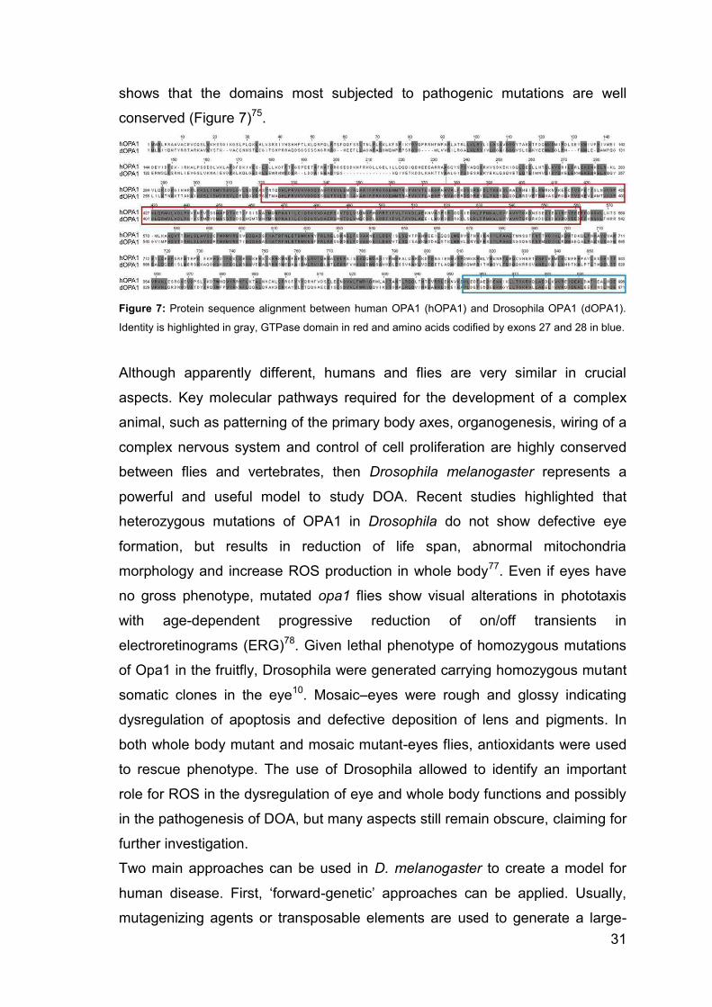
shows that the domains most subjected to pathogenic mutations are well
conserved (Figure 7)75.
Figure 7: Protein sequence alignment between human OPA1 (hOPA1) and Drosophila OPA1 (dOPA1).
Identity is highlighted in gray, GTPase domain in red and amino acids codified by exons 27 and 28 in blue.
Although apparently different, humans and flies are very similar in crucial
aspects. Key molecular pathways required for the development of a complex
animal, such as patterning of the primary body axes, organogenesis, wiring of a
complex nervous system and control of cell proliferation are highly conserved
between flies and vertebrates, then Drosophila melanogaster represents a
powerful and useful model to study DOA. Recent studies highlighted that
heterozygous mutations of OPA1 in Drosophila do not show defective eye
formation, but results in reduction of life span, abnormal mitochondria
morphology and increase ROS production in whole body77. Even if eyes have
no gross phenotype, mutated opa1 flies show visual alterations in phototaxis
with age-dependent progressive reduction of on/off transients in
electroretinograms (ERG)78. Given lethal phenotype of homozygous mutations
of Opa1 in the fruitfly, Drosophila were generated carrying homozygous mutant
somatic clones in the eye10. Mosaic–eyes were rough and glossy indicating
dysregulation of apoptosis and defective deposition of lens and pigments. In
both whole body mutant and mosaic mutant-eyes flies, antioxidants were used
to rescue phenotype. The use of Drosophila allowed to identify an important
role for ROS in the dysregulation of eye and whole body functions and possibly
in the pathogenesis of DOA, but many aspects still remain obscure, claiming for
further investigation.
Two main approaches can be used in D. melanogaster to create a model for
human disease. First, ‘forward-genetic’ approaches can be applied. Usually,
mutagenizing agents or transposable elements are used to generate a large-

32
scale number of mutant flies. Screenings for the desired disease phenotype (for
example, brain degeneration) are set up. Once the desired mutant flies have
been selected, one can proceed to the identification of mutant genes,
presumably involved in the generation of the phenotype of interest. Human
homologues of the identified Drosophila gene products are plausible candidates
for involvement in the disease that is being investigated. Many disease models
were developed through this approach79.
Alternately, when the disease genetic agent is known, ‘reverse genetics’ can be
applied. Overexpression of dominant negative mutation or downregulation of a
gene product may be used to screen for genetic interactors, after identification
of a “scorable” phenotype (such as alteration of organization of cells in the eye).
This approach is accomplished thanks to The Vienna Drosophila RNAi Center
(VDRC), a joint initiative of the Institute of Molecular Biotechnology (IMBA) and
the Research Institute of Molecular Pathology (IMP), which developed a
Drosophila transgenic RNAi library. Moreover, the use of the binary system
GAL4/UAS is a major tool in reverse genetics, because it allows the ectopic
expression of a transgene in a specific tissue or cell type. Geneticists created
genetic varieties of fruit flies, called GAL4 lines, each of which expresses the
yeast transcriptional activator GAL4 in some subset of the fly's tissues.
In this work I generated a new opa1 mutant in Drosophila using the new
genome editing system CRISPR/Cas9.

33
1.4 Genome Editing in Drosophila The advent of genome sequencing and genome-wide technologies for study of
gene expression, polymorphism and regulation has revolutionalised our ability
to associate genes with particular cellular functions or disease states. They
have also allowed us to make predictions about the function of a large
proportion of both coding and non-coding sequences. Although various
techniques such as homologous gene targeting have allowed us to selectively
mutagenise or alter gene function in a desired manner, the difficulty of applying
these techniques on a large scale has restricted our ability to test hypotheses
generated from such genome-wide analyses80.
Genome editing technologies have been developed over the past decade that
allow us to selectively mutagenise specific regions of the genome, and allow
sophisticated and detailed mechanistic studies to be performed in a variety of
organisms including Drosophila81. These technologies rely on specific DNA
binding factors that can be used to target various functional domains to defined
regions of the genome. Most experiments have used these reagents to
generate a double strand break (DSB) in the DNA at the target site, that can
then be repaired by nonhomologous end joining (NHEJ) or homologous
recombination (HR)82. NHEJ is somewhat errorprone, and can result in the
deletion or insertion of a few bases at the cut site, resulting in mutation of the
DNA82. HR normally results in precise repair from the sister chromatid, but if an
excess of a desired homologous template is supplied, this may be used to
introduce defined changes in the underlying DNA62,83.
CRISPR/Cas9 system The clustered regularly interspaced short palindromic repeat (CRISPR/Cas9)
system acts as a bacterial defense system against invading viruses and
plasmids in many different bacterial species84-87. The best studied system is
that from Streptococcus pyogenes. Here, the Cas9 endonuclease is targeted to
sequences from the invading pathogen by a crRNA (CRISPR RNA), that
provides specificity to the endonuclease by base pairing with a 20 nt
complimentary sequence within the DNA88, 89.
Endogenously, a further component, known as the tracrRNA (trans-acting
crRNA) forms a complex with the crRNA and targets its incorporation into the

34
Cas9 complex. Recently, this system has been shown to work in many other
organisms, including mammalian90, 91, insect92,93, plant94 and fungal95 cells.
Fusion of the crRNA and tracrRNA into a ~100 nt synthetic single guide or
chimeric RNA (sgRNA or chiRNA) has further simplified this system, which then
only requires two components to be expressed89, 90. The specificity is
determined by a 20 nt sequence at the 50 end of the sgRNA, which can be
altered to match any desired sequence in the DNA. The only limitation upon this
targeting is that the 20 nt guide sequence has to be followed by a protospacer
adjacent motif (PAM) of NGG in the DNA in order for efficient cleavage to occur
(Figure 8)96. This sequence should occur on average every 8 bases in the DNA,
but recent reports have suggested that this requirement may be relaxed to
include NAG sequences97, increasing the number of potential target sites still
further. CRISPR systems from other bacterial species have different PAM
requirements98, 99 and this suggests that it will be possible to engineer Cas
proteins to bind to essentially any sequence in the future.
Figure 8: Schematic of the 2-component CRIRISPR/Cas9 system. A target site in the yellow locus is
shown as an example. Cas9 is guided to a cleavage site by a chimeric RNA containing critical crRNA and
tracrRNA sequences, including 20-nt of homology to a target site. This RNA has alternately been referred
to as a guide RNA (gRNA), a single-guide RNA (sgRNA) or a chimeric RNA (chiRNA). Cas9 (gray)
contains 2 distinct endonuclease domains, a HNH domain and a RuvC-like domain, that independently
cleave both stands at the target site to generate a DSB (red arrowheads). Cleavage of target sites
requires a high degree of homology to the gRNA and a 3-bp PAM (NGG) immediately 3′ of the target
sequence.
35
Drosophila CRISPR system Several groups have used the CRISPR/Cas9 system to induce targeted
mutations in Drosophila93, 96, 100-102, but differ in their approach to supplying the
Cas9 protein and sgRNA components of the system. The first description of
mutagenesis with CRISPR/Cas9 involved coinjection of two plasmids into
syncytial blastoderm stage Drosophila embryos96. One plasmid expresses the
Cas9 gene under the Hsp70 promoter, and the second produces the sgRNA,
driven by a pol III promoter from the U6 gene. This was tested at the yellow
gene, and resulted in mutagenesis of the gene that was capable of being
transmitted to subsequent generations. The efficiency of mutagenesis due to
inefficient NHEJ was fairly low, with 5.9% of the injected flies giving rise to at
least one mutant offspring96. However, the authors further demonstrated that if
two sgRNAs are supplied, targeting either end of the yellow gene, this can
result in deletion of the intervening sequence, and that integration of short
sequences at the cleavage site is possible by coinjection with a short single
stranded oligonucleotide donor sequence96.
A second technique that has been applied by two groups independently
involves coinjection of in vitro transcribed Cas9 mRNA and sgRNA into early
stage embryos, and achieves much higher mutagenesis rates due to inefficient
NHEJ93, 100. Bassett et al. showed that up to 88% of injected flies gave rise to
mosaic expression of the yellow gene implying that this technique is highly
efficient. A second study by Yu et al. showed a similar efficiency (80%) at the
yellow gene, but also showed successful mutagenesis at six other target loci
spread throughout the genome, demonstrating the general applicability of this
approach. The difference in efficiency between plasmid and mRNA injection
techniques may be explained by the expression levels of the Cas9 protein and
sgRNA, or by the timing of expression relative to the specification of germ cells
in the embryo.
A third system has also been developed whereby two transgenic flies are
produced, one expressing Cas9 in the germline under the nanos promoter, and
a second with ubiquitous expression of the sgRNA again driven by the U6
promoter103. When these two flies are crossed together, highly efficient
mutagenesis can be achieved, giving rise to up to more than 90% of flies with at
least one mutant offspring, and allowing longer deletions of up to 1.6 kb to be

36
made efficiently by coexpression of two sgRNAs. Although efficient, this
requires the time consuming step of producing a new transgenic fly for each
sgRNA required, and removal of the Cas9 and sgRNA transgenes after mutant
generation. However, this technique will have advantages in certain
applications, since it is more reproducible than the techniques involving embryo
injection.
The final technique uses injection of plasmids encoding the sgRNA into
transgenic lines in which Cas9 is expressed specifically in the germline under
the vasa102 or nanos104 promoters. These techniques avoid potentially
problematic somatic mutagenesis by limiting Cas9 expression to the germline
cells. Sebo et al. demonstrated high rates of mutagenesis in the G1 offspring
derived from flies injected with plasmids encoding sgRNAs, but a significant
proportion of the injected flies were infertile. By using the nanos promoter to
drive Cas9 expression, Ren et al. achieved higher rates of fertility, and
generated high rates of mutagenesis in G1 offspring.
In addition to the injection of vectors expressing guide RNAs (gRNAs), the
production of fly lines that express Cas9 either ubiquitously or in the germline
will also allow direct injection of in vitro transcribed sgRNA into these embryos.
However, the relative efficiency of this technique has not been established.
Recently, expression vectors for Cas9 expression in Drosophila cell lines have
also been described. The Actin5c and U6 promoters were use to drive
expression of the Cas9 and sgRNA components, respectively. This results in
highly efficient mutagenesis inmore than 80%cells due to the indels generated
by inefficient NHEJ. The authors also demonstrated that homologous
integration is possible using short oligonucleotide donors to insertsmall
sequences, or longer homology arms to insert a 1.8 kb cassette at up to 4%
efficiency92.
Application of CRISPR/Cas9 system The majority of applications of CRISPR/Cas9 in genome engineering use its
ability to introduce DSBs at specific sites within the genome. The DSBs can be
repaired by either NHEJ or HR, and both repair mechanisms can be used to
generate mutations and manipulate the genome in a defined manner (Figures 9
and 10).

37
Figure 9 Double strand break repair can be used to target defined genomic changes. The double strand
break (DSB) induced by the Cas9/sgRNA complex can be repaired by non-homologous end joining
(NHEJ) or homologous recombination (HR). This can result in small insertions or deletions at the target
site (left), deletions of larger genomic regions when two cuts are made (middle) or homologous repair with
a desired template (right). This can be used to alter the genome in a variety of different ways (bottom).
Figure 10 Uses of genome editing within protein coding genes. NHEJ repair (top) of Cas9-induced DSBs
can be used to remove functional elements or disrupt genes in a variety of different ways. HR repair
(bottom) can be used to insert or replace sequences present within the gene for a variety of different uses.
Exons are indicated as boxes, with coding sequence (CDS) in dark blue and untranslated region (UTR) in
grey. Enhancers are also indicated in light blue.

38
Creation of a DSB increases the rate of homologous repair at that site by
several orders of magnitude105, and this enables gene targeting to produce
defined genetic changes much more rapidly and quickly than with classical
techniques106 (Figure 9). This relies on supplying a large excess of a
homologous repair template with the desired changes107. The donor DNA can
take two forms: single stranded DNA (ssDNA) oligonucleotides synthesised up
to 200 nt in length and used to integrate short sequences, or longer double
stranded DNA (dsDNA) constructs containing hundreds to thousands of
nucleotides of homologous sequence on either side of the DSB site107. The
latter are capable of integrating longer sequences at higher efficiency (Figure
10-11)80, 108.
Figure 11: (a) Targeted DNA sequence consists of the DNA target (red bar) directly upstream of a
requisite 5′-NGG adjacent motif (PAM; green). Cas9 mediates a DSB ∼3 bp upstream of the PAM for (+)
strand (blue triangle) or (−) strand (red triangle). (b) The guide oligos contain overhangs for ligation into
the BbsI sites in px330, a G-C base pair (blue) added at the 5′ end of the guide sequence for T7
transcription and the 20-bp sequence preceding 5′-NGG in genomic DNA. (c) ssDNA for point mutation
consists of a point-mutation site (purple), flanked by 60 base sequences on each side adjoining the DSBs.
(d) ssDNA for tag/loxP insertion consists of tag/loxP site (purple), flanked by 60-bp sequences on each
side adjoining the DSBs. (e) A circular donor vector for large fragment insertion consists of a large
fragment, flanked by homology arm sequences on each side adjoining the DSBs.

39
2.AIM OF WORK For a century, Drosophila has been a favored organism for genetic research.
However, the array of materials and methods available to the Drosophila,
worker has expanded dramatically in the last decade. Genetic engineering
techniques have been optimized for studying several disease including defects
of mitochondrial dynamics and thus also Dominant Optic Atrophy.
In this work I used the CRISPR/Cas9 system to generate the only mutant of
DOA which can be helpful to understand the pathogenesis of this disease.
As describe above we generated two different dOPA1 mutants: OPA1 R417H,
a mutant that carries in endogenous dOPA1 gene the mutation corresponding
to R445H in humans; and OPA1null carrying a microdeletion leading to
production of a inactive truncated protein of 482 amino acids.
We described the phenotypic effects observed in these mutants to determined if
they have pathological features. We also analyzed the mitochondrial
morphology in the nervous and muscular systems using confocal microscopy
and the mitochondria functions through biochemical assays.

40
3.METHODS
3.1 Molecular Biology
Production of the chiRNAs We chose the target PAM site NGG using the online tool
http://tools.flycripr.molbio.wisc.edu/targetFinder/. The sequence adjacent to the
PAM sites was used to cloned into pU6-BbsI-chiRNA.
We have done synthesize the oligos with BbsI site by Bio-Fab Research:
5’gRNA: Sense oligo CTTCGGAGTTCCGAATGAAGGCGT
Antisense oligo AAACACGCCTTCATTCGGAACTCC
3’gRNA: Sense oligo CTTCGTCATCCT TTCGTCCACGAC
Antisense oligo AAACGTCGTGGACGAAAGGATGAC
For annealing the oligos were diluted in TE or 1X ligation buffer, then the
following program was run in a thermocycler 95°C for 5 min, than ramp to 25°C
at a rate of -5°C/min.
The pU6-BbsI-chiRNA plasmid was cut with BbsI and de-phosphorylated finally
the annealed oligos were ligated into the cut pU6-BbsI-chiRNA and the ligation
transformed in E.Coli.
Production of the donor plasmids The two dsDNAs were synthesize by Eurofins Genomics, these fragments have
the following sequence:
dsDNA OPA1_R417H


43
Transformation of chemiocompetent cells We transformed competent cells NEB 5-alpha Competent E. coli (High
Efficiency, NEB #C2987) following the protocol:
Thaw chemically competent cells on ice.
Add 2 l of the chilled assembly product to the competent cells. Mix
gently by pipetting up and down or by flicking the tube 4–5 times. Do not
vortex.
Place the mixture on ice for 30 minutes. Do not mix.
Heat shock at 42°C for 30 seconds. Do not mix.
Transfer tubes to ice for 2 minutes.
Add λ50 l of room-temperature SOC media to the tube.
Incubate the tube at 37°C for 60 minutes. Shake vigorously (250 rpm) or
rotate.
Warm selection plates to 37°C.
Spread 100 l of the cells onto the selection plates. Use Amp plates for
positive control sample.
Incubate overnight at 37°C.
Drosophila genomic DNA extraction protocol
Obtain 1-5 flies per tube and keep on ice
Add 100µl of Buffer A and grind with tissue grinder ~5 min
Incubate at 65°C for 30 min
Add 200µl of Buffer B
Incubate on ice for 1 hour
Centrifuge at 12000 rpm for 15 min
Transfer supernatant to a new eppendorf tube
Add 150µl of Isopropanol
Centrifuge at 12000 rpm for 15 min
Remove supernatant
Wash with 200µl of cold Ethanol 70%
Centrifuge at 12000 rpm for 15 min
Remove supernatant
Dry the pellet and resuspend in 30µl of H2O
Add 1µl of RNase

44
3.2 Drosophila genetics
Embryo injection The plasmids were sent to BestGene Inc. for Drosophila embryo injection. The
line vas-Cas9-III (BDSC#51324) was chosen for microinjection.
Generation of stable stocks Individuals of the F2 carrying the dOPA1 mutation and the SM6a balancer
chromosome, were crossed to generate the stable mutant lines
OPA1_R417H/SM6a and OPA1null/SM6a. the stable mutant lines were crossed
to generate other two lines OPA1 R417H/CyoGFP and OPA1null/CyoGFP.
Drosophila strains used
elav-Gal4
D42-Gal4
UAS-mito-GFP (Bloomington Drosophila stock center)
Control genotypes was W1118
3.3 Microscopy
Immunohistochemistry Immunostaining was performed on wandering third instar larvae raised at 25°C.
After harvesting larvae, they were dissected dorsally in standard saline and
fixed in 4% paraformaldehyde for 10 min and then washed in PBS containing
0.3% Triton-X and incubated overnight at 4°C with Goat Anti-Horseradish
Peroxidase HRP antibody conjugate with Cyanine Cy™3 red (dilution 1:500,
product by Jackson ImmunoResearch, Amax: 550nm, Emax: 570nm).
Preparations were then washed 3 time in PBS. Coverslips were mounted with a
drop of Mowiol mounting medium (Sigma).
Live imaging of mitochondrial network on muscles of Drosophila larvae Experimental larvae were dissected dorsally in HL3 solution containing 7µM
Glutamate, in order to reduce muscle contractions. After the larvae were
incubated 30min at RT with MitoTracker® Orange CMTMRos 0.5µM in HL3
(Mitochondrion-Selective Probes produce by ThermoFisher scientific, Amax:

45
550nm, Emax: 570nm). Preparations were then washed in HL3 and analyzed
using a Nikon C1 confocal microscope with a 60X water immersion objective.
Mitochondria density analysis Larvae were dissected as previously described. To quantify mitochondria
distribution we compared mitochondrial density in 40µm long proximal and
distal regions of the same segmental nerve. Mitochondria density was
evaluated by collecting a series of confocal z-stacks to determine the volume of
mitochondria (volume of mitoGFP) and the volume of the nerve (volume
enclosed by HRP fluorescence). The volume of mitochondria was normalized to
the volume of the HRP labeled nerve and the ratio between mitochondria
density in distal region and mitochondria density in proximal region was
calculated for each nerve.
Image analysis Confocal images were acquired through x40 or x60 CFI Plan Apochromat Nikon
objectives with a Nikon C1 confocal microscope and analyzed using either
Nikon EZC1 (version 3.91), Volocity (PaerkinElmer Company, Santa Clara, CA)
or NIH ImageJ softwares.
3.4 Biochemical Assays
Mitochondrial Respiration Assay Oxygen consumption measurements were performed using a Clark type
electrode (Hansatech Instruments, King’s Lynn, England). Equal weight of
larvae were cut up and added to 1 ml of Respiration Buffer (see Appendix A) at
room temperature. Total oxygen consumption was calculated by subtracting the
rate measured under physiological conditions with that measured after addition
of 5 µM Rotenone and 5 µM Antimycin A. Mitochondrial respiration was
normalized to total larvae weight.
Activity of respiratory complexes The redox enzymatic activities were performed on crude mitochondria obtained
from third instar larvae of Drosophila. Equal weight of larvae was used and
frozen in liquid nitrogen and stored at -80°C.
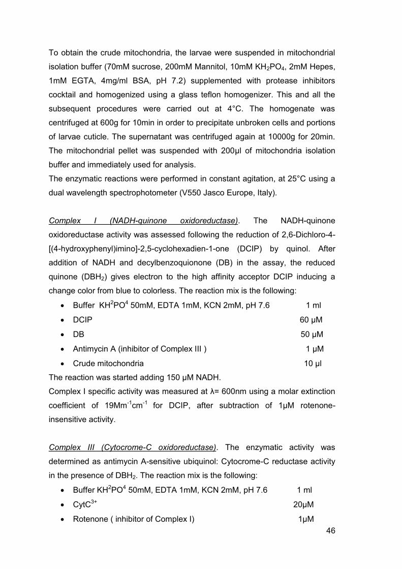
46
To obtain the crude mitochondria, the larvae were suspended in mitochondrial
isolation buffer (70mM sucrose, 200mM Mannitol, 10mM KH2PO4, 2mM Hepes,
1mM EGTA, 4mg/ml BSA, pH 7.2) supplemented with protease inhibitors
cocktail and homogenized using a glass teflon homogenizer. This and all the
subsequent procedures were carried out at 4°C. The homogenate was
centrifuged at 600g for 10min in order to precipitate unbroken cells and portions
of larvae cuticle. The supernatant was centrifuged again at 10000g for 20min.
The mitochondrial pellet was suspended with 200µl of mitochondria isolation
buffer and immediately used for analysis.
The enzymatic reactions were performed in constant agitation, at 25°C using a
dual wavelength spectrophotometer (V550 Jasco Europe, Italy).
Complex I (NADH-quinone oxidoreductase). The NADH-quinone
oxidoreductase activity was assessed following the reduction of 2,6-Dichloro-4-
[(4-hydroxyphenyl)imino]-2,5-cyclohexadien-1-one (DCIP) by quinol. After
addition of NADH and decylbenzoquionone (DB) in the assay, the reduced
quinone (DBH2) gives electron to the high affinity acceptor DCIP inducing a
change color from blue to colorless. The reaction mix is the following:
Buffer KH2PO4 50mM, EDTA 1mM, KCN 2mM, pH 7.6 1 ml
DCIP 60 µM
DB 50 µM
Antimycin A (inhibitor of Complex III ) 1 µM
Crude mitochondria 10 µl
The reaction was started adding 150 µM NADH.
Complex I specific activity was measured at = 600nm using a molar extinction
coefficient of 19Mm-1cm-1 for DCIP, after subtraction of 1µM rotenone-
insensitive activity.
Complex III (Cytocrome-C oxidoreductase). The enzymatic activity was
determined as antimycin A-sensitive ubiquinol: Cytocrome-C reductase activity
in the presence of DBH2. The reaction mix is the following:
Buffer KH2PO4 50mM, EDTA 1mM, KCN 2mM, pH 7.6 1 ml
CytC3+ 20µM
Rotenone ( inhibitor of Complex I) 1µM

47
Crude mitochondria 10µl
The reaction was started by adding 50µM DBH2.
Complex III specific activity was measured at at = 550nm using a molar
extinction coefficient of 19Mm-1cm-1 for Cyt-C2+.
Complex IV (Cytocrome-C oxidase). The enzymatic activity was assessed by
the capacity to oxidize Cyt-C2+ in Cyt-C3+ in presence of oxygen. The reaction
mix is the following:
Buffer KH2PO4 50mM, EDTA 1mM, pH 7.6 1 ml
Cyt-C2+ 20µM
The reaction was started by adding 10µl of crude mitochondria.
Complex IV specific activity was measured at = 550nm using a molar
extinction coefficient of 19Mm-1cm-1 for Cyt-C2+.
Citrate Synthase activity. The activity of different ETC enzymes was normalized
to Citrate Synthase (CS) activity which is generally considered a mitochondrial
mass index109. The enzymatic activity was assessed by the ability of CoaSH
derived from the reaction to cleave the disulfide bond of DTNB, producing 2-
nitro-5-thiobenzoate (TNB−), which ionizes to the TNB2− dianion in water and
has a yellow color.
The reaction mix is the following:
Buffer TRIS pH8 125mM, 0.1% Tiriton-X 100 0.85 ml
AcetylCoA (enzyme substrate) 0.3mM
DTNB 0.1mM
Crude mitochondria 10µl
After one minute in agitation Oxaloacetic Acid 0.5mM was added to starting the
reaction.
CS specific activity was measured at = 412nm using a molar extinction
coefficient of 13.6 mM-1cm-1 for DTNB.
48
APPENDIX A: Stock and Solutions LB Medium (Luria-Bertani Medium)
Bacto-tryptone 10g
Yeast extract 5g
NaCl 10g
H2O to 1 Liter
Autoclave.
LB Agar
Bacto-tryptone 10g
Yeast extract 5g
NaCl 10g
Agar 20g
H2O to 1 Liter
Adjust pH to 7.0 with 5N NaOH. Autoclave.
LB–Ampicillin Agar
Cool 1 Liter of autoclaved LB agar to 55° and then add 100 µg/ml filter-sterilized
ampicillin. Pour into petri dishes (~30 ml/100 mm plate).
SOC medium
Bacto-tryptone 20g
Yeast extract 5g
NaCl 0.5g
KCl 1M 2.5 ml
H2O to 1 Liter
Adjust pH to 7.0 with 10N NaOH, autoclave to sterilize and add 20 ml of sterile
1M glucose immediately before use.

49
Drosophila’s food
Agar 7 g
Yeast extract 26.4 g
Sucrose 26.4 g
H2O to 600ml
Autoclave and then add 1.5 g of Nipagin dissolved in 90% ethanol.
Phoshate Buffered Saline (PBS)
KH2PO4 15 g/L
NaCl 9 g/L
Na2HPO4 8 g/L
Mitochondrial isolation buffer pH 7.2
Sucrose 70 mM
Mannitol 200 mM
KH2PO4 10 mM
Hepes 2 mM
EGTA 1 mM
BSA 4 mg/ml
HL3
NaCl 70 mM
KCl 5 mM
CaCl2 1.5 mM
MgCl2 20 mM
NaHCO3 10 mM
Trealosio 5 mM
Sucrosio 115 mM
HEPES 5 mM
Adjust pH to 7.0 with 10N NaOH

51
APPENDIX B: Plasmids maps pBluescript II SK+ (BSSK+)
pBS-U6-BbsI-chiRNA
52
4. RESULTS
4.1 Drosophila OPA1 MUTANTS: GENERATION AND PHENOTYPIC CHARACTERIZATION
Generation of mutants To generate a model of Dominant Optic Atrophy, we introduced disease
mutations in the Drosophila dOPA1 ortholog using the CRISPR/Cas9 system
(described previously). Two different dOPA1 mutants were generated: OPA1
R417H carrying a severe missense mutation and OPA1null which carries a stop
codon in position 482 of the dOPA1 protein. Availability of these mutants
allowed us to compare the phenotypic effects of a completely null mutation with
those induced by the R417H mutations responsible for the DOA plus phenotype
in humans.
The experimental design to generate mutants using CRISPR/Cas9 system
requires the following steps: (a) generation of the gRNAs responsible for
precisely targeting the genomic region where recombination should take place;
(b) generation of the dsDNA templates containing the desired genomic
modifications to be introduced and homology arms for accurate recombination;
(c) choice of a screening method.
(a) To generate the gRNAs we first chose two PAM sites using the online tool
FlyCrispr_TargetFinder. The NGG PAM sites have some important features.
They should be adjacent or as close as possible to the region of the dOPA1
gene where the desired mutation is introduced, they must have no potential off-
target matches elsewhere in the Drosophila genome and they should permit
replacement of one of their two Gs by silent mutagenesis such that the
fragment introduced by recombination no longer contains a PAM site in that
position thereby increasing the efficiency of template incorporation. Figure 12
shows the PAM sites chosen for this project.

53
Figure 12: Genomic sequence of portion of the dOPA1 gene. Greenμ 5’ and 3’ PAM sites, the respective
target sequences of the gRNA are underlined. Red: c.1250G>A mutation to generate OPA1 R417H
mutant. Gray: c.1415delT mutation to generate OPA1null mutant.
After choosing the PAM sites targeting gRNAs were completed by adding
appropriate genomic targeting sequences. The genomic target sequences
should fulfill the following requirements: (1) 20-nt long, (2) followed by a 3-nt
PAM sequence NGG, (3) begin with a G to optimize U6-driven transcription.
The resulting sequences of the 5’ and 3’ gRNA are the following:
5’gRNAμ 5’- GGAGTTCCGAATGAAGGCGTCGG - 3’
3’gRNAμ 5’- GTCATCCTTTCGTCCACGACCGG – 3’ Targeting gRNAs were cloned into pU6-BbsI-chiRNA plasmid by annealed
oligos via the BbsI restriction sites (Figure 13).
Figure 13: Targeting gRNA: annealing and cloning into pU6-BbsI-chiRNA

54
(b) To generate the dsDNA templates necessary for homologous recombination
we synthesized two DNA fragments each containing the desired disease
mutation and the silent mutations required to eliminate the PAM sites present in
the wild type genome sequence. (c) In addition to these features, in the
synthesized DNA we added a silent mutation that introduces a novel BamHI
restriction site necessary for screening the occurrence of homologous
recombination events by restriction digest. dsDNA templates also require the
presence of two 1500bp homology arms at the 5’-end and 3’-end of the mutated
DNA segment that are responsible for homologous pairing during the
recombination event. These arms were very simply generated by PCR
amplification on genomic Drosophila DNA of the two regions adjacent on either
side of the DNA fragment to be replaced.
Completion of the dsDNA templates was achieved by assembling the
synthesized DNA with the two homology arms. We used a technology known as
Gateway assembly cloning where after amplification of each fragment with
specific primers containing homologous ends, they were all ligated together in
one step into pBluescript II SK+. The following plasmids were produced:
BSSK+OPA1_R417H
BSSK+OPA1null
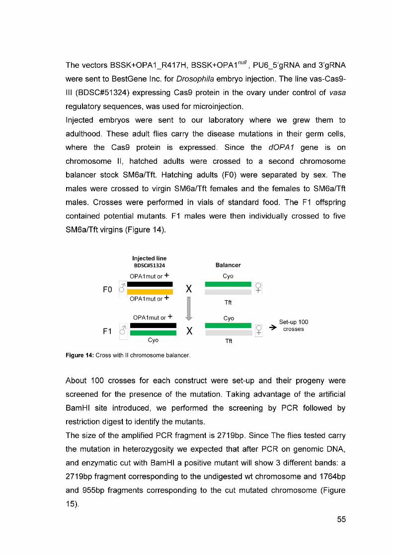


59
As shown in Figure 16, the mutants strains display a robust reduction in
lifespan. In controls 50% of survival is reached at 47 days, in OPA1null and
OPA1 R417H mutants this parameter goes down to 28 and 24 days
respectively. No obvious differences between the two mutants were detectable
using our assay.
Figure 18: Heterozygous dOPA1 mutation shortens lifespan in Drosophila.
The lifespan reduction of both dOPA1 mutants indicate that the heterozygous
mutations of OPA1 is likely to cause systemic consequences probably affecting
multiple processes. This dominant effect on lifespan is interesting in
consideration of the fact that OPA1 pathology is also dominant and suggests
that these flies can potentially offer a useful disease model.
4.2 ANALYSIS OF MITOCHONDRIAL DYNAMICS It is known that the OPA1 protein is involved in mitochondrial dynamics, the
literature suggests that in DOA patients the mitochondrial network is altered in
different tissues, therefore we performed a series of experiments to analyze
mitochondrial morphology in the neuronal and muscular systems of both
dOPA1 mutants.
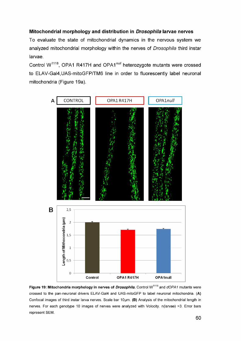

61
As shown in figure 19a, mitochondrial fragmentation is not evident at first sight.
However, analysis of the length of individual fluorescent spots with a specific
software reveals that a reduction of the average length of mitochondria was
detectable in dOPA1 mutants when compared to controls (Figure 19b). This
result indicates that mitochondria morphology is affected by mutation of a single
copy of the fly dOPA1 gene.
Existing evidence intimates that dysfunction of the mitochondrial dynamics
alters mitochondrial axonal distribution. We thus went on to investigate the
distribution of mitochondria in segmental nerves of Drosophila third instar
larvae. Mitochondria were labeled in control and dOPA1 mutants using
mitoGFP expressed under the control of the D42-gal4 driver line which
specifically expresses specifically in motor neurons thus labeling the larva
segmental nerves. were crossed to UAS-mitoGFP,D42-Gal4/TM6. The nerves
where identified by simultaneous labeling of axonal membranes using the
neuronal marker HRP. Mitochondria distribution along the nerves was then
evaluated by measuring mitochondrial density in proximal (250 µm away from
the cell bodies) and distal (1500 µm away from the cell bodies) regions of the
segmental nerves.
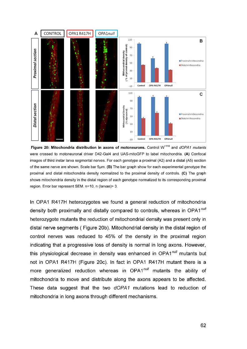


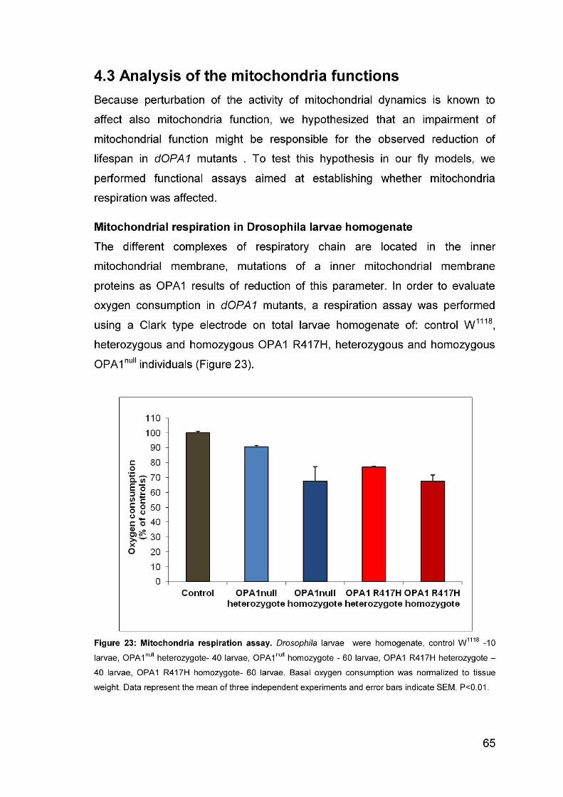

66
Analysis of mitochondrial respiratory capacity shows that in larvae homozygous
for dOPA1 mutations oxygen consumption was reduced by approximately 35%
compared to controls; whereas in heterozygous larvae OPA1null and OPA1
R417H , oxygen consumption was reduced by 20% and 10% respectively of
that of controls (Figure 23).
These data indicate that mutations on dOPA1 protein with morphological
alterations of mitochondria shape cause reduction of the mitochondrial
functions. Although this was an expected result based on the available
literature, it is interesting to underscore the observation that the OPA1 R417H
mutation in heterozygosity causes a greater reduction of mitochondrial
respiration compared to an heterozygote OPA1null mutant.
Analysis of the activity of respiratory complexes In order to confirm the results observed on mitochondrial respiration, the redox
activity of respiratory complexes was measured in a spectrometry assay using
crude mitochondria extracted from third instar larvae of control W1118,
heterozygous and homozygous OPA1 R417H, heterozygous and homozygous
OPA1null individuals. The activities of complex I (CI), complex III (CIII) and
complex IV (CIV) were normalized to Citrate Synthase (CS) activity. CS activity
is universally accepted as an index of mitochondrial mass since it is encoded in
the nucleus and localized in the mitochondrial matrix.
The bar graph in figure 24 shows the redox activity of different ETC complexes.
CI activity was reduced in both homozygous dOPA1 mutants, especially in
OPA1null homozygote mutant, as well as in heterozygous OPA1 R417H.
Surprisingly, CI activity of OPA1null heterozygotes was similar to controls,
suggesting that the mutated copy of OPA1 R417H alters CI activity more than a
reduced expression of OPA1. Furthermore, also complex IV activity was
strongly affected in OPA1 R417H heterozygote and slightly reduced in the other
mutants. Finally, CIII activity resulted similar to controls or enhanced in OPA1null
heterozygotes.
These data confirm that mutation of dOPA1 causes a reduction of mitochondrial
function. The most interesting result concerns the heterozygous mutant OPA1
R417H in which the activities of CI and CIV are drastically reduced. Again, this
result suggests that some functional parameters are more severely altered by

68
5. DISCUSSION The mechanisms by which OPA1 mutations produce DOA or DOA plus in
humans are not clear. A broad mutational spectrum has been observed for
OPA1 mutations. Over 250 OPA1 mutations have so far been reported and
they can be grouped into two main categories depending on whether they are
predicted to cause disease due to haploinsufficiency (deletions, insertions,
splice site and nonsense mutations) or can potentially act through a dominant-
negative mechanism (missense mutations)68. Although optic atrophy is the
defining feature of DOA, a specific missense mutation within the OPA1gene,
R455H, has been consistently associated with a number of additional clinical
manifestations including sensorineural deafness, chronic progressive external
ophthalmoplegia ataxia, myopathy and peripheral neuropathy thus giving rise to
the so called DOA plus phenotype.
To address the pathophysiological mechanism of OPA1 mutations we have
taken advantage of the outstanding sequence homology between the human
and Drosophila OPA1 (dOPA1) proteins has allowed us to use Drosophila as
model system to attempt an investigation of the mode of action of different
categories of OPA1 mutations, a nonsense mutation predicted to be
haploinsufficient and a missense mutation R417H homologous to human
R445H, thought to exert a dominant negative effect. Because the R455H
mutation has been linked to a DOA plus phenotype, it is believed to be a more
severe mutation, at least in a heterozygous state, than a null. To model these
mutations we have used the latest genome engineering technology, in vivo
CRISPR/Cas9 to modify the endogenous Drosophila OPA1 gene. To our
knowledge this is the first reported instance of a model organism that carries a
systemic pathogenic OPA1 mutation. Comparison of the two dOPA1 mutants
generated has allowed an initial assessment of the phenotypes elicited by the
mutations fundamentally revealing that two types of mutations show a number
of differences in their in vivo behavior. The first important observation concerns
viability. Since we are dealing with a fully mutant organism we have been able
to examine the effects of the mutations on viability. Not surprisingly, we found
that both mutants are lethal in homozygosity thus underscoring the importance
of OPA1 function for the organism. However, lethality occurs at a different

69
developmental stage. Homozygous null mutants (carrying the nonsense
mutation) die at the first instar larva stage while homozygous R417H mutants
die later, around the second instar larva stage. This result shows that not
having the OPA1 protein at all turns out to be a more severe condition than
having a R417H mutant OPA1 under this paradigm. However, both DOA and
DOA plus are dominant diseases and therefore caused by mutation of a single
copy of the gene in the heterozygous state66. Examination of both heterozygous
Drosophila mutants shows that they are viable but revealed also that they
display a similar shortening of lifespan, suggesting that under an heterozygosity
paradigm they behave similarly. However, the presence of more subtle
differences in lifespan may be hindered by the rather gross experimental
approach. Confirmation of the observation that heterozygous dOPA1 mutants
display defects has come from the analysis of mitochondria morphology which
has shown that both in nerves and muscles mitochondria network shape is
characterized by mild fragmentation and clusterization. Yet, it remains
absolutely clear that a single copy mutation of the OPA1 gene in flies displays a
dominant effect likely due to perturbation of mitochondria morphology which in
turn causes systemic defects that result in shortened lifespan. This is
reminiscent of the condition in humans where onset of DOA or DOA plus
disease demonstrates that alteration of a single copy of OPA1 has harmful
consequences for the organism.
How does perturbation of mitochondria morphology lead to the dominant
systemic consequences observed in our dOPA1 mutants? It has been reported
that loss or mutation of OPA1 is associated with impairment of oxidative
phosphorylation. In agreement with these reports, we found that both
homozygous mutants display a substantial decrease in oxygen consumption.
However, it is interesting to note that despite a similar effect on mitochondrial
morphology the null mutation and the R417 mutation behaved differently in the
respiration assay. Indeed, the observation that R417 heterozygote individual
show a greater decrease of oxygen consumption when compared to null
heterozygotes seem to suggest that that some functional parameters are more
severely altered by the presence of a single copy of a missense pathological
mutation than by the presence of a single copy complete loss of function allele.
This result appears to be in line with the fact that R417H has been linked to

70
DOA plus, a more severe pathology than DOA which is normally associated
with OPA1 haploinsufficiency66, 68, 110. This would indicate that also in an
organism model R417H may lead to a dominant interfering effect on the wild
type OPA1 protein thus accounting for the greater severity of the R417H
mutation compared to the null mutation. Therefore, the Drosophila models we
have generated appear to reproduce to some extent features that are typical of
OPA1 affected individuals. A previous report111 has shown that in fibroblasts
derived from patients carrying a R445H mutation (homologous to the R417H fly
mutant) the rate of mitochondria ATP synthesis was significantly decreased at
the level of complex I thus pointing to complex I as the culprit for the defective
oxidative phosphorylation. Although we also found that the activity of complex I
was generally reduced for both dOPA1 null and R417H homozygous mutants, it
was surprisingly normal in dOPA1 null heterozygous individuals while it was
reduced to about 50% of controls in R417H heterozygous individuals. Even
more striking are the results regarding complex IV activity since essentially both
homozygous mutants as well as the dOPA1 null heterozygote are unaffected.
Unexpectedly, however, complex IV activity was largely diminished in dOPA1
R417H heterozygous individuals. These results further point to a different
behavior for the two mutations in vivo in a whole organism, with the DOA plus
causing mutation R417H leading to more severe effects than the nonsense, null
mutation.
In this thesis work we have investigated the role of two types of pathogenic
mutations in the endogenous OPA1 gene using Drosophila as a model system.
One mutation completely obliterates OPA1 function and has been linked in
humans to DOA pathology through a haploinsufficient mechanism. The other
type is a severe missense mutation whose occurrence in humans is thought to
give rise to the more severe DOA plus pathology by functioning as a dominant
negative allelic form. Our novel animal models appear to suggest that indeed
the two types of mutation differentially affect some of the morphological and
functional parameters of mitochondria when heterozygous. We have observed
that in none of the experiments conducted the null heterozygous mutation had
worse consequences than the R417H mutation. It either behaved the same way
or was obviously milder. This result is in apparent contrast with the observation
that in homozygosity the null mutant dies at a very early developmental stage
71
while the R417H homozygote carries on until a later stage but can be explained
by viewing the R417H as a partially though severely inactivating mutation and
thus by definition weaker than a completely inactivating one. However, in
heterozygosity R417H could interfere with the activity of the wild type copy of
dOPA1 resulting in more severe phenotypes than those caused by the
presence of a single loss of function allele. Furthermore, the fact that some
tests do not show differences between the two mutations can easily be ascribed
to the coarseness of the test itself that would not permit to discriminate
relatively subtle divergences.
We believe that we have produced a rather good model to study the
etiopathology of different forms of DOA caused by different classes of
mutations within the OPA1 gene. Further validation of these models is
obviously still required and is already under way.

72
6. Reference
1. Westermann, B. Mitochondrial fusion and fission in cell life and death. Nat. Rev. Mol. Cell Biol. 11, 872-884 (2010).
2. Burté, F., Carelli, V., Chinnery, P. F. & Yu-Wai-Man, P. Disturbed mitochondrial dynamics and neurodegenerative disorders. Nat. Rev. Neurol. 11, 11-24 (2015).
3. Nisoli, E. & Carruba, M. O. Nitric oxide and mitochondrial biogenesis. J. Cell. Sci. 119, 2855-2862 (2006).
4. Hales, K. G. & Fuller, M. T. Developmentally Regulated Mitochondrial Fusion Mediated by a Conserved, Novel, Predicted GTPase. Cell 90, 121-129 (1997).
5. Fritz, S., Rapaport, D., Klanner, E., Neupert, W. & Westermann, B. Connection of the mitochondrial outer and inner membranes by Fzo1 is critical for organellar fusion. J. Cell Biol. 152, 683-692 (2001).
6. Rojo, M., Legros, F., Chateau, D. & Lombès, A. Membrane topology and mitochondrial targeting of mitofusins, ubiquitous mammalian homologs of the transmembrane GTPase Fzo. J. Cell. Sci. 115, 1663-1674 (2002).
7. Meeusen, S. et al. Mitochondrial Inner-Membrane Fusion and Crista Maintenance Requires the Dynamin-Related GTPase Mgm1. Cell 127, 383-395 (2006).
8. McQuibban, G. A., Saurya, S. & Freeman, M. Mitochondrial membrane remodelling regulated by a conserved rhomboid protease. Nature 423, 537-541 (2003).
9. Cipolat, S., De Brito, O. M., Dal Zilio, B. & Scorrano, L. OPA1 requires mitofusin 1 to promote mitochondrial fusion. Proc. Natl. Acad. Sci. U. S. A. 101, 15927-15932 (2004).
10. Yarosh, W. et al. The molecular mechanisms of OPA1-mediated optic atrophy in Drosophila model and prospects for antioxidant treatment. PLoS Genet. 4, 0062-0071 (2008).
11. Koshiba, T. et al. Structural basis of mitochondrial tethering by mitofusin complexes. Science 305, 858-862 (2004).
12. DeVay, R. M. et al. Coassembly of Mgm1 isoforms requires cardiolipin and mediates mitochondrial inner membrane fusion. J. Cell Biol. 186, 793-803 (2009).
13. Smirnova, E., Shurland, D. -., Ryazantsev, S. N. & Van Der Bliek, A. M. A human dynamin-related protein controls the distribution of mitochondria. J. Cell Biol. 143, 351-358 (1998).
73
14. Mozdy, A. D., McCaffery, J. M. & Shaw, J. M. Dnm1p GTPase-mediated mitochondrial fission is a multi-step process requiring the novel integral membrane component Fis1p. J. Cell Biol. 151, 367-379 (2000).
15. Zhang, Y. & Chan, D. C. Structural basis for recruitment of mitochondrial fission complexes by Fis1. Proc. Natl. Acad. Sci. U. S. A. 104, 18526-18530 (2007).
16. Tieu, Q., Okreglak, V., Naylor, K. & Nunnari, J. The WD repeat protein, Mdv1p, functions as a molecular adaptor by interacting with Dnm1p and Fis1p during mitochondrial fission. J. Cell Biol. 158, 445-452 (2002).
17. Lackner, L. L., Horner, J. S. & Nunnari, J. Mechanistic analysis of a dynamin effector. Science 325, 874-877 (2009).
18. Ingerman, E. et al. Dnm1 forms spirals that are structurally tailored to fit mitochondria. J. Cell Biol. 170, 1021-1027 (2005).
19. James, D. I., Parone, P. A., Mattenberger, Y. & Martinou, J. -. hFis1, a novel component of the mammalian mitochondrial fission machinery. J. Biol. Chem. 278, 36373-36379 (2003).
20. Yoon, Y., Krueger, E. W., Oswald, B. J. & McNiven, M. A. The mitochondrial protein hFis1 regulates mitochondrial fission in mammalian cells through an interaction with the dynamin-like protein DLP1. Mol. Cell. Biol. 23, 5409-5420 (2003).
21. Lee, Y. -., Jeong, S. -., Karbowski, M., Smith, C. L. & Youle, R. J. Roles of the mammalian mitochondrial fission and fusion mediators Fis1, Drp1, Opa1 in apoptosis. Mol. Biol. Cell 15, 5001-5011 (2004).
22. Breckenridge, D. G. et al. Caenorhabditis elegans drp-1 and fis-2 Regulate Distinct Cell-Death Execution Pathways Downstream of ced-3 and Independent of ced-9. Mol. Cell 31, 586-597 (2008).
23. Gandre-Babbe, S. & Van Der Bliek, A. M. The novel tail-anchored membrane protein Mff controls mitochondrial and peroxisomal fission in mammalian cells. Mol. Biol. Cell 19, 2402-2412 (2008).
24. Merz, S. & Westermann, B. Genome-wide deletion mutant analysis reveals genes required for respiratory growth, mitochondrial genome maintenance and mitochondrial protein synthesis in Saccharomyces cerevisiae. Genome Biol. 10 (2009).
25. Chen, H., McCaffery, J. M. & Chan, D. C. Mitochondrial Fusion Protects against Neurodegeneration in the Cerebellum. Cell 130, 548-562 (2007).
26. Li, Z., Okamoto, K., Hayashi, Y. & Sheng, M. The Importance of Dendritic Mitochondria in the Morphogenesis and Plasticity of Spines and Synapses. Cell 119, 873-887 (2004).
74
27. Amchenkova, A. A., Bakeeva, L. E., Chentsov, Y. S., Skulachev, V. P. & Zorov, D. B. Coupling membranes as energy-transmitting cables. I. Filamentous mitochrondia in fibroblasts and mitochondrial clusters in cardiomyocytes. J. Cell Biol. 107, 481-495 (1988).
28. Balaban, R. S., Nemoto, S. & Finkel, T. Mitochondria, Oxidants, and Aging. Cell 120, 483-495 (2005).
29. Nakada, K. et al. Inter-mitochondrial complementation: Mitochondria-specific system preventing mice from expression of disease phenotypes by mutant mtDNA. Nat. Med. 7, 934-940 (2001).
30. He, C. & Klionsky, D. J. Regulation mechanisms and signaling pathways of autophagy. Annu. Rev. Genet. 43, 67-93 (2009).
31. Twig, G. et al. Fission and selective fusion govern mitochondrial segregation and elimination by autophagy. EMBO J. 27, 433-446 (2008).
32. Youle, R. J. & Karbowski, M. Mitochondrial fission in apoptosis. Nat. Rev. Mol. Cell Biol. 6, 657-663 (2005).
33. Suen, D. -., Norris, K. L. & Youle, R. J. Mitochondrial dynamics and apoptosis. Genes Dev. 22, 1577-1590 (2008).
34. Fannjiang, Y. et al. Mitochondrial fission proteins regulate programmed cell death in yeast. Genes Dev. 18, 2785-2797 (2004).
35. Goyal, G., Fell, B., Sarin, A., Youle, R. J. & Sriram, V. Role of Mitochondrial Remodeling in Programmed Cell Death in Drosophila melanogaster. Developmental Cell 12, 807-816 (2007).
36. Jagasia, R., Grote, P., Westermann, B. & Conradt, B. DRP-1-mediated mitochondrial fragmentation during EGL-1-induced cell death in C. elegans. Nature 433, 754-760 (2005).
37. Frank, S. et al. The Role of Dynamin-Related Protein 1, a Mediator of Mitochondrial Fission, in Apoptosis. Developmental Cell 1, 515-525 (2001).
38. Poos, G. I. et al. Structure-activity studies with the selective rat toxicant norbormide. J. Med. Chem. 9, 537-540 (1966).
39. Rennison, D. et al. Synthesis and activity studies of analogues of the rat selective toxicant norbormide. Bioorg. Med. Chem. 15, 2963-2974 (2007).
40. Ricchelli, F. et al. Species-specific modulation of the mitochondrial permeability transition by norbormide. Biochim. Biophys. Acta 1708, 178-186 (2005).
41. Zulian, A. et al. Assessing the molecular basis for rat-selective induction of the mitochondrial permeability transition by norbormide. Biochim. Biophys. Acta 1767, 980-988 (2007).
75
42. Bernardi, P. et al. The mitochondrial permeability transition from in vitro artifact to disease target. FEBS J. 273, 2077-2099 (2006).
43. Rasola, A. & Bernardi, P. The mitochondrial permeability transition pore and its involvement in cell death and in disease pathogenesis. Apoptosis 12, 815-833 (2007).
44. Pletjushkina, O. Y. et al. Effect of oxidative stress on dynamics of mitochondrial reticulum. Biochim. Biophys. Acta 1757, 518-524 (2006).
45. Legros, F., Lombes, A., Frachon, P. & Rojo, M. Mitochondrial fusion in human cells is efficient, requires the inner membrane potential, and is mediated by mitofusins. Mol. Biol. Cell 13, 4343-4354 (2002).
46. De Vos, K. J., Allan, V. J., Grierson, A. J. & Sheetz, M. P. Mitochondrial function and actin regulate dynamin-related protein 1-dependent mitochondrial fission. Curr. Biol. 15, 678-683 (2005).
47. Liot, G. et al. Complex II inhibition by 3-NP causes mitochondrial fragmentation and neuronal cell death via an NMDA- and ROS-dependent pathway. Cell Death Differ. 16, 899-909 (2009).
48. Benard, G. et al. Mitochondrial bioenergetics and structural network organization. J. Cell. Sci. 120, 838-848 (2007).
49. Sauvanet, C., Duvezin-Caubet, S., di Rago, J. P. & Rojo, M. Energetic requirements and bioenergetic modulation of mitochondrial morphology and dynamics. Semin. Cell Dev. Biol. 21, 558-565 (2010).
50. Rossignol, R. et al. Energy substrate modulates mitochondrial structure and oxidative capacity in cancer cells. Cancer Res. 64, 985-993 (2004).
51. Westermann, B. Bioenergetic role of mitochondrial fusion and fission. Biochimica et Biophysica Acta (BBA) - Bioenergetics 1817, 1833-1838 (2012).
52. Chen, H., Chomyn, A. & Chan, D. C. Disruption of fusion results in mitochondrial heterogeneity and dysfunction. J. Biol. Chem. 280, 26185-26192 (2005).
53. Chen, H. et al. Mitofusins Mfn1 and Mfn2 coordinately regulate mitochondrial fusion and are essential for embryonic development. J. Cell Biol. 160, 189-200 (2003).
54. Scheckhuber, C. Q. et al. Reducing mitochondrial fission results in increased life span and fitness of two fungal ageing models. Nat. Cell Biol. 9, 99-105 (2007).
55. Scheckhuber, C. Q., Wanger, R. A., Mignat, C. A. & Osiewacz, H. D. Unopposed mitochondrial fission leads to severe lifespan shortening. Cell. Cycle 10, 3105-3110 (2011).

76
56. Sato, A., Nakada, K. & Hayashi, J. Mitochondrial dynamics and aging: Mitochondrial interaction preventing individuals from expression of respiratory deficiency caused by mutant mtDNA. Biochim. Biophys. Acta 1763, 473-481 (2006).
57. Tondera, D. et al. SLP-2 is required for stress-induced mitochondrial hyperfusion. EMBO J. 28, 1589-1600 (2009).
58. Twig, G., Hyde, B. & Shirihai, O. S. Mitochondrial fusion, fission and autophagy as a quality control axis: the bioenergetic view. Biochim. Biophys. Acta 1777, 1092-1097 (2008).
59. Yu-Wai-Man, P., Bailie, M., Atawan, A., Chinnery, P. F. & Griffiths, P. G. Pattern of retinal ganglion cell loss in dominant optic atrophy due to OPA1 mutations. Eye 25, 596-602 (2011).
60. Yu-Wai-Man, P. et al. Genetic screening for OPA1 and OPA3 mutations in patients with suspected inherited optic neuropathies. Ophthalmology 118, 558-563 (2011).
61. Alexander, C. et al. OPA1, encoding a dynamin-related GTPase, is mutated in autosomal dominant optic atrophy linked to chromosome 3q28. Nat. Genet. 26, 211-215 (2000).
62. Beumer, K. J. et al. Efficient gene targeting in Drosophila by direct embryo injection with zinc-finger nucleases. Proc. Natl. Acad. Sci. U. S. A. 105, 19821-19826 (2008).
63. Wang, A., Fann, M., Yu, H. & Yen, M. OPA1 expression in the human retina and optic nerve. Exp. Eye Res. 83, 1171-1178 (2006).
64. Yu-Wai-Man, P. et al. The Prevalence and Natural History of Dominant Optic Atrophy Due to OPA1 Mutations. Ophthalmology 117, 1538-1546.e1 (2010).
65. Marchbank, N. J. et al. Deletion of the OPA1 gene in a dominant optic atrophy family: evidence that haploinsufficiency is the cause of disease. J. Med. Genet. 39 (2002).
66. Amati-Bonneau, P. et al. OPA1 R445H mutation in optic atrophy associated with sensorineural deafness. Ann. Neurol. 58, 958-963 (2005).
67. Leruez, S. et al. Sensorineural hearing loss in OPA1-linked disorders. Brain 136 (2013).
68. Amati-Bonneau, P. et al. OPA1 mutations induce mitochondrial DNA instability and optic atrophy 'plus' phenotypes. Brain 131, 338-351 (2008).
69. Yu-Wai-Man, P. et al. Multi-system neurological disease is common in patients with OPA1 mutations. Brain 133, 771-786 (2010).

77
70. Liskova, P. et al. Novel OPA1 missense mutation in a family with optic atrophy and severe widespread neurological disorder. Acta Ophthalmol. 91, e225-e231 (2013).
71. Frezza, C. et al. OPA1 Controls Apoptotic Cristae Remodeling Independently from Mitochondrial Fusion. Cell 126, 177-189 (2006).
72. Jackson, G. R. Guide to understanding Drosophila models of neurodegenerative diseases. PloS Biol. 6, 0236-0239 (2008).
73. Cauchi, R. J. & Van Den Heuvel, M. The fly as a model for neurodegenerative diseases: Is it worth the jump? Neurodegenerative Dis. 3, 338-356 (2006).
74. Chan, H. Y. E. & Bonini, N. M. Drosophila models of human neurodegenerative disease. Cell Death Differ. 7, 1075-1080 (2000).
75. Debattisti, V. & Scorrano, L. D. melanogaster, mitochondria and neurodegeneration: Small model organism, big discoveries. Mol. Cell. Neurosci. 55, 77-86 (2013).
76. Dorn II, G. W. et al. MARF and Opa1 control mitochondrial and cardiac function in Drosophila. Circ. Res. 108, 12-17 (2011).
77. Tang, S., Le, P. K., Tse, S., Wallace, D. C. & Huang, T. Heterozygous mutation of Opa1 in Drosophila shortens lifespan mediated through increased reactive oxygen species production. PLoS ONE 4 (2009).
78. Shahrestani, P. et al. Heterozygous mutation of Drosophila Opa1 causes the development of multiple organ abnormalities in an age-dependent and organ-specific manner. PLoS ONE 4 (2009).
79. Brachmann, C. B. & Cagan, R. L. Patterning the fly eye: the role of apoptosis. Trends in Genetics 19, 91-96 (2003).
80. Bassett, A. R. & Liu, J. CRISPR/Cas9 and Genome Editing in Drosophila. Journal of Genetics and Genomics 41, 7-19 (2014).
81. Liu, J. et al. Efficient and Specific Modifications of the Drosophila Genome by Means of an Easy TALEN Strategy. Journal of Genetics and Genomics 39, 209-215 (2012).
82. Bibikova, M., Golic, M., Golic, K. G. & Carroll, D. Targeted chromosomal cleavage and mutagenesis in Drosophila using zinc-finger nucleases. Genetics 161, 1169-1175 (2002).
83. Bibikova, M., Beumer, K., Trautman, J. K. & Carroll, D. Enhancing gene targeting with designed zinc finger nucleases. Science 300, 764 (2003).
84. Ishino, Y., Shinagawa, H., Makino, K., Amemura, M. & Nakatura, A. Nucleotide sequence of the iap gene, responsible for alkaline phosphatase

78
isoenzyme conversion in Escherichia coli, and identification of the gene product. J. Bacteriol. 169, 5429-5433 (1987).
85. Jansen, R., Van Embden, J. D. A., Gaastra, W. & Schouls, L. M. Identification of genes that are associated with DNA repeats in prokaryotes. Mol. Microbiol. 43, 1565-1575 (2002).
86. Barrangou, R. et al. CRISPR provides acquired resistance against viruses in prokaryotes. Science 315, 1709-1712 (2007).
87. Garneau, J. E. et al. The CRISPR/cas bacterial immune system cleaves bacteriophage and plasmid DNA. Nature 468, 67-71 (2010).
88. Gasiunas, G., Barrangou, R., Horvath, P. & Siksnys, V. Cas9-crRNA ribonucleoprotein complex mediates specific DNA cleavage for adaptive immunity in bacteria. Proc. Natl. Acad. Sci. U. S. A. 109, E2579-E2586 (2012).
89. Jinek, M. et al. A programmable dual-RNA-guided DNA endonuclease in adaptive bacterial immunity. Science 337, 816-821 (2012).
90. Cong, L. et al. Multiplex genome engineering using CRISPR/Cas systems. Science 339, 819-823 (2013).
91. Wang, H. et al. One-Step Generation of Mice Carrying Mutations in Multiple Genes by CRISPR/Cas-Mediated Genome Engineering. Cell 153, 910-918 (2013).
92. Bassett, A. R., Tibbit, C., Ponting, C. P. & Liu, J. -. Mutagenesis and homologous recombination in Drosophila cell lines using CRISPR/Cas9. Biol. Open 3, 42-49 (2014).
93. Yu, Z. et al. Highly efficient genome modifications mediated by CRISPR/Cas9 in Drosophila. Genetics 195, 289-291 (2013).
94. Shan, Q. et al. Targeted genome modification of crop plants using a CRISPR-Cas system. Nat. Biotechnol. 31, 686-688 (2013).
95. Dicarlo, J. E. et al. Genome engineering in Saccharomyces cerevisiae using CRISPR-Cas systems. Nucleic Acids Res. 41, 4336-4343 (2013).
96. Gratz, S. J. et al. Genome engineering of Drosophila with the CRISPR RNA-guided Cas9 nuclease. Genetics 194, 1029-1035 (2013).
97. Mali, P. et al. RNA-guided human genome engineering via Cas9. Science 339, 823-826 (2013).
98. Esvelt, K. M. et al. Orthogonal Cas9 proteins for RNA-guided gene regulation and editing. Nat. Methods 10, 1116-1123 (2013).
79
99. Mojica, F. J. M., Díez-Villaseñor, C., García-Martínez, J. & Almendros, C. Short motif sequences determine the targets of the prokaryotic CRISPR defence system. Microbiology 155, 733-740 (2009).
100. Bassett, A., Tibbit, C., Ponting, C. & Liu, J. Highly Efficient Targeted Mutagenesis of Drosophila with the CRISPR/Cas9 System. Cell Reports 4, 220-228 (2013).
101. Gratz, S. J., Wildonger, J., Harrison, M. M. & O'Connor-Giles, K. M. CRISPR/Cas9-mediated genome engineering and the promise of designer flies on demand. Fly 7 (2013).
102. Sebo, Z. L., Lee, H. B., Peng, Y. & Guo, Y. A simplified and efficient germline-specific CRISPR/Cas9 system for Drosophila genomic engineering. Fly 8 (2014).
103. Kondo, S. & Ueda, R. Highly Improved gene targeting by germline-specific Cas9 expression in Drosophila. Genetics 195, 715-721 (2013).
104. Ren, X. et al. Optimized gene editing technology for Drosophila melanogaster using germ line-specific Cas9. Proc. Natl. Acad. Sci. U. S. A. 110, 19012-19017 (2013).
105. Smih, F., Rouet, P., Romanienko, P. J. & Jasin, M. Double-strand breaks at the target locus stimulate gene targeting in embryonic stem cells. Nucleic Acids Res. 23, 5012-5019 (1995).
106. Rong, Y. S. & Golic, K. G. Gene targeting by homologous recombination in Drosophila. Science 288, 2013-2018 (2000).
107. Beumer, K. J., Trautman, J. K., Mukherjee, K. & Carroll, D. Donor DNA utilization during gene targeting with zinc-finger nucleases. G3 Genes Genome Genet. 3, 657-664 (2013).
108. Yang, H., Wang, H. & Jaenisch, R. Generating genetically modified mice using CRISPR/Cas-mediated genome engineering. Nat. Protoc. 9, 1956-1968 (2014).
109. Trounce, I. A., Kim, Y. L., Jun, A. S. & Wallace, D. C. [42] Assessment of mitochondrial oxidative phosphorylation in patient muscle biopsies, lymphoblasts, and transmitochondrial cell lines. Methods Enzymol. 264, 484-509 (1996).
110. Hudson, G. et al. Mutation of OPA1 causes dominant optic atrophy with external ophthalmoplegia, ataxia, deafness and multiple mitochondrial DNA deletions: A novel disorder of mtDNA maintenance. Brain 131, 329-337 (2008).
111. Zanna, C. et al. OPA1 mutations associated with dominant optic atrophy impair oxidative phosphorylation and mitochondrial fusion. Brain 131, 352-367 (2008).

80