
1,2-benzenedicarboxylic acid, bis (2-methyl propyl) ester ...
47
1,2-benzenedicarboxylic acid, bis (2-methyl propyl) ester isolated from Onosma bracteata Wall. inhibits MG-63 cells proliferation via Akt-p53-cyclin pathway Ajay Kumar Guru Nanak Dev University Sandeep Kaur Guru Nanak Dev University Sukhvinder Dhiman Guru Nanak Dev University Prithvi Pal Singh CSIR-IHBT: Institute of Himalayan Bioresource Technology CSIR Sharad Thakur CSIR-IHBT: Institute of Himalayan Bioresource Technology CSIR Upendra Sharma CSIR-IHBT: Institute of Himalayan Bioresource Technology CSIR Subodh Kumar Guru Nanak Dev University Satwinderjeet Kaur ( [email protected] ) Guru Nanak Dev University Research Article Keywords: Antioxidant, Apoptosis, Cytotoxic potential, Onosma bracteata, Reactive oxygen species Posted Date: June 4th, 2021 DOI: https://doi.org/10.21203/rs.3.rs-182390/v1 License: This work is licensed under a Creative Commons Attribution 4.0 International License. Read Full License
Transcript of 1,2-benzenedicarboxylic acid, bis (2-methyl propyl) ester ...

1,2-benzenedicarboxylic acid, bis (2-methyl propyl)ester isolated from Onosma bracteata Wall. inhibitsMG-63 cells proliferation via Akt-p53-cyclin pathwayAjay Kumar
Guru Nanak Dev UniversitySandeep Kaur
Guru Nanak Dev UniversitySukhvinder Dhiman
Guru Nanak Dev UniversityPrithvi Pal Singh
CSIR-IHBT: Institute of Himalayan Bioresource Technology CSIRSharad Thakur
CSIR-IHBT: Institute of Himalayan Bioresource Technology CSIRUpendra Sharma
CSIR-IHBT: Institute of Himalayan Bioresource Technology CSIRSubodh Kumar
Guru Nanak Dev UniversitySatwinderjeet Kaur ( [email protected] )
Guru Nanak Dev University
Research Article
Keywords: Antioxidant, Apoptosis, Cytotoxic potential, Onosma bracteata, Reactive oxygen species
Posted Date: June 4th, 2021
DOI: https://doi.org/10.21203/rs.3.rs-182390/v1
License: This work is licensed under a Creative Commons Attribution 4.0 International License. Read Full License

1,2-benzenedicarboxylic acid, bis (2-methyl propyl) ester isolated from Onosma bracteata 1
Wall. inhibits MG-63 cells proliferation via Akt-p53-cyclin pathway 2
3
Ajay Kumara, Sandeep Kaura, Sukhvinder Dhimanb, Prithvi Pal Singhc,d, Sharad Thakure, 4
Upendra Sharmac,d, Subodh Kumarb and Satwinderjeet Kaura* 5
aDepartment of Botanical & Environmental Sciences, Guru Nanak Dev University, Amritsar 6
(India) 7
bDepartment of Chemistry, Guru Nanak Dev University, Amritsar (India) 8
cChemical Technology Division, CSIR-IHBT, Palampur (India) 9
dAcademy of Scientific and Innovative Research (AcSIR), Ghaziabad-201002, India 10
eBiotechnology Division, COVID-19 Project, CSIR-IHBT, Palampur (India) 11
12
*Corresponding Author 13
Dr. Satwinderjeet Kaur, Professor, 14
Department of Botanical & Environmental Sciences, 15
Guru Nanak Dev University, 16
Amritsar-143005, Punjab (India) 17
Email: Correspondence: [email protected]; [email protected] 18
Contact: +91-8283808508 19
List of abbreviations: Apaf-1: apoptotic protease activating factor 1; Bcl-2: b cell lymphoma 20
2; BHT: butylated hydroxytoluene; CDK2: Cyclin-dependent kinase 2; CLMS: Confocal Laser 21
Scanning Microscopy; COX-2: cyclooxygenase-2; CO2: carbon dioxide; DCFH-DA: 2′,7′-22
dichlorofluorescein diacetate; DIBP: Di-isobutyl phthalate; DMEM: Dulbecco's Modified 23
Eagle Medium; DMSO: Dimethyl sulfoxide; EA: early apoptosis; EDTA: ethylenediamine 24
tetra acetic acid; FBS: Fetal bovine serum; FTIR: fourier-transform infrared spectroscopy; 25
HRMS: High-resolution mass spectroscopy; KCL: potassium chloride; LA: late apoptosis; L: 26
live; MG-63: human osteosarcoma cells; MMP: mitochondria membrane potential; MTT: 3-27

(4,5-dimethythiazol-2-yl)-2,5-diphenyl tetrazolium bromide; N: necrotic; NADH: 28
nicotinamide adenine dinucleotide; NBT: nitro blue tetrazolium; NCCS: National Centre for 29
Cell Science; NFκB: nuclear factor-kappa B; NMR: Nuclear magnetic Resonance; OS: 30
Osteosarcoma; p-Akt: phosphorylated-Akt; PMS: phenazine methosulfate; PVDF: 31
polyvinylidene fluoride; PAGE: polyacrylamide gel electrophoresis; PBS: phosphate buffer 32
saline; RIN: residue interaction networks; RING: residue interaction network generator; RT: 33
room temperature; RT-qPCR: quantitative real-time polymerase chain reaction; RMSD: root 34
mean square deviation; ROS: reactive oxygen species; Rh123: rhodamine123; rpm: revolutions 35
per minute; SDS: sodium dodecyl sulphate; SDS page: dodecyl sulphate-polyacrylamide gel 36
electrophoresis; TBA: 2-thiobarbituric acid; TCA: trichloroacetic acid; TLC: thin layer 37
chromatography; WB: western blotting. 38
39
Abstract 40
Onosma bracteata Wall. (Boraginaceae family) is one of the important constituents of 41
Ayurvedic drugs which enhance immunity. Among all the fractions isolated from O. bracteata, 42
ethyl acetate fraction (Obea) showed good antioxidant activity in Superoxide radical 43
scavenging assay and Lipid peroxidation assay with EC50 value of 95.12 and 80.67 µg/ml, 44
respectively. Silica gel column chromatography of Obea yielded ObD1 fraction which was 45
characterized as Di-isobutyl phthalate (DIBP) using NMR, FTIR and HRMS spectroscopic 46
techniques. DIBP showed antiproliferative activity in human osteosarcoma MG-63, human 47
neuroblastoma IMR-32 and A549 cell lines with GI50 value of 37.53, 56.05 and 47.12 μM, 48
respectively, in MTT assay. In Flow cytometric studies, DIBP has shown disruption of 49
mitochondrial membrane potential (MMP) and enhancement of ROS, indicating the apoptosis 50
induction. The cells were found to be delayed at G0/G1 phase which might be due to the 51
downregulation of Cyclin E and CDK2 as shown in RT-PCR studies. Western blotting analysis 52
revealed an increased expression of p53, caspase 3 and caspase 9 and downregulation of p-NF-53

kB, p-Akt and Bcl-xl. Molecular docking studies also displayed the interaction of DIBP with 54
p53 (−151.13 kcal/mol) and CDK1 (−133.96 kcal/mol). Thus, DIBP has exhibited great 55
potential as chemopreventive/chemotherapeutic agent against osteosarcoma. 56
57
Keywords: Antioxidant, Apoptosis, Cytotoxic potential, Onosma bracteata, Reactive oxygen 58
species 59
60
Introduction 61
Cancer is a complicated disease in which cells multiply and grow uncontrollably due to 62
altered signaling pathways in different tumor types. Osteosarcoma (OS) is a primary malignant 63
bone sarcoma that occurs in children and adolescents with ~10% of OS in individuals older 64
than 60 years (Durfee et al., 2016). Corre et al., 2020 reported the yearly occurrence of 65
osteosarcoma as 1 to 3 cases per million in 15–19 years of age. OS generally develops via 66
unbalanced cell proliferation, dysregulation of cell cycle, mutations in DNA and around 70% 67
of osteosarcoma cases showed chromosomal aberrations (Misaghi et al., 2018). Numerous 68
types of treatment strategies are available for dealing with carcinogenesis such as 69
chemotherapy, immunotherapy, radiation therapy, targeted therapy and gene therapy which 70
help to cure cancer still these strategies have severe side effects (Siamof et al., 2020). So, there 71
is an urgent need to develop effective treatment strategies to prevent cancer. Although, there 72
are a variety of modern medicine options available for treating cancer, still these strategies have 73
limitations such as re-occurrence, metastasis, low success rate and several side effects (Bielack 74
et al., 2009; Huang et al., 2017). The cancer cells undergo incessant mutations that reduce the 75
effectiveness of cancer-targeting approaches (Loeb and Loeb 2000). Moreover, oxidative stress 76
is one of the factors which trigger cancer progression (Milkovic et al., 2017). However, 77
generation of ROS may lead to oxidative stress that interrupts redox signaling and causes 78

damage to biomolecules (Kim et al., 2015). Wang et al., 2017 demonstrated the crucial role of 79
mitochondrial-dependent pathways in ROS-mediated apoptosis. Manifestation of apoptosis 80
includes cell shrinkage, nuclear condensation and DNA fragmentation (Saraste and Pulkki, 81
2000; Joselin et al., 2006). Existing chemo-therapeutic medication does not show a significant 82
effect on cancer cells (Bao et al., 2019). Cancer cells can be metastasized to the distant parts of 83
the body organs, even after the tumor is completely removed from the primary site (Li et al., 84
2018). Therefore, considering the above-mentioned circumstances, the challenge is to 85
selectively eliminate cancer-promoting cells via tumor-specific biomarkers that are involved 86
in carcinogenesis. Natural compounds have been recognized as valuable sources of drugs, 87
especially for cancer treatment (Lin et al., 2020). Around 60% of anticancer drugs, explored 88
clinically, have been isolated from natural products (Elias et al., 2019). Phytoconstituents that 89
exhibit antioxidant and anticancer properties, act via molecular mechanisms targeting receptors 90
and enzymes in signal transduction pathways associated with cell proliferation (Bcl-2), 91
inflammation (p-NF-κB), apoptosis (p53, caspases) and multidrug resistance (Sun et al., 2019; 92
Choudhari et al., 2020). Anchusa italica (Boraginaceae) showed various types of biological 93
activities due to the presence of phytoconstituents including di-isobutyl phthalate (Kazemi, 94
2013). 95
Onosma bracteata Wall. (Boraginaceae) is an important medicinal herb which is 96
largely found in high altitude areas of India and Nepal (Ved et al., 2016). It is used in the 97
synthesis of various drugs in Unani and Ayurvedic medicinal systems due to its beneficial 98
health effects (Zeb et al., 2015). O. bracteata has been shown to possess various types of 99
pharmacological properties (Kumar et al., 2013; Albaqami et al., 2018; Farooq et al., 2019). 100
Ethyl acetate fraction (Obea) from O. bracteata was demonstrated to effectively inhibit the 101
proliferation of MG-63 cells (Kumar et al., 2020). The present study is planned to isolate the 102
phytoconstituent(s) responsible for the anticancer potential and aims at the isolation of effective 103

active compound(s) from Obea fraction with excellent anticancer potential. Silica gel Column 104
chromatography of Obea fraction yielded 1,2-benzene dicarboxylic acid, bis (2-methyl propyl) 105
ester, also known as di-isobutyl phthalate (DIBP), which possess good anti-proliferative 106
activity and was further studied for its role in induction of apoptosis in MG-63 cells. This is 107
the first study to report the anticancer potential of DIBP against osteosarcoma. 108
109
Materials and Methods 110
Chemicals and Reagents 111
Dulbecco’s modified Eagle’s medium (DMEM), Hoechst 33342, Fluoromount, 2′,7′- 112
Dichlorodihydrofluorescein diacetate (DCFH-DA), and Rhodamine-123 and Fetal Bovine 113
Serum (FBS) were purchased from Sigma (St. Louis, MO, USA). 3-(4,5-dimethylthiazol-2-yl)-114
2,5-diphenyl tetrazolium bromide (MTT) and trypsin were procured from Hi-media Pvt. 115
Limited, Mumbai (India). Rabbit monoclonal Bcl-xl, p-53, p-NF-κB, Caspase3 and Caspase9 116
antibodies, and anti-rabbit- HRP secondary antibody were obtained from Cell Signaling 117
Technology, Danvers, MA, USA. (PVDF) membrane (MDI, Ambala). RT-PCR chemical kit, 118
was purchased from Bio-Rad, California, USA. The BD Cycletest plus DNA Kit was from BD 119
Biosciences, San Jose, CA, USA. Chemicals and reagents of analytical (AR) grade were used 120
to perform the experiments. 121
122
Plant procurement, identification and authentication of plant material 123
The plant O. bracteata, with accession no. GAZ-03, was purchased from the Herbal 124
Health Research Consortium (HHRC) Pvt. Ltd. Amritsar, Punjab (India) associated to National 125
Medicinal Plant Board (NMPB), Ministry of AYUSH, Government of India. The plant material 126
was deposited in the Herbarium of the Department of Botanical and Environmental Sciences, 127
Guru Nanak Dev University, Amritsar (Accession no: 7576). 128

Extraction and fractionation 129
The plant material was washed with distilled water and kept at 40°C. The dried plant was 130
coarse grinded (2 kg) and soaked in ethanol (80%) by maceration method. The supernatant was 131
decanted off into a flask and ethanol was distilled using Rotavapor (Buchi Rotavapor R-210, 132
Switzerland) to get Ethanolic extract (Obeth). Further, fractionation was done using different 133
organic solvents with increasing polarity, viz. hexane to yield Obhex fraction (6g), chloroform 134
to yield Obcl fraction (10g), ethyl acetate to yield Obea (7g), n- butanol to yield Obbu (15g) and 135
remaining extract to yield Obaq fraction (18g) (Flow chart 1). 136
137
Column Chromatography of the Obea fraction 138
Isolation of OBD1 139
The slurry of Obea (3 g) of O. bracteata was packed in a column with silica (mesh size 140
60-120) using n-hexane. The gradient of n-hexane (Hex): ethyl acetate (EtAc) was used as 141
eluent. With increasing polarity, a total of 50 fractions of 50 ml each were collected and 142
concentrated based on their thin layer chromatography (TLC) results. Pooled fractions 11-15 143
were further subjected to preparatory TLC with a gradient eluent of Hex: EtAc (9:1), (8:2), 144
(7:3), (6:4) and (5:5) (Flow chart 2). The blue fluorescence single spot was collected, 145
concentrated and lyophilized. The compound was named as ObD1 (100 mg) which was further 146
characterized using spectroscopic techniques. 147
148
Structure elucidation and Characterization of ObD1 149
1H and 13C NMR spectra were recorded on Bruker NMR 500 MHz instruments using 150
CDCl3 as solvent. Chemical shift in ppm was measured relative to TMS as internal standard 151
and coupling constant J, was measured in Hz, multiplicity is indicated as: s = singlet, d = 152

doublet, t = triplet, m = multiplets. The HRMS spectra were recorded on Bruker Micro Toff/ 153
QII (Germany). IR spectra was recorded on FTIR Agilent machine. 154
155
Antioxidant Activity 156
Superoxide anion radical scavenging assay 157
Superoxide radical (O2•-), is a reactive radical which functions as a precursor of reactive 158
oxygen species (ROS) and is mediator in oxidative chain reactions (Jing et al., 2015). The 159
radical scavenging potential of different fractions was investigated based on a method 160
suggested by Nishikimi et al., 1972. 0.06 M NBT and 0.156 M NADH were added to the 161
various fraction concentrations (25-400 μg/ml), followed by the addition of 0.468 M PMS. 162
After the addition of PMS, the mixture was kept for 20 min. Finally, the yellow-colored NBT 163
solution changed to blue colored solution and the absorbance was measured at 560 nm. 164
Responses in terms of percentage inhibition obtained from NBT reduction depicted by color 165
change were represented as: 166
𝑃𝑃𝑃𝑃𝑃𝑃𝑃𝑃𝑃𝑃𝑃𝑃𝑃𝑃 𝑖𝑖𝑃𝑃ℎ𝑖𝑖𝑖𝑖𝑖𝑖𝑃𝑃𝑖𝑖𝑖𝑖𝑃𝑃 = 𝑂𝑂𝑂𝑂𝐶𝐶 − 𝑂𝑂𝑂𝑂𝑆𝑆𝑂𝑂𝑂𝑂𝐶𝐶 × 100 167
where ODC is the absorbance of control solution. 168
ODS is the absorbance of the sample solution. 169
170
Lipid peroxidation assay 171
The protocol proposed by Ohkawa et al., (1972) was followed for the evaluation of the 172
lipid peroxidation inhibitory potential of O. bracteata. For this, 500 µl of lipid source (10% 173
homogenous egg), 1000 µl of 150 mM KCL and 1000 µl of different concentration of fractions 174
(25-400 µg/ml) were mixed to make the reaction mixture. To continue lipo-oxidation, the above 175
reaction mixture was dissolved in 100 µl of 10 mM FeCl3 and kept for 30 min at 37 °C. 176
Thereafter, 2000 µl mixture of HCl, TCA, TBA and BHT was added and the resultant mixture 177

was heated at 95 °C for 1 h, followed by cooling and centrifugation. Lastly, supernatant having 178
pink color was collected and the absorbance was measured at 532 nm. 179
The percent inhibition (anti-lipoperoxidation activity) was calculated by given formula below: 180
𝑃𝑃𝑃𝑃𝑃𝑃𝑃𝑃𝑃𝑃𝑃𝑃𝑃𝑃 𝑖𝑖𝑃𝑃ℎ𝑖𝑖𝑖𝑖𝑖𝑖𝑃𝑃𝑖𝑖𝑖𝑖𝑃𝑃 = 𝑂𝑂𝑂𝑂𝐶𝐶 − 𝑂𝑂𝑂𝑂𝑆𝑆𝑂𝑂𝑂𝑂𝐶𝐶 × 100 181
where, 182
ODC is absorbance of control. 183
ODS is absorbance of sample mixture. 184
185
Cell Culture 186
IMR-32 (Human neuroblastoma), A-549 (Human alveolar basal epithelial), MG-63 187
(Human osteosarcoma) and HL-7702 (normal human hepatocyte) cell lines were purchased 188
from the NCCS, Pune, India. Cells were cultured in DMEM with 10% FBS and maintained 189
with CO2 (5%) incubator at 37 °C. Media was changed with fresh media at regular intervals. 190
191
MTT Assay 192
The cytotoxic potential of isolated compound ObD1 was evaluated via MTT assay 193
based on the method prescribed by Liu et al., (2006). Suspensions of the various cell lines (8 × 194
103 cells/0.1 ml) were seeded in 96 well microplates and incubated till confluency. Thereafter, 195
cells were treated with ObD1 using the serial dilution method. After 24 h, 20 μl of MTT was 196
added into 96 well plate and incubated for 4 h. The supernatant was discarded and 100 µl 197
DMSO was added to each well. Finally, absorbance was recorded at 570 nm. 198
% 𝐺𝐺𝑃𝑃𝑖𝑖𝐺𝐺𝑃𝑃ℎ 𝑖𝑖𝑃𝑃ℎ𝑖𝑖𝑖𝑖𝑖𝑖𝑃𝑃𝑖𝑖𝑖𝑖𝑃𝑃 = 𝑂𝑂𝑂𝑂𝐶𝐶 − 𝑂𝑂𝑂𝑂𝑆𝑆𝑂𝑂𝑂𝑂𝐶𝐶 × 100 199
where, 200
ODC= untreated control; 201
ODS= treated sample 202
203
Nuclear morphological studies 204
205
Hoechst staining using Confocal Laser Scanning Microscopy (CLSM) 206
The change in the nuclear morphology of cells was observed by Hoechst staining, as 207
per the method suggested by Woo et al., (1972). The MG-63 cells (2×10⁵ cells/well) were 208
cultured in six-well plates with 12 mm coverslips in each well. The cells were treated with GI50 209
(37.53 µM) concentration of ObD1 for 24 h. Thereafter, cells were fixed using 4% 210
paraformaldehyde and 2.5% glutaraldehyde solution for 30 min followed by the addition of 211
Hoechst dye (5 μg/ml) for staining of cells. After 10 min, cells were again washed with 1x 212
PBS. Coverslips were mounted on the slides using Fluoromount. Nuclear morphological 213
changes were observed under a Nikon eclipse Ti-2 fluorescence microscope (Nikon 214
Corporation, Tokyo, Japan). 215
216
Flow Cytometric Studies 217
ROS generation Analysis 218
The changes in ROS generation after treatment with ObD1 in MG-63 cells were 219
evaluated as per the method reported by Deeb et al., (2010). The MG-63 cells (4×10⁵ cells/well) 220
were cultured for 24 h in a six-well plate followed by the 37.53 µM concentration of DIBP. 221
After 24 hr, DCFH-DA (5µM) was added to MG-63 cells and incubated for 30 min and a pellet 222
was obtained using centrifugation. The pellet of cells was dissolved in 1x PBS (500 μl). Finally, 223
level of ROS generation was measured by a Flow cytometer. The results obtained were 224
analyzed by using the software provided by BD Biosciences, USA (version 1.0.264.21). 225
226
MMP (ΔΨm) Analysis 227
MG-63 cells were treated with DIBP (37.53 µM) in six-well plate for 24 hr and further 228
analyzed using protocol recommended by Pajaniradje et al., (2010). Rhodamine-123 (10 229
μg/ml) was added to the cells and kept for 30 min in the dark. Finally, the cell-pellet was 230

obtained and dissolved in 500 μl of 1x PBS. The suspension of cells was analyzed by flow 231
cytometry for determination of MMP. 232
233
Cell-Cycle Phase Distribution Analysis 234
BD Cycle test plus DNA Kit was used for the determination of the distribution of cell-235
cycle phase in MG-63 cells. The MG-63 cells (5×10⁵ cells/well) were cultured and treated with 236
DIBP (37.53 µM) for 24 h. Further, cells were trypsinized and centrifugated for 5 min at 1500 237
rpm to obtain a pellet. Then, cells were fixed by using 70% ethanol solution followed by 238
double-washing with 1x PBS. After fixation, the cell pellet was double-washed with 1x PBS 239
followed by addition of 250 μl of solution A and kept at room temperature (RT) for 10 min and 240
then solution B (200 μl) and solution C (200 μl) were added. The cells were analyzed by flow 241
cytometry to determine the cell cycle phase distribution using FlowJo software (version 242
10.7.1). 243
244
Western Blotting 245
The expression levels of proteins involved directly in the cell signaling pathway and 246
apoptotic proteins (p-Akt, p53, caspase3- 9, and Bcl-xl and p-NF-κB) were analyzed by 247
Western blotting. Firstly, MG-63 cells (5 × 10⁶) were cultured and treated with DIBP (37.53 248
µM) for 24 hr. The cells were collected using a cell scraper and cell pellet was obtained by 249
centrifugation at 1500 rpm for 5 min. For cell lysis, 150 μL of RIPA buffer was added to the 250
cell pellet and kept in ice for 25 min then centrifuged for 25 min, supernatant was collected 251
and protein concentration was quantified by Bradford method. Equal amount of protein (40 μg) 252
from DIBP treated and untreated cells was resolved by SDS-PAGE and was transferred to 253
Polyvinylidene difluoride (PVDF) membrane using a wet transfer apparatus (Biorad, CA, US). 254
After that, the PVDF membrane was blocked using BSA (5% in TBST, 0.1 % Tween-20) for 255

2 h at RT and incubated with antibodies p53 (1:1,000), caspase3 (1:1,500), caspase9 (1:1,500), 256
p-NFκB (1:2,000) and Bcl-xl (1:1,000). The membrane was washed thrice with TBST and 257
HRP-conjugated secondary antibody (1:1,500) was added and the membrane was incubated for 258
2 h at room temperature. The blot was imaged under Image-Quant LAS 4000, GE Healthcare. 259
Band densities were quantified with Alphaease FC Software (version 4.0). β-actin (1:500) as 260
endogenous control was used for stabilizing the expression of the protein of interest. 261
262
Quantitative Real-Time polymerase chain reaction (RT-qPCR) 263
Total RNA was isolated with Trizol Reagent from the untreated and DIBP (37.53 µM) 264
treated MG-63 cells, as per manufacturer’s protocol. The RNA samples were dissolved with 265
TE buffer and incubated at 60°C for 5 min. DNA impurities were removed with DNase-I 266
solution and the resulting solution was incubated for 30 min at 37°C. Finally, quantification of 267
RNA was performed using the Nano-Drop spectrophotometer (Thermo). Further, equal 268
concentrations of RNAs were used for cDNA preparation using iScriptTM cDNA (Biorad) 269
synthesis kit as per the manufacturer protocol. cDNA was used to perform RT-qPCR in a step 270
one RT-qPCR system using iQ SYBR Supermix (Biorad). The genes used as the biomarkers 271
for RT-qPCR analysis and their primer sequences (Table 1) were the following: p53, Bcl-2, 272
CDK2, CyclinE and Caspase3. The expression of each gene was quantified by using threshold 273
cycle method 2-ΔΔCt ± SEM. 274
275
Molecular Docking Studies 276
PatchDock server, a geometry-based molecular docking algorithm, was used for 277
docking analysis of ligand (DIBP) with CDK1 and p53 (Schneidman-Duhovny et al., 2005). 278
The PDB files of ligand and CDK1 and p53 proteins were uploaded to PatchDock server for 279
docking analysis, using cluster Root Mean Square Deviation (RMSD) at default value of 4.0 280

and protein-small ligand complex type as the analysis parameters. The Residue Interaction 281
Networks (RIN) profile was obtained and representative structures of ligand and CDK/p53 282
complexes were generated using Residue Interaction Networks Generator (RING) 2.0 283
webserver (Piovesan et al., 2016). RIN analysis represents various interactions in the form of 284
a detailed network model. 285
286
Statistical Analysis 287
One-way analysis of variance ANOVA was used to calculate statistical significance of 288
all the values. The difference among the means was further compared by high-range statistical 289
domain (HSD) using Tukey’s test. For all the experiments, the values were represented as mean 290
± standard errors in triplicate values. The probability p ≤ 0.05 was used to demonstrate that all 291
the values were statistically significant at a 5% level. 292
293
Results 294
Antioxidant Activity 295
Superoxide anion radical scavenging assay 296
Among all the fractions of O. bracteata, the Obea showed effective radical-scavenging 297
potential (Fig 1). As shown in Table 2, the EC50 values of Obea and rutin on scavenging 298
superoxide radical were 95.12 µg/ml and 46.18 µg/ml, respectively. At 400 µg/ml, the 299
percentage inhibition of the Obea was 85.36 ± 2.60 and that of rutin was 90.16 ± 1.07. 300
301
Lipid peroxidation assay 302
The Obea exhibited maximum potential to inhibit lipid peroxidation with lower EC50 303
value of 80.67 µg/ml as compared to EC50 value of 76.77 µg/ml of the reference compound, 304
rutin (Table 3). The Obea has potent lipid inhibition percentage of 16.50 ± 2.10 at 25 µg/ml 305

concentration whereas rutin has 20.06 ± 1.36 percentage inhibition. Therefore, keeping in mind 306
their antioxidant potentials, Obea was further subjected to column chromatography. 307
308
Identification and isolation of bioactive compound 309
Isolation of pure compound from silica gel chromatography 310
ObD1 was isolated from Ob 4 fraction of obea using silica gel chromatography and 311
gave blue fluorescence on TLC plate when exposed to UV light (Plate I) (Fig 2). 312
313
Characterization and Structure Elucidation of ObD1 314
ObD1 was isolated from the ethyl acetate fraction of Onosma brateata. The structure 315
of ObD1 was confirmed by NMR, HRMS and IR. In 1H NMR spectrum, two aromatic signals 316
at δ 7.71-7.73 ppm (m, 2H), and δ 7.52 - 7.54 ppm (m, 2H) were observed corresponding to 317
two sets of equivalent protons confirming the presence of an aromatic ring with two quaternary 318
carbons (Fig 3). The presence of doublet at 0.99 (d, 12H, J=6.7 Hz) indicated that four 319
equivalent methyl groups are attached to methine. Another doublet presence at δ 4.09 ppm (4H, 320
J= 6.7 Hz) suggested the presence of two equivalent methylene attached with methine and an 321
electronegative atom (Fig 4). The presence of δ 167.6 ppm in carbon spectrum showed the 322
presence of carbonyl ester which was further identified by the IR analysis (Fig 5). The HRMS 323
spectrum of ObD1 displayed single peak at m/z 301.1449 [M + Na]+ (calculated 301.1410) 324
indicated the molecular formula to be C16H22O4Na+. (Fig 6). Hence, from the above spectral 325
data and comparison with literature (Garg et al., 2014), the compound ObD1 was identified as 326
1,2-benzene dicarboxylic acid, bis (2-methyl propyl) also known as di-isobutyl phthalate 327
(DIBP). 328
329

Antiproliferative Activity ObD1 330
In MG-63 cell line, ObD1 has strong cytotoxic potential with GI50 value of 37.53 μM. 331
The ObD1 also showed GI50 values of 56.05 μM and 47.12 μM against IMR-32 and A549, 332
respectively (Table 4). Standard anticancer compound (Camptothecin) showed 333
antiproliferative potential with GI50 value of 52.80 µM against MG-63 cell line. ObD1 334
exhibited high antiproliferative activity in MG-63 cells as compared to IMR-32 and A549 (Fig 335
7), which was further explored for its apoptosis mechanism of action. 336
337
Confocal Laser Scanning Microscopy (CLSM) Studies 338
Hoechst 33342 is a DNA-specific fluorescent dye that penetrates the cell and 339
intercalates at A-T regions of DNA (Kumar et al., 2015). CLMS studies showed significant 340
differences among the DIBP treated and untreated MG-63 cells. Typical apoptosis signs such 341
as the disintegration, shrinking, fragmented nuclei and chromatin condensation in MG-63 cells 342
after the treatment with ObD1 (37.53 µM), were seen in Fig 8. However, untreated cells 343
showed non-apoptotic features, i.e., uniformly dispersed chromatin. 344
345
Flow Cytometric Analysis 346
ROS Analysis 347
ROS play a significant role in apoptosis and mitochondrial-mediated pathway (Redza-348
Dutordoir and Averill-Bates, 2016). ROS generation was observed by DCFH-DA probe that is 349
deacetylated by cell esterase enzyme and oxidized by ROS into the fluorescent 2′,7′-350
dichlorofluorescein (DCF) (Jia et al., 2020). The treatment of MG-63 cells with GI50 (37.53 351
µM) concentration of ObD1 resulted in an increase in the ROS generation by 78.6 % as 352
compared to untreated Mg-63 cells (37.4 %) (Fig. 9A). MG-63 cells with ObD1 displayed an 353
increase in the DCF fluorescence, which showed the generation of ROS as well as apoptosis-354
inducing potential. 355
356
Measurement of MMP (ΔΨm) Analysis 357
Mitochondria play a significant role in apoptosis pathways with apoptogenic factors 358
like apoptosis-inducing factor, release of cytochrome c into cytoplasm and decrease in 359
membrane potential (Ψm) (Wang and Youle, 2009). Rh-123 probe was used to detect MMP in 360
MG-63 cells treated with ObD1 (37.53 µM). These results showed that MG-63 cancer cells 361
increased depolarization of mitochondrial membrane by 69.8% on exposure to ObD1 in 362
comparison to untreated cells (25.7%) (Fig 9B). 363
364
Cell Cycle Distribution 365
MG-63 cells treated with ObD1 (37.53 µM) showed significant cell-cycle delay at 366
G0/G1 phase (50.36 ±4.48%) as compared to the untreated cells (25.3 ±1.84%), as shown in 367
Fig 9C. The results showed that a dose-dependent effect with concomitant decrease in the S 368
and G2/M phases. 369
370
Western Blotting 371
There was a decrease in the expression of p-AKT, Bcl-xl and p-NF-κB in MG-63 cells 372
treated with ObD1 (37.53 µM), but the expression of Caspase 3, Caspase 9 and p53 was 373
upregulated in comparison to the untreated control cells. (Fig 10). 374
375
RT-qPCR Analysis 376
The RT-qPCR analysis of MG-63 cells with ObD1 (37.53 µM) displayed an 377
enhancement of 5.43-fold and 7.51-fold in p53 and caspase-3 expression, respectively, whereas 378

decrease of 0.51-fold, 0.37-fold and 0.69-fold in Bcl-2, CDK2 and Cyclin E, respectively, as 379
compared to untreated MG-63 cells, as shown in Fig 11. 380
381
Molecular Docking 382
Material PatchDock server was used for docking analysis of ligand with CDK1 and 383
p53. We observed the docking energy for ligand-CDK-1 and ligand-p53 complex to be -133.96 384
kJ/mol and -151.13, respectively, that indicated the stability of the docked complex (Fig.12). 385
To identify key residues involved in the interactions, the residue interaction network 386
(RIN) profiles of docked complex was generated using RING 2.0 web server. Analysis of 387
docked structure and RIN plot showed that Leu 67, Phe 82, Gly 16, Ala 31, Gly 11, Phe 80, 388
Asp 145, Ala 144, Asn 132, Gln 131 and Asp 127 amino acids were predicted CDK residues 389
which are involved in binding with DIBP in complex structure. Similarly, we observed that Ple 390
232, His 233, Glu 221, Val 225, Glu 224, Gly 199, Pro 219, Thr 231, Pro 223, Asn 200, and 391
Val 218 were predicted p53 residues for interaction with DIBP in docked complex. (Fig. 12D). 392
393
Discussion 394
Secondary plant metabolites play a crucial role in chemoprevention strategies. 395
Numerous natural compounds have shown the potential of altering the cellular signaling 396
pathways due to their antioxidant, anti-metastatic, pro-apoptotic and anti-proliferative 397
properties (Gali-Muhtasib et al., 2015; Shi et al., 2018). These natural compounds specifically 398
halt the progress of carcinogenesis by repairing DNA damage and reducing inflammation 399
(Costea et al., 2019; Kopustinskiene et al., 2020). DIBP effectively control the proliferation of 400
MG-63 cells with a concentration of 37.53 μM (GI50). Khatiwora et al. (2013) reported a 401
bioactive secondary metabolite - Dibutyl Phthalate isolated from ethyl acetate extract of 402
Ipomoea carnea which showed antibacterial activity against Klebseilla pneumonia, Proteus 403

mirabilis and Pseudomonas aeruginosa. Maskovic and co-workers (2015) reported that water 404
extract of Onosma aucheriana has effective cytotoxic potential with GI50 values of 50.57, 40.34 405
and 25.24 µg/ml in RD (human rhabdomyosarcoma), Hep2c (human cervix carcinoma) and 406
L2OB (murine fibroblast) cell lines, respectively. Natural compounds with antioxidant 407
properties upsurge oxidative stress in cancer cells disabling various pro-survival signals 408
including ROS-scavenging mechanism, and signaling pathways suppressing cancer cell growth 409
(Carneiro and El-Deiry, 2020). DIBP treatment induced nuclear condensation and nuclear 410
fragmentation in MG-63 cells which is a sign of apoptosis. Kundakovic et al. (2006) 411
demonstrated Onosma arenaria to possess potent cytotoxicity against human cervix 412
adenocarcinoma cells (HeLa) and leukaemia K562 cells. Ukwubile et al. (2020) reported di–413
butyl phthalate isolated from ethyl acetate extract of Melastomastrum capitatum to show 414
cytotoxic potential against breast cancer cell line (MCF-7) and ovarian cancer cell line (OV-7) 415
with IC50 values of 22.71 µg/ml and 24.13 µg/ml, respectively. The production of reactive 416
oxygen species (ROS) and disruption of mitochondrial membrane potential play a key role in 417
the induction of apoptosis via activation of caspase pathway (Simon et al., 2000). DIBP from 418
O. bracteata exhibited 78.6 % increase in intracellular ROS production, as evident from flow 419
cytometer studies. Abnormal ROS generation is identified as a strong mediator of inflammation 420
and consequential cell injury leading to apoptosis (Kehrer Klotz. 2015). ROS generation 421
activates apoptosis by triggering the mitochondrial-dependent apoptotic pathway, mitogen-422
activated protein kinase (MAPK) pathway and induces proapoptotic signals resulting in cell 423
death (Li ZY et al., 2011). Thus, ROS act as key signaling messengers in determining apoptosis 424
or cell survival. Dilshara and co-authors (2018) reported the subsequent inhibition of growth 425
of colon cancer (HCT 116) cells at sub-G1 phase with treatment of β-hydroxyisovaleryl 426
shikonin isolated from roots of Lithospermum erythrorhizon Siebold & Zucc., (Boraginaceae) 427
via triggering ROS production and promoting the apoptosis by activating capase8/9. The DIBP 428

successfully reduced the MMP (ΔΨm) by 69.68% at the GI50 concentration and delayed the 429
growth of MG-63 cancer cells at the G0/G1 phase by 50.36 %. Chan and coworkers (2017) 430
reported that triptolide (natural compound) has the potential to arrest the Murine Leukemia 431
Cells (WEHI-3) cells at G0/G1 phase via production of Ca2+, ROS generation and reduction in 432
mitochondria membrane potential that eventually led to apoptosis. Kumar et al. (2020) reported 433
that ethanolic extract of Onosma bracteata has hepatoprotective potential against hepatic 434
damage induced by carbon tetra chloride (CCl4) in male Wistar rats due to the presence of 435
phytoconstituents in it. Kaur et al. (2020) reported that Epiafzelechin isolated from ethyl acetate 436
fraction of Cassia fistula showed antiproliferative activity due to increased ROS generation, 437
decreased in MMP and G0/G1 phase arrest. 438
Dysfunction of proto-oncogenes and tumor suppressor genes is one of the pathogenic 439
factors for osteosarcoma (OS). Like most other malignancies, OS involves multiple oncogenes 440
activations and tumor suppressor gene mutations, including proto-oncogene c-myc, ras, fos, 441
etc., and tumor suppressor gene p53, etc (Xia et al., 2015). DIBP compound upregulated the 442
activity of mutant p53 to induce apoptosis in MG-63 cells because the accumulation of p53 443
promotes apoptosis induction in cancer cells. Preventing mutant p53 oncogenic potential can 444
be an effective approach to treat human cancers as it can control many cellular functions 445
including cell cycle arrest, apoptosis and senescence (Blandino and Di Agostino. 2018; Cheng 446
et al., 2020). In the present study, DIBP significantly upregulated the level of p53, caspase3 447
and caspase9 and downregulated the expression of p-Akt, p-NF-κB and Bcl-xl as indicated in 448
western blot studies. DIBP treatment (37.53 µM) downregulated the expression of the Bcl-xl 449
showing the sign of apoptosis in osteosarcoma MG-63 cells. DIBP increased the gene 450
expression levels of caspase3, Bcl-2 and p53 but decreased the expression of CDK2 and Cyclin 451
E as detected in RT-qPCR studies. NFκB which is generally over-expressed in different type 452
of cancers and is responsible for transcription of several genes involved in tumor cell 453

proliferation, inflammation and metastasis (Yan et al., 2010). Stress signals stimulate the 454
release of cytochrome c from mitochondria which then associates with 47 kDa procaspase-455
9/Apaf-1 oligomer. This binding of procaspase-9 to apaf-1 initiates processing and activation 456
of procaspase-9 resulting in cleavage at Asp315 and Asp330 producing p35 and p37 subunits 457
which amplify the apoptotic response. Cleaved caspase-9 further activates caspase-3 resulting 458
in apoptosis (Noori et al., 2021). Bands corresponding to 35 kDa confirmed our findings. 459
Jannus et al., 2020, reported that diamine-PEGylated Oleanolic Acid (OADP) showed strong 460
anti-cancer effects in Human Hepatoma Cells (HepG2) causing cell cycle arrest in the G0/G1 461
phase and the loss of the mitochondrial membrane potential (MMP). Apoptosis induction 462
ability of OADP was related to the upregulated expression of caspase-8, caspase-9, caspase-3, 463
Bak, p21 and p53 and downregulated expression of Bcl-2. Cheng et al. (2020) reported that 464
mulberry water extract had cytotoxic ability against human liver cancer cell line (HepG2) and 465
human hepatocellular carcinoma cell line (Hep3B) via activation of caspase-3, -9, -8 and 466
downregulation in Bcl-2 via apoptotic mediated pathways. Molecular docking studies also 467
indicate that DIBP stably binds to p53 (-151.13 kJ/mol) and CDK1 (-133.96 kJ/mol). These 468
results recommend that DIBP is an effective molecule with potent antiproliferative activity via 469
apoptosis-inducing mechanisms, viz. disruption of ΔΨm, cell cycle delayed at G0/G1 with 470
downregulation of Cyclin E and CDK2, increase in the expression levels of p53, caspase-3 and 471
caspase-9 and decrease in the expression of Bcl-xL, p-NFκB, Bcl-2 and p-Akt (Fig.13). 472
473
Conclusion 474
DIBP exhibited strong cytotoxic property in MG-63 cell line (osteosarcoma) and 475
induced apoptosis via Akt/p53-cyclin pathways. DIBP increased ROS, decreased MMP and 476
delayed the cell cycle at G0/G1 phase. It decreased the expression of p-NF-κB, Bcl-2, p-Akt, 477
CDK2, cyclin E and upregulated anti-apoptotic protein (Bcl-xl), Caspase 3-9 and p53 genes. 478

This is the first report which unveils the cytotoxic potential of DIBP obtained from O. bracteata 479
against osteosarcoma cell line (MG-63). The results showed that the compound DIBP has a 480
unique ability to target the aberrant signaling pathways of MG-63 cells leading to apoptosis. 481
482
Declarations 483
Compliance with ethical standards: This manuscript is original, has not been published 484
before and is not currently being considered for publication elsewhere. Accepted principles of 485
ethical and professional conduct have been followed while executing this research work. NO 486
experiment was carried out on humans or animals to accomplish this research work. 487
Conflicts of Interest: No potential conflict of interest was reported by all author(s). 488
Consent to participate: No human participants were required/used to carry out the reported 489
research work. As there are no participants, so consent to participate is not required. 490
Consent to publish: We the undersigned declare that this manuscript is original, has not been 491
published before and is not currently being considered for publication elsewhere. We confirm 492
that the manuscript has been read and approved by all named authors and that there are no other 493
persons who satisfied the criteria for authorship but are not listed. We further confirm that the 494
order of authors listed in the manuscript has been approved by all of us. We understand that 495
the Corresponding Author is the sole contact for the Editorial process. He/she is responsible 496
for communicating with the other authors about progress, submissions of revisions and final 497
approval of proofs. 498
Author Contributions: 499
AK and SJK designed the research. AK, SD, ST and SJK performed the experiments and 500
analyzed data. AK and SD isolated the compounds. AK, SD performed NMR experiments. 501
AK, SD, PPS, US and SK2 collected NMR data, solved and refined the structures. AK and ST 502
performed RT-qPCR analysis. AK, SK1, SD and SJK wrote the manuscript. All authors have 503
read and agreed to the published version of the manuscript. 504

Acknowledgment 505
The authors are thankful to the University Grants Commission (UGC)- Basic Scientific 506
Research (BSR), DST-PURSE, DST-FIST programme for providing financial assistance. We 507
also like to acknowledge UGC, New Delhi for the instrumentation facility provided under 508
UGC-DRS V, RUSA 2.0 scheme, CPEPA and UPE program and Centre of Emerging Life 509
Sciences, Guru Nanak Dev University, Amritsar (India) for providing the required support and 510
facilities. The authors are also thankful to the Director, CSIR-IHBT, Palampur. 511
Availability of data and materials: The raw data supporting the conclusions of this article 512
will be made available by the authors, without undue reservation, to any qualified researcher. 513
Competing interests 514
The authors declare that they have no known competing financial interests or personal 515
relationships that could have appeared to influence the work reported in this paper 516
517
Reference 518
• Albaqami J, Myles LE, Tiriveedhi V (2018) The Effect of Onosma bracteatum in 519
cancer cells. MOJ Bioequiv 5:321–325. doi: 10.15406/mojbb.2018.05.00122. 520
• Bao J, Zeng J, Song C, Yu H, Shi Q, Mai W, Qu G (2019) A Retrospective 521
Clinicopathological Study of Osteosarcoma Patients with Metachronous Metastatic 522
Relapse. Journal of Cancer 10:2982-2990. doi:10.7150/jca.30750. 523
• Bielack S, Carrle D, Casali PG, ESMO Guidelines Working Group (2009) 524
Osteosarcoma: ESMO clinical recommendations for diagnosis, treatment and 525
follow-up. Annals of Oncology, 20:137-139. doi: 10.1093/annonc/mdp154. 526
• Blandino G, Di Agostino S (2018) New therapeutic strategies to treat human 527
cancers expressing mutant p53 proteins. Journal of Experimental & Clinical 528
Cancer Research 37:1-13. doi: /10.1186/s13046-018-0705-7. 529
• Carneiro BA, El-Deiry WS (2020) Targeting apoptosis in cancer therapy. Nature 530
Reviews Clinical Oncology 17:395–417. doi: 10.1038/s41571-020-0341-y. 531
• Chan SF, Chen YY, Lin JJ, Liao CL, Ko YC, Tang NY, Kuo CL, Liu KC, Chung 532
JG (2017) Triptolide induced cell death through apoptosis and autophagy in murine 533
leukemia WEHI‐3 cells in vitro and promoting immune responses in WEHI‐3 534
generated leukemia mice in vivo. Environmental toxicology 32:550-568. doi: 535
10.1002/tox.22259. 536
• Chen Y, Li H, Zhang W, Qi W, Lu C, Huang H, Yang Z, Liu B, Zhang L (2020) 537
Sesamin suppresses NSCLC cell proliferation and induces apoptosis via Akt/p53 538
pathway. Toxicology and Applied Pharmacology 387:1-11. doi: 539
10.1016/j.taap.2019.114848. 540
• Cheng KC, Wang CJ, Chang YC, Hung TW, Lai CJ, Kuo CW, Huang HP (2020) 541
Mulberry fruits extracts induce apoptosis and autophagy of liver cancer cell and 542
prevent hepatocarcinogenesis in vivo. Journal of Food and Drug Analysis 28:84-93. 543
doi: 10.1016/j.jfda.2019.06.002 544
• Choudhari AS, Mandave PC, Deshpande M, Ranjekar P, Prakash O (2020) 545
Phytochemicals in cancer treatment: From preclinical studies to clinical practice. 546
Frontiers in pharmacology 10:1-17. doi: 10.3389/fphar.2019.01614. 547
• Corre I, Verrecchia F, Crenn V, Redini F, Trichet V (2020) The Osteosarcoma 548
Microenvironment: A Complex but Targetable Ecosystem. Cells 9:1-25. 549
doi:10.3390/cells9040976. 550
• Costea T, Nagy P, Ganea C, Szöllősi J, Mocanu MM (2019) Molecular mechanisms 551
and bioavailability of polyphenols in prostate cancer. International journal of 552
molecular sciences 20:1-39. doi: 10.3390/ijms20051062. 553
• Deeb D, Gao X, Jiang H, Janic B, Arbab AS, Rojanasakul Y, Dulchavsky SA, 554
Gautam SC (2010) Oleanane triterpenoid CDDO-Me inhibits growth and induces 555
apoptosis in prostate cancer cells through a ROS-dependent mechanism. 556
Biochemical pharmacology 79:350-360. doi: 10.1016/j.bcp.2009.09.006. 557
• Dilshara MG, Karunarathne WAHM, Molagoda IMN, Kang CH, Jeong JW, Choi 558
YH, Kim GY (2018) β-Hydroxyisovalerylshikonin promotes reactive oxygen 559
species production in HCT116 colon cancer cells, leading to caspase-mediated 560
apoptosis. Revista Brasileira de Farmacognosia 28:344-351. doi: 561
10.1016/j.bjp.2018.03.003. 562
• Durfee RA, Mohammed, M, Luu, HH (2016) Review of osteosarcoma and current 563
management. Rheumatology and therapy 3:221-243. doi: 10.1007/s40744-016-564
0046-y. 565

• Elias A, Shebaby WN, Nehme B, Faour W, Bassil BS, El Hakim J, Iskandar R, Dib-566
Jalbout N, Mroueh M, Daher C, Taleb RI (2019). In Vitro and In Vivo Evaluation 567
of the Anticancer and Anti-inflammatory Activities of 2-Himachelen-7-ol isolated 568
from Cedrus libani. Scientific reports, 9:1-9. doi: 10.1038/s41598-019-49374-9. 569
• Farooq U, Pan Y, Disasa D, Qi J (2019) Novel Anti-Aging Benzoquinone 570
Derivatives from Onosma bracteatum Wall. Molecules 24:1-9. doi: 10.3390/ 571
molecules24071428. 572
• Gali-Muhtasib H, Hmadi R, Kareh M, Tohme R, Darwiche N (2015) Cell death 573
mechanisms of plant-derived anticancer drugs: beyond apoptosis. Apoptosis 20: 574
1531-1562. doi: 10.1007/s10495-015-1169-2. 575
• Garg B, Bisht T, Ling YC (2014) Sulfonated graphene as highly efficient and 576
reusable acid carbocatalyst for the synthesis of ester plasticizers. RSC Adv 4:57297–577
57307. doi: 10.1039/c4ra11205a. 578
• Huang CY, Ju DT, Chang C F, Reddy PM, and Velmurugan BK (2017) A review 579
on the effects of current chemotherapy drugs and natural agents in treating non–580
small cell lung cancer. Biomedicine 7:12-23. doi: 10.1051/bmdcn/2017070423. 581
• Jannus F, Medina-O’Donnell M, Rivas F, Díaz-Ruiz L, Rufino-Palomares EE, 582
Lupiáñez JA, Parra A, Reyes-Zurita, FJ (2020). A Diamine-PEGylated Oleanolic 583
Acid Derivative Induced Efficient Apoptosis through a Death Receptor and 584
Mitochondrial Apoptotic Pathway in HepG2 Human Hepatoma Cells. 585
Biomolecules, 10:1375. doi: 10.3390/biom10101375. 586
• Jia P, Dai C, Cao P, Sun D, Ouyang R, Miao Y (2020) The role of reactive oxygen 587
species in tumor treatment. RSC Advances 10:7740-7750. doi: 588
10.1039/C9RA10539E. 589
• Jing L, Ma H, Fan P, Gao, R, Jia Z (2015). Antioxidant potential, total phenolic and 590
total flavonoid contents of Rhododendron anthopogonoides and its protective effect 591
on hypoxia-induced injury in PC12 cells. BMC complementary and alternative 592
medicine 15:1-12. doi: 10.1186/s12906-015-0820-3. 593
• Joselin AP, Schulze-Osthoff K, Schwerk C (2006) Loss of Acinus inhibits 594
oligonucleosomal DNA fragmentation but not chromatin condensation during 595
apoptosis. Journal of Biological Chemistry 281:12475-12484. doi: 596
10.1074/jbc.m509859200. 597

• Kaur S, Kumar A, Thakur S, Kumar K, Sharma R, Sharma A, Singh P, Sharma U, 598
Kumar S, Landi M, Brestič, M, Kaur S (2020) Antioxidant, Antiproliferative and 599
Apoptosis-Inducing Efficacy of Fractions from Cassia fistula L. Leaves. 600
Antioxidants 9:1-31. doi:10.3390/antiox9020173. 601
• Kazemi M (2013) Essential oil composition of Anchusa italica from Iran. Chemistry 602
of Natural Compounds 49:369-370. doi: 10.1007/s10600-013-0611-3. 603
• Kehrer JP, Klotz LO (2015) Free radicals and related reactive species as mediators 604
of tissue injury and disease: implications for health. Critical reviews in toxicology 605
45:765-798. doi: 10.3109/10408444.2015.1074159. 606
• Khatiwora E, Adsula VB, Kulkarni M, Deshpande NR, Kashalkar RV (2013) 607
Isolation and characterization of substituted dibutyl phthalate from Ipomoea carnea 608
stem. Der Pharma Chemica 5:5-10. 609
• Kim YS, Hwang JW, Sung SH, Jeon YJ, Jeong JH, Jeon BT, MoonSH, Park PJ 610
(2015). Antioxidant activity and protective effect of extract of Celosia cristata L. 611
flower on tert-butyl hydroperoxide-induced oxidative hepatotoxicity. Food 612
chemistry 168:572-579. doi: 10.1016/j.foodchem.2014.07.106. 613
• Kopustinskiene DM, Jakstas V, Savickas A, Bernatoniene J (2020) Flavonoids as 614
anticancer agents. Nutrients 12:1-26. doi: 10.3390/nu12020457. 615
• Kumar A, Kaur S, Pandit K, Kaur V, Thakur S and Kaur S (2020) Onosma 616
bracteata Wall. induces G0/G1 arrest and apoptosis in MG-63 human osteosarcoma 617
cells via ROS generation and AKT/GSK3β/cyclin E pathway. Environmental 618
Science and Pollution Research 1-22. doi: 10.1007/s11356-020-11466-9. 619
• Kumar A, Kaur V, Pandit K, Tuli HS, Sak K, Jain SK, Kaur S (2020) Antioxidant 620
Phytoconstituents From Onosma bracteata Wall. (Boraginaceae) Ameliorate the 621
CCl4 Induced Hepatic Damage: In Vivo Study in Male Wistar Rats. Frontiers in 622
pharmacology 11:1-18. doi: 10.3389/fphar.2020.01301. 623
• Kumar N, Kumar R and Kishore K (2013) Onosma L.: A review of phytochemistry 624
and ethnopharmacology. Pharmacognosy Rev. 7:140. doi: 10.4103/0973-625
7847.120513. 626
• Kumar, M., Kaur, P., Kumar, S., & Kaur, S. (2015). Antiproliferative and apoptosis 627
inducing effects of non-polar fractions from Lawsonia inermis L. in cervical (HeLa) 628
cancer cells. Physiology and Molecular Biology of Plants 21:249-260. doi: 629
10.1007/s12298-015-0285-3. 630

• Kundakovic T, Stanojković T, Juranić Z, Kovačević N (2006) Cytotoxicity in vitro 631
of naphthazarin derivatives from Onosma arenaria. Phytotherapy Research 632
20:602-604. doi: 10.1002/ptr.1899. 633
• Li SW, Hu KZ, Chen SC, Liu SL, Wang YH (2018) High expression of long non-634
coding RNA LOC730101 correlates with distant metastasis and exhibits a poor 635
prognosis in patients with osteosarcoma. Eur Rev Med Pharmacol Sci 22:4115-636
4120. doi: 10.26355/eurrev_201807_15403. 637
• Li ZY, Yang Y, Ming M, Liu B (2011) Mitochondrial ROS generation for 638
regulation of autophagic pathways in cancer. Biochem. Biophys. Res. Commun 414: 639
5-8. doi: 10.1016/j.bbrc.2011.09.046. 640
• Lin SR, Chang CH, Hsu CF, Tsai MJ, Cheng H, Leong MK, Sung PJ, Chen JC, 641
Weng CF (2020) Natural compounds as potential adjuvants to cancer therapy: 642
Preclinical evidence. British journal of pharmacology 177:1409-1423. doi: 643
10.1111/bph.14816. 644
• Liu J, Li Y, Ren W, Hu WX (2006) Apoptosis of HL-60 cells induced by extracts 645
from Narcissus tazetta var. chinensis. Cancer letters 242:133-140. doi: 646
10.1016/j.canlet.2005.11.023. 647
• Loeb KR, Loeb, LA (2000) Significance of multiple mutations in cancer. 648
Carcinogenesis 21: 379-385. doi: 10.1093/carcin/21.3.379. 649
• Maskovic PZ, Diamanto LD, Vujic JM, Cvetanović AD, Radojković MM, Gadžurić 650
SB,Zengin G (2015) Onosma aucheriana: A source of biologically active 651
molecules for novel food ingredients and pharmaceuticals. Journal of functional 652
foods 19:479-486. doi: 10.1016/j.jff.2015.09.054. 653
• Milkovic L, Zarkovic, N, Saso L (2017) Controversy about pharmacological 654
modulation of Nrf2 for cancer therapy. Redox biology 12:727-732. doi: 655
10.1016/j.redox.2017.04.013. 656
• Misaghi A, Goldin A, Awad M, Kulidjian AA (2018) Osteosarcoma: a 657
comprehensive review. Sicot-j, 12:1–8. doi: 10.1051/sicotj/2017028. 658
• Nishikimi M, Rao NA and Yagi K (1972) The occurrence of superoxide anion in 659
the reaction of reduced phenazine methosulfate and molecular oxygen. Biochemical 660
and biophysical research communications 46:849-854. doi: 10.1016/S0006-661
291X(72)80218-3. 662
• Noori AR, Tashakor A, Nikkhah, M, Eriksson LA, Hosseinkhani S, Fearnhead HO 663
(2021) Loss of WD2 subdomain of Apaf-1 forms an apoptosome structure which 664
blocks activation of caspase-3 and caspase-9. Biochimie, 180:23-29. doi: 665
10.1016/j.biochi.2020.10.013. 666
• Ohkawa H, Ohishi N, Yagi K (1979) Assay for lipid peroxides in animal tissues by 667
thiobarbituric acid reaction. Analytical biochemistry 95:351-358. doi: 668
10.1016/0003-2697(79)90738-3. 669
• Pajaniradje S, Mohankumar K, Pamidimukkala R, Subramanian S, Rajagopalan R, 670
(2014) Antiproliferative and apoptotic effects of Sesbania grandiflora leaves in 671
human cancer cells. BioMed research international 1:1-11. 672
doi:10.1155/2014/474953. 673
• Piovesan D, Minervini G, Tosatto SC (2016) The RING 2.0 web server for high 674
quality residue interaction networks. Nucleic acids research 44:367-374. doi: 675
10.1093/nar/gkw315. 676
• Redza-Dutordoir M, Averill-Bates DA (2016) Activation of apoptosis signalling 677
pathways by reactive oxygen species. Biochimica et Biophysica Acta (BBA)-678
Molecular Cell Research 1863:2977-2992. doi: 10.1016/j.bbamcr.2016.09.012. 679
• Saraste A, Pulkki K (2000) Morphologic and biochemical hallmarks of apoptosis. 680
Cardiovascular research 45:528-537. doi: 10.1016/S00086363(99)00384-3. 681
• Schneidman-Duhovny D, Inbar Y, Nussinov R, Wolfson HJ (2005) PatchDock and 682
SymmDock: servers for rigid and symmetric docking. Nucleic acids research 33: 683
363-367. doi: 10.1093/nar/gki481. 684
• Shi L, Qin H, Jin X, Yang X, Lu X, Wang H, Wang R, Yu D, Feng, B (2018) The 685
natural phenolic peperobtusin A induces apoptosis of lymphoma U937 cells via the 686
Caspase dependent and p38 MAPK signaling pathways. Biomedicine & 687
Pharmacotherapy 102:772-781. doi: 10.1016/j.biopha.2018.03.141. 688
• Siamof CM, Goel S, Cai W (2020) Moving beyond the pillars of cancer treatment: 689
perspectives from nanotechnology. Frontiers in Chemistry, 8:1088. doi: 690
10.3389/fchem.2020.598100. 691
• Simon HU, Haj-Yehia A, Levi-Schaffer F (2000) Role of reactive oxygen species 692
(ROS) in apoptosis induction. Apoptosis, 5:415-418. doi: 693
10.1023/A:1009616228304. 694

• Sun LR, Zhou W, Zhang HM, Guo QS, Yang W, Li BJ, Sun ZH, Gao SH, Cui RJ 695
(2019) Modulation of multiple signaling pathways of the plant-derived natural 696
products in cancer. Frontiers in Oncology, 9:1-15. doi: 10.3389/fonc.2019.01153. 697
• Ukwubile CA, Ikpefan EO, Malgwi TS, Bababe AB, Odugu JA, Angyu AN, Otalu 698
O, Bingari MS and Nettey HI (2020) Cytotoxic effects of new bioactive compounds 699
isolated from a Nigerian anticancer plant Melastomastrum capitatum Fern. leaf 700
extract. Scientific African 8:1-9. doi: 10.1016/j.sciaf.2020.e00421. 701
• Ved DK, Sureshchandra ST, Barve V, Srinivas V, Sangeetha, S, Ravikumar K, et 702
al. (2016). (envis. frlht. org/frlhtenvis. nic. in). (Bengaluru: FRLHT’s ENVIS 703
Centre on Medicinal Plants), 1475–1484. 704
• Wang C, Youle RJ (2009) The role of mitochondria in apoptosis. Annual review of 705
genetics 43:95-118. doi: 10.5483/bmbrep.2008.41.1.011. 706
• Wang JP, Hsieh CH, Liu CY, Lin KH, Wu PT, Chen KM and Fang K (2017) 707
Reactive oxygen species-driven mitochondrial injury induces apoptosis by 708
teroxirone in human non-small cell lung cancer cells. Oncology Letters 14, 3503-709
3509. doi: 10.3892/ol.2017.6586. 710
Web link: https://www.derpharmachemica.com/pharma-chemica/isolation-and-711
characterization-of-substituted-dibutyl-phthalate-from-ipomoea-carnea-stem.pdf. 712
• Woo M, Hakem R, Soengas MS, Duncan GS, Shahinian A, Kägi D, Hakem A, 713
McCurrach M, Khoo W, Kaufman SA, Senaldi G (1998) Essential contribution of 714
caspase 3/CPP32 to apoptosis and its associated nuclear changes. Genes & 715
development 12:806-819. doi: 10.1101/gad.12.6.806. 716
• Xia P, Sun Y, Zheng C, Hou T, Kang M, Yang X. (2015) p53 mediated apoptosis 717
in osteosarcoma MG-63 cells by inhibition of FANCD2 gene expression. 718
International journal of clinical and experimental medicine, 8:11101. ISSN:1940-719
5901/IJCEM0010324. 720
• Yan M, Xu Q, Zhang P, Zhou X, Zhang Z, Chen W (2010). Correlation of NF-721
kappa B signal pathway with tumor metastasis of human head and neck squamous 722
cell carcinoma. BMC Cancer, 10:437-441. doi: 10.1186/1471-2407-10-437. 723
• Zeb MA, Sajid M, Rahman TU, Khattak KF, Ullah S, Pandey S, Salahuddin M, 724
Begum Z (2015) Phytochemical Screening and Antibacterial Activity of Opuntia 725
dillenii and Onosma bracteatum. J Microbiol Exp 3:216-219. doi: 726
10.15406/jmen.2015.02.00074. 727

Flow chart 1: Schematic representation of extraction from O. bracteata using maceration method.

Flow chart 2: Schematic representation of ObD1 isolation from O. bracteata using coloumn chromatography.

Figure. 1. Antioxidant potential of various fractions obtained from O. bracteata using Superoxide anion radical scavenging assay and
lipid peroxidation assay. Result showed mean ± SE of performed three experiments independent in triplicates. Data labels with different
letters represent significant difference among them at (p ≤ 0.05).

Figure 2. Thin layer chromatography of Obea fraction (A) of O. bracteata, (B) compound ObD1 after column chromatography
under 365 nm light, (C) ObD1 after exposure of iodine ((Solvent system- n-hexane: ethyl acetate (7:3)).

Figure 3. 1H NMR of the ObD1 from Obea fraction of O. bracteata.

Figure 4. 13C NMR of the ObD1 from Obea fraction of O. bracteata.
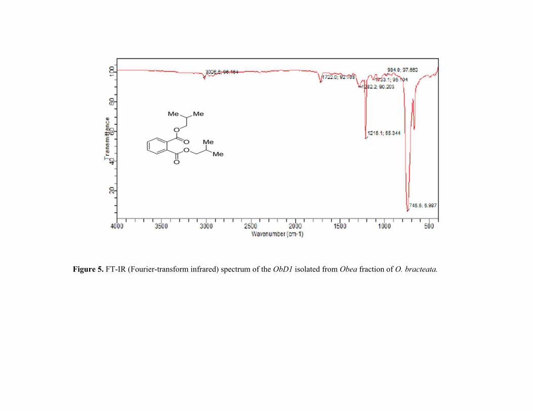
Figure 5. FT-IR (Fourier-transform infrared) spectrum of the ObD1 isolated from Obea fraction of O. bracteata.

Figure 6. The HRMS (High-Resonance Mass Spectroscopy) chromatogram of ObD1 isolated from Obea of O. bracteata.

Figure 7. Cytotoxic potential of ObD1 obtained from Obea of O. bracteata on MG-63, A549, IMR32 and HL-7702 cells after 24 h of treatment. Values are expressed as Mean ± SE at level of significance p ≤ 0.05. Data labels with different letters represent significant
difference among them.

Figure 8. Confocal micrographs of DAPI stained MG-63 cells treated with ObD1 from O. bracteata for 24 h. Arrows show nuclear condensation/fragmentation and formation of apoptotic bodies at a magnification 100× oil immersion objective lens.

Figure 9. (A) The generation of intracellular ROS in MG-63 detected by DCFH-DA staining (M1 represents intact cell population and
M2 represents cells with accumulation of intracellular ROS) (B) Histogram showing generation of intracellular ROS in MG-63 cells (24 h) exposed to ObD1 from O. bracteata. (C) The disruption of mitochondrial membrane potential (ΔΨm) in Mg-63 cells detected by
staining with Rhodamine 123 (M1 represents cells with the disruption of ΔΨm and M2 represents the intact cells.). (D) Histogram showing disruption of mitochondrial membrane potential (ΔΨm) in MG-63 cells (24 h) exposed to ObD1 from O. bracteata. (E) The
treatment of ObD1 from O. bracteata (24 h) induced cell cycle arrest at G0/G1 phase in MG-63 cells detected by Cell cycle analysis kit.
(F) Histogram showing different phases of G0/G1, S, G2/M in MG-63 cells using flow cytometer. (a) Untreated MG-63 cells, (b) MG-
63 cells treated with ObD1 (37.53 µM) for 24 h. Data labels with different letters represent significant difference among them at (p ≤ 0.05).
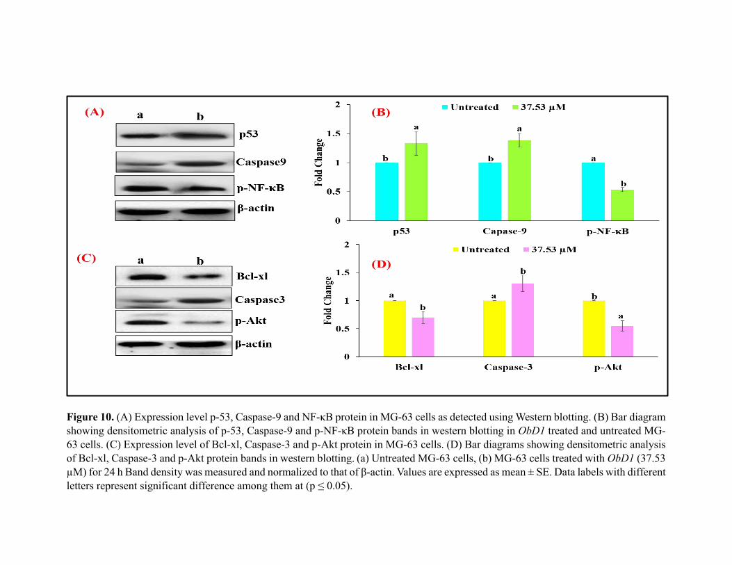
Figure 10. (A) Expression level p-53, Caspase-9 and NF-κB protein in MG-63 cells as detected using Western blotting. (B) Bar diagram
showing densitometric analysis of p-53, Caspase-9 and p-NF-κB protein bands in western blotting in ObD1 treated and untreated MG-
63 cells. (C) Expression level of Bcl-xl, Caspase-3 and p-Akt protein in MG-63 cells. (D) Bar diagrams showing densitometric analysis
of Bcl-xl, Caspase-3 and p-Akt protein bands in western blotting. (a) Untreated MG-63 cells, (b) MG-63 cells treated with ObD1 (37.53 µM) for 24 h Band density was measured and normalized to that of β-actin. Values are expressed as mean ± SE. Data labels with different
letters represent significant difference among them at (p ≤ 0.05).

Figure 11. Effect of ObD1 from O. bracteata on the gene expression for p53, Bcl-2, Caspase-3, CDK2 and Cyclin E genes in MG-63
cells as detected using RT-PCR. Values are expressed as mean ± SE. Data labels with different letters represent significant difference
among them at (p ≤ 0.05).

Figure 12. Docking conformations (PatchDock server) showing interaction of ObD1 with minimum binding energy p-53 and CDK2
binding site, respectively (A) CDK-2 with the binding energy of −133.96 kcal/mol, (B) showed CDK2-ObD1 RIN plot, (C) p53 with
the binding energy of −151.13 kcal/mol, (D) showed P53-ObD1 RIN plot. RIN analysis (RING 2.0 web server) used to showed
interactions among proteins.

Figure 13. Schematic diagram showed the effect of ObD1 isolated from Obea of Onosma bracteata induced apoptosis in osteosarcoma
(MG-63 cells).

Table 1. RT-qPCR primers sequence analysis.
S. No. Primer Name [Acession No.] Product Size Oligonucleotides (5′-3′) sequence Source
1. p53 [NM_000546.5] 199 Forward-TCACTGAAGACCCAGGTCCA
Reverse -TTGGCTGTCCCAGAATGCAA
NCBI
2. Bcl-2 [NM_000633.2] 123 Forward-AGTCTGGGAATCGATCTGGA
Reverse-GGCAACGATCCCATCAATCT
NCBI
3. Cyclin E [NM_001238.3] 150 Forward- GGTATCAGTGGTGCGACATAG
Reverse- CCAAGCTGTCTCTGTGGGTC
NCBI
4. CDK2 [NM_001798] 180 Forward- GGCCCTATTCCCTGGAGATTC
Reverse- CGTCCATCTTCATCCAGGGG
NCBI
5. β-actin [T25383] 166 Forward- GTCCTCTCCCAAGTCACACA
Reverse- GCTCATACATCTCAAGTTGGGAC
NCBI
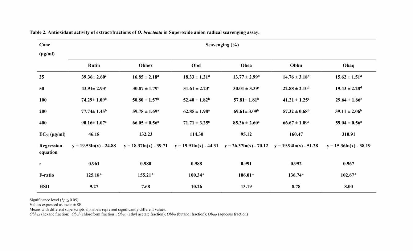
Table 2. Antioxidant activity of extract/fractions of O. bracteata in Superoxide anion radical scavenging assay.
Conc
(μg/ml)
Scavenging (%)
Rutin Obhex Obcl Obea Obbu Obaq
25 39.36± 2.60c 16.85 ± 2.18d 18.33 ± 1.21d 13.77 ± 2.99d 14.76 ± 3.18d 15.62 ± 1.51d
50 43.91± 2.93c 30.87 ± 1.79c 31.61 ± 2.23c 30.01 ± 3.39c 22.88 ± 2.10d 19.43 ± 2.28d
100 74.29± 1.09b 50.80 ± 1.57b 52.40 ± 1.82b 57.81± 1.81b 41.21 ± 1.25c 29.64 ± 1.66c
200 77.74± 1.45b 59.78 ± 1.69a 62.85 ± 1.98a 69.61± 3.09b 57.32 ± 0.68b 39.11 ± 2.06b
400 90.16± 1.07a 66.05 ± 0.56a 71.71 ± 3.25a 85.36 ± 2.60a 66.67 ± 1.09a 59.04 ± 0.56a
EC50 (μg/ml) 46.18 132.23 114.30 95.12 160.47 310.91
Regression
equation
y = 19.53ln(x) - 24.88 y = 18.37ln(x) - 39.71 y = 19.91ln(x) - 44.31 y = 26.37ln(x) - 70.12 y = 19.94ln(x) - 51.28 y = 15.36ln(x) - 38.19
r 0.961 0.980 0.988 0.991 0.992 0.967
F-ratio 125.18* 155.21* 100.34* 106.01* 136.74* 102.67*
HSD 9.27 7.68 10.26 13.19 8.78 8.00
Significance level (*p ≤ 0.05). Values expressed as mean ± SE.
Means with different superscripts alphabets represent significantly different values.
Obhex (hexane fraction); Obcl (chloroform fraction); Obea (ethyl acetate fraction); Obbu (butanol fraction); Obaq (aqueous fraction)

Table 3. Antioxidant activity of extract/fractions of O. bracteata in Lipid peroxidation assay.
Conc
(μg/ml)
Scavenging (%)
Rutin Obhex Obcl Obea Obbu Obaq
25 20.06 ±1.36e 7.91 ± 1.14e 11.38 ± 1.85d 16.50 ± 2.10e 9.79 ± 2.18d 8.83 ± 1.75e
50 39.12 ± 1.76d 19.33 ± 2.23d 19.33 ± 2.68d 31.74 ± 2.78d 16.28 ± 1.06d 16.92 ± 0.99d
100 59.28 ± 3.48c 37.73 ± 4.22c 39.05 ± 3.91c 63.23 ± 2.80c 38.74 ± 1.69c 38.51 ± 1.62c
200 75.09 ± 3.73b 69.68 ± 1.54b 68.73 ± 1.01b 77.32 ± 2.32b 58.55 ± 1.51b 58.96 ± 1.32b
400 89.88 ± 0.83a 90.23 ± 0.44a 90.55 ± 2.50a 91.50 ± 1.04a 92.30 ± 1.19a 92.84 ± 0.95a
EC50 (μg/ml) 76.77 117.52 114.30 80.67 125.79 127.03
Regression
equation y = 25.33ln(x) - 59.97 y = 31.01ln(x) - 97.84 y = 29.96ln(x) - 92.19 y = 28.21ln(x) - 73.88 y = 29.91ln(x) - 94.58 y = 30.02ln(x) - 95.40
r 0.997 0.987 0.983 0.988 0.976 0.975
F-ratio 121.80* 222.03* 167.18* 195.39* 450.18* 604.67*
HSD 11.75 10.76 12.03 10.43 7.37 6.38
Significance level (*p ≤ 0.05). Values expressed as mean ± SE.
Means with different superscripts alphabets represent significantly different values.
Obhex (hexane fraction); Obcl (chloroform fraction); Obea (ethyl acetate fraction); Obbu (butanol fraction); Obaq (aqueous fraction)

Table 4. Cytotoxic effects of ObD1 on A549, IMR-32 and MG-63 cancer cell line in MTT assay.
Conc. (μM) Percent Inhibition (ObD1)
A549 Lung cancer
cell line
IMR-32 neuroblastoma
cell line
MG-63 osteosarcoma
cell line
HL-7702 normal human
hepatocyte cell line
15.625 28.99 ± 2.09d 22.52 ± 1.71c 30.07 ± 1.21d 4.06 ± 0.30d
31.25 38.40 ± 1.45c 28.50 ± 1.87c 42.27 ± 0.82c 8.20 ± 0.68c
62.5 49.77 ± 1.11b 51.58 ± 0.36b 60.75 ± 1.14b 18.63 ± 0.9b
125 76.71 ± 1.19a 72.99 ± 0.90a 82.23 ± 1.24a 26.97 ± 0.60a
Regression equation y = 22.39ln(x) – 36.26 5.24ln(x) – 51.61 y = 25.03ln(x) – 40.73 y = 11.30ln(x) - 28.25
r 0.968 0.976 0.992 0.986
GI50 (μM) 47.12 56.05 37.53 1013.35
Camptothecin (GI50) (µM)
53.37 64.34 52.80 74.84
F-ratio 186.15* 287.77* 412.29* 231.88*
HSD 6.85 6.16 5.07 3.07
Significance level (*p ≤ 0.05). Values expressed as mean ± SE.
Means with different superscripts alphabets represent significantly different values.


![K ]P]vo o Hydroxypropyl-β-Cyclodextrin (HBC ... then prepared complex hydroxyl propyl methyl cellulose controlled released matrix tablets. The ... carrier materials such as Hydroxypropyl](https://static.fdocuments.net/doc/165x107/5ac37c707f8b9af91c8c06a9/k-pvo-o-hydroxypropyl-cyclodextrin-hbc-then-prepared-complex-hydroxyl.jpg)