1. Why do live cell microscopy? 2. Maintaining living ...
78
Live cell microscopy 1. Why do live cell microscopy? 2. Maintaining living cells on a microscope stage. 3. Considerations for imaging living cells. 4. Fluorescence labeling of living cells. 5. Imaging polarized cells on permeable filters.
Transcript of 1. Why do live cell microscopy? 2. Maintaining living ...
Live cell microscopy
1. Why do live cell microscopy?2. Maintaining living cells on a microscope stage.3. Considerations for imaging living cells.4. Fluorescence labeling of living cells.5. Imaging polarized cells on permeable filters.
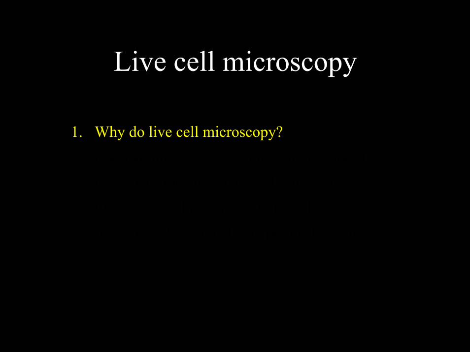
Why Do Live Cell Imaging? We want to understand the rules of biology, but
most of our information comes from fixed samples
Before After
What are the Rules?- Dave Piston, Vanderbilt

Why Do Live Cell Imaging? Or we grind up the sample and run a gel
Nose Tackles
Linemen
LinebackersRunning BacksReceiversDefensive Backs
Now what do we know about the rules?
- Dave Piston, Vanderbilt

Why Do Live Cell Imaging? If you want to learn the rules, you have to watch
- Dave Piston, Vanderbilt

Live cell microscopy – imaging cell dynamics
Focal adhesion dynamics imaged by reflection interference contrast
- Marysse Bailly, Einstein

Live cell microscopy – imaging cell physiology
Tsien and Miyawaki, 1998

Live cell microscopy – poorly fixed probes
Live cell microscopy
1. Why do live cell microscopy?2. Maintaining living cells on a microscope stage.3. Considerations for imaging living cells.4. Fluorescence labeling of living cells.5. Imaging polarized cells on permeable filters.

Culturing live cells on the microscope stage
• pH control (gassing your cells)• medium manipulations - perfusion• temperature control• where did the cells go? - focus drift

Commercial stage heaters

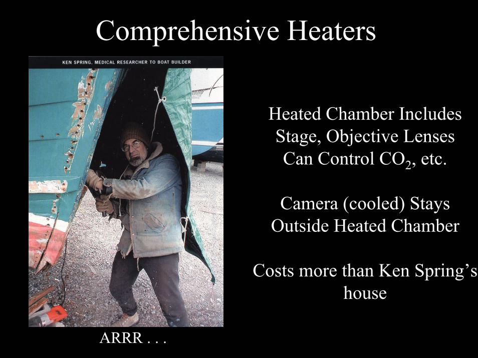
Comprehensive Heaters

Comprehensive Heaters
Heated Chamber IncludesStage, Objective LensesCan Control CO2, etc.
Camera (cooled) StaysOutside Heated Chamber
Costs more than Ken Spring’s house
Comprehensive Heaters
ARRR . . .
Heated Chamber IncludesStage, Objective LensesCan Control CO2, etc.
Camera (cooled) StaysOutside Heated Chamber
Costs more than Ken Spring’s house

Live cell microscopy
1. Why do live cell microscopy?2. Maintaining living cells on a microscope stage.3. Considerations for imaging living cells.4. Fluorescence labeling of living cells.5. Imaging polarized cells on permeable filters.
Live cell microscopy
1. Why do live cell microscopy?2. Maintaining living cells on a microscope stage.3. Considerations for imaging living cells.
EVERYTHING IS THAT MUCH HARDER.1. Fluorescence labeling of living cells.2. Imaging polarized cells on permeable filters.

Limits to fluorescence microscopy
• Limited number of photons
• Limited flux of photons
• Limited resolution

Fluorescence runs out - photobleaching always accompanies fluorescence excitation
• For every photon absorbed, there is a fixed probability of either fluorescence emission or photobleaching.
•The total number of fluorescence photons emitted is given by the ratio of the quantum efficiency of fluorescence to the quantum efficiency of photobleaching or Qe/Qb
• For fluorescein this works out to around 30,000 photons.
• Since we collect ~1% of the photons - “every photon is sacred”
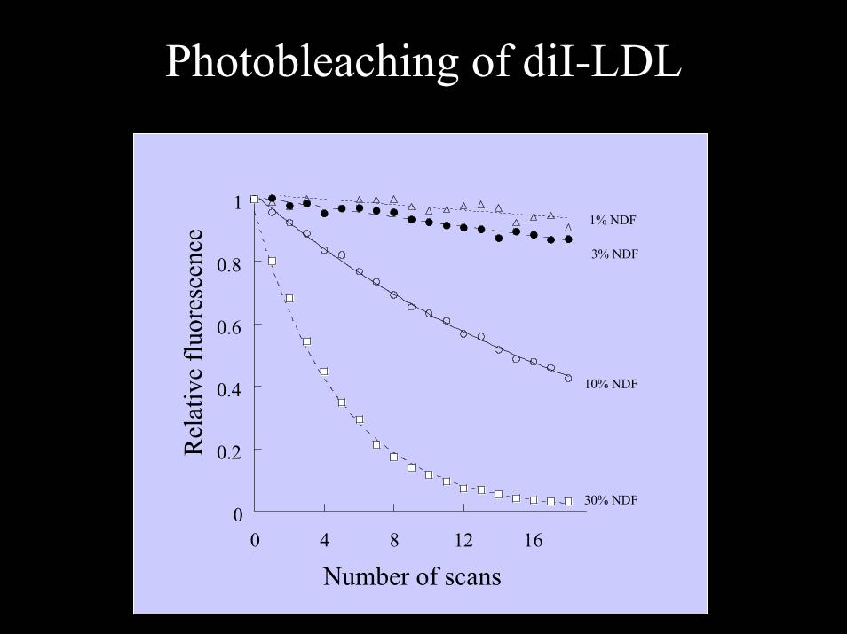
Photobleaching of diI-LDL
0
0.2
0.4
0.6
0.8
1
0 4 8 12 16
Rel
ativ
e flu
ores
cenc
e
Number of scans
1% NDF
3% NDF
10% NDF
30% NDF
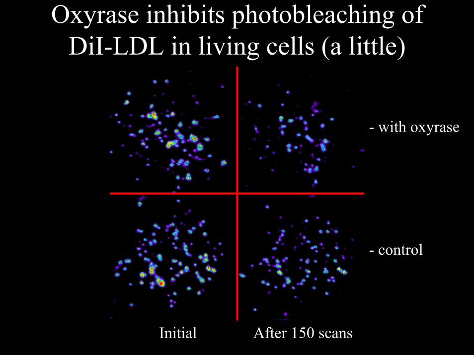
Oxyrase inhibits photobleaching of DiI-LDL in living cells (a little)
- control
- with oxyrase
Initial After 150 scans
Limits to fluorescence microscopy
• Limited number of photons
• Limited flux of photons
• Limited resolution

Limits to photon flux –Excitation Saturation
• The rate of emission is dependent upon the time the molecule remains within the excitation state (the excited state lifetime τf)
• Optical saturation occurs when the rate of excitation exceeds the reciprocal of τf
• In a scanned image of 512 x 768 pixels (400,000 pixels) if scanned in 1 second requires a dwell time per pixel of 2 x 10⁻⁶ sec.
• Molecules that remain in the excitation beam for extended periods have higher probability of interstate crossings and thus phosphorescence
• Usually, increasing dye concentration can be the most effective means of increasing signal when energy is not the limiting factor (ielaser based confocal systems)
Limits to fluorescence microscopy
• Limited number of photons
• Limited flux of photons
• Limited resolution

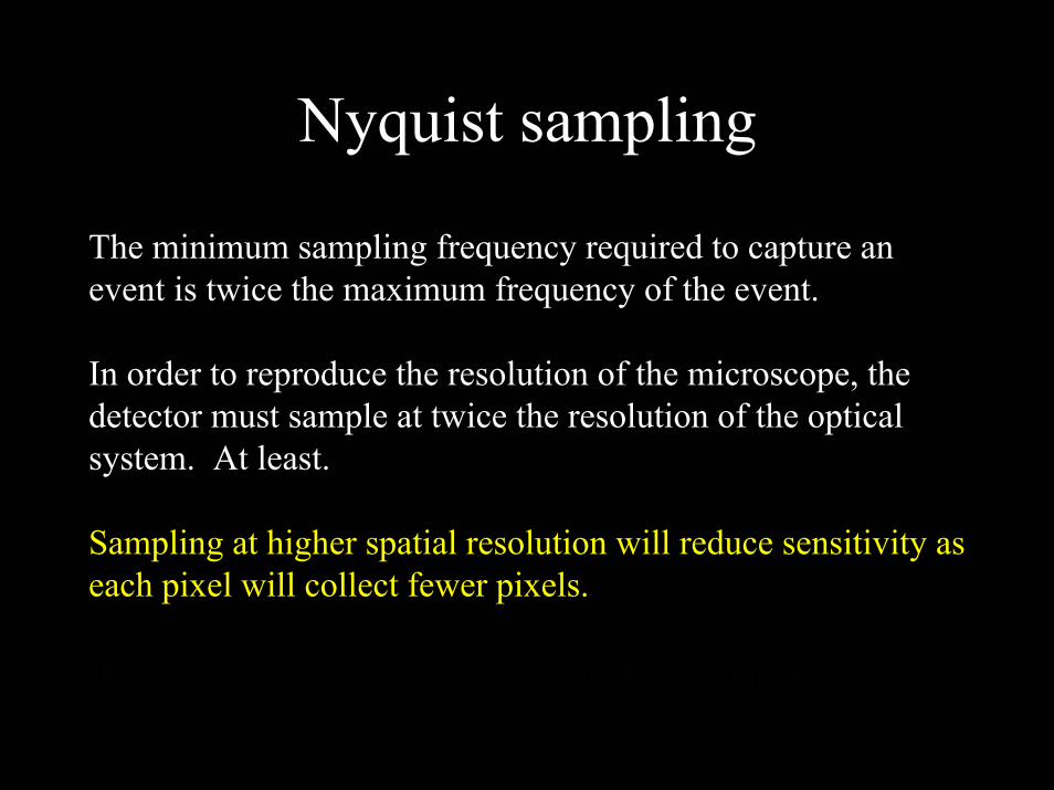
Nyquist sampling
The minimum sampling frequency required to capture an event is twice the maximum frequency of the event.
In order to reproduce the resolution of the microscope, the detector must sample at twice the resolution of the optical system. At least.
Sampling at higher spatial resolution will reduce sensitivity aseach pixel will collect fewer pixels.
The same rule, and consequences applie to temporal resolution.
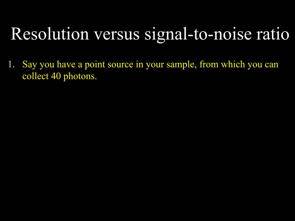
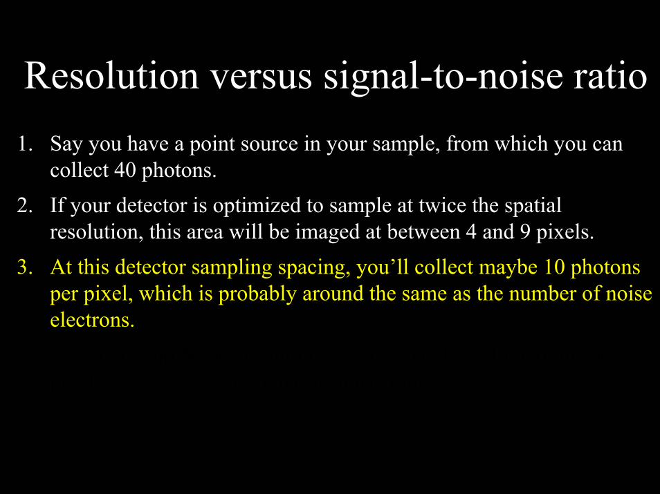
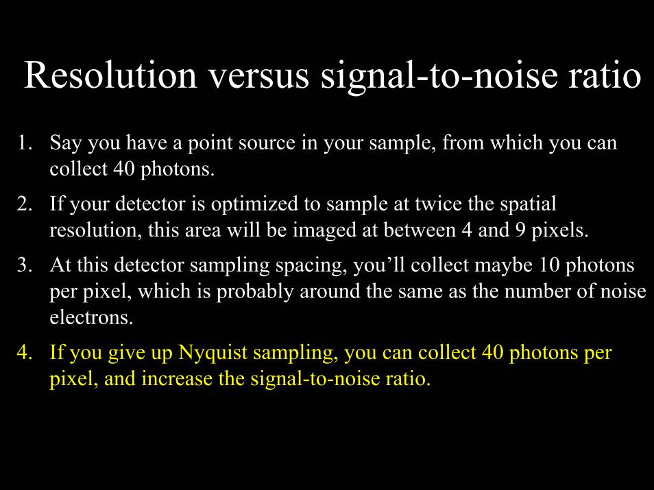
Resolution versus signal-to-noise ratio1. Say you have a point source in your sample, from which you can
collect 40 photons.2. If your detector is optimized to sample at twice the spatial
resolution, this area will be imaged at between 4 and 9 pixels.3. At this detector sampling spacing, you’ll collect maybe 10 photons
per pixel, which is probably around the same as the number of noise electrons.
4. If you give up Nyquist sampling, you can collect 40 photons per pixel, and increase the signal-to-noise ratio.

Live cell microscopy
So, fluorescence microscopy is limited by the number of photons that can be collected from a minimal volume.
The problem is aggravated in live cell microscopy, since cells are dynamic, they move, they move things, they change their constitution. Quickly.
So now you have to trade resolution, signal-to-noise and speed off of one another.

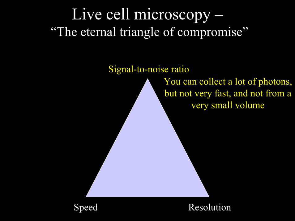
Live cell microscopy –“The eternal triangle of compromise”
Signal-to-noise ratio
Speed Resolution
Live cell microscopy –“The eternal triangle of compromise”
You can collect a lot of photons, but not very fast, and not from a
very small volume
Signal-to-noise ratio
Speed Resolution

Live cell microscopy –“The eternal triangle of compromise”
Signal-to-noise ratio
You can collect photons rapidly, but
not very many and not from a small area.
Speed Resolution

Live cell microscopy –“The eternal triangle of compromise”
Signal-to-noise ratio
You can collect photons rapidly, but
not very many and not from a small area.And maybe not in
more than one colorSpeed Resolution

Live cell microscopy –“The eternal triangle of compromise”
Signal-to-noise ratio
You can collect photons from a small area, but not very many, and not very
fast.
Speed Resolution
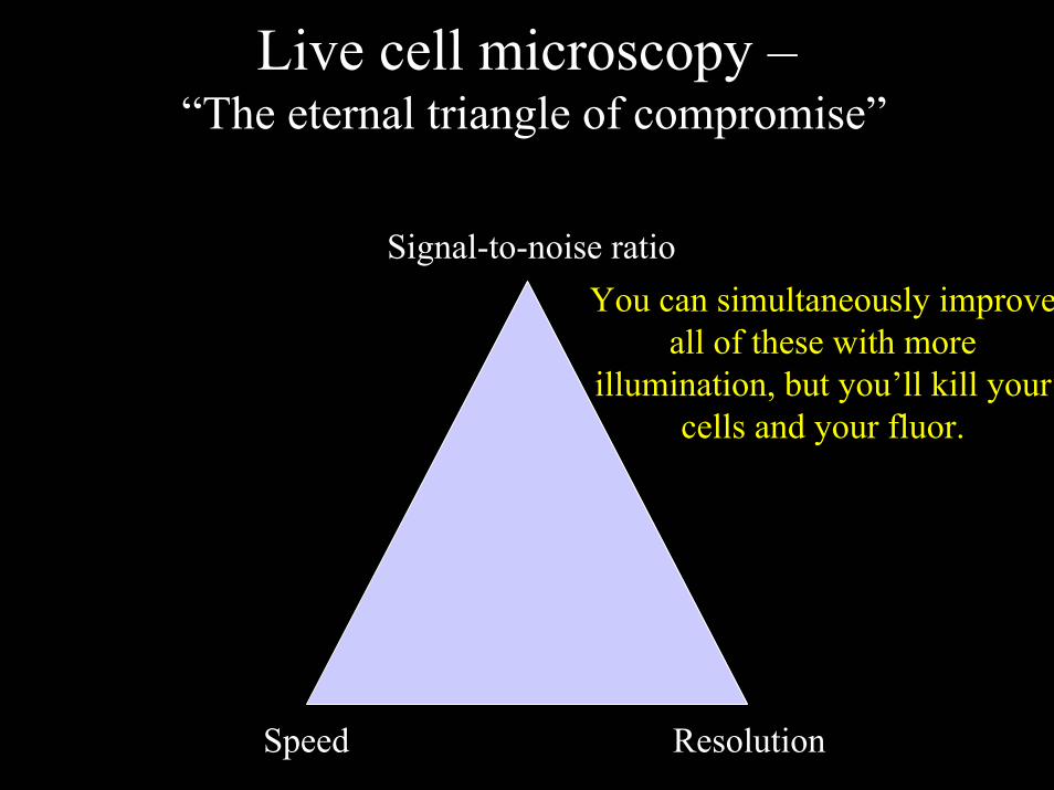
Live cell microscopy –“The eternal triangle of compromise”
Signal-to-noise ratioYou can simultaneously improve
all of these with more illumination, but you’ll kill your
cells and your fluor.
Speed Resolution

PhototoxicityPhototoxicity is the absolute limiting factor in live cell
imaging
– Excitation of fluorescent molecules in the presence of oxygen leads to fluorochrome bleaching and free radical generation.
– Free radicals kill cells.– The interaction of light with cells > heat– Heat kills cells.
If individual images are well separated temporally this effect can be minimized.

How do you recognize photodamage when imaging dynamics in living cells?
Compare your imaged cells to their non-imaged neighbors.
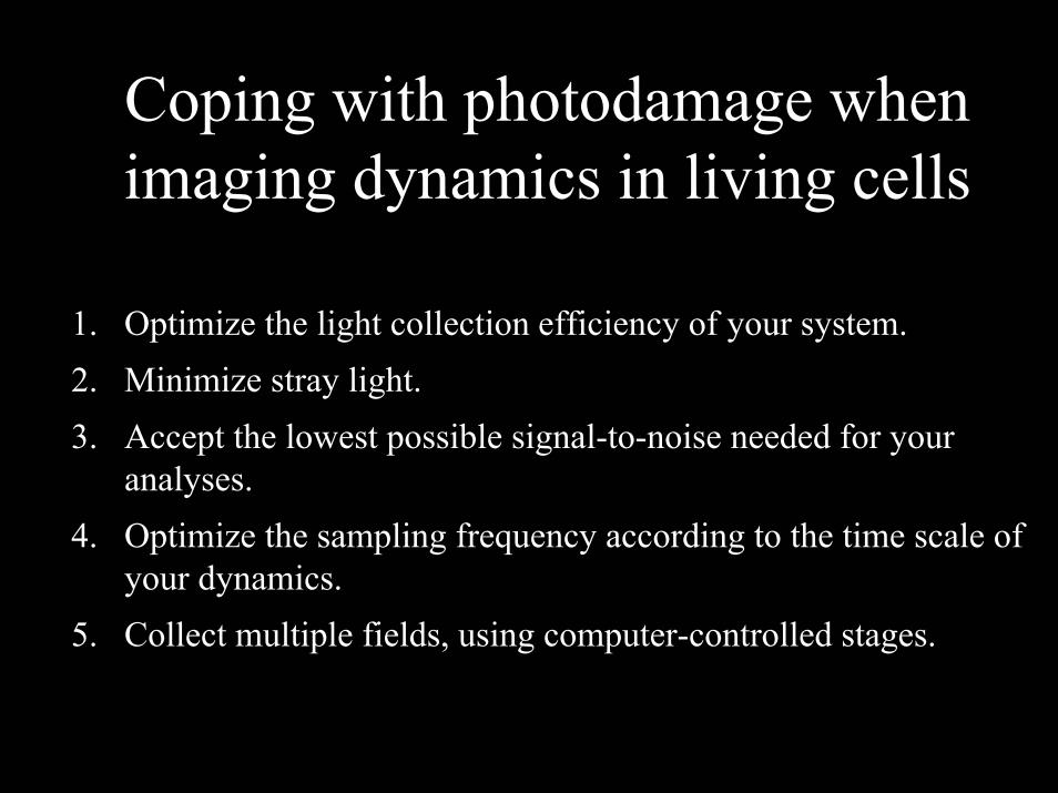
Coping with photodamage when imaging dynamics in living cells
1. Optimize the light collection efficiency of your system.2. Minimize stray light.3. Accept the lowest possible signal-to-noise needed for your
analyses.4. Optimize the sampling frequency according to the time scale of
your dynamics.5. Collect multiple fields, using computer-controlled stages.

Computer-driven stage controllers
Marzhauser, Inc.

Live cell microscopy
1. Why do live cell microscopy?2. Maintaining living cells on a microscope stage.3. Considerations for imaging living cells.4. Fluorescence labeling of living cells.5. Imaging polarized cells on permeable filters.
Fluorescent labeling of cells
• Immunofluorescence• Protein-binding toxins• DNA intercalating dyes• DNA hybridization• Permeant dyes• Membrane partitioning dyes• Endocytosis of fluorescent molecules• Microinjection of fluorescent molecules• Expression of fluorescent chimeras
Fluorescent labeling of cells
• Immunofluorescence• Protein-binding toxins• DNA intercalating dyes• DNA hybridization• Permeant dyes• Membrane partitioning dyes• Endocytosis of fluorescent molecules• Microinjection of fluorescent molecules• Expression of fluorescent chimeras
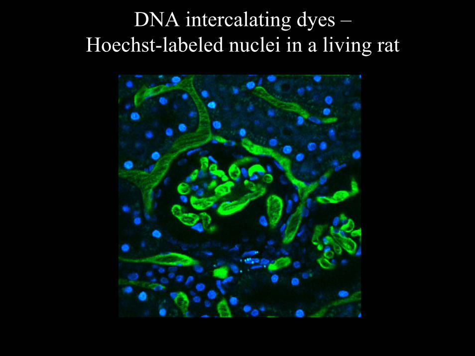
DNA intercalating dyes –Hoechst-labeled nuclei in a living rat
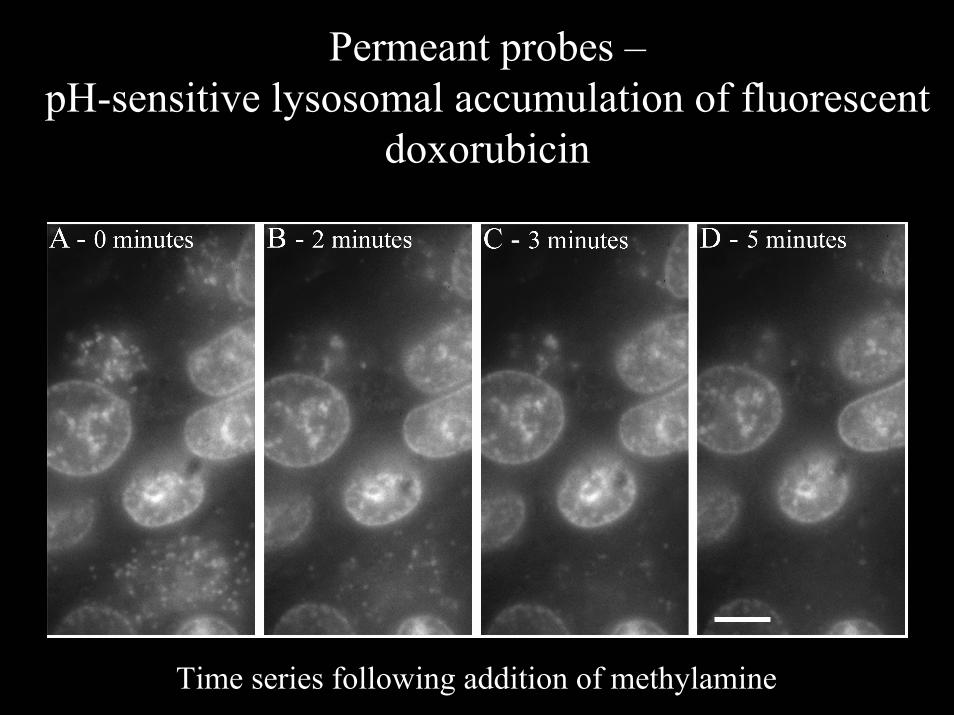
Permeant probes –pH-sensitive lysosomal accumulation of fluorescent
doxorubicin
Time series following addition of methylamine

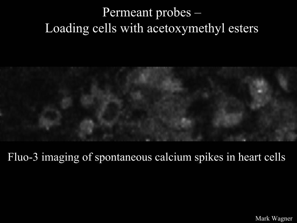
Permeant probes –Loading cells with acetoxymethyl esters
380nm
340nm
340/380
- UniversalImaging

Permeant probes –Loading cells with acetoxymethyl esters
- Molecular Probes, Inc.
Permeant probes –Loading cells with acetoxymethyl esters
Fluo-3 imaging of spontaneous calcium spikes in heart cells
Mark Wagner
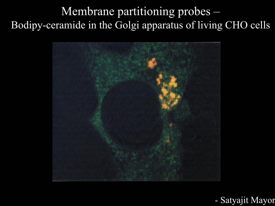
Membrane partitioning probes –Bodipy-ceramide in the Golgi apparatus of living CHO cells
- Satyajit Mayor

Membrane partitioning probes –Detecting changes in membrane polarity with FRET
Coumarin labeled phospholipid – immobile in outer leafletOxonol – charged, mobile in membrane
Gonzalez et al., 1997

Endocytosis of fluorescent probes by living cells
Internalized transferrin and GFP-Rab25 in living MDCK cells

Endocytosis of fluorescent probes by living cells
Optical sectioning of confocal allows distinction of extracellular (neutral) FR-Tf from endosomal (acidic) FR-Tf

Microinjection of fluorescent probes
Ratio of rhodamine myosin II to cy2dextranDeBiasio et al., 1996. MBC (Lance Taylor lab)

Expression of fluorescent protein chimeras –Green Fluorescent Protein
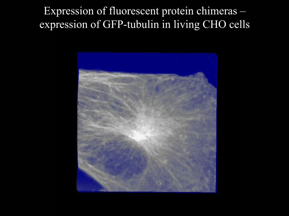
Expression of fluorescent protein chimeras –expression of GFP-tubulin in living CHO cells
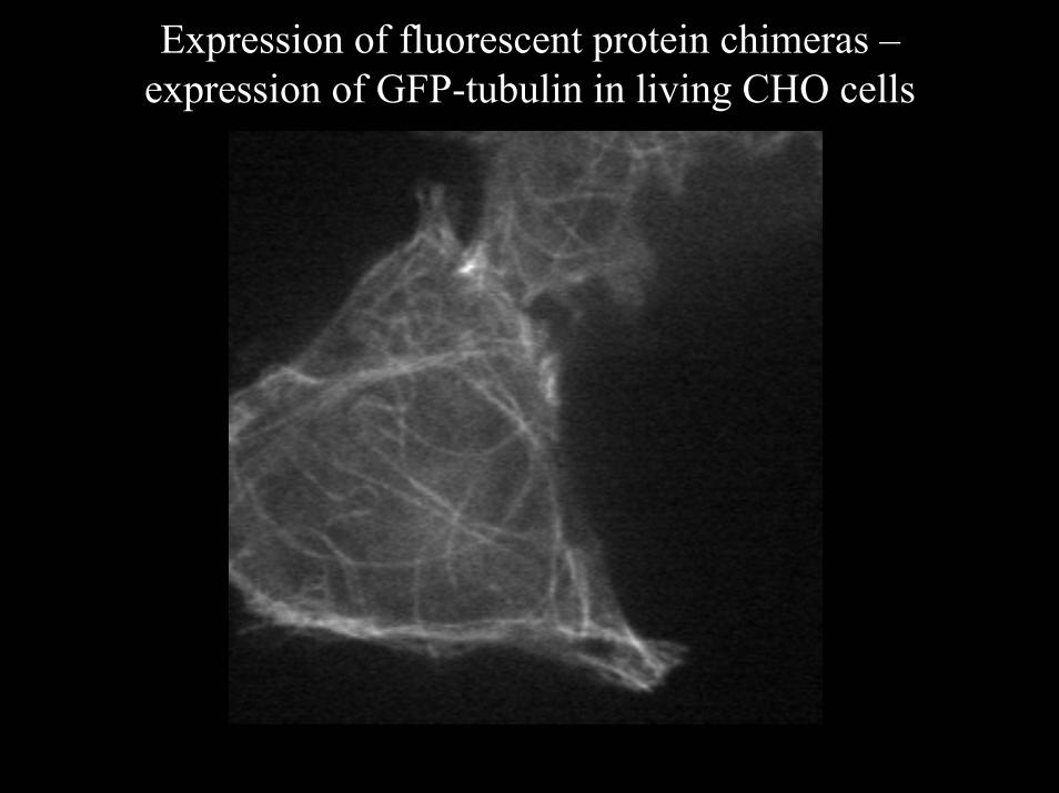
Expression of fluorescent protein chimeras –expression of GFP-tubulin in living CHO cells
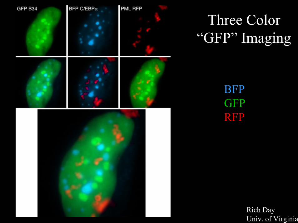
Three Color “GFP” Imaging
BFPGFPRFP
Rich Day Univ. of Virginia

Live cell microscopy
1. Why do live cell microscopy?2. Maintaining living cells on a microscope stage.3. Considerations for imaging living cells.4. Fluorescence labeling of living cells.5. Imaging polarized cells on permeable filters.

Approach – Fluorescence microscopy of cells incubated with fluorescent ligands
Microscopeobjective
Permeablegrowth insert
45 µ spacer
coverslip
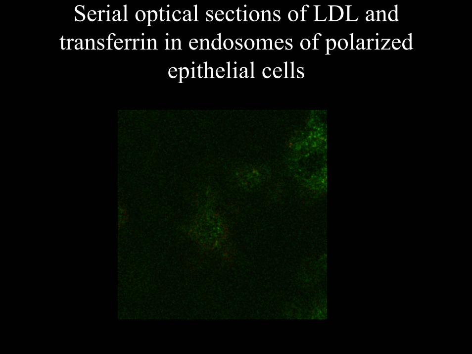
Serial optical sections of LDL and transferrin in endosomes of polarized
epithelial cells

3D microscopy of polarized MDCK cells –internalized IgA and anti-cadherin

Transferrin endocytosis in polarized MDCK cells
After 2 minutes exposureto fluorescent transferrin

Live cell microscopy of polarized MDCK cells on permeable supports
Apical IgA (green) and basalTf (red) - medial plane
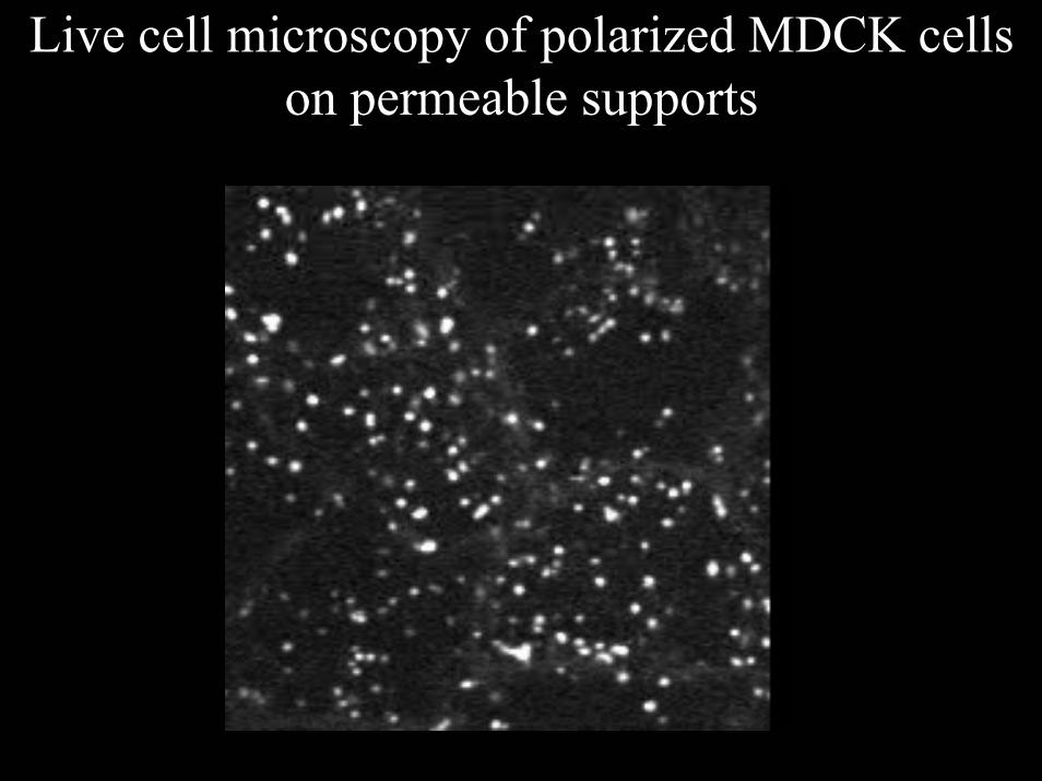
Live cell microscopy of polarized MDCK cells on permeable supports
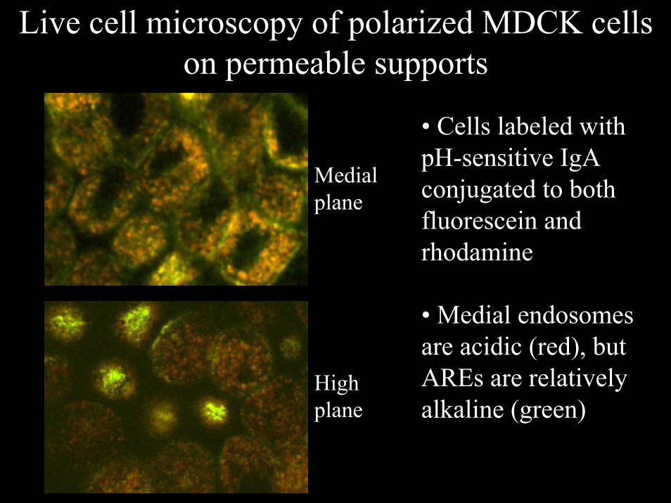
Live cell microscopy of polarized MDCK cells on permeable supports
• Cells labeled with pH-sensitive IgA conjugated to both fluorescein and rhodamine
• Medial endosomes are acidic (red), but AREs are relatively alkaline (green)
Medialplane
Highplane
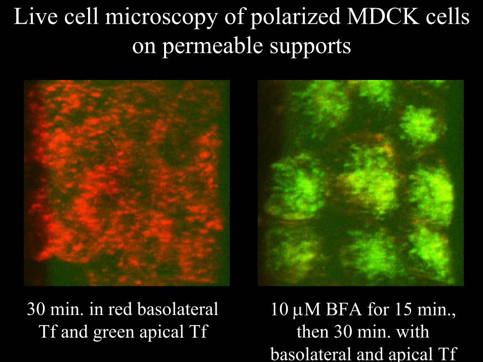
Live cell microscopy of polarized MDCK cells on permeable supports
10 µM BFA for 15 min., then 30 min. with
basolateral and apical Tf
30 min. in red basolateral Tf and green apical Tf
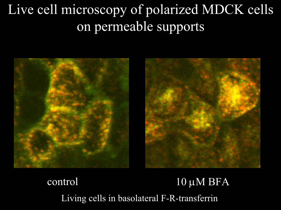
Live cell microscopy of polarized MDCK cells on permeable supports
control 10 µM BFALiving cells in basolateral F-R-transferrin
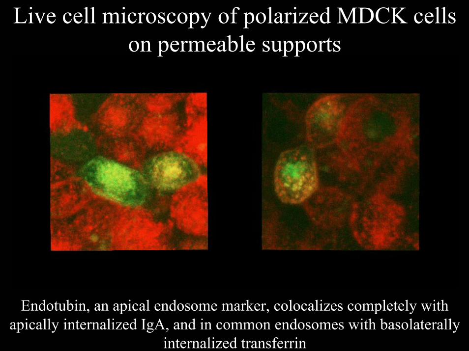
Live cell microscopy of polarized MDCK cells on permeable supports
Endotubin, an apical endosome marker, colocalizes completely with apically internalized IgA, and in common endosomes with basolaterally
internalized transferrin

Live cell microscopy of polarized MDCK cells on permeable supports
Rme-1 and IgA

Imaging living cells on filters – use a water immersion objective
60X water immersion
0 microns 50 microns 60 microns
100X Oil immersion
0 microns 40 microns 50 microns

Which kind of microscope for living cells?
•Inverted.•Widefield or confocal?•Single-point or multi-point scanning confocal?•Confocal or Multiphoton?

Which kind of microscope for living cells?
•Inverted.•Widefield or confocal?•Single-point or multi-point scanning confocal?•Confocal or Multiphoton?

Which kind of microscope for living cells?
•Inverted.•Widefield or confocal?•Single-point or multi-point scanning confocal?•Confocal or Multiphoton?

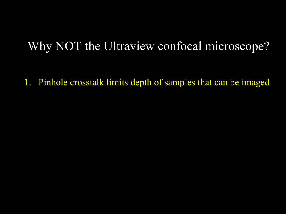
Why NOT the Ultraview confocal microscope?
1. Pinhole crosstalk limits depth of samples that can be imaged.2. Multicolor images must be collected sequentially.3. Single mode fiber optic – non-uniform illumination.
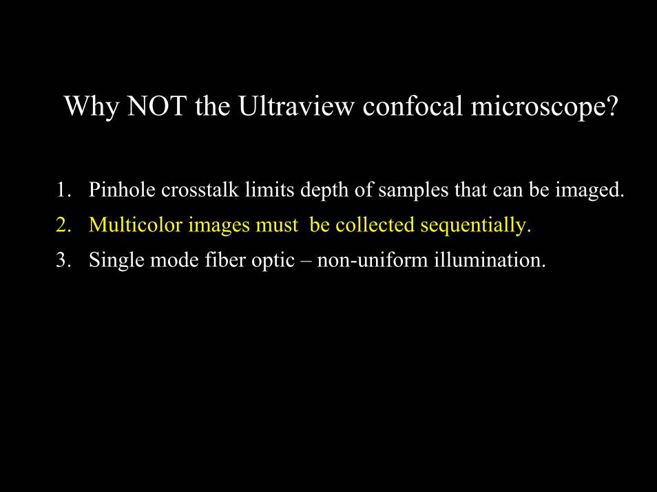
Why NOT the Ultraview confocal microscope?
1. Pinhole crosstalk limits depth of samples that can be imaged.2. Multicolor images must be collected sequentially.3. Single mode fiber optic – non-uniform illumination.

Spinning disk systems
Pinhole crosstalk
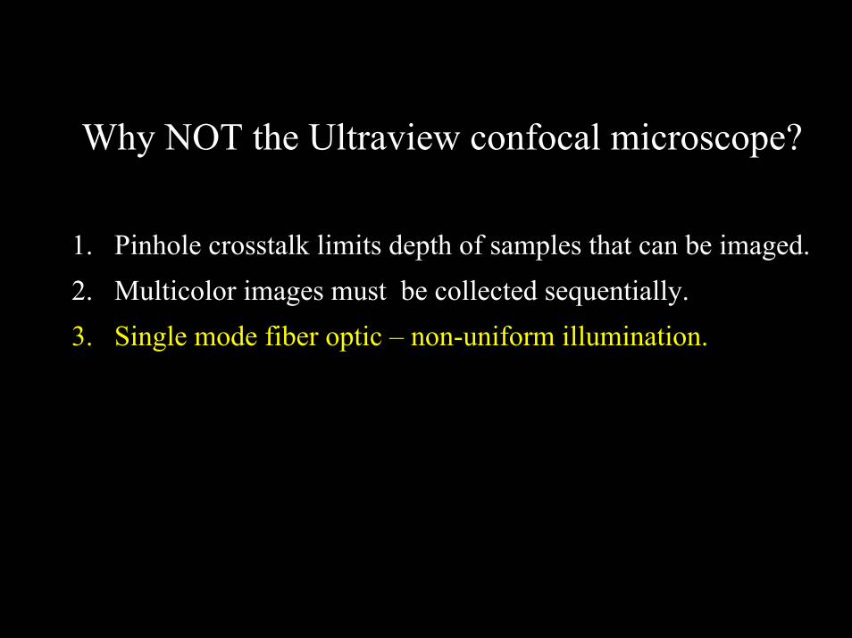
Why NOT the Ultraview confocal microscope?
1. Pinhole crosstalk limits depth of samples that can be imaged.2. Multicolor images must be collected sequentially.3. Single mode fiber optic – non-uniform illumination.
Why NOT the Ultraview confocal microscope?
1. Pinhole crosstalk limits depth of samples that can be imaged.2. Multicolor images must be collected sequentially.3. Single mode fiber optic – non-uniform illumination.

Which kind of microscope for living cells?
•Inverted.•Widefield or confocal?•Single-point or multi-point scanning confocal?•Confocal or Multiphoton?