1 Chromosomes Dr Pupak Derakhshandeh, PhD Ass Prof Medical Science of Tehran University.
108
1 Chromosomes Chromosomes Dr Pupak Derakhshandeh, PhD Ass Prof Medical Science of Tehran University
-
Upload
eugene-banks -
Category
Documents
-
view
217 -
download
1
Transcript of 1 Chromosomes Dr Pupak Derakhshandeh, PhD Ass Prof Medical Science of Tehran University.

1
ChromosomChromosomeses
Dr Pupak Derakhshandeh, PhD
Ass Prof Medical Science of Tehran University
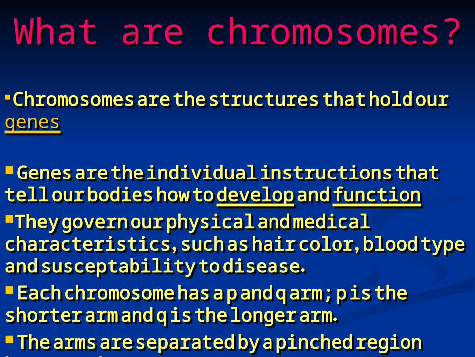
What are chromosomes?What are chromosomes?
Chromosomes are the structures that hold Chromosomes are the structures that hold our our genesgenes Genes are the individual instructions that Genes are the individual instructions that tell our bodies how to tell our bodies how to developdevelop and and functionfunctionTheThey govern our physical and medical y govern our physical and medical characteristics, such as hair color, blood characteristics, such as hair color, blood type and susceptability to disease.type and susceptability to disease. Each chromosome has a p and q arm; p is Each chromosome has a p and q arm; p is the shorter arm and q is the longer arm.the shorter arm and q is the longer arm. The arms are separated by a pinched The arms are separated by a pinched region known as the region known as the centromerecentromere
3

4
How many chromosomes do How many chromosomes do humans have?humans have?
The typical number of The typical number of chromosomes in a human cell is 46 chromosomes in a human cell is 46 - two pairs of 2- two pairs of 22 + XX/XY2 + XX/XY
HHolding an estimated 30,000 to olding an estimated 30,000 to 35,000 genes.35,000 genes.
One set of 23 chromosomes is One set of 23 chromosomes is inherited from the biological inherited from the biological mother (from the egg), and the mother (from the egg), and the other set is inherited from the other set is inherited from the biological father (from the sperm).biological father (from the sperm).

5
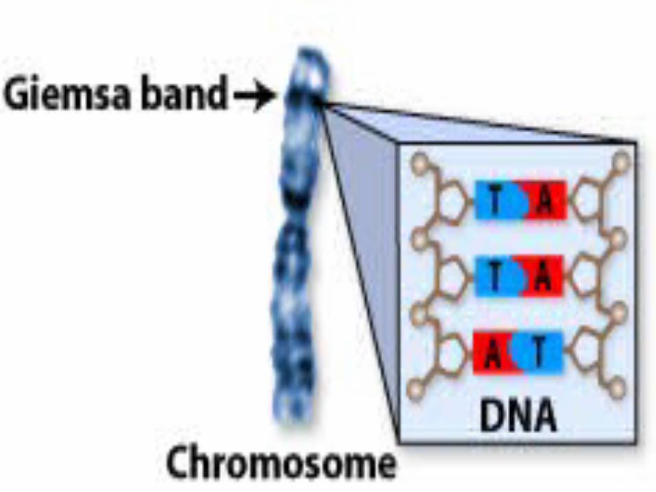
study of the study of the chromosomeschromosomes
with a microscope with a microscope , then , then StainStainningning TThe chromosomes look like strings he chromosomes look like strings
with light and dark "bands" with light and dark "bands" A picture, or chromosome map, of A picture, or chromosome map, of
all 46 chromosomes is called a all 46 chromosomes is called a karyotypekaryotype
The karyotype The karyotype :: identify chromosome identify chromosome abnormalitiesabnormalities:: that are evident that are evident in in either the structure or the number of either the structure or the number of chromosomes.chromosomes.
6
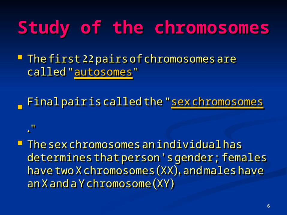
Study of the Study of the chromosomeschromosomes
The first 22 pairs of chromosomes are The first 22 pairs of chromosomes are called "called "autosomesautosomes" "
FFinal pair is called the "inal pair is called the "sex chromosomessex chromosomes." ."
The sex chromosomes an individual has The sex chromosomes an individual has determines that person's gender; determines that person's gender; females have two X chromosomes (XX), females have two X chromosomes (XX), and males have an X and a Y and males have an X and a Y chromosome (XY)chromosome (XY)

7

8
Karyotype Karyotype 4646)), Xy), Xy)
9
10
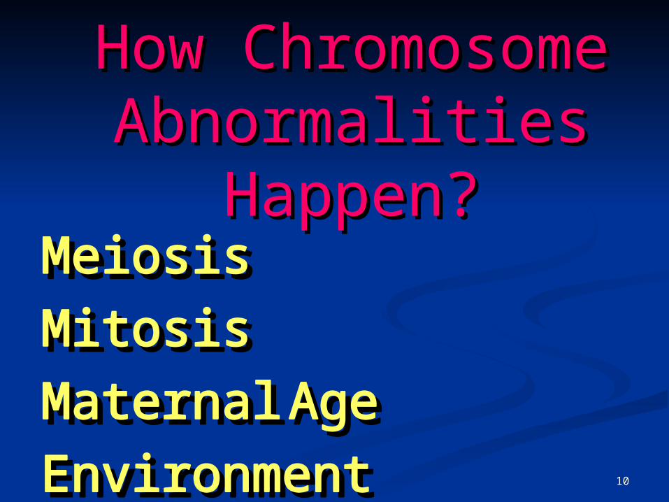
How Chromosome How Chromosome Abnormalities Abnormalities
Happen?Happen?MeiosisMeiosis
MitosisMitosis
Maternal AgeMaternal Age
EnvironmentEnvironment
11
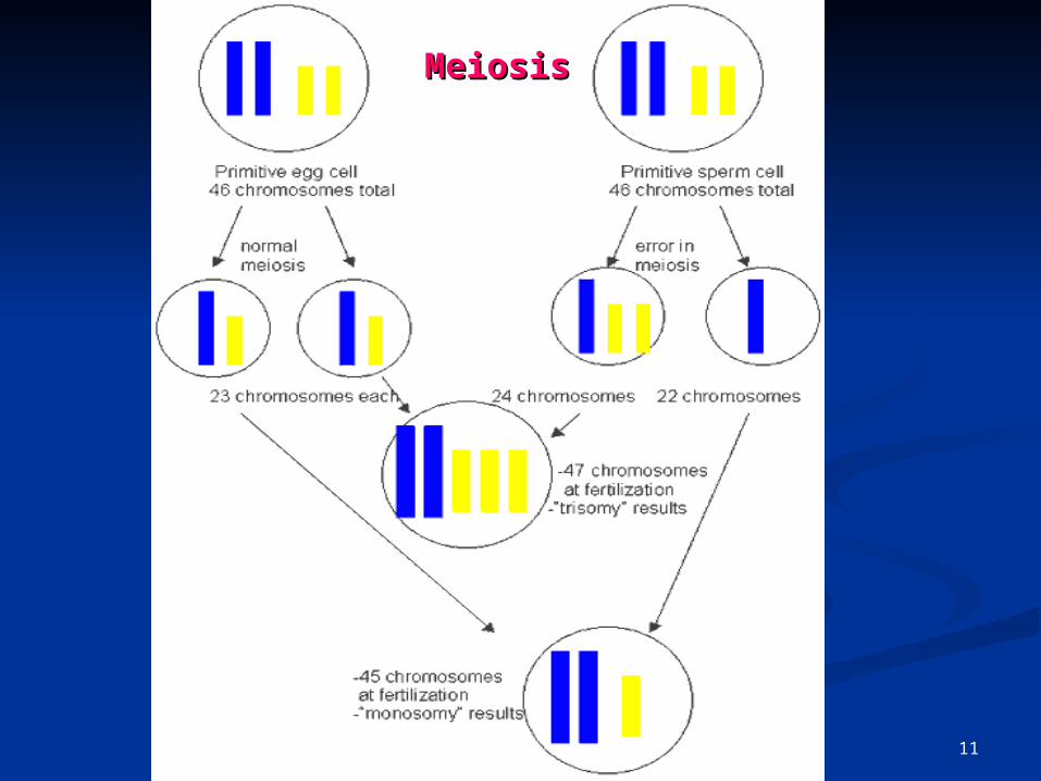
MeiosisMeiosis

12
MeiosisMeiosis

13

14
Chromosome Chromosome abnormalitiesabnormalities
AAbnormality of chromosome bnormality of chromosome number or structure:number or structure:
Numerical Numerical AbnormalitiesAbnormalities
Structural AbnormalitiesStructural Abnormalities

15
Numerical AbnormalitiesNumerical Abnormalities When an individual is missing either a When an individual is missing either a
chromosome from a pair (chromosome from a pair (monosomymonosomy) or ) or has more than two chromosomes of a pair has more than two chromosomes of a pair ((trisomytrisomy).).
An exampleAn example: : Down Syndrome, also known Down Syndrome, also known as Trisomy 21 (an individual with Down as Trisomy 21 (an individual with Down Syndrome has three copies of Syndrome has three copies of chromosome 21, rather than two).chromosome 21, rather than two).
Turner Syndrome is an example of Turner Syndrome is an example of monosomy the individual is born with only monosomy the individual is born with only one sex chromosome, an X.one sex chromosome, an X.
Kleinfelter Kleinfelter Syndrome is an example ofSyndrome is an example of tritrisomy the individual is born with somy the individual is born with threethree sex chromosome,sex chromosome, XXY XXY..

16
Trisomy 18, 47 Ch.Trisomy 18, 47 Ch.

17
Trisomy 18, 47 Ch.Trisomy 18, 47 Ch. incidence of about 1 in 3,000 incidence of about 1 in 3,000 There is a 3:1 preponderance of females There is a 3:1 preponderance of females
to malesto males Thirty percent of affected newborns die Thirty percent of affected newborns die
within the first monthwithin the first month 50% by two months50% by two months and 90% by one year.and 90% by one year. severe mental retardationsevere mental retardation microcephalymicrocephaly overlapping fingers, and rocker bottom overlapping fingers, and rocker bottom
feetfeet Neurologically they are hypertonic Neurologically they are hypertonic Other common malformations include Other common malformations include
congenital heart, kidney, .... congenital heart, kidney, .... abnormalities.abnormalities.

18
Trisomy 18, 47 Ch.Trisomy 18, 47 Ch.

19
20
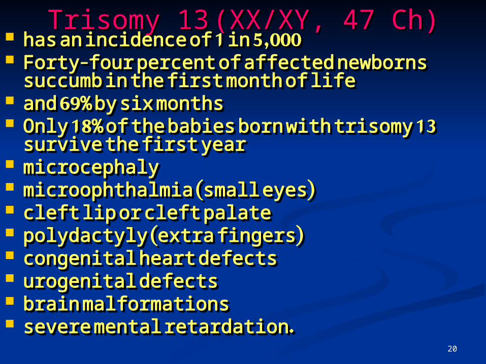
Trisomy 13Trisomy 13 (XX/XY, 47 Ch)(XX/XY, 47 Ch) has an incidence of 1 in 5,000has an incidence of 1 in 5,000 Forty-four percent of affected newborns Forty-four percent of affected newborns
succumb in the first month of life succumb in the first month of life and 69% by six monthsand 69% by six months Only 18% of the babies born with trisomy Only 18% of the babies born with trisomy
13 survive the first year13 survive the first year microcephalymicrocephaly microophthalmia (small eyes)microophthalmia (small eyes) cleft lip or cleft palatecleft lip or cleft palate polydactyly (extra fingers)polydactyly (extra fingers) congenital heart defectscongenital heart defects urogenital defectsurogenital defects brain malformationsbrain malformations severe mental retardation.severe mental retardation.

21
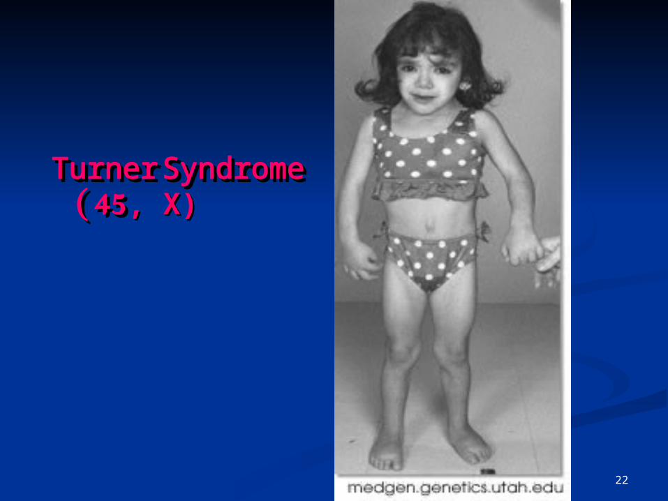
Turner SyndromeTurner Syndrome ( ( 45 45, , X)X)
4545, X, X
22
Turner Turner SyndromeSyndrome ( (4545, , X)X)

23
Turner syndromeTurner syndrome• OOnly femalesnly females• OOne X chromosome ne X chromosome • OOr has two X chromosomes but r has two X chromosomes but one is one is damageddamaged• SShort staturehort stature• DDelayed growth of the skeletonelayed growth of the skeleton• SSometimes heart abnormalitiesometimes heart abnormalities• UUsually infertile due to ovarian sually infertile due to ovarian failurefailure• Diagnosis is by blood test Diagnosis is by blood test (karyotype)(karyotype)• 1 out of every 2,500 female live 1 out of every 2,500 female live births births worldwideworldwide• SShort neck with a webbed hort neck with a webbed appearanceappearance

24
KleinefelterKleinefelterXXYXXY

25
KleinefelterKleinefelter/47/47XXYXXY
26
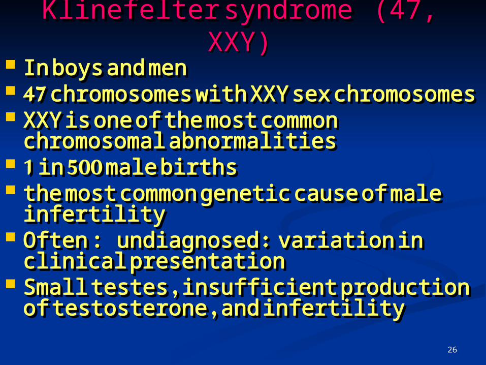
Klinefelter syndrome Klinefelter syndrome (47, XXY) (47, XXY) IIn boys and men n boys and men 47 chromosomes with XXY sex 47 chromosomes with XXY sex
chromosomeschromosomes XXY is one of the most common XXY is one of the most common
chromosomal abnormalitieschromosomal abnormalities 1 in 500 male births1 in 500 male births the most common genetic cause of the most common genetic cause of
male infertilitymale infertility OOften ften : : undiagnosed undiagnosed :: variation in variation in
clinical presentation clinical presentation SSmall testes , insufficient production mall testes , insufficient production
of testosterone , and infertility of testosterone , and infertility

27
Klinefelter syndrome Klinefelter syndrome (47, XXY) (47, XXY)
BBreast enlargement, lack of facial reast enlargement, lack of facial and body hair, a rounded body type and body hair, a rounded body type , to be overweight , and be taller , to be overweight , and be taller than their fathers and brothersthan their fathers and brothers
LLearning and/or behavioral earning and/or behavioral problemsproblems
Testosterone replacement corrects Testosterone replacement corrects the symptoms of androgen the symptoms of androgen deficiency deficiency
28
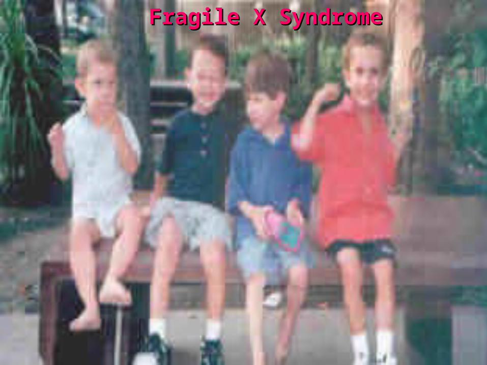
Fragile X SyndromeFragile X Syndrome 1 in 3,600 males and 1 in 4,000 to 1 in 3,600 males and 1 in 4,000 to
6,000 females with the full mutation 6,000 females with the full mutation worldwideworldwide
It is estimated that 1 in 250 females It is estimated that 1 in 250 females and 1 in 700 males are carriers of and 1 in 700 males are carriers of the premutation. the premutation.
It is second only to Down Syndrome It is second only to Down Syndrome as a cause of mental retardation as a cause of mental retardation
Fragile X syndrome appears in Fragile X syndrome appears in children of all ethnic, racial and children of all ethnic, racial and socio-economic backgrounds socio-economic backgrounds

29
Fragile X SyndromeFragile X Syndrome most common most common inheritedinherited form of familial mental form of familial mental
retardationretardation (CGG)n trinucleotide expansion in the FMR1 (CGG)n trinucleotide expansion in the FMR1
gene leading to the typical Martin-Bell gene leading to the typical Martin-Bell phenotypephenotype
Clinical features vary depending on age and Clinical features vary depending on age and seseXX
Expansion of a (CCG)n repeat in the FMR2 gene Expansion of a (CCG)n repeat in the FMR2 gene corresponds to the FRAXE fragile site which corresponds to the FRAXE fragile site which lies distal to FRAXA and is also associated with lies distal to FRAXA and is also associated with mental retardation, but it is less frequent and mental retardation, but it is less frequent and lacks a consistent phenotype lacks a consistent phenotype
30
Fragile X Fragile X SyndromeSyndrome

31
Fragile X Fragile X SyndromeSyndrome
Down Syndrome Down Syndrome ((Trisomy 21(Trisomy 21(
Trisomy 13 & 18Trisomy 13 & 18

33
Down Syndrome Down Syndrome ((Trisomy 21(Trisomy 21(
Trisomy 2(Trisomy 2(

34
Down Down Syndrome Syndrome ((Trisomy Trisomy 21(21(

35
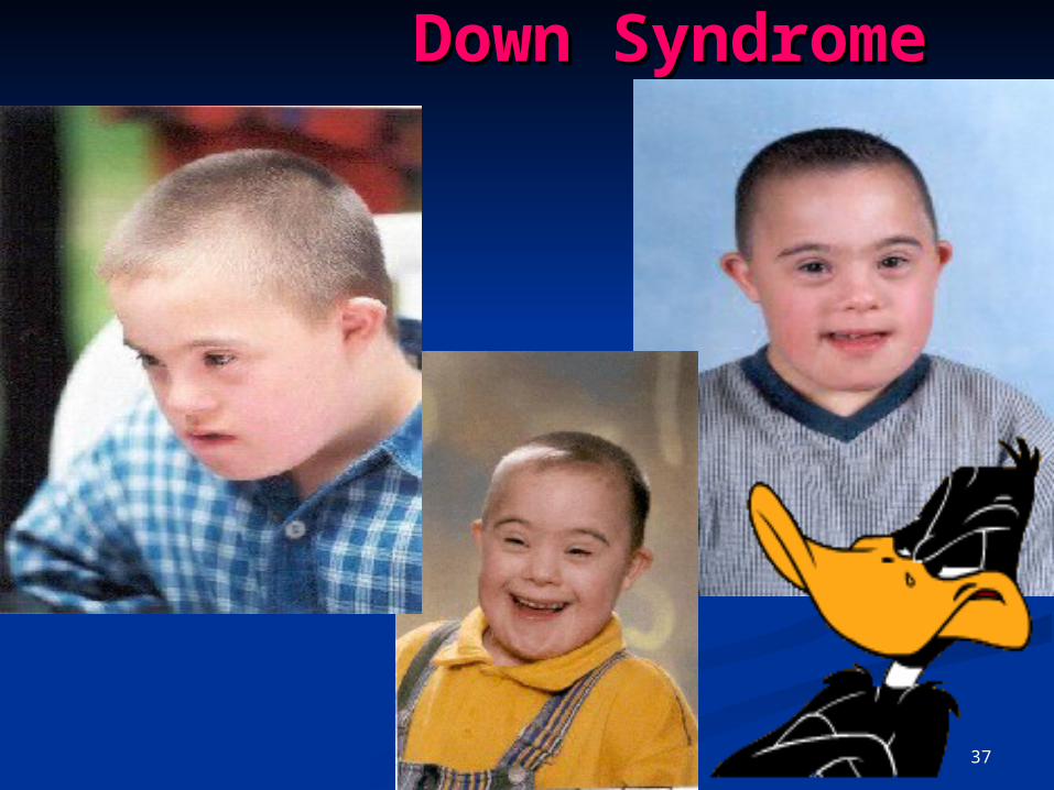
Down syndrom) Down syndrom) Trisomy 21, Trisomy 21, 46)46)
critical region:critical region: A region on the long (q) arm of A region on the long (q) arm of
chromosome 21chromosome 21 Down syndrome causes mental Down syndrome causes mental
retardation, a characteristic facial retardation, a characteristic facial appearance, and multiple appearance, and multiple malformationsmalformations
AAssociated with a major risk for ssociated with a major risk for heart malformations a small but still heart malformations a small but still significant risk of acute leukemia . significant risk of acute leukemia .
3 copies of chromosome number 21 3 copies of chromosome number 21

36
incidence of 1 in 660 and is by far the incidence of 1 in 660 and is by far the most common chromosomal most common chromosomal abnormalitabnormalityy
SSlight flattening of the facelight flattening of the face AA low bridge of the nose (lower than low bridge of the nose (lower than
the usually flat nasal bridge of the the usually flat nasal bridge of the normal newborn)normal newborn)
AAn epicanthal fold (a fold of skin over n epicanthal fold (a fold of skin over top of the inner corner of the eye, top of the inner corner of the eye, which can also be seen less which can also be seen less frequently in normal babies)frequently in normal babies)
A A ring of tiny harmlessring of tiny harmless white spots white spots around the irisaround the iris
mental retardationmental retardation
37
Down SyndromeDown Syndrome

38
Down SyndromeDown Syndrome: : Prenatal Prenatal RiskRisk
The risk of trisomy 21 is The risk of trisomy 21 is directly related to maternal directly related to maternal age age
Patients who will be 35 years Patients who will be 35 years or older on their due date or older on their due date should be offered chorionic should be offered chorionic villus sampling or second-villus sampling or second-trimester amniocentesis trimester amniocentesis

39
The use of ultrasound to The use of ultrasound to estimate gestational age estimate gestational age improves the sensitivity and improves the sensitivity and specificity of maternal serum specificity of maternal serum screeningscreening. (. (Am Fam Am Fam Physician 2000;62:825-Physician 2000;62:825-32,837-832,837-8.) .)

40
Etiology and Clinical Etiology and Clinical ManifestationsManifestations
Trisomy 21 is present in 95 Trisomy 21 is present in 95 percent of persons with Down percent of persons with Down syndrome.syndrome.
Mosaicism, a mixture of normal Mosaicism, a mixture of normal diploid and trisomy 21 cells, diploid and trisomy 21 cells, occurs in 2 percent.occurs in 2 percent.

41
Etiology and Clinical Etiology and Clinical ManifestationsManifestations
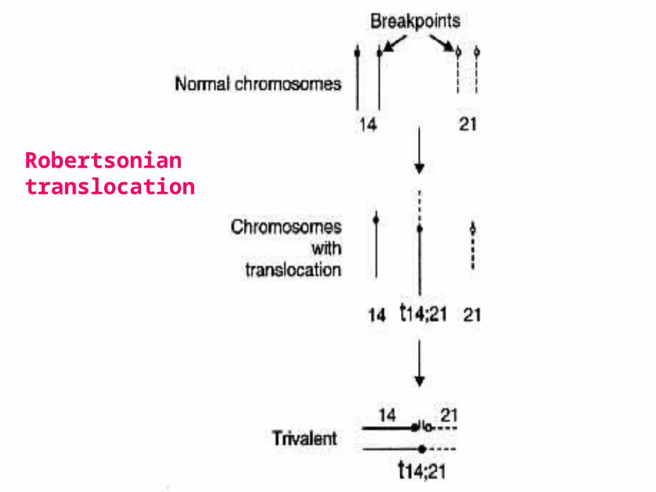
The remaining 3 percent The remaining 3 percent have a Robertsonian have a Robertsonian translocation in which all or translocation in which all or part of an extra chromosome part of an extra chromosome 21 is fused with another 21 is fused with another chromosomechromosome. .

42
Robertsonian Robertsonian translocationtranslocation
TThe reciprocal transfer of he reciprocal transfer of the long arms of two of the the long arms of two of the acrocentric chromosomes: acrocentric chromosomes: 13, 14, 15, 21 or 2213, 14, 15, 21 or 22
On rare occasions, other On rare occasions, other non-acrocentric non-acrocentric chromosomes undergo chromosomes undergo Robertsonian translocationRobertsonian translocation
43
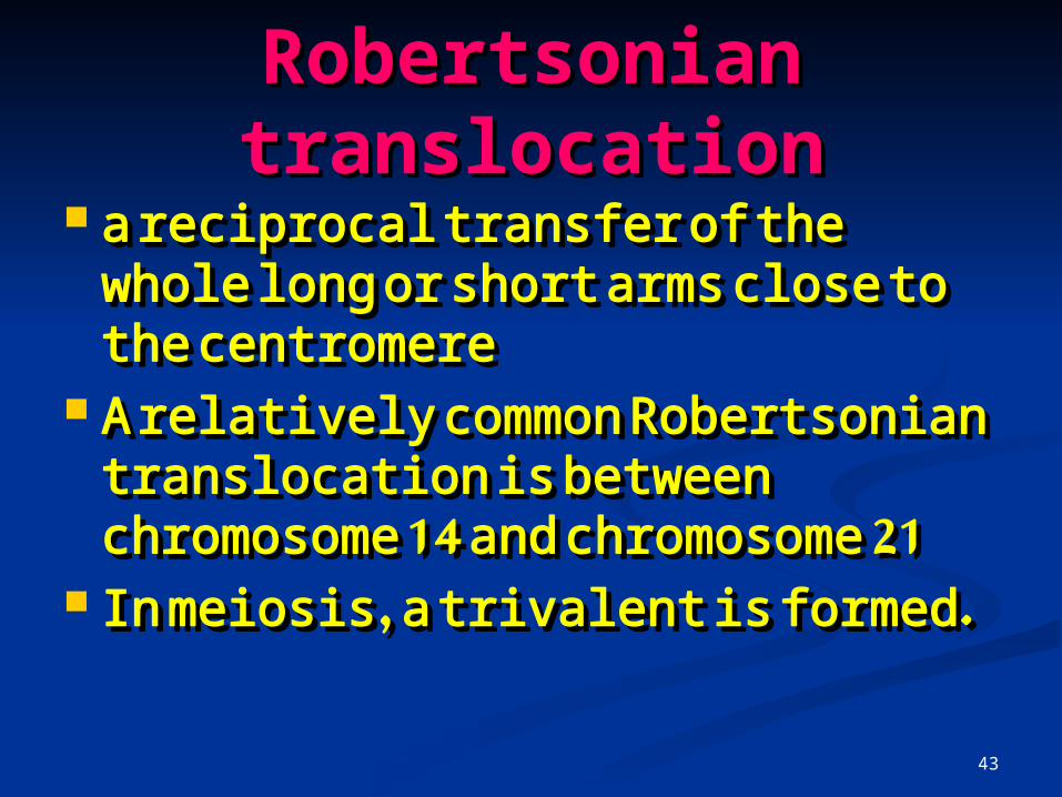
Robertsonian Robertsonian translocationtranslocation
a reciprocal transfer of the whole a reciprocal transfer of the whole long or short arms close to the long or short arms close to the centromerecentromere
A relatively common A relatively common Robertsonian translocation is Robertsonian translocation is between chromosome 14 and between chromosome 14 and chromosome 21chromosome 21
In meiosis, a trivalent is formedIn meiosis, a trivalent is formed..
44
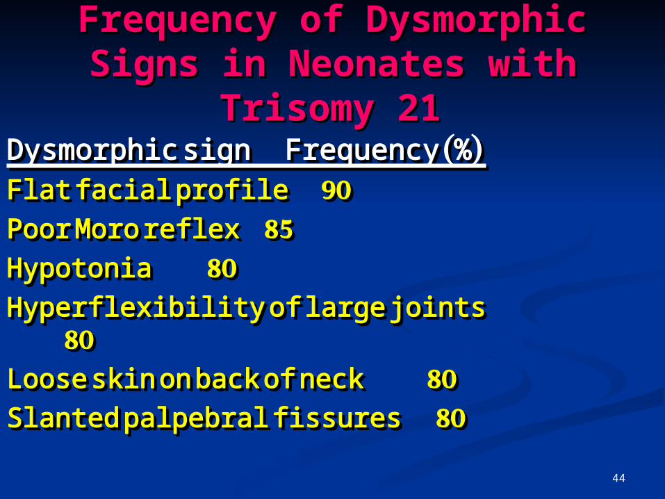
Frequency of Dysmorphic Frequency of Dysmorphic Signs in Neonates with Signs in Neonates with
Trisomy 21Trisomy 21 Dysmorphic sign Frequency (%) Dysmorphic sign Frequency (%) Flat facial profileFlat facial profile 90 90
Poor Moro reflexPoor Moro reflex 85 85
HypotoniaHypotonia 80 80
Hyperflexibility of large jointsHyperflexibility of large joints 80 80
Loose skin on back of neck Loose skin on back of neck 8080
Slanted palpebral fissuresSlanted palpebral fissures 80 80

45
Frequency of Dysmorphic Frequency of Dysmorphic Signs in Neonates with Signs in Neonates with
Trisomy 21Trisomy 21Dysmorphic sign Frequency Dysmorphic sign Frequency
(%) (%)
Dysmorphic pelvis on radiographDysmorphic pelvis on radiograph 7070
Small round ears Small round ears 6060
Hypoplasia of small fingerHypoplasia of small finger 60 60

46
Persons with Down syndrome Persons with Down syndrome usually have mild to moderate usually have mild to moderate mental retardation mental retardation
School-aged children with Down School-aged children with Down syndrome often have difficulty syndrome often have difficulty with language, communicationwith language, communication
Adults with Down syndrome have a Adults with Down syndrome have a high prevalence of early high prevalence of early Alzheimer's disease Alzheimer's disease

47
Adult Adult Down Down
SyndroSyndromeme

48
Incidence of Some Incidence of Some Associated Medical Associated Medical
Complications in Persons Complications in Persons with Down Syndrome with Down Syndrome
DisorderDisorder Incidence Incidence (%)(%)Mental retardationMental retardation >95 >95
Growth retardationGrowth retardation >95 >95Early Alzheimer's disease Early Alzheimer's disease 75% 75% by age 60by age 60Congenital heart defectsCongenital heart defects (atrioventricular canal defect, (atrioventricular canal defect, ventricular septal defect, atrial septalventricular septal defect, atrial septal defectdefect 4 400

49
DisorderDisorder Incidence Incidence (%)(%)
Hearing loss Hearing loss 40 to 75 40 to 75Ophthalmic disorders (congenital Ophthalmic disorders (congenital
cataracts,cataracts,glaucoma(glaucoma( 60 60EpilepsyEpilepsy 5 to 10 5 to 10Gastrointestinal malformations Gastrointestinal malformations
(duodenal atresia,(duodenal atresia, Hirschsprung disease)Hirschsprung disease) 55HypothyroidismHypothyroidism 55LeukemiaLeukemia 55

50
DisorderDisorder Incidence Incidence (%)(%)
Increased susceptibility to Increased susceptibility to
infection (pneumonia, otitis infection (pneumonia, otitis media, sinusitis, pharyngitis(media, sinusitis, pharyngitis(
1-61-6
InfertilityInfertility >99% in men>99% in men
anovulation in anovulation in 30% of 30% of womenwomen

51
Estimated risk of Down syndrome according to maternal
age

52
The risk of having a child The risk of having a child with Down syndromewith Down syndrome
1/1,300 for a 25-year-old 1/1,300 for a 25-year-old woman;woman;
at age 35, the risk increases at age 35, the risk increases to 1/365to 1/365
At age 45, the risk of a At age 45, the risk of a having a child with Down having a child with Down syndrome increases to 1/30syndrome increases to 1/30

53
Maternal Serum ScreeningMaternal Serum Screening
If all pregnant women 35 years or If all pregnant women 35 years or older chose to have amniocentesisolder chose to have amniocentesis
about 30 percent of trisomy 21 about 30 percent of trisomy 21 pregnancies would be detectedpregnancies would be detected
Women younger than 35 years Women younger than 35 years give birth to about 70 percent of give birth to about 70 percent of infants with Down syndromeinfants with Down syndrome

54
The risk of having a child The risk of having a child with Down syndromewith Down syndrome
Maternal serum screening Maternal serum screening ((multiplemultiple--marker screeningmarker screening) ) can allow the detection of can allow the detection of trisomy 21 pregnancies in trisomy 21 pregnancies in women in this younger age women in this younger age groupgroup..

55
Maternal Serum ScreeningMaternal Serum Screening"triple test" or "triple screen" "triple test" or "triple screen"
"Multiples of the Median "Multiples of the Median (MoM)"(MoM)"
Alpha-fetoprotein (AFP)Alpha-fetoprotein (AFP) unconjugated estriolunconjugated estriol human chorionic gonadotropin human chorionic gonadotropin
(hCG) (hCG)

56
""Multiples of the Median Multiples of the Median ((MoMMoM)")"
AFP is produced in the yolk sac AFP is produced in the yolk sac and fetal liver.and fetal liver.
Unconjugated estriol and hCG are Unconjugated estriol and hCG are produced by the placenta.produced by the placenta.
The maternal serum levels of each The maternal serum levels of each of these proteins and of steroid of these proteins and of steroid hormones vary with the hormones vary with the gestational age of the pregnancy.gestational age of the pregnancy.

57
""Multiples of the Median Multiples of the Median ((MoMMoM)")"
With trisomy 21, second-With trisomy 21, second-trimester maternal serum levels trimester maternal serum levels of AFP and unconjugated estriol of AFP and unconjugated estriol are about are about 25 percent lower than 25 percent lower than normal levelsnormal levels
maternal serum hCG is maternal serum hCG is approximately two times higherapproximately two times higher than the normal hCG levelthan the normal hCG level

58
Maternal Serum ScreeningMaternal Serum Screening"triple test" or "triple "triple test" or "triple
screen"screen"
The triple test can detect The triple test can detect approximately 60 percent of approximately 60 percent of the pregnancies affected by the pregnancies affected by trisomy 21, with a false-trisomy 21, with a false-positive rate of about 5 positive rate of about 5 percent. percent.

59
Recurrence Risk and Family Recurrence Risk and Family HistoryHistory
If a patient has had a trisomy If a patient has had a trisomy 21 pregnancy in the past, the 21 pregnancy in the past, the risk of recurrence in a risk of recurrence in a subsequent pregnancy subsequent pregnancy increases to approximately 1increases to approximately 1-3-3
percent above the baseline percent above the baseline risk determined by maternal risk determined by maternal ageage

60
Diagnosis of a Diagnosis of a chromosome-21 chromosome-21 translocation in the fetus or translocation in the fetus or newborn is an indication for newborn is an indication for karyotype analysis of both karyotype analysis of both parentsparents
If both parents have normal If both parents have normal karyotypes, the recurrence karyotypes, the recurrence risk is risk is 11 to 3 percent to 3 percent

61
Ultrasonographic Findings Ultrasonographic Findings Associated with Fetal Down Associated with Fetal Down
SyndromeSyndrome
Chorionic villus samplingChorionic villus sampling
10 to 12 10 to 12 weeks weeks 0.5 to 0.5 to 1.5 %1.5 %
Early amniocentesisEarly amniocentesis
12 to 15 12 to 15 weeksweeks 1.0 to 2.0 %1.0 to 2.0 %
Second-trimester amniocentesisSecond-trimester amniocentesis
15 to 20 15 to 20 weeksweeks 0.5 to 1.0 %0.5 to 1.0 %

62
a a womanwoman having amniocentesis having amniocentesis
63
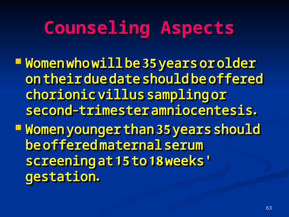
Counseling AspectsCounseling Aspects
Women who will be 35 years or Women who will be 35 years or older on their due date should older on their due date should be offered chorionic villus be offered chorionic villus sampling or second-trimester sampling or second-trimester amniocentesis. amniocentesis.
Women younger than 35 years Women younger than 35 years should be offered maternal should be offered maternal serum screening at 15 to 18 serum screening at 15 to 18 weeks' gestation. weeks' gestation.

64
UltrasoundUltrasound
During the first trimester of the During the first trimester of the majority of pregnancies, it is majority of pregnancies, it is possible to measure the size of possible to measure the size of the fluid area at the back of the the fluid area at the back of the fetus’s neck, known as the fetus’s neck, known as the nuchal translucency or NT The nuchal translucency or NT The increasing size of the NT increasing size of the NT indicates a greater risk of the indicates a greater risk of the fetus having Down’s syndrome.fetus having Down’s syndrome.

65
UltrasoundUltrasound

66
Fluorescent In Situ Hybridisation techniques

67
female fetus with trisomy-female fetus with trisomy-2121
chromosomes chromosomes 18 (aqua), X 18 (aqua), X (green), and Y (green), and Y (red).(red).
• chromosomes chromosomes 13 (green), 13 (green), and 21 (red)and 21 (red)

68
Quantitative fluorescent Quantitative fluorescent polymerase chain reactionpolymerase chain reaction
2:1 ratio (Down's 2:1 ratio (Down's Syndrome)Syndrome)
1:1 ratio (normal fetus)1:1 ratio (normal fetus)
69
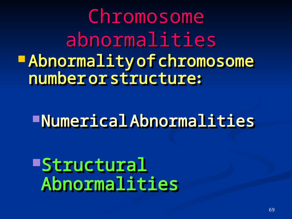
Chromosome Chromosome abnormalitiesabnormalities
AAbnormality of chromosome bnormality of chromosome number or structure:number or structure:
Numerical AbnormalitiesNumerical Abnormalities Structural AbnormalitiesStructural Abnormalities
70
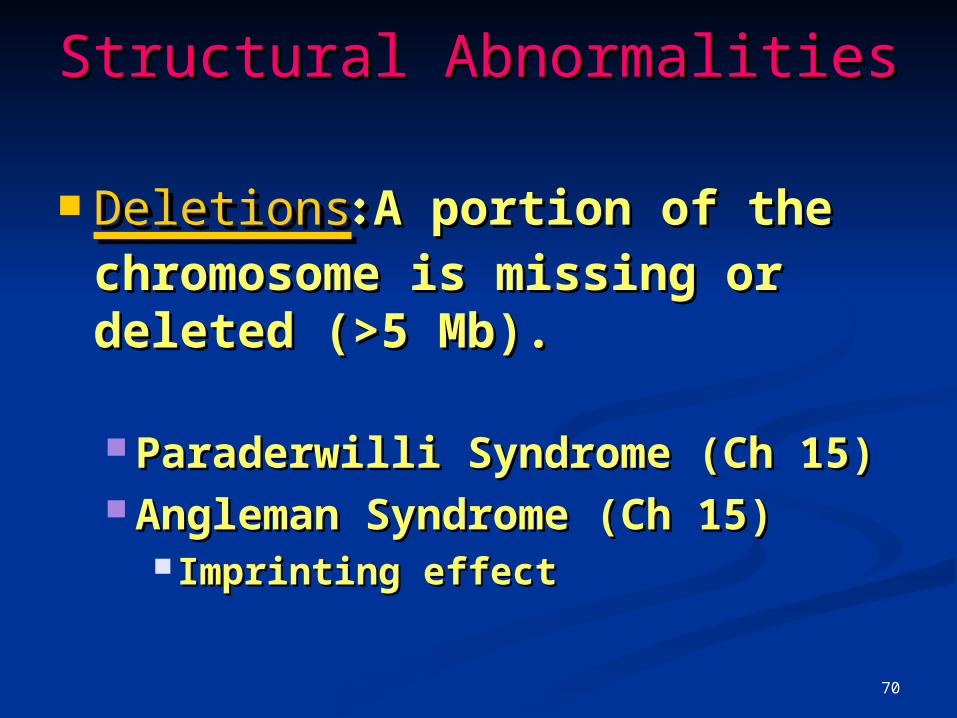
Structural AbnormalitiesStructural Abnormalities
DeletionsDeletions: : A portion of the A portion of the chromosome is missing or chromosome is missing or deleted (>5 Mb)deleted (>5 Mb)..
Paraderwilli Syndrome (Ch 15)Paraderwilli Syndrome (Ch 15) Angleman Syndrome (Ch 15)Angleman Syndrome (Ch 15)
Imprinting effectImprinting effect

71
DELETIONSDELETIONS
Deletion refers to the loss of a Deletion refers to the loss of a segment of a chromosomesegment of a chromosome
This can be terminal (close to This can be terminal (close to the end of the chromosome on the end of the chromosome on the long arm or the short arm)the long arm or the short arm)
or it can be interstitial (withinor it can be interstitial (within)) ig.DGS IIig.DGS II

72
DELETIONSDELETIONS

73
Structural AbnormalitiesStructural Abnormalities
• DuplicationsDuplications: A portion of the : A portion of the chromosome is duplicated, chromosome is duplicated, resulting in extra genetic resulting in extra genetic material.material.
• Oncogenes (c-onc, c-fos, c-myc)Oncogenes (c-onc, c-fos, c-myc)
74
DUPLICATIONSDUPLICATIONS refers to an extra chromosomal segment refers to an extra chromosomal segment
within the same homologous chromosome within the same homologous chromosome or an extra chromosomal segment on or an extra chromosomal segment on another nonhomologous chromosome.another nonhomologous chromosome.
Again, the clinical findings are highly Again, the clinical findings are highly variable depending upon the chromosomal variable depending upon the chromosomal segments involved. segments involved. Gene expantion:Gene expantion:
in Huntington Disease/ Fragile X, ….in Huntington Disease/ Fragile X, ….

75
DUPLICATIONSDUPLICATIONS
76
Structural AbnormalitiesStructural Abnormalities
TranslocationsTranslocations: : When a portion When a portion of one chromosome is of one chromosome is transferred to another transferred to another chromosomechromosome. .

77
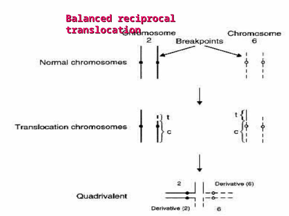
There are two main types of There are two main types of translocations. translocations. In a In a reciprocal translocationreciprocal translocation, ,
segments from two different segments from two different chromosomes have been chromosomes have been exchanged. In a exchanged. In a Robertsonian translocationRobertsonian translocation, an , an entire chromosome has attached entire chromosome has attached to another at the centromere.to another at the centromere.

78
TRANSLOCATIONSTRANSLOCATIONS
79
Balanced reciprocal translocation
Balanced reciprocal Balanced reciprocal translocationtranslocation

80

81
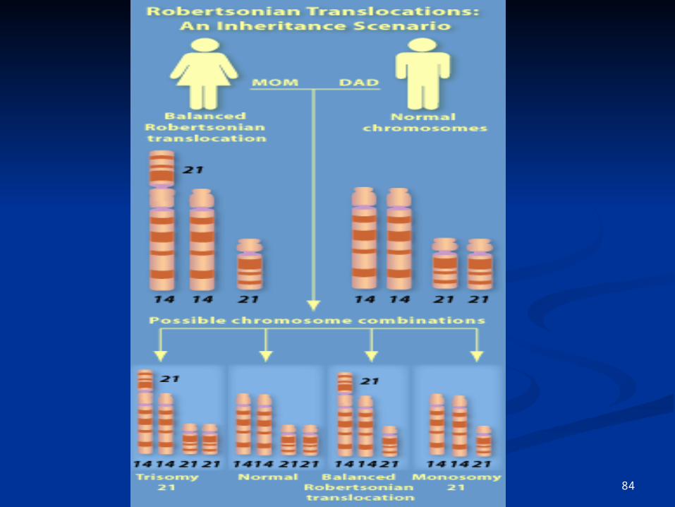
Robertsonian translocationRobertsonian translocation TThe reciprocal transfer of the long he reciprocal transfer of the long
arms of two of the acrocentric arms of two of the acrocentric chromosomes: 13, 14, 15, 21 or 22chromosomes: 13, 14, 15, 21 or 22
On rare occasions, other non-On rare occasions, other non-acrocentric chromosomes undergo acrocentric chromosomes undergo Robertsonian translocationRobertsonian translocation
a reciprocal transfer of the whole a reciprocal transfer of the whole long or short arms close to the long or short arms close to the centromerecentromere
A relatively common Robertsonian A relatively common Robertsonian translocation is between translocation is between chromosome 14 and chromosome 21chromosome 14 and chromosome 21
In meiosis, a trivalent is formedIn meiosis, a trivalent is formed..
82
Robertsonian translocation

83
84

85
Structural AbnormalitiesStructural Abnormalities
InversionsInversions: : A portion of the A portion of the chromosome has broken chromosome has broken off, turned upside down off, turned upside down and reattached, therefore and reattached, therefore the genetic material is the genetic material is invertedinverted..eg Ch9 inv in Iraneg Ch9 inv in Iran

86
involve only one chromosomeinvolve only one chromosome the intervening segment is rejoined in an the intervening segment is rejoined in an
inverted or opposite manner.inverted or opposite manner. Since there is no loss nor gain of Since there is no loss nor gain of
chromosomal material, inversion carriers chromosomal material, inversion carriers are normalare normal
ParacentricParacentric: : does not include the does not include the centromerecentromere
pericentric:inverted segment contains the pericentric:inverted segment contains the centromerecentromere
In meiosis, the normal chromosome and In meiosis, the normal chromosome and the inverted chromosome will form a loop the inverted chromosome will form a loop to allow pairing of specific DNA sequencesto allow pairing of specific DNA sequences
that occur within the inversion loop result that occur within the inversion loop result in gametes with both deletions and in gametes with both deletions and duplications duplications
inversion carriers have a relatively low risk inversion carriers have a relatively low risk of having abnormal offspring.of having abnormal offspring.
Inversions

87
Inversions

88

89
RingsRings: : A portion of a chromosome A portion of a chromosome has broken off and formed a has broken off and formed a circle or ringcircle or ring. . This can happen This can happen with or without loss of genetic with or without loss of genetic materialmaterial. .

90
Ring

91
OncologyOncology Chronic Myelogenous Leukemia
(CML) a clonal expansion of transformed
hematopoietic progenitor cells: Myeloid Monocytic Erythroid Megakaryocytic lymphoid lineages

92
HematologyHematology Bone Bone marrowmarrow

93
Chronic myelogenous Chronic myelogenous leukemia leukemia ((CMLCML))
15% to 20% of leukemias in adults incidence of 1 to 2 cases per 100,000 population

94
Chronic myelogenous Chronic myelogenous leukemialeukemia occurs more frequently in males than in
females (ratio of 1.3 to 1) Incidence: increases with age the median age at presentation is between
45 and 55 years Up to 30% of patients with CML are 60
years or older which is an important consideration for the
selection of therapeutic strategies stem-cell transplantation treatment with interferon-alfa (Intron A,
Roferon-A)

95
96
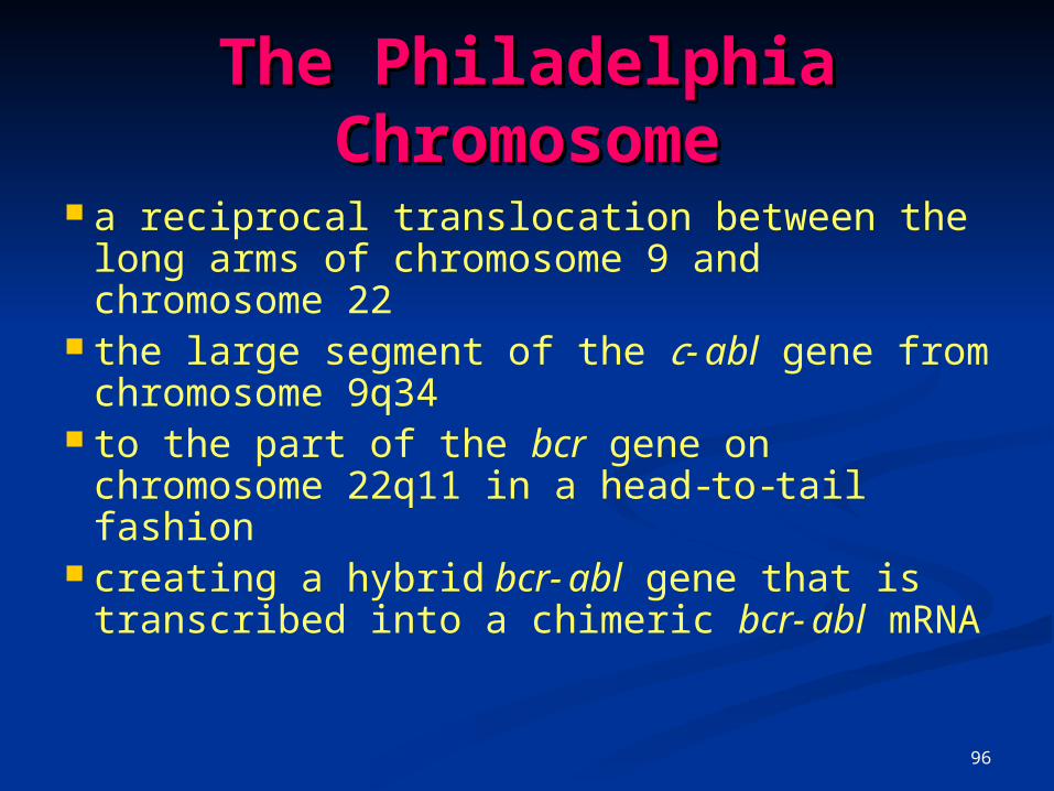
The Philadelphia The Philadelphia ChromosomeChromosome
a reciprocal translocation between the long arms of chromosome 9 and chromosome 22
the large segment of the c -abl gene from chromosome 9q34
to the part of the bcr gene on chromosome 22q11 in a head-to-tail fashion
creating a hybrid bcr-abl gene that is transcribed into a chimeric bcr-abl mRNA

97

98

99
Role of the bcr-abl Fusion Gene in CML Pathogenesis
Ch 9: c-abl gene :a proto-oncogene Encodes: a nonreceptor tyrosine
kinase with a molecular mass of 145 kd
(p145c-abl) It is localized in both cytoplasm and
nucleus It consists of 11 exons 230 kilobases (kb)

100
Role of the bcr-abl Fusion Gene in CML Pathogenesis
c -abl gene : Exon 1 has two alternative forms 1a and 1b
In most cases, the breakpoint : within the segment between exons
1a and 1b creating a bcr-abl fusion mRNA of
8.5 kb The fusion mRNAs are translated
into a 210-kd chimeric protein called p210bcr-abl

101
Detection of Detection of bcrbcr--ablabl Cytogenetic analysis Ph chromosome in 90% of
patients with CML Such analysis is tedious and
time-consuming allows the examination of only 20
to 25 metaphases per bone marrow sample
misses the 5% of patients who are Ph-negative but bcr-abl-positive
102
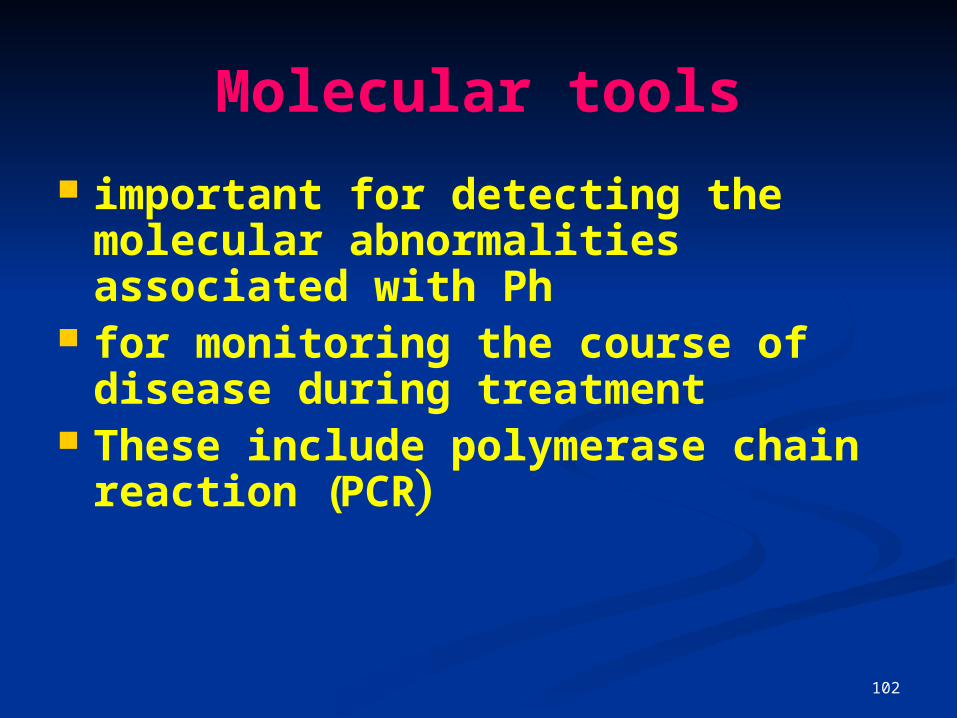
Molecular tools
important for detecting the molecular abnormalities associated with Ph
for monitoring the course of disease during treatment
These include polymerase chain reaction (PCR)

103
RTRT--PCRPCR
Quantitative reverse Quantitative reverse transcriptase–polymerase chain transcriptase–polymerase chain reactionreaction
following patients with CML after following patients with CML after stemstem--cell transplantationcell transplantation
Its use for monitoring patients Its use for monitoring patients receiving interferonreceiving interferon--alphaalpha

104
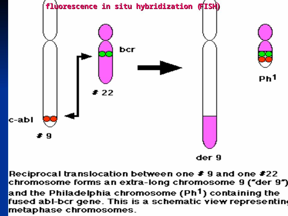
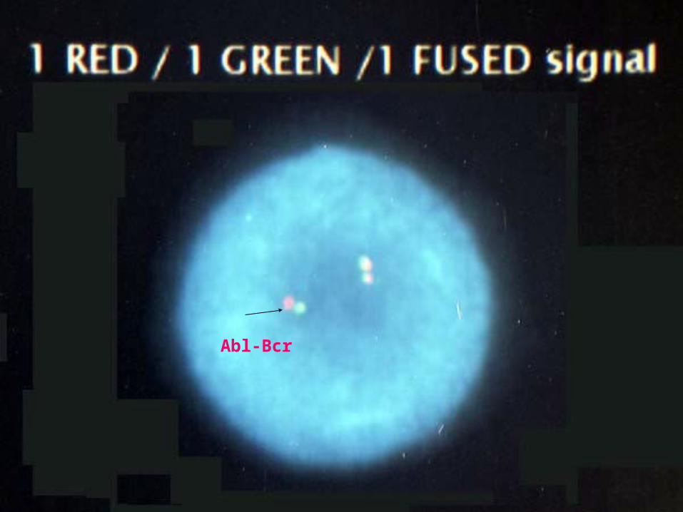
FISHFISH
Fluorescence in situ hybridization allows for the analysis of
metaphase
and nondividing interphase cells
Results of FISH studies are easily quantifiable
105
fluorescence in situ hybridization fluorescence in situ hybridization ((FISHFISH))

106
fluorescence in situ hybridizationfluorescence in situ hybridization ((FISHFISH) )

107
FISHFISH
Abl-Bcr
Abl
Bcr
108
Abl-Bcr