1 Chapter 10 Anatomy of the Muscular System. Introduction There are over 600 muscles in the body ...
98
1 Chapter 10 Anatomy of the Muscular System
-
Upload
margaret-watts -
Category
Documents
-
view
215 -
download
0
Transcript of 1 Chapter 10 Anatomy of the Muscular System. Introduction There are over 600 muscles in the body ...

1
Chapter 10
Anatomy of the Muscular System

Introduction
There are over 600 muscles in the body Muscles make up 40-50% of our weight Muscle helps to determine the form and contours of
our body
2

Structure of Skeletal Muscle
Epimysium Layer of connective tissue that surrounds the entire muscle
Perimysium Separates the muscle into fasicles
Fasicle A group of muscle fibers
Endomysium Surrounds an individual muscle fiber
Tendon Connect muscle to bone
3

Structure of Skeletal Muscle
• Sometimes muscles are connected to each other by broad sheets of connective tissue called aponeuroses.
4


6

Types of Fiber Arrangements
• Parallel Fibers- run across
• Convergent- converge to a narrow attachment
• Oblique- diagonal– Pennate- one feather– Bipennate- two feathers
• Direction of fibers important for strength and range of motion
7

Fiber Arrangements

Skeletal Muscle Actions A. Origin and Insertion
1. The immovable end of a muscle is the origin, while the movable end is the insertion; contraction pulls the insertion toward the origin.
2. Some muscles have more than one insertion or origin.

10

Muscle Actions
• Prime mover- primary muscle being moved
• Antagonist- muscles that oppose the prime movers
• Synergist- (Syn= together Erg= work) muscles that contract along with the prime mover
• Fixator Muscles- joint stabilizers
11
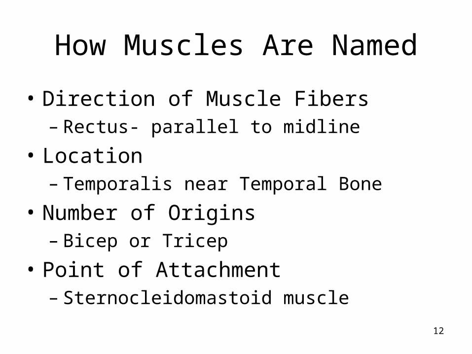
How Muscles Are Named
• Direction of Muscle Fibers– Rectus- parallel to midline
• Location– Temporalis near Temporal Bone
• Number of Origins– Bicep or Tricep
• Point of Attachment– Sternocleidomastoid muscle
12
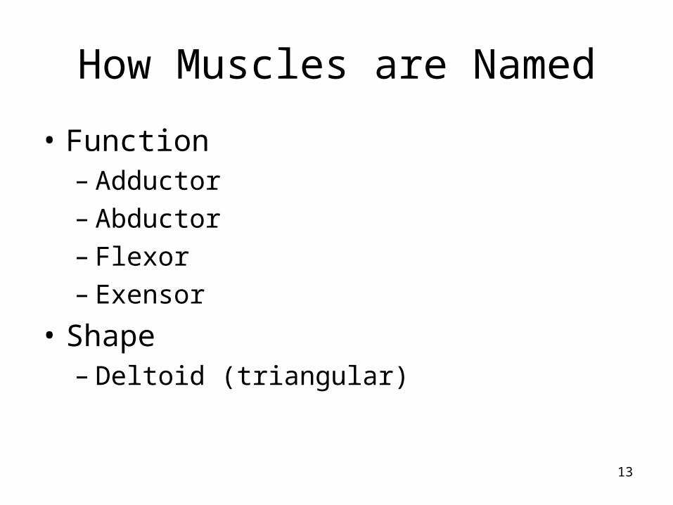
How Muscles are Named
• Function– Adductor– Abductor– Flexor – Exensor
• Shape– Deltoid (triangular)
13

How Muscles Are Named
• Size– Maximus– Medius– Minimus– Longus – Brevis
14

Head and Neck Muscles
• Two Large Categories– Facial Muscles
• Unique because at least one of their attachments is to the deep layers of the skin
• Allows for facial expression
– Chewing Muscles
15

16

Facial Muscles
• Frontalis– Covers frontal bone– Raise eyebrows and wrinkle forehead
• Occipitalis– Covers the posterior aspect of the skull and
pulls the scalp back posteriorly
• Epicranial Aponeurosis– connects the Frontalis and Occipitalis
17

18

Facial Muscles
• Obicularis Oris– Circular muscles of the lips– Closes the mouth– Protrudes the lips– Called the kissing muscle
• Obicularis Oculi– Circular muscle around the eyes– Allows you to close your eyes, squint, blink
and wink19

Facial Muscles
• Buccinator– Flattens the cheek to whistle or blow trumpet– Also a chewing muscle
• Zygomaticus– Smiling muscle that raises the corners of the
mouth upwards
20

21

Chewing Muscles
• Called muscles of mastication
• Buccinator belongs in this group
• Masseter– Closes jaw
• Temporalis– Lies over the temporal bone– Inserts into the mandible and acts as a
synergist of the masseter in closing the jaw
22

Neck Muscles
• Platysma– Covers the neck anterolateral
23

24

Neck Muscles
• Sternocleidomastoid Muscle– Two heads of origin
• Sternum and clavicle
– Inserts in the
mastoid process
of temporal bone
25

Neck Muscles
• Sternocleidomastoid Muscle– Both flex (angle is decreased) causes bowing
of head• Called the prayer muscles
– If only one is flexed, the head turns to the side
• In some difficult births, muscle can be injured and spasms develop– Called torticollis or wryneck
26

Trunk Muscles
• Anterior Muscles– Pectoralis Major
• Large fan muscle of the chest• Origin- from the sternum, shoulder girdle, and first
six ribs• Inserts proximal end of the humerus• Adducts and flexes the arm
– Pectoralis Minor• Under the pectoralis major
27

28

Anterior Trunk Muscles
• Intercostal Muscles– Found between the ribs
• External Intercostals– Elevates the rib cage for breathing
• Internal Intercostals– Depresses the rib cage for breathing
29

Anterior Trunk Muscles
• Diaphram– Large flat muscle that separates the thoracic
from the abdominal cavity
30
Muscles of the Abdominal Wall
• This group of muscles connects the rib cage and vertebral column to the pelvic girdle
• A band of tough connective tissue, the linea alba, extending from the xiphoid process to the symphysis pubis, serves as an attachment for certain abdominal wall muscles
31

Muscles of Abdominal Wall
•Rectus Abdominus•Outer layer•Runs from pubis to rib cage•Flexes vertebral column•Compresses abdominal cavity

Muscles of the Abdominal Wall
33

Muscles of Abdominal Wall• External Oblique
– Make up the lateral walls of the abdomen– Flex the vertebrae– Rotates the trunk – Bend the trunk laterally
• Internal Obliques– Fibers run at right angles to external oblique
• Transverse Abdominus– Parallel fibers
Muscles of the Pelvic Floor
• Muscles of pelvic floor support the pelvic cavity
• Diamond shaped outlet is called the perineum
35

36

Posterior Trunk Muscles
• Trapezius– Diamond shaped muscle of back– Shrug the shoulders– Hold head up– Adducts the arm
37

38

Posterior Trunk Muscles
• Latissimus Dorsi– Large flat muscle of lower back– Originates in lower spine– Inserts at the distal end of the humerus– Extends and _____ the arm
39

40

Posterior Trunk Muscles
• Erector Spinae– Deep muscles of the back
• Longissimus• Iliocostalis• Spinalis
– Powerful back extensors (erectors)– Can go into spasms and are a common
source of lower back pain
41

ADD
• Picture of Erector Spinae
42

Posterior Trunk Muscles
• Deltoid– Triangle shoulder muscle– Inserts in the deltoid tuberosity of the
humerus– Multifunctional muscle– Abducts the arm
43

44

Muscles of the Upper Limb
• 6 muscles attach arm so lots of movement
• 4 muscles form a cap or cuff around shoulder joint- rotator cuff– Infraspinatus– Supraspinatus– Subscapularis– Teres minor

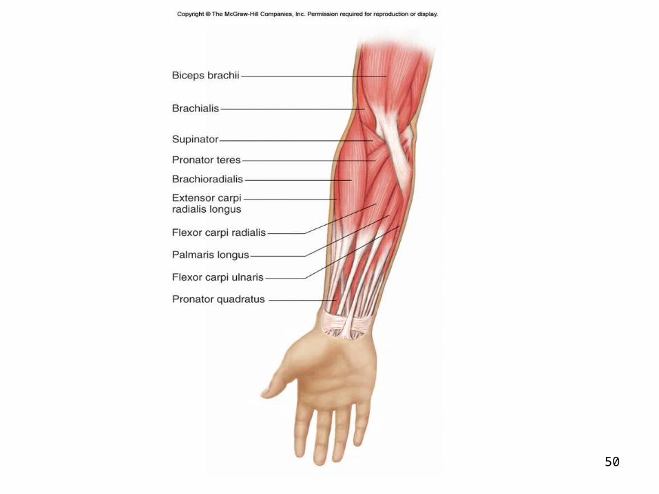
Muscles That Act On The Forearm
• Cause elbow flexion
• Biceps Brachii– Two origins- scapula coracoid and
superglenoid tuberosity– Inserts in the radial tuberosity– Supinates the forearm– “It turns the corkscrew and pulls the cork”
46

47

Muscles That Act on the Forearm
• Brachialis– Lies deep to the bicep muscle and helps with
flexion
• Brachioradialis– Weak muscle in the forearm
48

49
50
Muscles That Act On The Forearm
• Tricep Brachii– Has three heads– Is the prime mover of elbow extension– Atagonist of the bicep– Called the boxer’s muscle
51

52

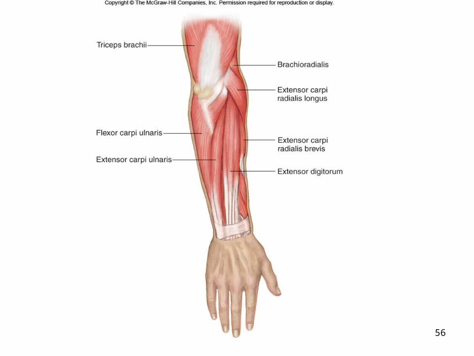
Muscles that Move The Wrist, Hand, and Fingers
• Are located on the forearm
• Many intrinsic muscles– 8 separate muscles serve the thumb
• As a general rule, forearm names reflect their activities– Flexor carpi and flexor digitorum muscles
cause flexion of wrist and fingers
53
54
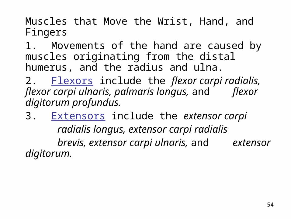
Muscles that Move the Wrist, Hand, and Fingers1. Movements of the hand are caused by
muscles originating from the distal humerus, and the radius and ulna.
2. Flexors include the flexor carpi radialis, flexor carpi ulnaris, palmaris longus, and flexor digitorum profundus.
3. Extensors include the extensor carpi radialis longus, extensor carpi radialis brevis, extensor carpi ulnaris, and
extensor digitorum.

55
56

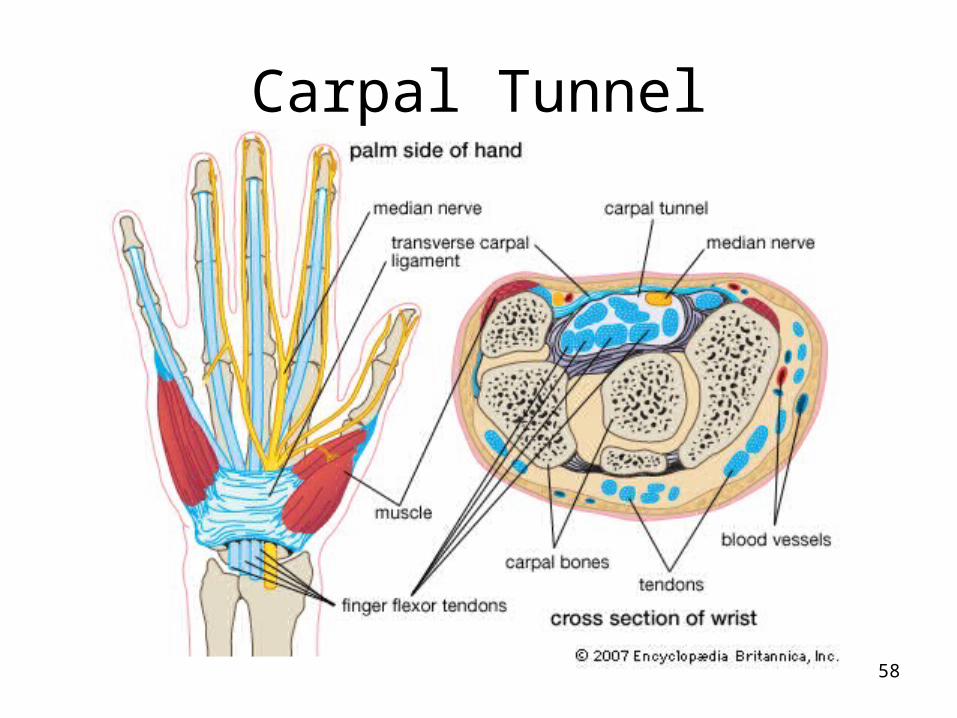
Carpal Tunnel Syndrome
• Tendon sheath becomes inflamed and puts pressure on the median nerve
• Characterized by weakness, pain, and tingling in hand
57
Carpal Tunnel
58

Carpal Tunnel
59

Causes of Carpal Tunnel
• Repetitive movements– Playing piano– Typing– Swinging a hammer– Meat cutters– Quilting, cross-stitch, handicrafts– Some factory work
60

Carpal Tunnel Treatment
• Can be relieved with anti-inflammatory injections
• Can wear braces to help
• Use specially designed keyboards
61

Carpal Tunnel Treatment
• Permanent cure is surgical cutting or removing swollen tissue pressing on nerve
• Will come back if you continue to do the same tasks that caused it.
62

Carpal Tunnel Treatment
63

Muscles Of The Lower Limb
• Among the strongest muscles in the body
• Because pelvic girdle is composed of heavy, fused bone that allow little movement, no special group of muscles is necessary to stabilize it
• Muscle that move leg form the flesh of the thigh
• Muscles originating on leg move the ankle and foot
64

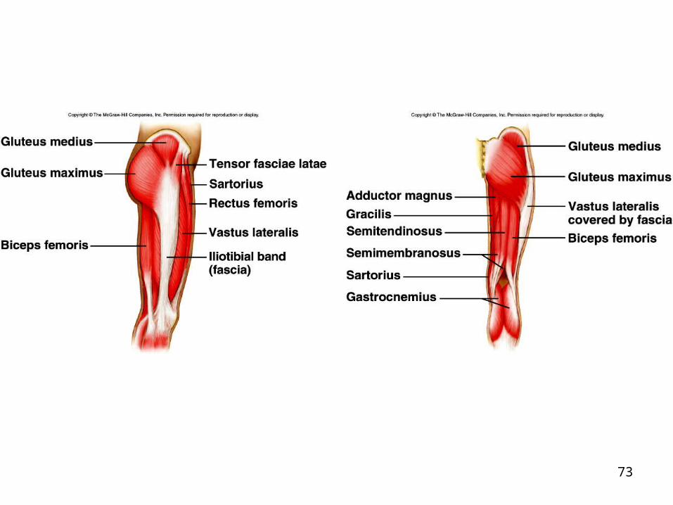
Muscles Causing Movement At The Hip
• Gluteus Maximus– Forms most of the buttocks
• Gluteus Medius– Is a hip abductor– Beneath the gluteus maximus
• Gluteus Minimus– Under the gluteus medius
65

66

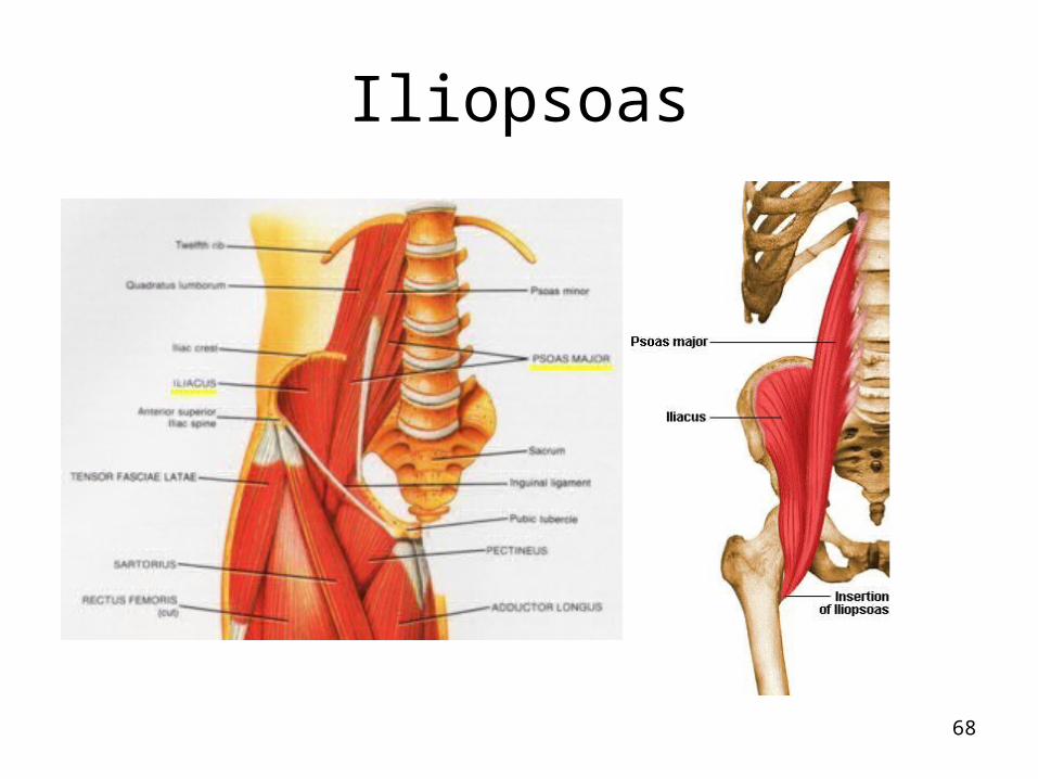
Muscles Causing Movement At The Hip
• Iliopsoas– Formed by iliacus and psoas major– Runs from iliac crest to insert on greater
trochanter of the femur– Prime mover of hip flexion
67
Iliopsoas
68

69

Adductor Muscles
• Form the medial side of each thigh
• Since gravity does most of the work for them, special exercises are usually needed to keep them toned– Adductor Longus– Adductor Magnus– gracilis
70

71
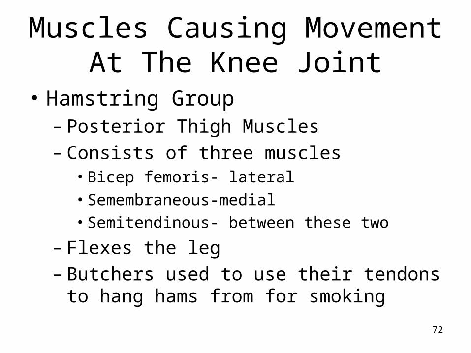
Muscles Causing Movement At The Knee Joint
• Hamstring Group– Posterior Thigh Muscles– Consists of three muscles
• Bicep femoris- lateral• Semembraneous-medial• Semitendinous- between these two
– Flexes the leg– Butchers used to use their tendons to hang
hams from for smoking
72
73

Muscles Causing Movement At The Knee Joint
• Sartorius– Thin straplike muscle
• Most superficial of thigh muscles
• Runs oblique across the thigh
• Called “tailor’s muscle” since it is a synergist in the crossed-leg position
74

75

Muscles Causing Movement At The Knee Joint
• Quadriceps– Rectus Femoris– Vastus Lateralis– Vastus Medialis– Vastus Intermedius
• All 4 muscles insert into the tibial tuberosity of the patellar ligament
76

77

Muscles Causing Movement At The Knee Joint
• Quadriceps act to extend the knee powerfully
78

Muscles Causing Movement At The Ankle And Foot
• Tibialis Anterior– Dorsiflexes and inverts the foot– Inflammation of this muscle is the cause of
shin splints
• Extensor Digitorium Longus– Lateral to the tibialis anterior– Prime mover of toe extention and dorsiflexor
of the foot
79

80

Muscles Causing Movement At The Ankle and Foot
• Fibularis Muscles- found on lateral part of leg– Longus– Brevis– Tertius– Plantar flexes and everts the foot
• Soleus
81
Muscles Causing Movement At The Ankle and Foot
• Gastrocnemius– Bulging calf muscle– Connects to the heal (calcaneus) by the
Achilles tendon– Plantar flexes the foot– Called the “toe dancer’s muscle”
82

83

84

85

Intramuscular Injection
• Allows the Dr. to inject a large amount of the drug at one time yet have it enter the circulation gradually
• 5 ml or less- deltoid
• More than 5 ml- gluteus medius
• Vastus lateralis in children
86
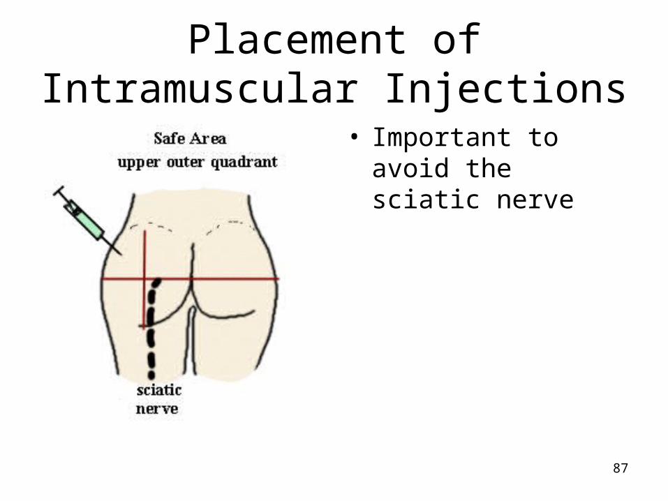
Placement of Intramuscular Injections
• Important to avoid the sciatic nerve
87
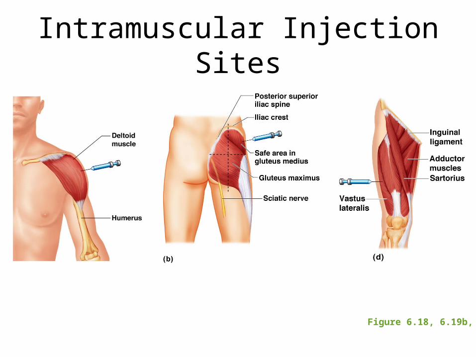
Intramuscular Injection Sites
Figure 6.18, 6.19b, d

R.I.C.E. Treatment For Injuries
• Rest• Ice
– On for 15 min. off for 20 min.
• Compression– Limits swelling
• Elevation– Reduces swelling– Should be above level of heart
• Referral89

Effect of Exercise On Muscle
• Muscle burns calories at three times the rate of fat– Lean body is more energetically efficient
• Aerobic or endurance exercises result in stronger, more flexible muscles with greater resistance to fatigue
• Resistance or isometric exercises increase muscle size and strength
90

Weight Training and Muscles
• Trapezius
• Pectorals
• Abdominals– Rectus abdominus v. obliques
• Latissimus Dorsi
• Erector Spinae
• Deltoid
91

Weight Training and Muscles
• Rotator Cuff
• Biceps
• Triceps
• Flexor and extensor muscles of forearm
• Gluteal Muscles
• Adductor Thigh Muscles
• Hamstring Group
92
Weight Training and Muscles
• Rotator Cuff
• Biceps
• Triceps
• Flexor and extensor muscles of forearm
• Gluteal Muscles
• Adductor Thigh Muscles
• Hamstring Group
93

Weight Training and Muscles
• Quadriceps
• Gastrocnemius
94
Weight Training and Muscles
• Does the order of your weight training exercises matter?
• Yes, lift major muscle groups first and then small ones
• The small groups tire more easily and may affect your ability to do the large groups if they are tired.
95

Anabolic Steroids
• Synthetically produced versions of male sex hormone testosterone
• Banned by international athletic competitions
• Advantages– Increased muscle mass and strength– Increased oxygen carrying capacity of blood– Increased aggressive behavior
96

Anabolic Steroids
• Complications– Bloated faces– Shriveled testes and infertility– Damage to liver and liver cancer– Increased blood cholesterol– Increased risk of heart disease– Roid rage– Withdrawal symptoms similar to heroine– Reduced production of natural testosterone
97
Massage Therapy
• Lots of skill involved
• Can help recover from injuries and prevent further problems
• Training varies greatly
• All programs require understanding of anatomy and physiology
• Look for an accredited program of the American Massage Therapy Association.
98