1 antoniazzi et al, 2011. histopathological changes induced by extracts from the tissue covering
7
Histopathological changes induced by extracts from the tissue covering the stingers of Potamotrygon falkneri freshwater stingrays Marta M. Antoniazzi a , Luiz A. Benvenuti b , Marcela S. Lira c , Simone G.S. Jared a , Domingos Garrone Neto d , Carlos Jared a , Katia C. Barbaro c, * a Laboratório de Biologia Celular, Instituto Butantan, Av. Vital Brazil 1500, 05503-900 São Paulo, SP, Brazil b Instituto do Coração (InCor), Faculdade de Medicina da Universidade de São Paulo, Av. Dr. Enéas Carvalho de Aguiar 44, 05403-000 São Paulo, SP, Brazil c Laboratório de Imunopatologia, Instituto Butantan, Av. Vital Brazil 1500, CEP 05503-900 São Paulo, SP, Brazil d Instituto de Biociências, UNESP, Botucatu, Brazil UNESP,18618-000 Botucatu, SP, Brazil article info Article history: Received 20 June 2010 Received in revised form 9 August 2010 Accepted 6 December 2010 Available online 14 December 2010 Keywords: Stingrays Potamotrygon Venom Toxin Dermonecrosis Histopathology abstract Pain is the most conspicuous symptom observed in patients wounded by stingrays, and skin necrosis is common in accidents by freshwater stingrays. The extract from the stinger integumentary tissue of Potamotrygon falkneri containing toxic components (venom) was tested for its ability to induce histopathological changes in the dorsal skin of mice at different times. 3–6 h after injection, foci of necrosis in isolated basal epidermal cells were observed. Full coagulative necrosis of the skin, subcutaneous tissue and skeletal muscle was evident as soon as 24 h after venom exposure, with a clear demarcation from the normal skin. After 48 h, round collections of necrotic cells start to coalesce originating extensive skin necrotic plaques that detach from viable tissue after 72–96 h. Inflammatory infiltrate was observed after 6 h, but was always mild. Acute vascular thrombosis was rare, and hemorrhage was not present at any time. Superficial bacterial infection was present in two of the examined cases. In conclusion, the venom of P. falkneri is responsible for the development of an early necrosis with mild inflammatory reaction, probably due to direct action of the venom. The severe local damage is probably worsened by the mechanical trauma caused by the stinger. Ó 2010 Elsevier Ltd. All rights reserved. 1. Introduction Envenomations by freshwater stingrays are character- ized by intense pain and pathological alterations at the injury site. These include edema, erythema and, in most cases, necrosis (Haddad et al., 2004). The damage is caused by the stinger located in the back of the stingray tail, which is used by the animal to defend itself (Charvet-Almeida et al., 2002; Garrone Neto et al., 2007). Integumentary and glan- dular tissues cover the stinger where the toxins are produced (Pedroso et al., 2007). The anatomical regions most afflicted in injuries caused by stingrays are the hands and feet (Haddad et al., 2004; Brisset et al., 2006; Lim and Kumarasinghe, 2007; Garrone Neto and Haddad, 2009). Lethal injuries rarely occur except for cases where the stinger reaches vital organs (Isbister, 2001; Garrone Neto and Haddad, 2009). Specific antivenom is not available for the treatment of stingray injuries, and the therapeutic approach is based on the use of analgesic and anti-inflam- matory drugs, hot water to relieve the excruciating pain and antibiotics to prevent secondary infection (Haddad et al., 2004; Clark et al., 2007; Dehghani et al., 2009; Garrone Neto and Haddad, 2010). In Brazil, the distribution of freshwater stingrays has gradually increased due to envi- ronmental alterations mainly represented by the construc- tion of hydroelectric power plants (Barbaro et al., 2007; Garrone Neto et al., 2007; Garrone Neto and Haddad, 2010). * Corresponding author. Tel.: þ55 11 37267222x2278/2134; fax þ55 11 37261505. E-mail addresses: [email protected], [email protected] (K.C. Barbaro). Contents lists available at ScienceDirect Toxicon journal homepage: www.elsevier.com/locate/toxicon 0041-0101/$ – see front matter Ó 2010 Elsevier Ltd. All rights reserved. doi:10.1016/j.toxicon.2010.12.005 Toxicon 57 (2011) 297–303
-
Upload
pryloock -
Category
Health & Medicine
-
view
105 -
download
0
Transcript of 1 antoniazzi et al, 2011. histopathological changes induced by extracts from the tissue covering
ilable at ScienceDirect
Toxicon 57 (2011) 297–303
Contents lists ava
Toxicon
journal homepage: www.elsevier .com/locate/ toxicon
Histopathological changes induced by extracts from the tissue coveringthe stingers of Potamotrygon falkneri freshwater stingrays
Marta M. Antoniazzi a, Luiz A. Benvenuti b, Marcela S. Lira c, Simone G.S. Jared a,Domingos Garrone Neto d, Carlos Jared a, Katia C. Barbaro c,*
a Laboratório de Biologia Celular, Instituto Butantan, Av. Vital Brazil 1500, 05503-900 São Paulo, SP, Brazilb Instituto do Coração (InCor), Faculdade de Medicina da Universidade de São Paulo, Av. Dr. Enéas Carvalho de Aguiar 44, 05403-000 São Paulo, SP, Brazilc Laboratório de Imunopatologia, Instituto Butantan, Av. Vital Brazil 1500, CEP 05503-900 São Paulo, SP, Brazild Instituto de Biociências, UNESP, Botucatu, Brazil UNESP, 18618-000 Botucatu, SP, Brazil
a r t i c l e i n f o
Article history:Received 20 June 2010Received in revised form 9 August 2010Accepted 6 December 2010Available online 14 December 2010
Keywords:StingraysPotamotrygonVenomToxinDermonecrosisHistopathology
* Corresponding author. Tel.: þ55 11 37267222x237261505.
E-mail addresses: [email protected](K.C. Barbaro).
0041-0101/$ – see front matter � 2010 Elsevier Ltddoi:10.1016/j.toxicon.2010.12.005
a b s t r a c t
Pain is the most conspicuous symptom observed in patients wounded by stingrays, andskin necrosis is common in accidents by freshwater stingrays. The extract from the stingerintegumentary tissue of Potamotrygon falkneri containing toxic components (venom) wastested for its ability to induce histopathological changes in the dorsal skin of mice atdifferent times. 3–6 h after injection, foci of necrosis in isolated basal epidermal cells wereobserved. Full coagulative necrosis of the skin, subcutaneous tissue and skeletal musclewas evident as soon as 24 h after venom exposure, with a clear demarcation from thenormal skin. After 48 h, round collections of necrotic cells start to coalesce originatingextensive skin necrotic plaques that detach from viable tissue after 72–96 h. Inflammatoryinfiltrate was observed after 6 h, but was always mild. Acute vascular thrombosis was rare,and hemorrhage was not present at any time. Superficial bacterial infection was present intwo of the examined cases. In conclusion, the venom of P. falkneri is responsible for thedevelopment of an early necrosis with mild inflammatory reaction, probably due to directaction of the venom. The severe local damage is probably worsened by the mechanicaltrauma caused by the stinger.
� 2010 Elsevier Ltd. All rights reserved.
1. Introduction
Envenomations by freshwater stingrays are character-ized by intense pain and pathological alterations at theinjury site. These include edema, erythema and, in mostcases, necrosis (Haddad et al., 2004). The damage is causedby the stinger located in thebackof the stingray tail,which isused by the animal to defend itself (Charvet-Almeida et al.,2002; Garrone Neto et al., 2007). Integumentary and glan-dular tissues cover the stinger where the toxins areproduced (Pedroso et al., 2007). The anatomical regions
278/2134; fax þ55 11
. All rights reserved.
most afflicted in injuries caused by stingrays are the handsand feet (Haddad et al., 2004; Brisset et al., 2006; Lim andKumarasinghe, 2007; Garrone Neto and Haddad, 2009).Lethal injuries rarely occur except for cases where thestinger reaches vital organs (Isbister, 2001; Garrone Netoand Haddad, 2009). Specific antivenom is not available forthe treatment of stingray injuries, and the therapeuticapproach is based on the use of analgesic and anti-inflam-matory drugs, hotwater to relieve the excruciating pain andantibiotics to prevent secondary infection (Haddad et al.,2004; Clark et al., 2007; Dehghani et al., 2009; GarroneNeto and Haddad, 2010). In Brazil, the distribution offreshwater stingrays has gradually increased due to envi-ronmental alterations mainly represented by the construc-tion of hydroelectric power plants (Barbaro et al., 2007;Garrone Neto et al., 2007; Garrone Neto and Haddad, 2010).
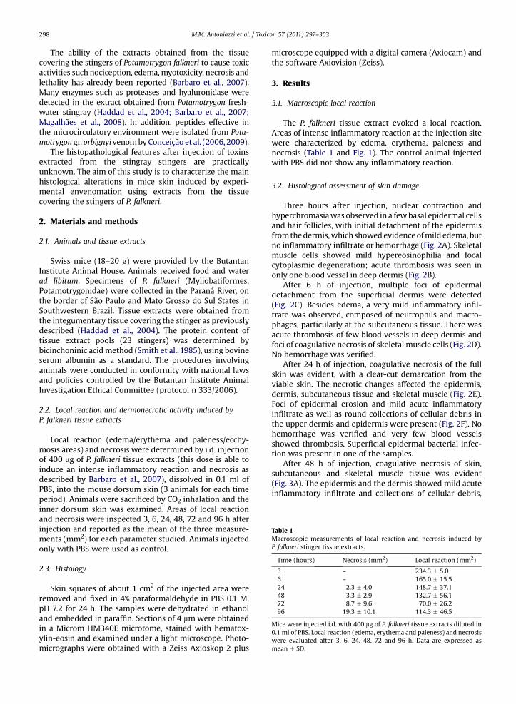
Table 1Macroscopic measurements of local reaction and necrosis induced byP. falkneri stinger tissue extracts.
Time (hours) Necrosis (mm2) Local reaction (mm2)
3 – 234.3 � 5.06 – 165.0 � 15.524 2.3 � 4.0 148.7 � 37.148 3.3 � 2.9 132.7 � 56.172 8.7 � 9.6 70.0 � 26.296 19.3 � 10.1 114.3 � 46.5
Mice were injected i.d. with 400 mg of P. falkneri tissue extracts diluted in0.1 ml of PBS. Local reaction (edema, erythema and paleness) and necrosiswere evaluated after 3, 6, 24, 48, 72 and 96 h. Data are expressed asmean � SD.
M.M. Antoniazzi et al. / Toxicon 57 (2011) 297–303298
The ability of the extracts obtained from the tissuecovering the stingers of Potamotrygon falkneri to cause toxicactivities such nociception, edema,myotoxicity, necrosis andlethality has already been reported (Barbaro et al., 2007).Many enzymes such as proteases and hyaluronidase weredetected in the extract obtained from Potamotrygon fresh-water stingray (Haddad et al., 2004; Barbaro et al., 2007;Magalhães et al., 2008). In addition, peptides effective inthe microcirculatory environment were isolated from Pota-motrygon gr. orbignyivenombyConceição et al. (2006, 2009).
The histopathological features after injection of toxinsextracted from the stingray stingers are practicallyunknown. The aim of this study is to characterize the mainhistological alterations in mice skin induced by experi-mental envenomation using extracts from the tissuecovering the stingers of P. falkneri.
2. Materials and methods
2.1. Animals and tissue extracts
Swiss mice (18–20 g) were provided by the ButantanInstitute Animal House. Animals received food and waterad libitum. Specimens of P. falkneri (Myliobatiformes,Potamotrygonidae) were collected in the Paraná River, onthe border of São Paulo and Mato Grosso do Sul States inSouthwestern Brazil. Tissue extracts were obtained fromthe integumentary tissue covering the stinger as previouslydescribed (Haddad et al., 2004). The protein content oftissue extract pools (23 stingers) was determined bybicinchoninic acidmethod (Smith et al., 1985), using bovineserum albumin as a standard. The procedures involvinganimals were conducted in conformity with national lawsand policies controlled by the Butantan Institute AnimalInvestigation Ethical Committee (protocol n 333/2006).
2.2. Local reaction and dermonecrotic activity induced byP. falkneri tissue extracts
Local reaction (edema/erythema and paleness/ecchy-mosis areas) and necrosis were determined by i.d. injectionof 400 mg of P. falkneri tissue extracts (this dose is able toinduce an intense inflammatory reaction and necrosis asdescribed by Barbaro et al., 2007), dissolved in 0.1 ml ofPBS, into the mouse dorsum skin (3 animals for each timeperiod). Animals were sacrificed by CO2 inhalation and theinner dorsum skin was examined. Areas of local reactionand necrosis were inspected 3, 6, 24, 48, 72 and 96 h afterinjection and reported as the mean of the three measure-ments (mm2) for each parameter studied. Animals injectedonly with PBS were used as control.
2.3. Histology
Skin squares of about 1 cm2 of the injected area wereremoved and fixed in 4% paraformaldehyde in PBS 0.1 M,pH 7.2 for 24 h. The samples were dehydrated in ethanoland embedded in paraffin. Sections of 4 mmwere obtainedin a Microm HM340E microtome, stained with hematox-ylin-eosin and examined under a light microscope. Photo-micrographs were obtained with a Zeiss Axioskop 2 plus
microscope equipped with a digital camera (Axiocam) andthe software Axiovision (Zeiss).
3. Results
3.1. Macroscopic local reaction
The P. falkneri tissue extract evoked a local reaction.Areas of intense inflammatory reaction at the injection sitewere characterized by edema, erythema, paleness andnecrosis (Table 1 and Fig. 1). The control animal injectedwith PBS did not show any inflammatory reaction.
3.2. Histological assessment of skin damage
Three hours after injection, nuclear contraction andhyperchromasiawas observed in a fewbasal epidermal cellsand hair follicles, with initial detachment of the epidermisfromthedermis,which showedevidenceofmild edema, butno inflammatory infiltrate or hemorrhage (Fig. 2A). Skeletalmuscle cells showed mild hypereosinophilia and focalcytoplasmic degeneration; acute thrombosis was seen inonly one blood vessel in deep dermis (Fig. 2B).
After 6 h of injection, multiple foci of epidermaldetachment from the superficial dermis were detected(Fig. 2C). Besides edema, a very mild inflammatory infil-trate was observed, composed of neutrophils and macro-phages, particularly at the subcutaneous tissue. There wasacute thrombosis of few blood vessels in deep dermis andfoci of coagulative necrosis of skeletal muscle cells (Fig. 2D).No hemorrhage was verified.
After 24 h of injection, coagulative necrosis of the fullskin was evident, with a clear-cut demarcation from theviable skin. The necrotic changes affected the epidermis,dermis, subcutaneous tissue and skeletal muscle (Fig. 2E).Foci of epidermal erosion and mild acute inflammatoryinfiltrate as well as round collections of cellular debris inthe upper dermis and epidermis were present (Fig. 2F). Nohemorrhage was verified and very few blood vesselsshowed thrombosis. Superficial epidermal bacterial infec-tion was present in one of the samples.
After 48 h of injection, coagulative necrosis of skin,subcutaneous and skeletal muscle tissue was evident(Fig. 3A). The epidermis and the dermis showed mild acuteinflammatory infiltrate and collections of cellular debris,

Fig. 1. Macroscopic aspect of mice skin (inner side) after injection of P. falkneri stinger tissue extract, at different times. Local reaction and necrosis were evaluatedafter 3, 6, 24, 72 and 96 h (A–E, respectively). Control animal (PBS) (F).
M.M. Antoniazzi et al. / Toxicon 57 (2011) 297–303 299
characterizing micro-abscesses (Fig. 3B). Few blood vesselsin the dermis and subcutaneous tissue presented throm-bosis. No hemorrhage was verified.
After 72 h of injection, the necrotic tissue presentedcellular debris in the form of numerous round collections ordiffuse infiltration, constituting a necrotic plaque focallydetached from the deep tissue (Fig. 3C). Regenerativehyperplasia of epidermal cells appeared at the lesionborders (Fig. 3D). A mild inflammatory infiltrate wasobserved around viable blood vessels in the deep subcu-taneous tissue.
After 96 h of injection, the regenerative hyperplasia ofepidermal cells at thenecrotic skin borderwasmore evident(Fig. 4A). The coagulative necrosis of the tissue was clear,affecting the skeletal muscle and presenting cellular debrisinfiltration. In one of the samples the epidermiswasmissingin some areas and superficial bacterial infection appeared(Fig. 4B). No hemorrhage or blood vessel thrombosis wasdetected.
In animals of the control group no evidence of necrosiswas noted although mild edema and mononuclear cellinfiltration of dermis and subcutaneous tissue were focallypresent. Moreover, control animals did not show anyhistological abnormalities in most of the skin, and subcu-taneous and skeletal muscle tissue (Fig. 4C,D).
4. Discussion
There are few reports in literature on the toxic effectsof freshwater stingray venom. Under our experimentalconditions, we verified that the tissue extract of P. falkneri
could induce necrosis and an inflammatory reaction at thesite of injection. These data are in agreementwith reports ofaccidents in humans (Haddad, 2000; Haddad et al., 2004;Garrone Neto and Haddad, 2010). They are also in agree-ment with an experimental model (Barbaro et al., 2007)demonstrating that necrosis and local inflammation aremuch more prominent in injuries caused by freshwaterstingrays when compared with those caused by marinespecies. Our histological study demonstrated that necrosisoccurs very soon after the exposure; foci of epidermalnecrosis with initial detachment from the dermis weredetected 3–6 h after extract injection. Moreover, at thesetimes, signs of initial necrosis of skeletal muscle wereobserved. Necrosis was clearly established 24 h after extractinjection, affecting all the soft tissues (skin, subcutaneousand skeletal muscle tissue), presenting a clear-cut demar-cation with the viable neighboring skin and showinga coagulative appearance. This appearance indicates theoccurrence of protein denaturation, which is compatiblewith theactionofproteases. Furthermore, our studyshowedno evidence of significant vascular thrombosis or hemor-rhage at any time, which reinforces the hypothesis that thevenom induces tissue necrosis probably by the direct actionof toxins/enzymes (Barbaro et al., 2007).
Envenomations caused by some species of snakes(Gutiérrez et al., 2005; Moura-da-Silva et al., 2007), spiders(Ospedal et al., 2002; Hogan et al., 2004) and fish (Limaet al., 2003; Pareja-Santos et al., 2009) are also character-ized by severe local tissue damage. The venom of theseanimals has enzymes involved in the pathogenesis of localmyonecrosis, skin damage with intense inflammatory
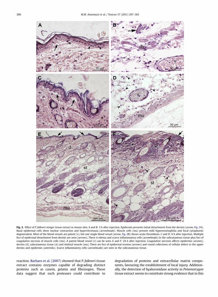
Fig. 2. Effect of P. falkneri stinger tissue extract in mouse skin. A and B: 3 h after injection. Epidermis presents initial detachment from the dermis (arrow, Fig. 2A).Basal epidermal cells show nuclear contraction and hyperchromasia (arrowheads). Muscle cells (mu) present mild hypereosinophilia and focal cytoplasmicdegeneration. Most of the blood vessels are patent (v), but one single blood vessel (arrow, Fig. 2B) shows acute thrombosis. C and D: 6 h after injection. Multiplefoci of epidermal detachment from dermis are seen (arrows). There is edema and scarce inflammatory cells (arrowheads) in the subcutaneous tissue plus foci ofcoagulative necrosis of muscle cells (mu). A patent blood vessel (v) can be seen. E and F: 24 h after injection. Coagulative necrosis affects epidermis (arrows),dermis (d), subcutaneous tissue (st) and skeletal muscle (mu). There are foci of epidermal erosion (arrows) and round collections of cellular debris in the upperdermis and epidermis (asterisks). Scarce inflammatory cells (arrowheads) are seen in the subcutaneous tissue.
M.M. Antoniazzi et al. / Toxicon 57 (2011) 297–303300
reaction. Barbaro et al. (2007) showed that P. falkneri tissueextract contains enzymes capable of degrading distinctproteins such as casein, gelatin and fibrinogen. Thesedata suggest that such proteases could contribute to
degradation of proteins and extracellular matrix compo-nents, favouring the establishment of local injury. Addition-ally, the detection of hyaluronidase activity in Potamotrygontissue extract seems to constitute strong evidence that in this
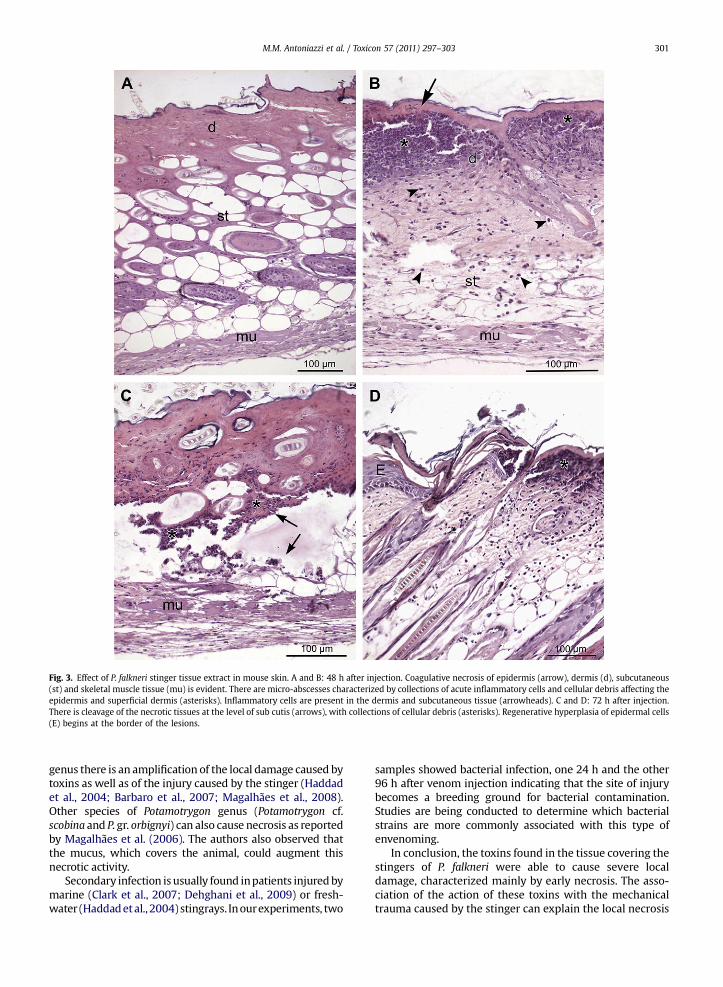
Fig. 3. Effect of P. falkneri stinger tissue extract in mouse skin. A and B: 48 h after injection. Coagulative necrosis of epidermis (arrow), dermis (d), subcutaneous(st) and skeletal muscle tissue (mu) is evident. There are micro-abscesses characterized by collections of acute inflammatory cells and cellular debris affecting theepidermis and superficial dermis (asterisks). Inflammatory cells are present in the dermis and subcutaneous tissue (arrowheads). C and D: 72 h after injection.There is cleavage of the necrotic tissues at the level of sub cutis (arrows), with collections of cellular debris (asterisks). Regenerative hyperplasia of epidermal cells(E) begins at the border of the lesions.
M.M. Antoniazzi et al. / Toxicon 57 (2011) 297–303 301
genus there is an amplification of the local damage caused bytoxins as well as of the injury caused by the stinger (Haddadet al., 2004; Barbaro et al., 2007; Magalhães et al., 2008).Other species of Potamotrygon genus (Potamotrygon cf.scobina and P. gr. orbignyi) can also cause necrosis as reportedby Magalhães et al. (2006). The authors also observed thatthe mucus, which covers the animal, could augment thisnecrotic activity.
Secondary infection is usually found inpatients injuredbymarine (Clark et al., 2007; Dehghani et al., 2009) or fresh-water (Haddadetal., 2004) stingrays. Inourexperiments, two
samples showed bacterial infection, one 24 h and the other96 h after venom injection indicating that the site of injurybecomes a breeding ground for bacterial contamination.Studies are being conducted to determine which bacterialstrains are more commonly associated with this type ofenvenoming.
In conclusion, the toxins found in the tissue covering thestingers of P. falkneri were able to cause severe localdamage, characterized mainly by early necrosis. The asso-ciation of the action of these toxins with the mechanicaltrauma caused by the stinger can explain the local necrosis
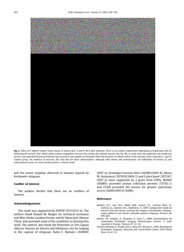
Fig. 4. Effect of P. falkneri stinger tissue extract in mouse skin. A and B: 96 h after injection. There is an evident regenerative hyperplasia of epidermal cells (E)bordering the necrotic skin, which shows intense coagulative necrosis that reaches the skeletal muscle (mu, Fig. 4B). In some areas the epidermis was totally lost(arrow) and superficial bacterial infection was present in one sample (arrowheads) Note the presence of cellular debris in the necrotic tissue (asterisks). C and D:Control group. No evidence of necrosis. The skin did not show abnormalities, although mild edema and mononuclear cell infiltration of dermis (d) andsubcutaneous tissue (st) were focally present. v–blood vessel.
M.M. Antoniazzi et al. / Toxicon 57 (2011) 297–303302
and the severe sequelae observed in humans injured byfreshwater stingrays.
Conflict of interest
The authors declare that there are no conflicts ofinterest.
Acknowledgements
This work was supported by FAPESP (07/55272-4). Theauthors thank Danieli M. Rangel, for technical assistanceand Miss Ottilie Carolina Forster and Dr Maria José AlencarVilela, who provided some of the conditions to develop thiswork. The authors also thank the fishermen in Três Lagoas(Marcos Teixeira da Silveira and Edmilson) city for helpingin the capture of stingrays. Katia C. Barbaro (304800/
2007-4), Domingos Garrone Neto (142985/2005-8), MartaM. Antoniazzi (307029/2009-3) and Carlos Jared (307247/2007-4) were supported by a grant from CNPq. IBAMA(SISBIO) provided animal collection permits (15702-1)and CGEN provided the license for genetic patrimonyaccess (02001.005111/2008).
References
Barbaro, K.C., Lira, M.S., Malta, M.B., Soares, S.L., Garrone Neto, D.,Cardoso, J.L., Santoro, M.L., Haddad Jr., V., 2007. Comparative study onextracts from the tissue covering the stingers of freshwater (Potamo-trygon falkneri) and marine (Dasyatis guttata) stingrays. Toxicon 50,676–687.
Brisset, I.B., Schaper, A., Pommier, P., Haro, L., 2006. Envenomation byamazoniam freshwater stingray Potamotrygon motoro: 2 casesreported in Europe. Toxicon 47, 32–34.
Charvet-Almeida, P., Araújo,M.L.G., Rosa, R.S., Rincón, G., 2002. Neotropicalfreshwater stingrays: diversity and conservation status. IUCN SharkNews 14, 47–51.

M.M. Antoniazzi et al. / Toxicon 57 (2011) 297–303 303
Clark, R.F., Girard, R., Rao, D., Ly, B.T., Davis, D.P., 2007. Stingray enveno-mation: a retrospective review of clinical presentation and treatmentin 119 cases. J. Emerg. Med. 33, 33–37.
Conceição, K., Konno, K., Melo, R.L., Marques, E.E., Hiruma-Lima, C.A.,Lima, C., Richardson, M., Pimenta, D.C., Lopes-Ferreira, M., 2006.Orpotrin: a novel vasoconstrictor peptide from this venom of theBrazilian stingray Potamotrygon gr. orbignyi. Peptides 27, 3039–3046.
Conceição, K., Santos, J.M., Bruni, F.M., Klitzke, C.F., Marques, E.E.,Borges, M.H., Melo, R.L., Fernandez, J.H., Lopes-Ferreira, M., 2009.Characterization of a new bioactive peptide from Potamotrygon gr.orbignyi freshwater stingray venom. Peptides 30, 2191–2199.
Dehghani, H., Sajjadi, M.M., Rajaian, H., Sajedianfard, J., Parto, P., 2009.Study of patient’s injuries by stingrays, lethal activity determinationand cardiac effects induced by Himantura gerrardi venom. Toxicon 54,881–886.
Garrone Neto, D., Haddad Jr., V., Vilela, M.J.A., Uieda, V.S., 2007. Registrode ocorrência de duas espécies de potamotrigonídeos na região doAlto do Rio Paraná e algumas considerações sobre biologia. BiotaNeotropica 7 (1). http://www.biotaneotropica.org.br/v7n1/pt/abstract?shortcommunicationþbn00707012007 1676–0603 Jan/Abr 2007.
Garrone Neto, D., Haddad Jr., V., 2009. Acidentes Por Raias. In: Cardoso, J.L.C., França, F.O.S., Wen, F.H., Málaque, C.M., Haddad Jr., V. (Eds.), Ani-mais Peçonhentos No Brasil: Biologia, Clínica E Terapêutica DosAcidentes, second ed. Sarvier, São Paulo, pp. 295–305.
Garrone Neto, D., Haddad Jr., V., 2010. Arraias em rios da região Sudestedo Brasil: locais de ocorrência e impactos sobre a população. Rev. Soc.Bras. Med. Trop. 43, 82–88.
Gutiérrez, J.M.,Rucavado,A.,Escalante,T.,Diaz, C., 2005.Hemorrhage inducedby snake venom metalloproteinases: biochemical and biophysicalmechanisms involved in microvessel damage. Toxicon 45, 997–1011.
Haddad Jr., V., 2000. Atlas de animais aquáticos perigosos do Brasil:guia médico de identificação e tratamento. Editora Rocca, São Paulo.pp. 123–128.
Haddad Jr., V., Garrone Neto, D., de Paula, N.J.B., Marques, F.P.L., Barbaro, K.C.,2004. Freshwater stingrays: study of epidemiologic, clinic andtherapeutic aspects based on 84 envenomings in humans and someenzymatic activities of the venom. Toxicon 43, 287–294.
Hogan, C.J., Barbaro, K.C., Winkel, K., 2004. Loxoscelism: old obstacles,new directions. Ann. Emerg. Med. 44, 608–624.
Isbister, G., 2001. Venomous fish stings in tropical northern Austrália. Am.J. Emerg. Med. 19, 561–565.
Lim, Y.L., Kumarasinghe, S.P.W., 2007. Cutaneous injuries from marineanimals. Singapore Med. J. 48, 25–28.
Lima, C., Clissa, P.B., Piran-Soares, A.A., Tanjoni, I., Moura-da-Silva, A.M.,Lopes-Ferreira, M., 2003. Characterisation of local inflammatoryresponse induced by Thalassophryne nattereri fish venom in a mousemodel of tissue injury. Toxicon 42, 499–507.
Magalhães, K.W., Lima, C., Piran-Soares, A.A., Marques, E.E., Hiruma-Lima, C.A., Lopes-Ferreira, M., 2006. Biological and biochemicalproperties of the Brazilian Potamotrygon stingrays: Potamotrygon cf.scobina and Potamotrygon gr. orbignyi. Toxicon 47, 575–583.
Magalhães, M.R., da Silva, N.J., Ulhoa Jr., C.J., 2008. A hyaluronidase fromPotamotrygon motoro (freshwater stingrays) venom: isolation andcharacterization. Toxicon 51, 1060–1067.
Moura-da-Silva, A.M., Butera, D., Tanjoni, I., 2007. Importance of snakevenom metalloproteinases in cell biology: effects on platelets,inflammatory and endothelial cells. Curr. Pharm. Des. 13, 2893–2905.
Ospedal, K.Z., Appel, M.H., Fillus Neto, J., Mangili, O.C., Veiga, S.S.,Gremski, W., 2002. Histopathological findings in rabbits after exper-imental acute exposure to the Loxosceles intermedia (brown spider)venom. Int. J. Exp. Pathol. 83, 287–294.
Pareja-Santos, A., Saraiva, T.C., Costa, E.P., Santos, M.F., Zorn, T.T., Souza, V.M., Lopes-Ferreira, M., Lima, C., 2009. Delayed local inflammatoryresponse induced by Thalassophryne nattereri venom is related toextracellular matrix degradation. Int. J. Exp. Pathol. 90, 34–43.
Pedroso, C.M., Jared, C., Charvet-Almeida, P., Almeida, M.P., GarroneNeto, D., Lira, M.S., Haddad Jr., V., Barbaro, K.C., Antoniazzi, M.M.,2007. Morphological characterization of the venom secretoryepidermal cells in the stinger of marine and freshwater stingrays.Toxicon 50, 688–697.
Smith, P.K., Krohn, R.I., Hermanson, G.T., Mallia, A.K., Gartner, F.H.,Provenzano, M.D., Fujimoto, E.K., Goeke, N.M., Olson, B.J., Klenk, D.C.,1985. Measurement of protein using bicinchoninic acid. Anal. Bio-chem. 150, 76–85.