Comparative Histopathological Evaluation of Permethrin
16
The Egyptian Journal of Hospital Medicine Vol., 11 : 58 –73 June 2003 I.S.S.N: 12084 1687-2002 Comparative Histopathological Evaluation of Permethrin, Pirimiphos Methyl and Bendiocarb Toxicities in Testes, Liver and kidney of rat. Afaf L. Nessiem; Nahed S. bassily and Salwa A. Metwally National Organization for Drug Control and Research NODCAR, Egypt Abstract The increasing use of insecticides in agriculture and in public health calls for greater attention for studying their possible toxic effect (s) on man and animals. Acute toxic effects have been relatively well known whereas chronic effects require further studies. The aim of the present work was, therefore, to study the histopathological changes in testes, liver and kidney of rats due to 30 days feeding on diet containing permethrin, pirimiphos methyl and bendiocarb. The dose used for each insecticide represented a concentration that equals ten times the acceptable human daily intake. These doses are far below the LD50, but represent possible exposure doses. Forty male Sprague- Dawley rats were divided into 4 equal groups. Animals of each group were fed either by normal diet, or diet mixed with permethrin (21.739 ppm), pirimiphos methyl (4.350 ppm) or bendiocarb (2ppm) for 30 days. Histological sections of testes, liver and kidneys were examined and histopathological changes and quantitative estimates were recorded. Incidence of spermatogenic suppression, Leydig cell atrophy and vacuolation of Sertoli cells were most prominent in testicular sections from primiphos methyl treated animal testis than in animals of the other groups. Peremethrin feeding resulted in the least deteriorative changes. In sections of liver, dilatation and congestion of blood sinusoids was most evident in the group treated with primiphos methyl and to less extent in those treated with bendiocarb. Swollen hepatocytes with pyknotic nuclei and incidence of apoptosis were also recorded. In kidney sections, vacuolar degeneration, tubular and capsular dilatation, and glomerular congestion were observed especially in primiphos methyl treated rats. In conclusion, the obtained changes were of different severity as a response of exposure to permethrin, pirimiphos methyl or bendiocarb at the same equivelant of human acceptable daily intake. Introduction The use of pesticides has been largely expanded during the last fifty years. More than 3 million tons of approximately 600 different chemicals are applied annually throughout the world. The WHO (1992), reported that 3 million pesticide poisoning cases occurred annually and resulted in 220 000 deaths allover the world. In the developing countries the situation is worse, since higher proportions of pesticidepoisoning and deaths occurred, the reasons behind this include inadeq - uate occupational safety standards and insufficient knowledge of pesticide hazards. Some pesticides are carcinog - enic, most are teratogenic, and others are mutagenic. All are attributed to normal agriculture use (US Geological survey 1997). So, it is safe to assume that sooner or later higher percentages of our people (especially in developing 58 Refree : Prof ; Dr. Fathey Matter .
Transcript of Comparative Histopathological Evaluation of Permethrin
The Egyptian Journal of Hospital Medicine Vol., 11 : 58 –73 June 2003
I.S.S.N: 12084
1687-2002
Comparative Histopathological Evaluation of Permethrin, Pirimiphos
Methyl and Bendiocarb Toxicities in Testes, Liver and kidney of rat.
Afaf L. Nessiem; Nahed S. bassily and Salwa A. Metwally National Organization for Drug Control and Research NODCAR, Egypt
Abstract
The increasing use of insecticides in agriculture and in public health calls for
greater attention for studying their possible toxic effect (s) on man and animals. Acute
toxic effects have been relatively well known whereas chronic effects require further
studies. The aim of the present work was, therefore, to study the histopathological
changes in testes, liver and kidney of rats due to 30 days feeding on diet containing
permethrin, pirimiphos methyl and bendiocarb. The dose used for each insecticide
represented a concentration that equals ten times the acceptable human daily intake.
These doses are far below the LD50, but represent possible exposure doses. Forty male
Sprague- Dawley rats were divided into 4 equal groups. Animals of each group were fed
either by normal diet, or diet mixed with permethrin (21.739 ppm), pirimiphos methyl
(4.350 ppm) or bendiocarb (2ppm) for 30 days. Histological sections of testes, liver and
kidneys were examined and histopathological changes and quantitative estimates were
recorded. Incidence of spermatogenic suppression, Leydig cell atrophy and vacuolation
of Sertoli cells were most prominent in testicular sections from primiphos methyl
treated animal testis than in animals of the other groups. Peremethrin feeding resulted in
the least deteriorative changes. In sections of liver, dilatation and congestion of blood
sinusoids was most evident in the group treated with primiphos methyl and to less
extent in those treated with bendiocarb. Swollen hepatocytes with pyknotic nuclei and
incidence of apoptosis were also recorded. In kidney sections, vacuolar degeneration,
tubular and capsular dilatation, and glomerular congestion were observed especially in
primiphos methyl treated rats.
In conclusion, the obtained changes were of different severity as a response of
exposure to permethrin, pirimiphos methyl or bendiocarb at the same equivelant of
human acceptable daily intake.
Introduction
The use of pesticides has been
largely expanded during the last fifty
years. More than 3 million tons of
approximately 600 different chemicals
are applied annually throughout the
world. The WHO (1992), reported that
3 million pesticide poisoning cases
occurred annually and resulted in 220
000 deaths allover the world. In the
developing countries the situation is
worse, since higher proportions of
pesticidepoisoning and deaths occurred,
the reasons behind this include inadeq -
uate occupational safety standards and
insufficient knowledge of pesticide
hazards. Some pesticides are carcinog -
enic, most are teratogenic, and others
are mutagenic. All are attributed to
normal agriculture use (US Geological
survey 1997). So, it is safe to assume
that sooner or later higher percentages
of our people (especially in developing
58 Refree : Prof ; Dr. Fathey Matter .
Afaf L. Nessiem et al
59
countries) will suffer from some serious
forms of diseases like cancer and kidney
failure (Cheraskin 2000).These diseases
will result from toxins in air we breathe,
food we eat and water we drink.
Among the potent synthetic
insecticides that have been increasingly
employed in recent years are synthetic
pyrethroids, organophosphates and car -
bamates. Pyrethroids administration
resulted in deleterious effects on liver
and blood parameters, and to induce
chromosomal aberrations (Ishmael and
Lithfield, 1988 and Institoris et al.,
1999a and b), to suppress erythropiosis
and hemoglobin synthesis and to
increase the number of leukocytes (Tos-
Luty et al., 2001). Pyrethroids were also
reported to cause slight activation of
cytochrom P 450 1A and 2B- mediated
reactions (Kostka et al., 1997 and
Moresseau et al. 1999) and to act as a
tumor promotor at a non-hepatotoxic
doses (Hemming et al., 1993). They
may inhibit the G2 phase in the cell
cycle and consequently may suppress
the cell entering into the stage of
mitosis (Kostka et al., 2000).
Pyrethroids were also found to affect
male and female reproductive system
(Eil and Nisuls, 1990).
Organophosphate insecticides
were in existence since 1854, but were
not recognized as having toxic
potentials until 1930 (Marrs, 1993).
These compounds induce significant fall
of body weight (Gajewski and
katkiewuz, 1981), and reduce glycogen
content in liver and kidney (Awasthi et
al., 1984). Pirimiphos methyl is known
to affect the proteolytic enzyme
activities in rat heart, kidney, brain and
liver (Mantle et al., 1997). It induces
significant inhibition of brain and
erythrocyte-acetyl cholinesterase, plas -
ma pseudo cholinesterase and non-
specific carboxyl esterase of brain,
plasma and kidney (Rajini et al.,1989).
Carbamates may represent a class
of chemicals which function through a
mechanism separate from ligand-
receptor binding, as they may act as
general endocrine modulators in
mammalian cells (Klotz et al., 1997).
They induce dose dependent decrease in
body weight (Pant et al., 1995a and b
and Kackar et al., 1997) and significant
change in the weight of testes,
epididymides and accessory sex organs
(Pant et al., 1995b). Carbamate
insecticides were found to inhibit both
aggregation and arachidonic acid
metabolism in human blood platelets
(Krug et al., 1988), to inhibit brain and
blood acetylcholinesterase, liver glucose
6 phosphatase and succinic dehyd -
rogenase (Fayez and Kilgore 1992).
The wide spread use of the
abovementioned insecticides in agricu -
lture and in public health drew our
attention for studying their possible
toxic action (s) in man and animals. The
aim of the present work is to study the
histopathological changes in liver,
kidney and testes due to daily oral
feeding for 30 days by diet containing
permethrin, pirimiphos methyl and
bendiocarb, each at a concentration that
equals ten times the acceptable daily
intake (a concentration that may
represent the real life situation
regarding exposure to the tested
compounds).
Material and Methods Animals:
male Sprague-Dawley rats of 110-
120 gm body weight were obtained
from the breading colony of the
National Organization for Drug Control
and Research (NODCAR), Cairo. They
were housed as 7 animals per cage.
Insecticides:
Permethrin represents pyrethroid
insecticides. Pirimiphos methyl repre-
sents organophosphorus insecticides.
Comparative Histopathological Evaluation of Permethrin…….
60
Bendiocarb represents carbamate
insecticides.
Experimental design:
rats were divided into 4 equal
groups (each consisted of 10 animals):
Control group: animals were fed norm -
al diet and served as a control.
Permethrin group: animals were
fed diet containing 10 times the human
maximal acceptable daily intake of
permethrin (21.739 ppm) for 30 days.
Pirimiphos methyl group: animals
were fed diet containing 10 times the
human maximal acceptable daily intake
of pirimiphos methyl (4.350 ppm) for
30 days.
Bendiocarb group: animals were
fed diet containing 10 times the human
maximal acceptable daily intake of
bendiocarb (2 ppm) for 30 days.
Histological manipulation:
Twenty- four hours after the last
day of feeding with insecticides,
animals of each group were killed by
narcosis. Testes, liver and kidneys were
removed, fixed in buffered formol and
prepared for paraffin sectioning. Six
micron sections were mounted on clean
slides, and stained by hematoxylin and
eosin (Culling, 1974).
Quantitative analysis:
Low magnification ten sections
from the testis were used to count the
number of seminiferous tubular sections
that do not contain all the spermato-
genic components including spermato-
zoa in their lumen. Such sections were
considered as having suppressed sperm-
atogenesis. High magnification sections
were used to count Sertoli cells with
vacuolar degeneration and Leydig cells
that lack the normal healthy appearance.
Liver sections were used to count
the percentage of hepatocytes with
vacuolar degeneration and those with
apoptotic morphology.
Results
The testis
Sections from the testes of the
different groups are represented in
plates (1,2.).
The sectioned seminiferous
tubules in control animal testis are
closely packed with regularly distrib-
uted interstitial tissues (plate 1).
Different stages of spermatogenesis are
represented in the arrangement and
number of layers in the tubule.
Spermatozoa are clearly visible in the
tubular lumen (plate 2).
Sections in the seminiferous tubul-
es of permethrin-treated animal testis
show almost normal arrangement of
spermatogenic cells (plate 1). The
Sertoli cells, however, are vacuolated.
Fewer spermatozoa are found in the
lumen compared with control. The
interstitial matrix is swelled with
aggregation of Leydig cells (plate 2).
The seminiferous tubules of
pirimiphos methyl treated rat testis are
smaller in diameter with irregular shape
and depressed spermatogenesis(plate 1).
The Sertoli cells are highly vacuolated.
The layers representing spermatogenic
cells are highly disturbed. Giant cells
are clearly represented in the lumen of
the tubules. There is a near complete
absence of spermatozoa (plate 2).
The contours of sections of
seminiferous tubules from the testes of
bendiocarb fed rats appear also irregular
and widely separated. Signs of swelling
can also be noticed in the interstitial
area. (plate 1). In some sections, the
spermatozoa appear in the middle of the
lumen without clearly differentiated
tails (plate 2).
Afaf L. Nessiem et al
61
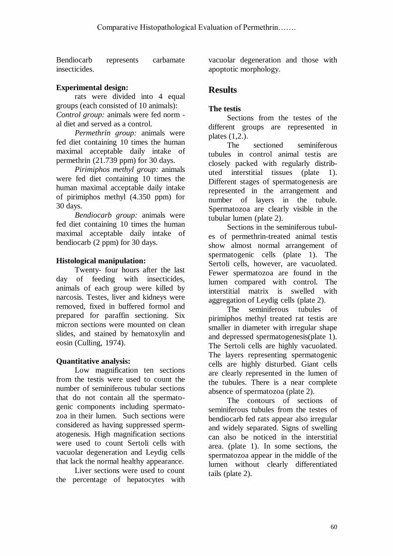
Table (1): Incidence of histological changes in testicular tissue of rats fed diet
containing permethrin (21.739 ppm), pirimiphos methyl (4.35 ppm) or bendiocarb
(2 ppm)
Incidence (%)
Treatment spermatogensis leydig cell atrophy vacuolar degeneration
of sertoli`s cells
normal supressed present absent present absent
normal diet 100% 0% 0% 100% 0% 100% permethrin containing diet 80% 20% * 0% 100% 10% 90% Pirimiphos methyl containing 10% 90% *** 90% *** 10% 90% *** 10%
diet bendiocarb containing diet 60% 40% ** 40% ** 60% 30% ** 70%
*, **, *** statistical difference at p<0.05, <0.01 and < 0.005, respectively compared with the control. Statistical analysis using chi-square test.
Table (1) represents the incidence
of the histological changes that occurred
in rat testes due to daily oral feeding by
diet containing permethrin, pirimiphos
methyl or bendiocarb. The data showed
that spermatogenesis was suppressed in
20, 90 and 40% in sections of seminife-
rous tubules in the testes of animals fed
by diet containing permethrin, pirimip-
hos methyl or bendiocarb, respectively.
Leydig cell atrophy was encountered in
90% and 40% of animals treated with
pirimiphos methyl or bendiocarb,
respectively. The obtained data showed
also that vacuolar degene-ration in
Sertoli cells was present in 10, 90 and
30% of animals due to daily oral
feeding with permethrin, pirimiphos
methyl or bendiocarb, containing diet,
respectively.
The liver
Compared with control, sections
in the liver of rats treated with
permethrin revealed normal lobular
architecture with mild changes in form.
Scattered inflammatory cell aggregates
and strands of fibrosis were observed in
the portal area (plate3). There are
scattered mildly dilated sinusoids with
prominent Kupffer cells and intrava -
scular leucocytes. The hepatocytes were
mildly swollen and showed few scat-
ered apoptotic cells. It also showed
occasional hepatocytes with pyknotic
nuclei and acidophilic cytoplasm toge-
ther with few scattered binucleated
hepatocytes (plate 4).
In pirimiphos methyl treated
animals, the histolgical changes in liver
tissue were severe. Portal areas were
markedly expanded, showing dilated
portal veins, areas of fibrosis and
inflammatory cellular infiltrate It also
showed areas with inflammatory cell
aggregates. Signs of hemorrhage can
also be seen (plate 3). Hepatocytes
showed marked ballooning and vacuoli-
zation, together with marked sinusoidal
dilatation and high incidence of
apoptotic body formation . Moreover,
binucleated hepatocytes could be also
seen Sections in liver of the animals fed
with diet containing bendiocarb showed
diffuse and prominent cytoplasmic
vacuolation, mildly dilated sinusoids
and scattered apoptotic body formation
(plate 4).
Comparative Histopathological Evaluation of Permethrin…….
62
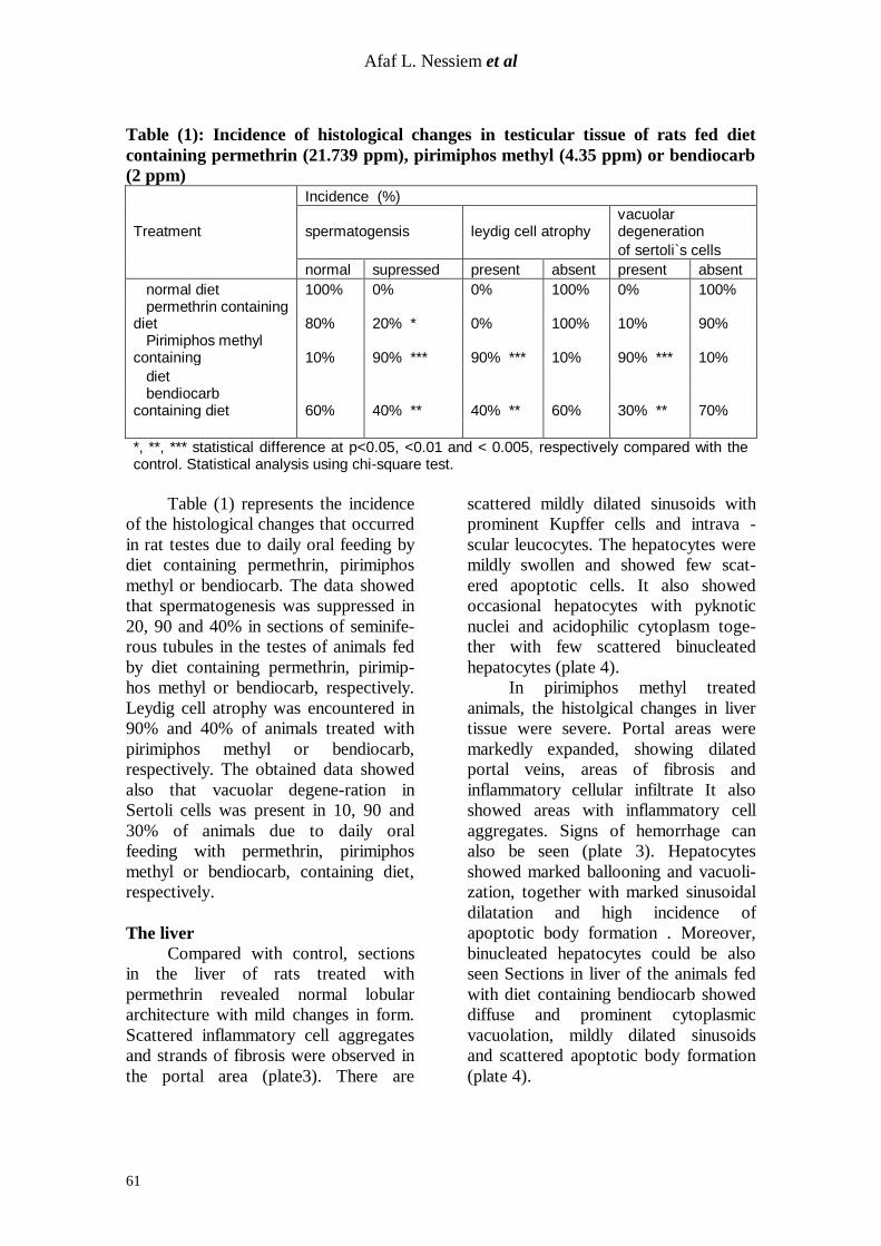
Table (2) shows the incidence of
vacuolar degeneration and the mean
apoptotic index in liver tissue due to
feeding with diet containing permethrin,
pirimiphos methyl or bendiocarb. The
data present in this table show that
permethrin feeding induced mild vacu-
olar degeneration in 10% of hepatoc-
ytes, irimiphos methyl induced severe
vacuolar degeneration in 100% of
hepatocytes, while bendiocarb induced
only moderate effect in 50%, of the
hepatocyte population. Moreover, the
table shows that the mean induced
apoptotic indices were 8% in permet-
hrin, 80% in pirimiphos methyl, and
40% in bediocarb fed animals.
The Kidney
Microscopic structure of kidney
tissue of rats treated with permethrin
was almost normal, with normal
appearing tubular and glomerular
structure, where as in rats fed on diet
contains pirimiphos methyl (plate 5),
the histological examination revealed
vacuolar degenerative changes in
epithelial cells lining some renal
tubules. Glomerular tufts were seen
showing hyper cellularity with evidence
of congestion The lumen of Bowmans
capsule was dilated.
Sections in the kidneys of rats
subjected to bendiocarb treatment,
showed widely dilated distal convoluted
tubules. Necrotic changes were predo -
minant in proximal convoluted tubules.
The Bowmans capsule was dilated with
some signs of congestion in the glome -
rular capillaries (Plate 5).
Table (2) : Incedence of histological changes in liver tissue of rats fed diet
containing permethrin(21.739ppm), pirimiphos methyl (4.350ppm) and
bendiocarb (2 ppm).
Incidence (%) (a)
Treatment vacuolar degeneration Mean apoptotic index
negative mild modrate severe
normal diet 100% 0 0 0 0
permethrin containing diet 90% 10% 0 0 8% pirimiphos methyl containing 0 0 0% 100% *** 80%
diet bendiocarb containing diet 50% 0 50% ** 0 40%
(a), apoptotic index = number of apoptotic cells in 1000 cells devided by 10 *, **, *** statistical difference at p<0.05, <0.01 and < 0.005, respectively compared with the control . values were analyzed using chi-square test.

Afaf L. Nessiem et al
63
Plate (1): Sections in the testes of control and insecticide treated animals. In the control, the
seminiferous tubules appear in the different phases of spermatogenesis, the lumen is loaded with mature spermatozoa and the interstitial area containing the Leydig cells is continuous. In
Permethrin treated group, signs of spermatogenesis are less prominent,the interstitial area
suggest signs of swelling. In perimiphos methyl treated group, the seminiferous tubules have smaller diameter and irregular in shape, most of the tubules have suppressed spermiogenesis
and the interstitium is swelled. In bendiocarb treated group, there is moderate change in tubular
diameter and shape irregularity with interstitial swelling. The areas in the squares are
represented at higher power in plate 2 ( Hx,E X200)
Comparative Histopathological Evaluation of Permethrin…….
64
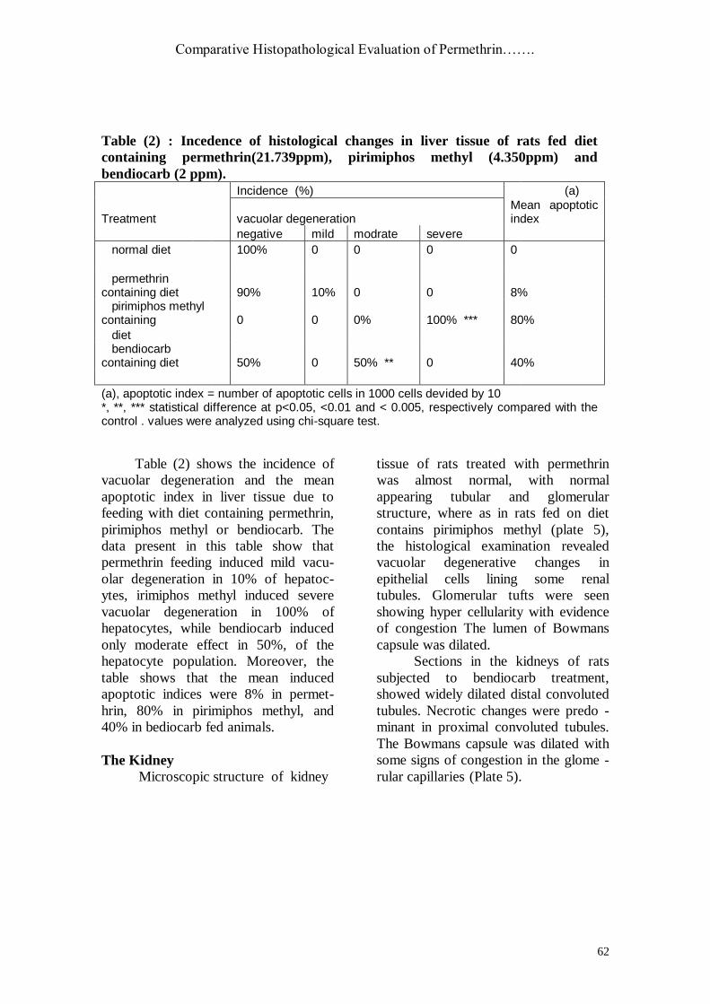
Plate (2): Sections in the testes of control and insecticide treated animals. In the control, the
lumen of the seminiferous tubules appear loaded with mature spermatozoa (SZ) spermatids
(SD) in different phases of normal differentiation. In Permethrin treated group, the spermatogonia (SG) form a continuous basal layer . The spermatids (SD) are more or less
normal while Sertoli cells (SC) are moderately vacuolated. Leydig cells (LC) are aggregated
near by the basement membrane with some swelling in the interstitium. In perimiphos methyl
treated group, the seminiferous tubules have suppressed spermiogenesis with formation of giant cells. The Sertoli cells (SC) are highly vacuolated, the spermatogenic cell layer is not
continuous and the Leydig cells(LC) are aggregated far from the basement membrane. The
interstitium is highly swelled. In bendiocarb treated group, The Sertoli cell (SC) is also vacuolted, the spermatogenic cells are not well differentiated and the interstitial Leydig
cells(LC) are aggregated far from the basement with interstitial swelling. ( Hx,E X400
Afaf L. Nessiem et al
65
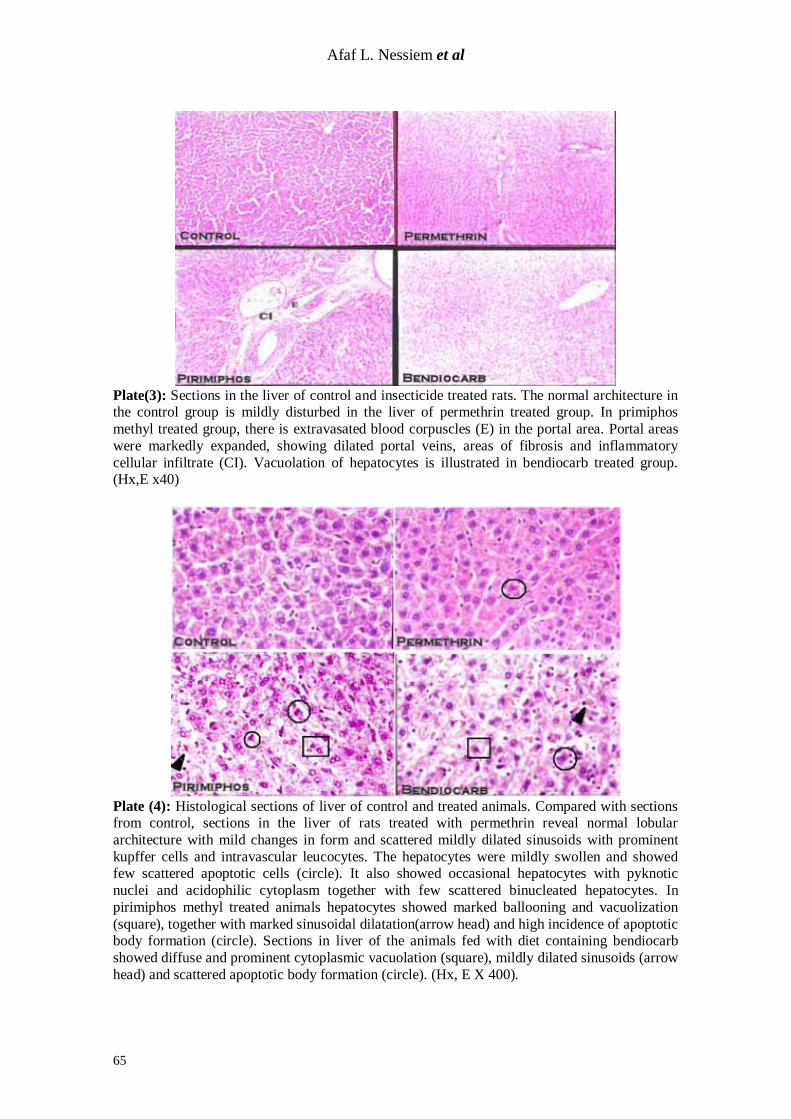
Plate(3): Sections in the liver of control and insecticide treated rats. The normal architecture in the control group is mildly disturbed in the liver of permethrin treated group. In primiphos
methyl treated group, there is extravasated blood corpuscles (E) in the portal area. Portal areas
were markedly expanded, showing dilated portal veins, areas of fibrosis and inflammatory
cellular infiltrate (CI). Vacuolation of hepatocytes is illustrated in bendiocarb treated group. (Hx,E x40)
Plate (4): Histological sections of liver of control and treated animals. Compared with sections from control, sections in the liver of rats treated with permethrin reveal normal lobular
architecture with mild changes in form and scattered mildly dilated sinusoids with prominent
kupffer cells and intravascular leucocytes. The hepatocytes were mildly swollen and showed few scattered apoptotic cells (circle). It also showed occasional hepatocytes with pyknotic
nuclei and acidophilic cytoplasm together with few scattered binucleated hepatocytes. In
pirimiphos methyl treated animals hepatocytes showed marked ballooning and vacuolization
(square), together with marked sinusoidal dilatation(arrow head) and high incidence of apoptotic body formation (circle). Sections in liver of the animals fed with diet containing bendiocarb
showed diffuse and prominent cytoplasmic vacuolation (square), mildly dilated sinusoids (arrow
head) and scattered apoptotic body formation (circle). (Hx, E X 400).

Comparative Histopathological Evaluation of Permethrin…….
66
Plate (5): Microscopic structure of kidney tissue of rats treated with permethrin was
almost normal, with mild capsular dilatation (DIL) and minor tubular cell degeneration
(DT), where as in rats fed on diet containing pirimiphos methyl, there is vacuolar
degenerative changes in epithelial cells lining some renal tubules (DT). Glomerular tufts
were seen showing hyper cellularity with evidence of congestion (CON) and capsular
dilatation (DIL). In the kidneys of rats subjected to bendiocarb treatment, there is
widely dilated distal convoluted tubules (DILT). Necrotic changes were predominant in
proximal convoluted tubules(DT). The Bowmans capsule was dilated (DIL) with some
signs of congestion in the glomerular capillaries. (Hx, E X 400).
Discussion Although insecticides represent
one of the most widely encountered
toxic pollutants, the need to its use in
agriculture and house insect control is
indispensable for the human life.
Different groups of insecticides are
used. The need for choosing the group
with the least hazards to human life
calls for active research towards this
goal. In the present study, represen -
tatives of three groups of insecticides
were compared as to their effect on
three vital organs in order to find out
their differential hazard on these organs.
The insecticides used represented one of
natural origin, the pyrethroid permeth-
rin, the organophosphate pirimiphos
methyl and the carbamate bendiocarb.
The organs were the testis, liver and
kidney.
The histopathological changes
obtained in the testicular tissue due to
feeding diet containing permethrin indu-
ced spermatogenic suppression, widen-
ing between spermatogoia and Sertoli
cell vacuolation,. Similar changes were
also reported by Yenilmez (1995);
Tyrkiel et al., (2001) and Abou-Donia
et al., (2003).
Abou-Donia et al. (2003) found
that dermal daily application of perme -
thrin (0.13mg/kg in ethanol) to human
was implicated in the development of
genitrourinary disorders among verter -
ans of Persian Gulf War. On performing
experiments on rats, these authors

Afaf L. Nessiem et al
67
observed incidence of histopathological
alterations in rat testes due to permet-
hrin administration. The alterations
included apoptosis of testicular germ,
sertoli and leydig cells. In an earlier
work, Yenilmez (1995), reported that
permethrin oral administration induced
widening between spermatogonia and
sertoli`s cells, extrusion of the germ
cells and increase in the number and
size of the lipid inclusion in the leydig`s
cell.
The obtained histopathological
changes in testicular tissues were
suggested to result from the binding of
permethrin to the receptors of the
androgen male sex hormone (Eil, et al.
1990) and/or binding to the benz-
odiazepine receptor that stimulates the
production of male sex hormone
testosterone (Ramadan et al., 1988).
Another explanation was that the above
mentioned histopathological changes
may be due to the decrease in the blood
testes barrier permeability (Abou-Donia
et al.,2001). Daily feeding with diet
containing pirimiphos methyl for 30
days was found to induce marked
spermatogenesis suppression where
primary and secondary spermatogonia
were markedly reduced, reduced num-
ber of sperms and spermatids. The obta-
ined changes included also interstitial
leydig cell atrophy (incidence 100%),
focal areas of widening between sperm-
atogonia and Sertoli cell as well as
scattered dilated vessels in different
areas.
These changes were also reported in
studies of Ray et al. (1988 and 1992);
El Nahas et al. (1989); Debnath and
Mandal, (2000)and Dutta and Meijer
(2003). Ray et al. (1988) found
impaired testicular functions due to
detrimental changes in the seminiferous
epithelium as the result of organophos-
phate insecticides. Massive degener-
ation of all varieties of germ cells, and
remarkable reduction of the sperm cou-
nts were shown to result in response to
organophosphates exposure (El Nahas
et al., 1989 and Ray, et al.,1992).
Debnath and Mandal, (2000) reported a
reduction in the tubular diameter and
testicular atrophy leading to degene-
rative changes in the germinal epithe-
lium. More recently Dutta and meijer,
(2003) showed that exposure to these
insecticides resulted in testes disruption,
enlarged sperm cells, the diameter of
the seminiferous tubules were more
widened and the number of viable
spermatogonia being suppressed.
The obtained changes in the
testicular tissue due to pirimiphos
methyl may result from blocking the
dihydrotesterone-dependent androgen
receptors in a concentration dependent
manner (Tamura, et al., 2001).
The present work showed that
daily oral feeding by diet containing
bendiocarb result in histopathological
changes in the rat’s testes. These
changes include marked suppression of
spermatogenesis (with 40% incidence),
reduction in number of sperms,
irregularity in shape and size of semine-
ferous tubules. Moreover exfoliated
clumps of degenerated spermatogenic
cell and focal areas of separation were
found. Pant et al. (1995a) reported that
oral administration of 0.1, 0.2, 0.4, or
0.8 mg/kg. Carbamate insecticides indu-
ced significant decrease in the weight of
epididymides, seminal vesicles, ventral
prostate and coagulating glands as well
as decrease in sperm motility and
epididymal sperm count. The changes
reported in the above- mentioned study
included also Sertoli cell damage, germ
cells alteration, accumulation of cellular
depris and presence of giant cells in the
lumen of seminiferous tubules (Pant et
al., 1995a). In another study (Kackar et
al., 1997), the authors reported that
carbamate insecticides induced degene-

Comparative Histopathological Evaluation of Permethrin…….
68
ration in seminiferous tubules and
epididymal tubules, with sperm loss.
According to Pant et al. (1995b), the
obtained histopathological changes in
the present work may be due to the
decrease in the testicular enzyme sorb-
ital dehydrogenase, to the increase in
the lactate dehydrogenase which
account to degeneration of the spermat-
ogenic cell, and/or to the increase of
gamma glutamyl transpeptidase and
decrease of 6 phosphate dehydrogenase
which was suggested to account for
similar observed declines in epididymal
sperm count, sperm motility and
increased number of abnormal sperm.
In the present study, the exposure
to oral daily feeding by diet containing
permethrin was found to induce liver
histopathological changes that include
scattered midly dilated blood sinusoids
with prominent kupffer cells, midly
swollen hepatocytes and mild cytopla-
smic vacuolation with few scattered
apoptotic cells. Moreover, the changes
were found to include hepatocytes with
pyknotic nuclei and acidophilic cytopl-
asm, scattered binucleated hepatocyets
as well as few mildy expanded portal
areas, scattered inflammatory cell agg-
regates and strands of fibrosis. Ishmael
and Lithfield, (1988), Kosta et al.
(2000) and Tos-Luty, (2001) reported
the effect of the insecticide on liver.
Actually, Ishmael and Lithfield (1988),
reported an increase in liver weight,
liver atrophy, increase in the
microsomal enzyme activity and
proliferation of smooth endoplasmic
reticulm due to 2500 ppm permethrin
oral feeding to male mice. Also Kosta et
al. (2000) showed that permethrin
caused liver enlargement and an
increase in binucleation in hepatocyets.
Tos-Luty et al. (2001) revealed that oral
administration of 5 and 25 mg/kg
deltamethrin caused degenerative
changes in the liver. Thus the obtained
histopathological changes in the present
work confirm that liver is a sensitive
organ for permethrin toxicities (U.S.E
PA, 1997). The observed changes may
be explained on basis of the work of
Gassner et al. (1997). These authors
found that perme-thrin affects the
energy coupling by mitochondria,
where a concentration-dependent inhibi-
tion of glutamate and succinate
sustained stat 3 respiration, a condition
that causes disturbance in hepatic cell
function and consequently hepatic
histopathological changes.
According to the present findings,
pirimiphos methyl induced severe chan-
ges in the liver tissues These changes
include marked ballooning, vacuolation,
marked sinusoidal dilation, and high
incidence of apoptotic formation. In
addition expanded portal areas, dilated
portal veins, areas of fibrosis and
inflammatory cellular infiltrate were
also observed. Focal areas of hepatic
necrosis with inflammatory cells
aggregates, acidophillic cytoplasm and
pyknotic nuclei were observed. Our
Gajewski and Kathiewicz, (1981) found
parenchymal cell atrophy due to i.p.
injection with pirimiphos methyl. Other
authors (Rajini and Krishnakumari,
1988), showed that dietary feeding at
10, 500, 1000 ppm for 28 days of
pirimiphos methyl induced slight
increase in the liver weight. According
to the work of Kaminski et al.(1997),
organophosphates were found to
produce nonspecific effect on the
morphology and enzymatic structure of
the liver. On the other hand, Ito, et
al.(1996) revealed that mixture of 20
organophosphate insecticides at 100
times the acceptable daily intake
induced significant increase in the
number and area of the preneoplastic
lesion developed by diethylnitrosamine.
On the other hand, unusual nonneo-
plastic lesions characterized by perice-
Afaf L. Nessiem et al
69
llular fibrosis, hepatocyte nuclear pleo-
morphism and intrasinusoidal foci of
macrophages with intracellular crysta-
lline structures were obtained as a result
of oral feeding with diet containing
8000 or 16000 ppm of teterachlorovin-
ophos (Ward, et al., 1979).
The obtained changes in the
structure of the liver, as a consequence
of expo-sure to pirimiphos methyl may
be due to the decrease in glycogen
content of the liver tissue (Awasthi
1984), to the significant inhibition in
esterases enzymes (Rajini, et al., 1989)
and/or to intracellular hypoxia in the
liver tissue (Hettwer 1975). Also, the
histological changes in the liver tissue
due to pirimiphos methyl daily oral
feeding may be explained to occur as a
result of dysfunction of intracellular
protein catabolic processes (Mantle et
al., 1997) and / or due to the significant
inhibition of activity of all of the
cytoplasmic proteases responsible for
the various stages of protein degrad-
ation cascade and essential for normal
cell function (Mantle et al.,1997).
The hepatic histopathological
changes induced by bendiocarb daily
oral feeding include diffuse and
prominent cytoplasmic vacuolation,
mild dilated blood sinusoids and
scattered apoptotic body formation. In
his study, Hunter et al. (1978), reported
vacuolated centrilobular hepatocytes.
Moreover, Ram and Singh, (1988)
showed cytoplasmolysis, nuclear
pyknosis and necrosis, extensive
degeneration of proliferated hepatocy-
tes, dark stained debris of hepatomass
due to the exposure of toleost fish to a
safe dose of carbofuran (4.5 ppm).
Changes obtained in the liver
tissue due to carbamate insecticides
exposure, may be due to their effect on
the liver ATPase activity, that may
inhibit several biochemical function of
phosphorylation of liver cells (El Tokhy
and Girgis, 1983).
Histopathological examination of
kidney tissue due to daily oral feeding
by diet containing permethrin showed
almost normal tissue with normal tubu -
lar and glomerular structure. Reports of
U.S. EPA, (1997) indicated no signif -
icant increase in the kidney weight. On
the other hand, as shown in the present
work, daily oral feeding with pirimiphos
methyl containing diet lead to induction
of vacuolar degenerative changes in the
epithelial cells lining renal tubules and
scattered glomerular tufts with hyperc -
ellularity together with mesangeal cell
proliferation. Hettwer`s (1975) in
similar experiments reported fatty
degeneration. Also, Zaleska-Freljam et
al. (1983), showed that organophos-
phates induced stellate shape lumen of
the proximal convoluted tubules and
vacuolation degenerative changes in the
wall of these tubules.
The obtained histopathological
changes in the rat’s kidney due to
pirimiphos methyl may be due to
intercellular hypoxia (Hettwer 1975),
inhibition of kidney esterase (Rajini et
al., 1989) and / or to decrease of kidney
mucoid content (Awasthi et al., 1984).
On the other hand, these changes may
occur as an outcome of direct tubular
cytotoxicity and / or oxidative stress at
the tubular level (Poovala, et al., 1999).
In the present work, daily oral
feeding with diet containing bendiocarb
for 30 days, resulted in mild histopa-
thological changes in kidney tissue.
These changes were vacuolation of
epithelial lining senal tubules and
glomerular tufts. Such could be due to
interference with metabolic activities
(Fayez and kelgoni, 1992; Pant, et
al.,1995a and Kackar, 1997).
In conclusion the results of this
comparative study emphasize the

Comparative Histopathological Evaluation of Permethrin…….
70
occurrence of histopathological chan-
ges of different severity in testes, liver
and kidney of rats as a response to
exposure to permethrin, pirimiphos met-
hyl or bendiocarb at the same equivelant
of human maximal acceptable daily
intake.
References
1. Abo-Donia, M.; Goldstein, L.;
Dechovskaia, A.; Bullman, S.; Jones,
K.; Herrick, E.; Abdei- Rahman, A.
and Khan, W. (2001): Effects of daily dermal application of DEET and
permethrin, alone and in combination,
on sensorimotor performance, blood-brain barrier, and blood-testicular
barrier in rats. J Toxicol Environ Health
A. 62 (7):523-41.
2. Abou-Donia,M; Suliman, H.; Khan,
W. and Abdel-Rahman, A.(2003):
Testicular germ cells apoptosis in
stressed rats following combined exposure to pyridostigmine bromide, N,
N-diethyl m-toluamide (DEET), and
permethrin. J.Toxicol and Environ –
Health. Part A 66: (1), 57-73.
3. Awasthi, M.; Shah, P.; Dubale, M.
and Gadhia, P. (1984): Metabolic
changes induced by organophosphates in the piscine organs. Environ Res. 35
(1): 320-25.
4. Cheraskin E (2000): Chemical pollut-ion. Detoxification: A must for the New
millennium. J. Orthomolecular Medicin
15 (2): 60-62.
5. Culling,CFA. (1974): Hand book of histological and histochemical techni-
ques. Butter worth co published Ltd P:
226-68.
6. Debnath, D. and Mandle, T. (2000): Study of quinalphos (an environmental
oestrogenic insecticide) formulation (Ekalux 25 EC.).Induced damage of the
testicular tissues and antioxidant defe-
nse systems in Sprague-Dawley albino
rats. Appl toxicol. 20(3); 197-204.
7. Dutta, H. M.,and Meijer, H.J.(2003):
Sublethal effects of diazinon on the
structure of testis of bluegill, Lepomis
macrochirus: a microscopic analysis.
Environ Pollut.125 (3): 355-60.
8. Eil, C. and Nisula. (1990): The bind-ing properties of pyrethroides to human
skin fibroblast androgen receptors and
to sex hormone binding globulin. J.
Steroid Biochem. 35:409-14.
9. El-Nahas, S.; De-Hondt, H. and
Abdou, H. (1989): Chromosome aber-
rations in spermatogonia and sperm abnormalities in Curacron-treated mice.
Mutat Res. 222(4): 409-14.
10. El-Toukhy, M.and Girgis,R.(1983): In vivo and in vitro studies on the effect of larvin and cypermethrin on adeno-
sine triphosphatase activity of male
rats. J. Environ Sci Health B. 28 (5): 599-619.
11. Fayez,V. and Kilgore, W. (1992):
Acute toxic effects of oxamyl in the rat. Fundam Appl Toxicol. 18 (10: 155-9.
12. Gajewski, D. and Katkiewicz, M.
(1981): Activity of certain enzymes and
histomorphological changes in subacute intoxication of rats with selected
organophsphates. Acta Physiol Pol. 32
(5): 507-20.
13. Gassner,B.; Wuthrich, A, Scholtysik,
G.; and Solioz, M. (1997): The pyret-
hroid permethrin and cyhalothrin are potent inhibitors of the mitochondrial
complex. J. Pharmacol. Exp.Ther. 281
(2): 855-60.
14. Hemming, H., Flodstrom sand wangard, L (1993): Enhancement of
altered hepatic foci in rat liver and inhi-
bition of intercellular communication in vitro by the pyrethroid insecticides fen-
valerate flucythrinate and cypermethrin.
Carcinogenesis 14(12) :2531-35.
15. Hettwer,H. (1975): Histological investigations on liver and kidney of rat
after intoxication with organophosp-
hates. Acta Histochem. 52(2) : 239-52.
16. Hunter, B.; Watson,M.; Heywood,
R,; Street, A.; Offer, J. and Gregson,
R.(1978): Toxicity to rats when administrated in the diet for 13 weeks.
Final Report from Huntingdon
Research Center, England, Submitted to
the World Health Organization by FBC Limited. http: // pubmed. gov/
Afaf L. Nessiem et al
71
17. Institoris, L.; Siroki,O. ; Undeger, U.
; Desi, I. And Nagymajtenyi, (1999a):
Immunotoxicological effects of repeat-ed combined exposure by cypermethrin
and the heavy metals, lead and cadmi-
um in rats. In J. Immuno-Pharmacol.21
(11): 735-43.
18. Institoris, L.; Undeger,U. ; ;Siroki,
O. ; Nahez, L. and Desi, I .(1999b): Comparison of detection sensitivity of immuno and genotoxicological effects
of subacute cypermethrin and perme-
thrin exposure in rats. Toxicology 137
(1): 47-55.
19. Ismael, J. and Lithfield, MH. (1988): Chronic toxicity and carcinogenic eval-
uation of permethrin in rats and mice. Fundam Appl Toxicol. 11 (2): 308-22.
20. Ito, N.; Hagiwara, A.; Tamano, S.;
Futacuchi, M.; Imaida, K. and Shirai, T. (1996): Effects of pesticides
mixtures at the acceptable daily intake
levels on rat carcinogenesis. Food
Chem Toxicol. 34:1091-6.
21. Kackar,R.; Srivastava, M. and
Raizada, R.(1997) Induction of
gonadal toxicity to male rats after chronic exposure to mancozeb. Ind
Health. 35 (1): 104-11.
22. Kaminski, M.; Wiaderkiewicz, R, and Siekierska, E. (1997): Effect of
chlorofenvinphos on rat liver subjected
to ischemia and reperfusion. Przegl
Lek. 54(10): 693-701.
23. Klotz, D; Arnold, S. and McLachlan,
J. (1997): Inhibition of 17 betaestradiol
and progesterone activity in human breast and endometrial cancer cells by
carbamate insecticide. Life Sci. 60(17):
1467-75.
24. Kostka, G.; Palut, D. and Wiadrowska, B.(1997): The effect of
permethrin and DDT on the activity of
cytochrom P540 1A and 2B molecular forms in rat liver.Roiz Panstw Zakl
Hig., 48 (3):229-237.
25. Kostka,G.; Palut, D; Kopec-Szlezak, J. and Ludwicki, J. K. (2000): Early
hepatic changes in rats by permethrin in
comparison with DDT. Toxicology 142
:134-42.
26. Krug,H. ; Hamm, U. and Berndt, J.
(1988): Mechanism of inhibition of
cyclo-oxygenase in human blood
platelets by carbamate insecticides.
Biochem. J. 250(1):103-10.
27. Mantle, D.; Saleem, M.; Williams, F.;
Wilkins, R. and Shakoori, A. (1997): Effect of pirimiphos methyl on prote-
olytic enzyme activities in rat heart, kidney, brain and liver tissues in vivo.
Clin Chim Acta. 27;262 (1-2): 89-97.
28. Marrs, T.C., (1993): Organophosp-hate poisoning. Pharmacol. Ther.
58:51- 66
29. Morisseau,C.; Derbel, M.; Lane,
T.;Stautamire,D. and Hammock,BD. (1999): Differential induction of
hepatic-drug metabolizing enzymes by
fenvaleric acid in male rats. Toxicol. Sci. 52 (2): 148-53.
30. Pant, N.; Prasad, A.; Srivastava, S.;
Shankar, R.and Srivastava, S. (1995): a Effect of oral administration of
carbofuran on male reproductive
system of rat. Hum Exp Toxicol. 14
(11): 889-94.
31. Pant, N.; Srivastava S.; Prasad, A.;
Shankar, R.and Srivastava, S.(1995):
b. Effects of carbaryl on the rat’s male reproductive system. Vet Hum Toxicol.
37 (5):421-5.
32. Poovala, V.; Huang, H. and Salahudeen, A. (1999):Role of reactive
oxygen metabolites in organophosp-
hate- bidrin- induced renal tubular
cytotoxicity. J. Am Soc Nephrol. 10(8): 1746-52.
33. Rajini, P. and Krishnakumari, M.
(1988): Toxicity of pirimiphos methyl: Theacute and subacute oral toxicity in
albino rats. J. Environ Sci. Health
B.23(2):127-44.
34. Rajini, P.; Muralidhara and Krishnakumari, M. (1989): Inhibit-
ory pattern of tissue esterases in rats fed
dietary pirimiphos methyl. J. Environ Sci Health B. 24(5): 509-24.
35. Ram, R. and Singh, S.(1988): Carbofuran-induced histopathological and biochemical changes in liver of the
teleost fish, Channa punctatus (Bloch).
Ecotoxicol Environ Saf. 16 (3):194-
201. 36. Ramadan, A.A. et al.(1988): Actions
of pyrethroids on the peripheral

Comparative Histopathological Evaluation of Permethrin…….
72
benzodizepine receptor. Pest. Biochem.
Physiol. 32: 106-13.
37. Ray, A,; Chatterjee, S.; Bagchi, P. Das, T. and Deb, C. (1988): Effect of
quinalphos on testicular steroidogenesis
in rats. Andrologia. 20(2): 163-8
38. Ray, A.; Chatterjee, S.;
Bhattacharya, K.; Pakrashi, A. and
Deb, C. (1992): Quinalphos- induced
suppression of spermatogenesis, plasma gonadotrophins, testicular testosterone
production, and secretion in adult
rats.Environ Res. 57(2): 181-9.
39. Tos-Luty, S.; Haratym-Maj, A.;
Latuszynska, J.; Obuchowska-
Przebirowska, D. and Tokarska-
Roda. (2001): Oral toxicity of deltam-ethrin and fenvalerate in Swiss mice.
Ann Agric Environ Med. 8 (2): 245-54.
40. Tyrkiel, E.; Wiadrowska, B. and Ludwicki, J. (2001): Comparative
study of the effect of pyrethroids on the
induction of gene changes in mice
somatic and sex cells depending on the exposure route. Rocz Panstw Zakl Hig.
52(2): 97-109.
41. U.S. EPA. Office of pesticides
Programs. Health Effects Division.
(1997): Tox one liners: Permethrin.
Washington D. C., June 24.
42. US Geological survey (1997): Pesticides in surface and ground water
of the United States: Preliminary results of the national water quality assessment
program (NAWQA). Pesticide National
Synthesis Project US Geological
survey.
43. Ward, J; Bernal, B.; Buratto, B.;
Goodman, D.; Strandberg, J. and
Schueier, R. (1979): Histopathology of neoplastic and nonneoplastic hepatic
lesions in mice fed diet containing
teterachlorvinphos. J. Natl Cancer Inst.
63(1):111 :118. 44. WHO (1992): Our planet, our health:
report of the WHO commission on
health and environment. Geneva World Health Organization. http : // www.
ciesin. Org/docs/ 001-0112/ 001-012
html. 45. Yenilmez, E. (1995): Effects of
permethrin on rat testis morphology.
ISTANB-TIP-FAK-MECM. Istanbul-
Tip-Fakultesi-Mecmuasi. 58(1): 78-82.
46. Zaleska-Freljan,KI; Kosicka, B.and
Zbiegieni, B.(1983):The histological
changes in some organs of the laboratory mice after intragastrically
given bromofenvinphos and mixture of
bromofenvinphos with methoxychlor. Pol. J. Pharmacol Pharm. 35(3);185-93.
Afaf L. Nessiem et al
73
التقييم المقبرن للتغيرات الهستىببثىلىجيه لسمية كل مه البيرمثريه
والبريميفىس ميثبيل والبىديىكبرة عل خصية وكليه وكبد الفئران
سلىي متىل –وبهد ببسيل –عفبف وسيم مصر –والبحىث الدوائية الهيئة القىمية للرقببة
هع اتشار اطتخدام الوبدات الحشرز ىر الشار ر ال رح ال اهر ن رد هري ر
لقرد جرزرد اراطرات ر رزى لر . الدراطات ل هد اثزا طوتا للاظاى الحاى ر ىأرا هاسالرد الظو الحااى لذ الوبدات اها أثز الجز رات ال ررزى لر الورد ال
. حتاج ال اراطات ار ز
قررد اطررتدىد ررذ الدراطرر تبررع الترررزات الظررت اثلر ىرر ررر هرري الخ رر
هررا لرر ررذا هيررال الرر ررر هرري 3ال لرر ال بررد ررد رذرر ىجررزاى التجررار لورردى
ا شرز هرزات الجز ر البزه زي ا البزو ىص ه ا ر ا البرد ررار جز ر هقردار
.الوظوح ا للإظاى الت هاسالد اق هي الجز ال قا ل
اطررتخدم ىرر ررذا البحرر ار ررى هرري رررر الثجررزاى الوررب رر قظرروا الرر ار ررع هجو ات هتظا ذد الوجو ر اليرا لر رذا الثجرزاى ال راا ا اهرا ال ار ىقرد
هي البزهتزي رذد ( م/ج)رش ل هلى 32..21يال ال ذد ثض الرذا ه
هري البزوىرص هتا ر ا جهرا الزا ر ىقرد رذد لر 4.35ال ال ل ذا هيال الر
رم حرد ىر اترا 30م هي البدرار ن رذ الثجزاى لوردى /ج 2 ذا هيال ال
ال لرر ال بررد رربرد الوا رظررلي ررن حيررز ق ا ررات مررو هرري ررر هرري الخ رر
.انطي
ىح د الق ا ات رت هجزا روا ن التقدز ال و ل هي هد بر ولر
الت ي الو اضوحلال خلاا لدج ى الثجات ى خرلال طرز ل ىر الخ ر اهرا ه اهر الوررت الثظررلر ىر ال بررد ىقرد ررن التقرردز ال ور ل رر هري انضرروحلال الثجرر
.للخلاا
قد جظزت التائج اى الوبد الحشز الثطثر ال ير زوىرص ه ار رراى
ل جطا اثرار لر رورع الورراز ىر رورع ات يرا رلا الوبرد الحشرز هري هجو ر
.رطبب الوبد الحشز البزه زي اق انضزا( درار )ال ار اهات
ظتتج هي البح اى الترزات الت ن اراطتا جظزت هظتات هختلث هي التأثز
د اطتخدام البزه زي البزوثص هتا البدرار رد رز رات و ري اى ت رز
. لا الإظاى