1. 2 CHAPTER 2 THE TECHNOLOGIES USED IN CELLULAR BIOLOGY.
54
1
-
Upload
beatrice-greer -
Category
Documents
-
view
216 -
download
1
Transcript of 1. 2 CHAPTER 2 THE TECHNOLOGIES USED IN CELLULAR BIOLOGY.

1
2
CHAPTER 2
THE TECHNOLOGIES USED IN CELLULAR BIOLOGY
3
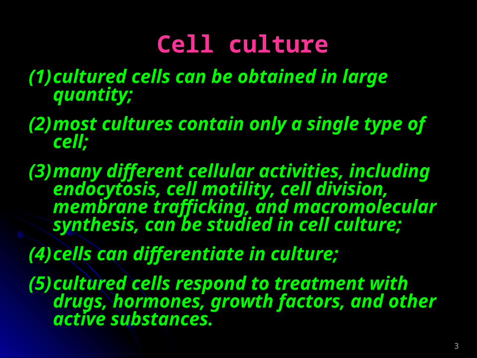
Cell culture(1) cultured cells can be obtained in large quantity;
(2) most cultures contain only a single type of cell;
(3) many different cellular activities, including endocytosis, cell motility, cell division, membrane trafficking, and macromolecular synthesis, can be studied in cell culture;
(4) cells can differentiate in culture;
(5) cultured cells respond to treatment with drugs, hormones, growth factors, and other active substances.
4
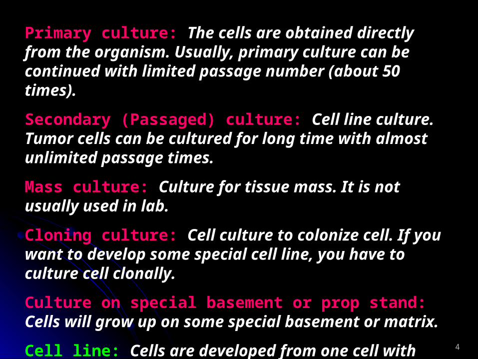
Primary culture: The cells are obtained directly from the organism. Usually, primary culture can be continued with limited passage number (about 50 times).
Secondary (Passaged) culture: Cell line culture. Tumor cells can be cultured for long time with almost unlimited passage times.
Mass culture: Culture for tissue mass. It is not usually used in lab.
Cloning culture: Cell culture to colonize cell. If you want to develop some special cell line, you have to culture cell clonally.
Culture on special basement or prop stand: Cells will grow up on some special basement or matrix.
Cell line: Cells are developed from one cell with some specific characters.

5
Basic Steps for Cell Culture
Construction of primary cell culture or cell line
Culture with medium and other help reagents
Observation and property checking
Change medium
Experimental treatments and results examination
Cell passage and cell line stock
Data analysis
6
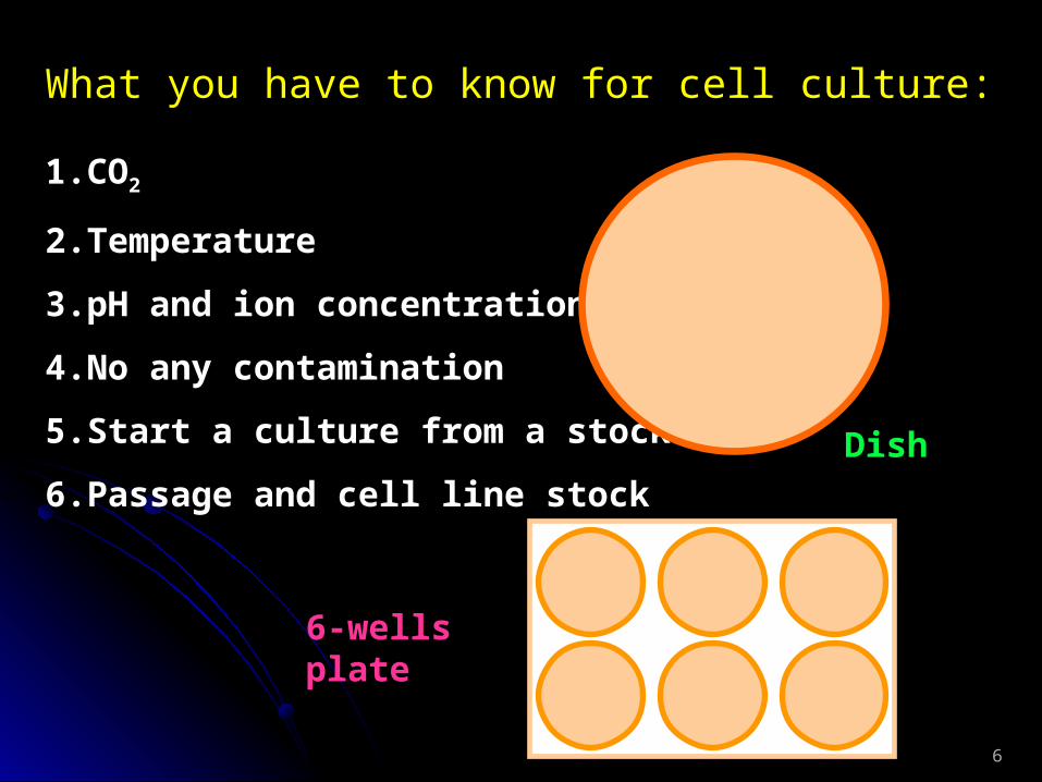
What you have to know for cell culture:
1. CO2
2. Temperature
3. pH and ion concentration
4. No any contamination
5. Start a culture from a stock
6. Passage and cell line stock
Dish
6-wells plate
7
Microscopes and ImagesOptical microscope
Optical inverted microscope
Fluorescence microscope
Electron microscope
Laser confocal scanning microscope
Polarized light microscope
Phase contrast microscope
Cell image station
8
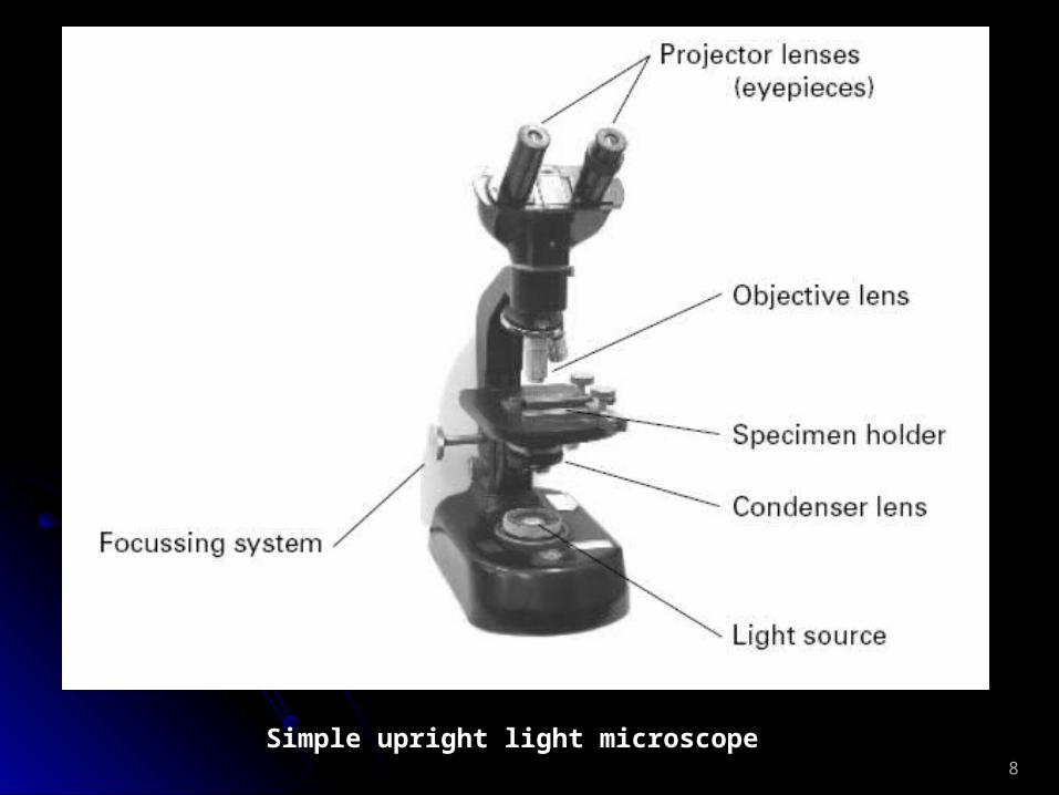
Simple upright light microscope

9
Optical microscopes

10
Fluorescence microscope
11
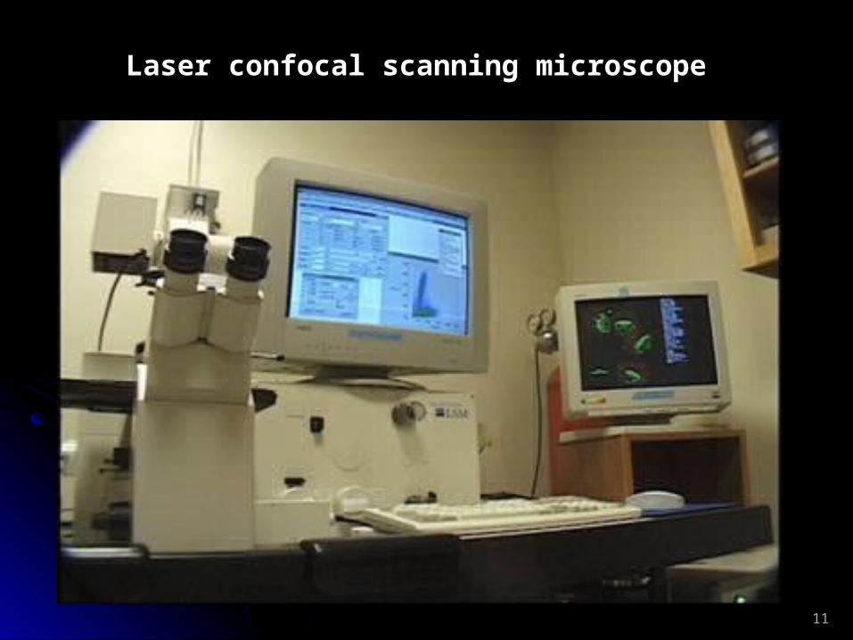
Laser confocal scanning microscope
12
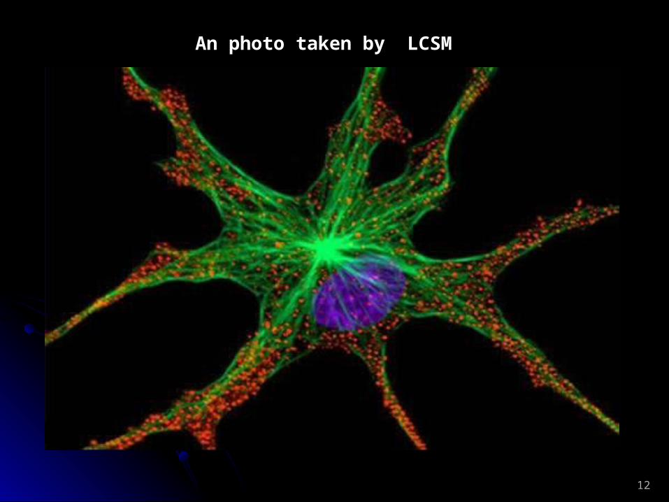
An photo taken by LCSM

13
Mechanism about phase contrast microscope
14
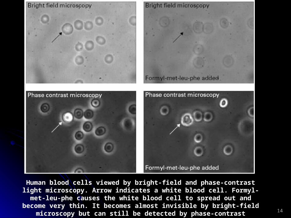
Human blood cells viewed by bright-field and phase-contrast light microscopy. Arrow indicates a white blood cell. Formyl-met-leu-phe causes the white blood cell to spread out and become very thin. It becomes almost invisible by bright-
field microscopy but can still be detected by phase-contrast microscopy
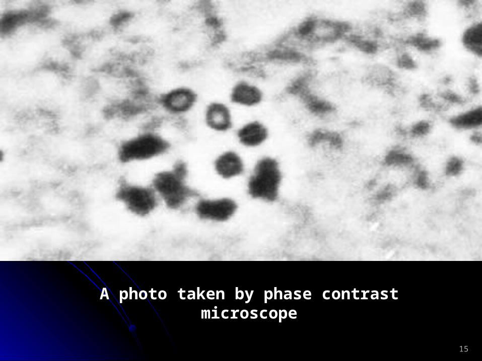
15
A photo taken by phase contrast microscope
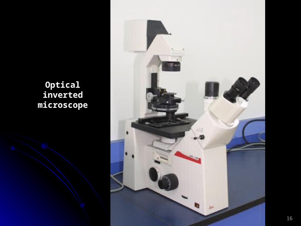
16
Optical inverted microscope

17
Transmission Electron
Microscope

18
A tissue section machine for transmission electron microscope
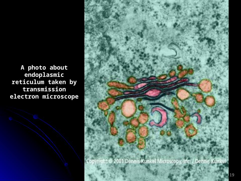
19
A photo about endoplasmic
reticulum taken by transmission
electron microscope
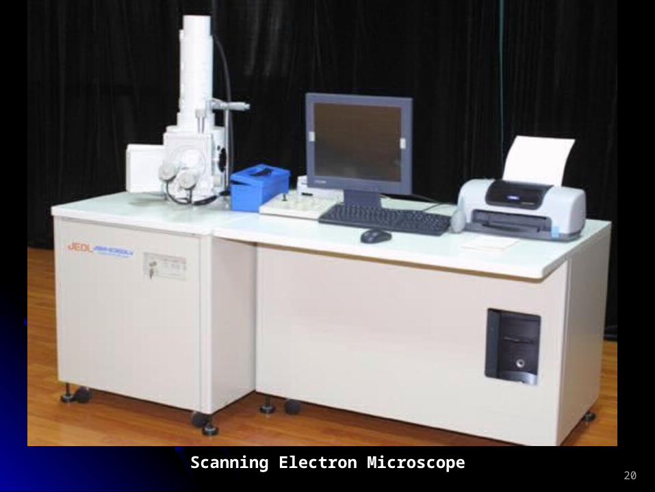
20Scanning Electron Microscope

21Human blood cells photo taken by scanning electron microscope
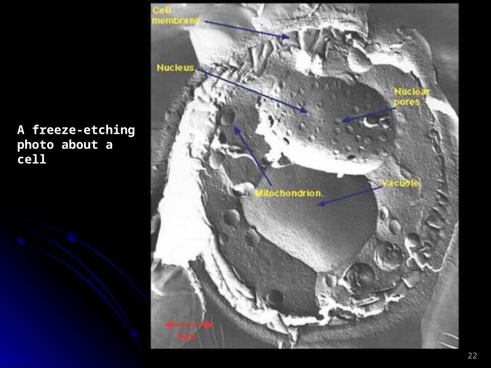
22
A freeze-etching photo about a cell
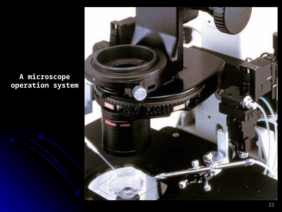
23
A microscope operation system
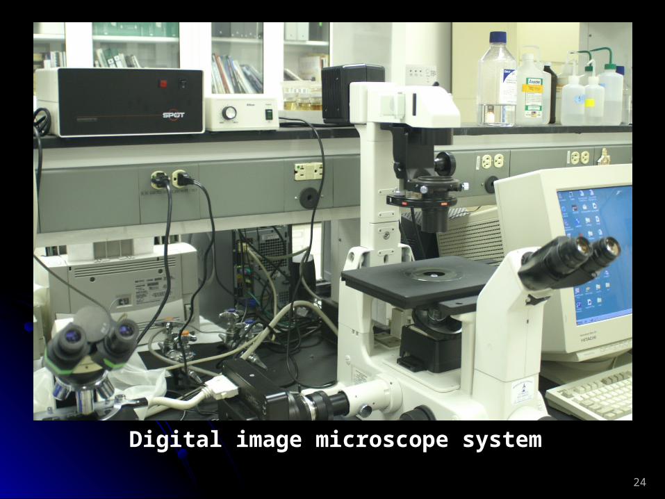
24
Digital image microscope system
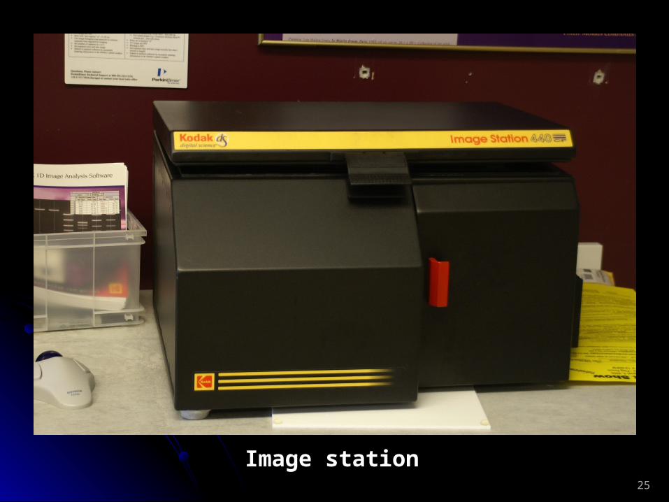
25
Image station
26
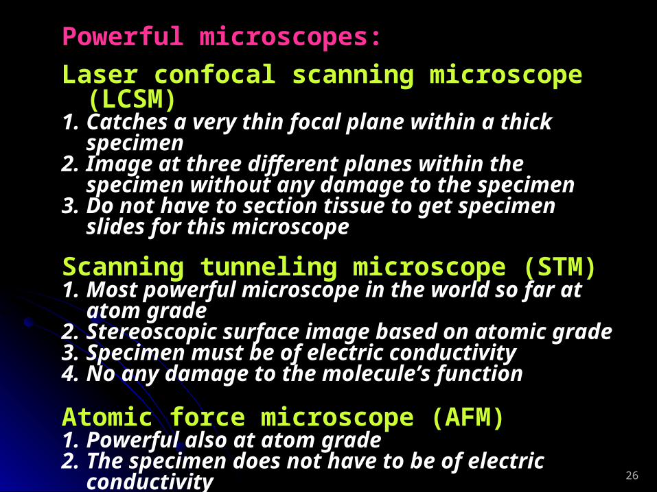
Powerful microscopes:
Laser confocal scanning microscope (LCSM)1. Catches a very thin focal plane within a thick specimen2. Image at three different planes within the specimen
without any damage to the specimen3. Do not have to section tissue to get specimen slides for this
microscope
Scanning tunneling microscope (STM)1. Most powerful microscope in the world so far at atom
grade2. Stereoscopic surface image based on atomic grade 3. Specimen must be of electric conductivity4. No any damage to the molecule’s function
Atomic force microscope (AFM)1. Powerful also at atom grade2. The specimen does not have to be of electric conductivity

27
Histochemistry, immunochemistry, display and other technologies
1. Isolation of the cell apparatus, biological macromolecules and complexes:
Centrifuge methods to isolate cell
components
Differential centrifugation
Density gradient
centrifugation
The cell components will
be divided following their
different sedimentation
coefficient, “S”If the both methods are used together, you will obtain the better isolation result than
using one method alone.

28
Differential centrifugation
Using different RPM you will spin down the particles with different sedimentation coefficient
29
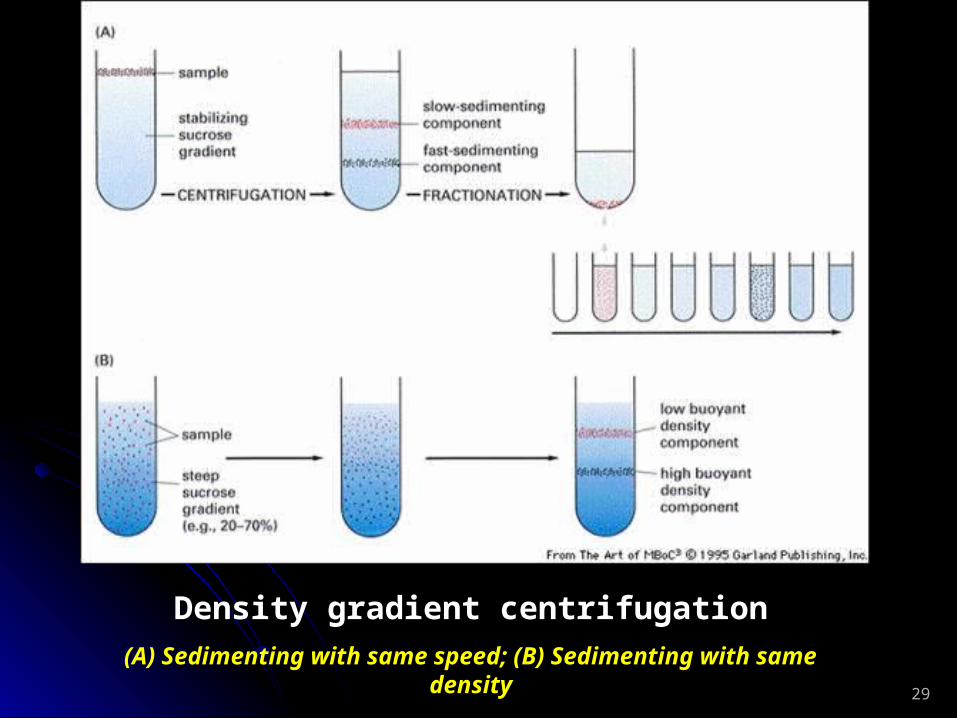
Density gradient centrifugation
(A) Sedimenting with same speed; (B) Sedimenting with same density

30
2. Display and stain the nucleic acids, protein, enzyme, sugar, and fat/lipid:
Nucleic acids: Feulgen reaction (Schiff reaction). Sugar: PAS reaction. Fatty/Lipid: Sudan III or Sudan Black reaction. Protein: Millon reaction, Ninhydrin staining. Enzyme: Use ubstrate reaction or the methods same to stain protein as above.
3. Locate and quantitate the antigen in cells:
Usually, we use HRP (Horseradish peroxidase), AP (Alkaline phosphatase), biotin or inflorescence to label the Ab against the specific Ab against the Ag you want to check or display. We call the labeled Ab as
second Ab and the Ag binding Ab as first Ab.
31
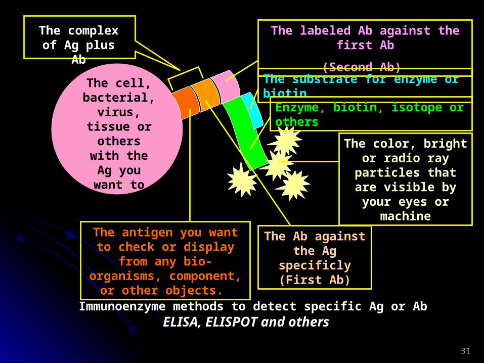
The cell, bacterial,
virus, tissue or others
with the Ag you want to
check
The antigen you want to check or display from any
bio-organisms, component, or other objects.
The Ab against the Ag specificly
(First Ab)
The labeled Ab against the first Ab
(Second Ab)
The substrate for enzyme or biotin
The complex of Ag plus Ab
Enzyme, biotin, isotope or others
The color, bright or radio ray particles that are visible by
your eyes or machine
Immunoenzyme methods to detect specific Ag or Ab ELISA, ELISPOT and others
32
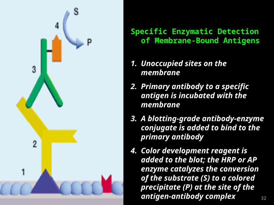
Specific Enzymatic Detection of Membrane-Bound Antigens
1. Unoccupied sites on the membrane
2. Primary antibody to a specific antigen is incubated with the membrane
3. A blotting-grade antibody-enzyme conjugate is added to bind to the primary antibody
4. Color development reagent is added to the blot; the HRP or AP enzyme catalyzes the conversion of the substrate (S) to a colored precipitate (P) at the site of the antigen-antibody complex

33
Western blotting
Western blotting is the most important and popularly used detection method for cell biology, molecular biology and molecular immunology to check gene expression, antigen, components of antigen, and antibody. The good western blotting performance skills are the basic requirements to every body who is working in a life science research laboratory. It is not so easy to get good data (images).

34
+ Anode
– Cathode
+Anode– Cathode
Protein migration direction
Develop each band of each lane to be visible with a serial steps as a
image result
Gel NC membrane
Basic steps for Western Blotting
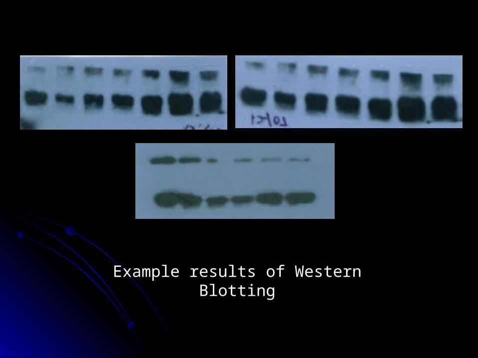
Example results of Western Blotting
36
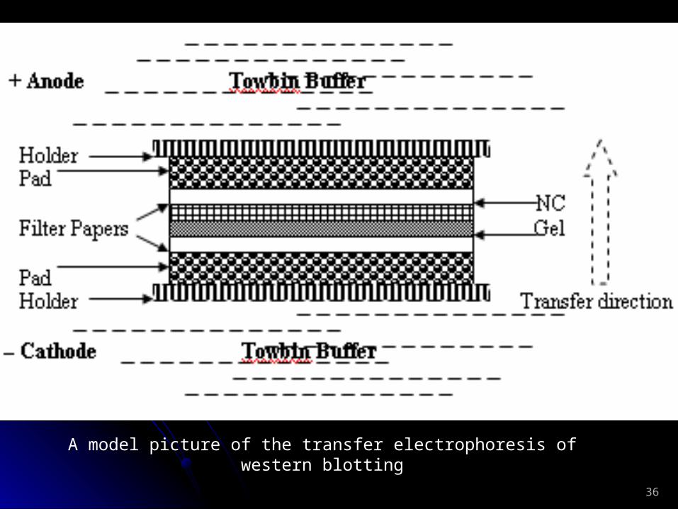
A model picture of the transfer electrophoresis of western blotting
37
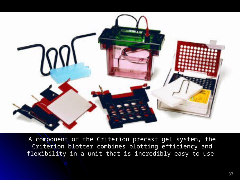
A component of the Criterion precast gel system, the Criterion blotter combines blotting efficiency and flexibility in a unit that is incredibly
easy to use

38
The Mini Trans-Blot cell provides rapid, high-quality blotting of mini gels. A component of the Mini-PROTEAN 3 electrophoresis system, the Mini Trans-
Blot cell accommodates two gel holder cassettes for electrophoretic transfer of both mini format gels run in the Mini-PROTEAN 3 cell.
39
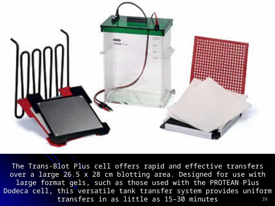
The Trans-Blot Plus cell offers rapid and effective transfers over a large 26.5 x 28 cm blotting area. Designed for use with large format gels, such as those used with the PROTEAN Plus Dodeca cell, this versatile tank transfer system
provides uniform transfers in as little as 15–30 minutes
40
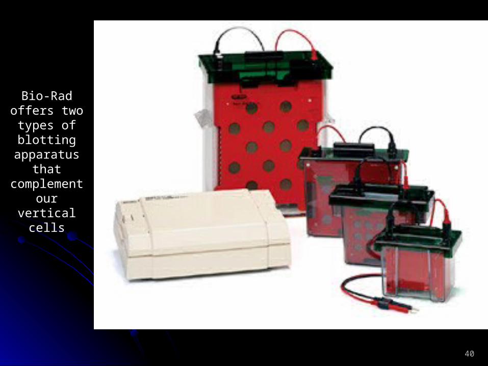
Bio-Rad offers two types of blotting
apparatus that
complement our vertical
cells
41
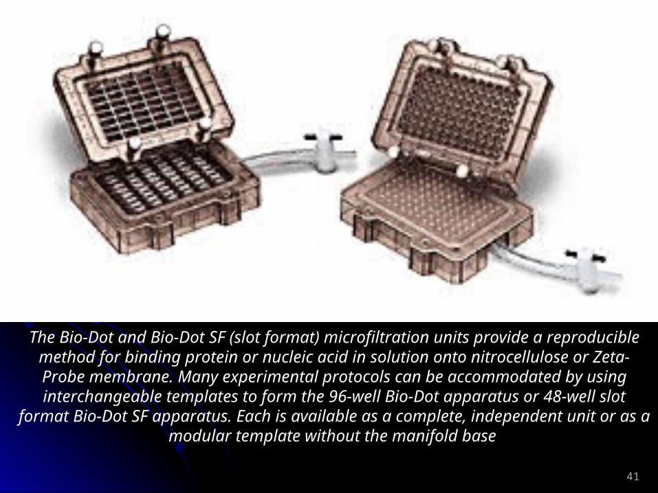
The Bio-Dot and Bio-Dot SF (slot format) microfiltration units provide a reproducible method for binding protein or nucleic acid in solution onto
nitrocellulose or Zeta-Probe membrane. Many experimental protocols can be accommodated by using interchangeable templates to form the 96-well
Bio-Dot apparatus or 48-well slot format Bio-Dot SF apparatus. Each is available as a complete, independent unit or as a modular template without
the manifold base
42
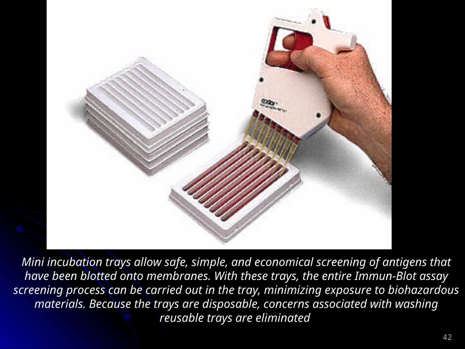
Mini incubation trays allow safe, simple, and economical screening of antigens that have been blotted onto membranes. With these trays, the
entire Immun-Blot assay screening process can be carried out in the tray, minimizing exposure to biohazardous materials. Because the trays are
disposable, concerns associated with washing reusable trays are eliminated

43
Using the Mini-PROTEAN II multiscreen apparatus, you can quickly and efficiently screen up to 40 different antibodies or sera without having to cut the western blot into individual strips. This unit clamps the blot securely in place, creating 40 leakproof channels. Individual samples can be screened
without cross-contamination. Additionally, procedures like screening monoclonal antibodies and monitoring antibody titers from multiple sources
are simplified
44
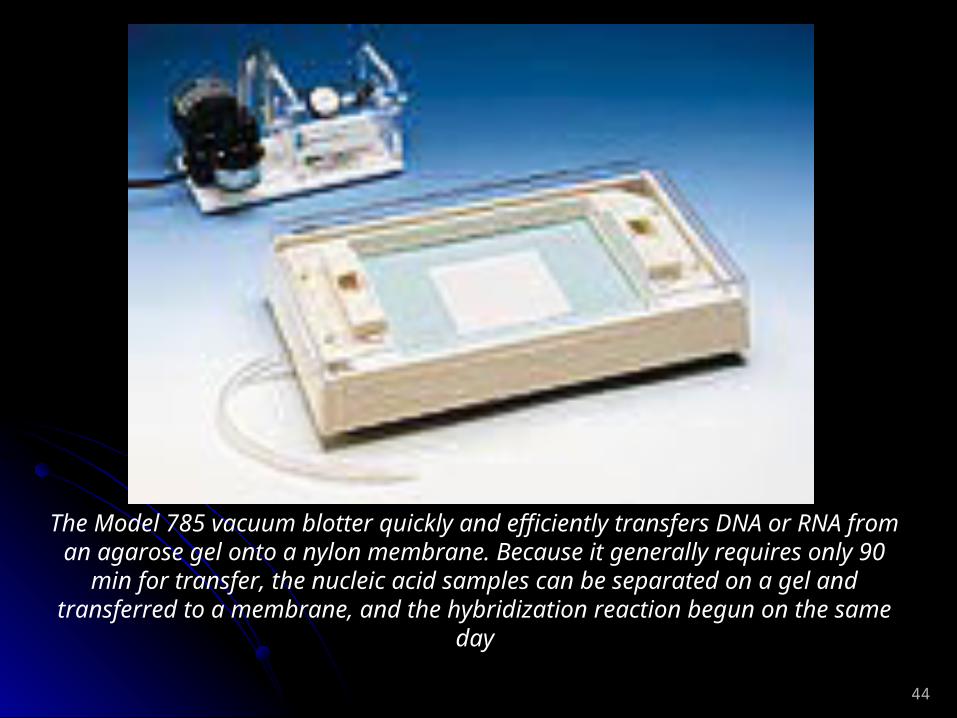
The Model 785 vacuum blotter quickly and efficiently transfers DNA or RNA from an agarose gel onto a nylon membrane. Because it
generally requires only 90 min for transfer, the nucleic acid samples can be separated on a gel and transferred to a membrane, and the
hybridization reaction begun on the same day

45
Northern transfer using the Model 785 vacuum blotter. Samples of 1.0, 2.5, 5, 10, and 20 µg of total RNA from CHO HA-1 cells were
separated on a glyoxyl gel and transferred onto a Zeta-Probe membrane using 3" Hg for 90 min. The blot was probed with a 32P-labeled hsp70 cDNA fragment* and exposed to X-ray film overnight.
46
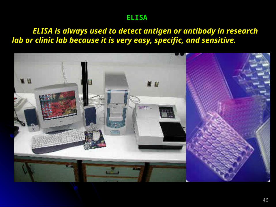
ELISA
ELISA is always used to detect antigen or antibody in research lab or clinic lab because it is very easy, specific, and sensitive.

47
Immunofluorescence Technology
Immunofluorescence Technology was designed based on the fluorescence labeled antibody technology. Stain the specific antigen, then you can view the stained antigen in cells or tissue under a fluorescence microscopy or take digital images as your experimental data.
4. Locate or quantitate the specific nucleic acid sequence in cells or tissue: Hybridization in situ is used for this object. Label specific oligonucleotide (DNA probe or RNA probe) with biotin, fluorescence, or isotope, make the probe hybridized with specific target sequence in cells or tissue, then check the samples under a fluorescence microscopy or electric microscope.
Biotin, Isotope, or fluorescence
Oligonucleotide probe
Target DNA/RNA sequence

48
Human chromosome telomeres were displayed by Hybridization in situ with fluorescence labeled probe

49
An photo taken by fluorescence microscope
Nucleus
Micro filament
Micro tubes
50
5. Analyze the synthesis of the macromolecules in cells with radio labeling technology3H inserting method:
1. One of the 4 dNTPs is labeled. Develop your sample as DNA/RNA image data.
2. One of the 20 amino acids to be labeled. Develop your sample as protein image data.
6. Flow cytometry (FCM)1. To quantitate some molecule in or on cells2. To check the cell subsets, immunity status3. To sort cells into tubes for the culturing
Usually, the antibody against some special marker (first antibody) on the cell surface is labeled by fluorescence.
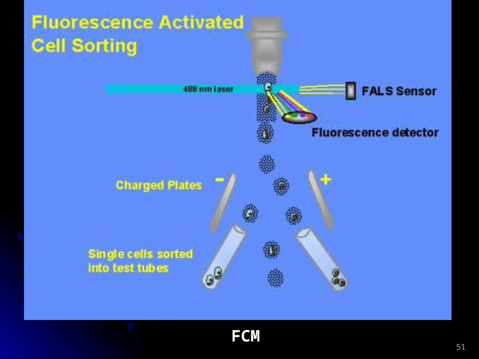
51FCM

52
7. Section and slides:
Paraffin section: Usually use to make slides for long time storage
Section
Freeze section: Usually use to diagnose quickly or keep molecule activity no changed

53

54
Section machine