046 Structure of the Deadly Grouper Fish Virus is Revealed the major farmed fish include milkfish,...
2
046 ACTIVITY REPORT 2015 mortality at the larval stage among diverse species (warm- and cold-water fish) or even across species from marine to freshwater fish in the aquaculture industry. The grouper nervous necrosis virus (GNNV) belongs to genus betanodavirs and is the constant appearance of the infectious diseases such as VNN. Notably, GNNV following VNN is a major factor that limited the expansion of the grouper fish culture in Taiwan (Fig. 1). To investigate the structure of T = 3 GNNV at atomic resolution, Chun- Jung Chen, Nai-Chi Chen, Masato Yoshimura from Life Science Group of the NSRRC, together with their research team pursued the huge and complicated structures of GNNV-LP in various forms using SP12B2, SP44XU, BL13C1 and BL15A1. The crystals of GNNV-LP with the size of 0.3 x 0.1 x 0.1 mm 3 were used for this study, and the mor- phology of GNNV-LP with negative-stained EM images reveal a T = 3 capsid of diameter 30 ~ 35 nm (Figs. 2(a) and 2(b)). The overall topological structure of the GNNV capsid protein (CP) consists of the N-terminal arm (N-arm) (residues 34−51), the shell domain (S-domain) (residues 52−213), the link region (residues 214−220) and the protrusion domain (P-domain) (residues 221−338) (Fig. 2(c)). Sixty trimeric S-domains participate in inter- subunit contacts, forming a continuous thin shell of the capsid with an empty inner cavity. Three neighboring P- domains per iASU embrace one another at quasi three- fold (Q3) axes to form 60 protrusions on the particle surface (Fig. 2(d)). 3 For the investigation of biological functions of each domain of GNNV CP, they first speculated that the N- ARM (N-terminal arginine-rich motif ) or P-domain of GNNV-LP might act as a major molecular switch in regulating T = 3 or T = 1 particle assembly. To address this issue, they constructed two sub-clones, including (i) the delta-P-domain mutant (residues 35−217) and (ii) the N-ARM deletion mutant (residues 35−338), and de- In many countries aquaculture, such as cultivation of marine fish and prawns, is a major global economic activity. In fact, Taiwan, an island surrounded by sea, is one of the major distant water fisheries and aquaculture producers in the world. In terms of production and value, aquaculture has long been far ahead of offshore and coastal fisheries combined, and has been just sec- ond to distant water fisheries. 1 Today, aquaculture in Taiwan, including island and marine cultures, is well correlated with the Taiwanese economy and society, such as income generation, food source and food supply. Furthermore, several species, such as oyster, farmed fish and grass carp, have long been reared as important food sources in Taiwan. Cur- rently, the major farmed fish include milkfish, tilapia, grouper and prawn with an export economic importance in Taiwan, 1 but viral disease in Taiwan has a major impact on aquaculture since fish farming became a significant commercial entity in this century. Alpha-nodaviruses and beta-nodaviruses are major genera in family Nodaviri- dae. 2 Alphanodaviruses infect primarily insects, and betanodaviruses are also called nervous necrosis viruses (NNV) because they cause an acute syn- drome of viral nervous necrosis (VNN). VNN is a serious syndrome disease that causes viral encephalopathy or retinopathy, and is responsible for great Structure of the Deadly Grouper Fish Virus is Revealed Fig. 2: Crystal and EM images and overall structure of GNNV-LP. (a) Crystal image of T = 3 GNNV-LP. (b) A representative negative-stained EM image of purified GNNV-LPs. (c) A ribbon presentation and topological diagram of subunit C of GNNV-LP. (d) Surface domain-colored diagram (leſt) and central cavity (right) representations of T = 3 GNNV-LP. [Reproduced from Ref. 3] (c) (a) (b) (d) 350Å 47Å P-domain Linker region Ca S-domain N-terminal arm N-terminal ARM 228Å Fig. 1: Orange-spoed Grouper fish and GNNV virus.
Transcript of 046 Structure of the Deadly Grouper Fish Virus is Revealed the major farmed fish include milkfish,...
046
AC
TIV
ITY R
EPORT
2015
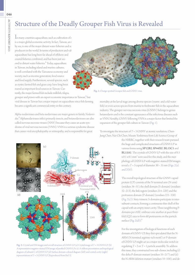
mortality at the larval stage among diverse species (warm- and cold-water fish) or even across species from marine to freshwater fish in the aquaculture industry. The grouper nervous necrosis virus (GNNV) belongs to genus betanodavirs and is the constant appearance of the infectious diseases such as VNN. Notably, GNNV following VNN is a major factor that limited the expansion of the grouper fish culture in Taiwan (Fig. 1).
To investigate the structure of T = 3 GNNV at atomic resolution, Chun-Jung Chen, Nai-Chi Chen, Masato Yoshimura from Life Science Group of
the NSRRC, together with their research team pursued the huge and complicated structures of GNNV-LP in various forms using SP12B2, SP44XU, BL13C1 and BL15A1. The crystals of GNNV-LP with the size of 0.3 x 0.1 x 0.1 mm3 were used for this study, and the mor-phology of GNNV-LP with negative-stained EM images reveal a T = 3 capsid of diameter 30 ~ 35 nm (Figs. 2(a) and 2(b)).
The overall topological structure of the GNNV capsid protein (CP) consists of the N-terminal arm (N-arm) (residues 34−51), the shell domain (S-domain) (residues 52−213), the link region (residues 214−220) and the protrusion domain (P-domain) (residues 221−338) (Fig. 2(c)). Sixty trimeric S-domains participate in inter-subunit contacts, forming a continuous thin shell of the capsid with an empty inner cavity. Three neighboring P-domains per iASU embrace one another at quasi three-fold (Q3) axes to form 60 protrusions on the particle surface (Fig. 2(d)).3
For the investigation of biological functions of each domain of GNNV CP, they first speculated that the N-ARM (N-terminal arginine-rich motif ) or P-domain of GNNV-LP might act as a major molecular switch in regulating T = 3 or T = 1 particle assembly. To address this issue, they constructed two sub-clones, including (i) the delta-P-domain mutant (residues 35−217) and (ii) the N-ARM deletion mutant (residues 35−338), and de-
In many countries aquaculture, such as cultivation of marine fish and prawns, is a major global economic activity. In fact, Taiwan, an island surrounded by sea, is one of the major distant water fisheries and aquaculture producers in the world. In terms of production and value, aquaculture has long been far ahead of offshore and coastal fisheries combined, and has been just sec-ond to distant water fisheries.1 Today, aquaculture in Taiwan, including island and marine cultures, is well correlated with the Taiwanese economy and society, such as income generation, food source and food supply. Furthermore, several species, such as oyster, farmed fish and grass carp, have long been reared as important food sources in Taiwan. Cur-rently, the major farmed fish include milkfish, tilapia, grouper and prawn with an export economic importance in Taiwan,1 but viral disease in Taiwan has a major impact on aquaculture since fish farming became a significant commercial entity in this century.
Alpha-nodaviruses and beta-nodaviruses are major genera in family Nodaviri-dae.2 Alphanodaviruses infect primarily insects, and betanodaviruses are also called nervous necrosis viruses (NNV) because they cause an acute syn-drome of viral nervous necrosis (VNN). VNN is a serious syndrome disease that causes viral encephalopathy or retinopathy, and is responsible for great
Structure of the Deadly Grouper Fish Virus is Revealed
Fig. 2: Crystal and EM images and overall structure of GNNV-LP. (a) Crystal image of T = 3 GNNV-LP. (b) A representative negative-stained EM image of purified GNNV-LPs. (c) A ribbon presentation and topological diagram of subunit C of GNNV-LP. (d) Surface domain-colored diagram (left) and central cavity (right) representations of T = 3 GNNV-LP. [Reproduced from Ref. 3]
(c)(a)
(b)
(d) 350Å47Å
P-domain
Linker region
Ca
S-domain N-terminal armN-terminal ARM
228Å
Fig. 1: Orange-spotted Grouper fish and GNNV virus.
047
Life Science
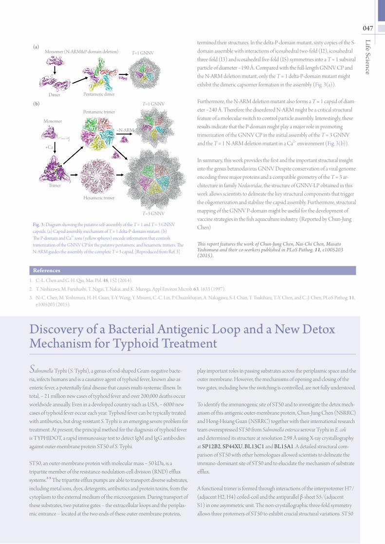
Fig. 3: Diagram showing the putative self-assembly of the T = 1 and T = 3 GNNV capsids. (a) Capsid assembly mechanism of T = 1 delta-P-domain mutant. (b) The P-domain and Ca2+ ions (yellow spheres) encode information that controls trimerization of the GNNV CP for the putative pentameric and hexameric trimers. The N-ARM guides the assembly of the complete T = 3 capsid. [Reproduced from Ref. 3]
Discovery of a Bacterial Antigenic Loop and a New Detox Mechanism for Typhoid Treatment
Salmonella Typhi (S. Typhi), a genus of rod-shaped Gram-negative bacte-ria, infects humans and is a causative agent of typhoid fever, known also as enteric fever, a potentially fatal disease that causes multi-systemic illness. In total, ~ 21 million new cases of typhoid fever and over 200,000 deaths occur worldwide annually. Even in a developed country such as USA, ~ 6000 new cases of typhoid fever occur each year. Typhoid fever can be typically treated with antibiotics, but drug-resistant S. Typhi is an emerging severe problem for treatment. At present, the principal method for the diagnosis of typhoid fever is TYPHIDOT, a rapid immunoassay test to detect IgM and IgG antibodies against outer-membrane protein ST50 of S. Typhi.
ST50, an outer-membrane protein with molecular mass ~ 50 kDa, is a tripartite member of the resistance-nodulation-cell division (RND) efflux systems.1-5 The tripartite efflux pumps are able to transport diverse substrates, including metal ions, dyes, detergents, antibiotics and protein toxins, from the cytoplasm to the external medium of the microorganism. During transport of these substrates, two putative gates – the extracellular loops and the periplas-mic entrance – located at the two ends of these outer-membrane proteins,
play important roles in passing substrates across the periplasmic space and the outer membrane. However, the mechanisms of opening and closing of the two gates, including how the switching is controlled, are not fully understood.
To identify the immunogenic site of ST50 and to investigate the detox mech-anism of this antigenic outer-membrane protein, Chun-Jung Chen (NSRRC) and Hong-Hsiang Guan (NSRRC) together with their international research team overexpressed ST50 from Salmonella enterica serovar Typhi in E. coli and determined its structure at resolution 2.98 Å using X-ray crystallography at SP12B2, SP44XU, BL13C1 and BL15A1. A detailed structural com-parison of ST50 with other homologues allowed scientists to delineate the immuno-dominant site of ST50 and to elucidate the mechanism of substrate efflux.
A functional trimer is formed through interactions of the interprotomer H7/(adjacent H2, H4) coiled-coil and the antiparallel β-sheet S5/(adjacent S1) in one asymmetric unit. The non-crystallographic three-fold symmetry allows three protomers of ST50 to exhibit crucial structural variations. ST50
(a)T=1 GNNV
Dimer
Trimer
+Ca
−N-ARM
Hexameric trimer
Monomer (N-ARM&P-domain deletion)
Pentameric dimer
Pentameric trimer
Monomer
T=1 GNNV
T=3 GNNV
(b)
termined their structures. In the delta-P-domain mutant, sixty copies of the S-domain assemble with interactions of icosahedral two-fold (I2), icosahedral three-fold (I3) and icosahedral five-fold (I5) symmetries into a T = 1 subviral particle of diameter ~190 Å. Compared with the full-length GNNV CP and the N-ARM deletion mutant, only the T = 1 delta-P-domain mutant might exhibit the dimeric capsomer formation in the assembly (Fig. 3(a)).
Furthermore, the N-ARM deletion mutant also forms a T = 1 capsid of diam-eter ~240 Å. Therefore the disordered N-ARM might be a critical structural feature of a molecular switch to control particle assembly. Interestingly, these results indicate that the P-domain might play a major role in promoting trimerization of the GNNV CP in the initial assembly of the T = 3 GNNV and the T = 1 N-ARM deletion mutant in a Ca2+ environment (Fig. 3(b)).
In summary, this work provides the first and the important structural insight into the genus betanodavirus GNNV. Despite conservation of a viral genome encoding three major proteins and a compatible geometry of the T = 3 ar-chitecture in family Nodaviridae, the structure of GNNV-LP obtained in this work allows scientists to delineate the key structural components that trigger the oligomerization and stabilize the capsid assembly. Furthermore, structural mapping of the GNNV P-domain might be useful for the development of vaccine strategies in the fish aquaculture industry. (Reported by Chun-Jung Chen)
This report features the work of Chun-Jung Chen, Nai-Chi Chen, Masato Yoshimura and their co-workers published in PLoS Pathog. 11, e1005203 (2015).
References
1. C.-L. Chen and G.-H. Qiu, Mar. Pol. 48, 152 (2014).
2. T. Nishizawa, M. Furuhashi, T. Nagai, T. Nakai, and K. Muroga, Appl Environ Microb. 63, 1633 (1997).
3. N.-C. Chen, M. Yoshimura, H.-H. Guan, T.-Y. Wang, Y. Misumi, C.-C. Lin, P. Chuankhayan, A. Nakagawa, S.-I. Chan, T. Tsukihara, T.-Y. Chen, and C.-J. Chen, PLoS Pathog. 11, e1005203 (2015).