What types of cells are found in muscle tissue? How are these cells specialized to carry out their...
69
THE MUSCULAR SYSTEM
-
Upload
dortha-merritt -
Category
Documents
-
view
216 -
download
1
Transcript of What types of cells are found in muscle tissue? How are these cells specialized to carry out their...

THE MUSCULAR
SYSTEM
Overview: Guiding Questions
What types of cells are found in muscle tissue?
How are these cells specialized to carry out their function?
What does BMI measure?

What types of cells are found in the muscle tissue? How are they
specialized to carry out their function?
FUNCTIONS CONTRACTILE CELLS
Pumping blood throughout the body
Moving the skeletal system
Passing food through the digestive system
Specialized cell membrane and cytoskeleton that permit them to change their shape
Cytoskeleton allows shortening in one or more planes (contraction)
Laid out as sheets of muscle tissue that produce coordinated contractions

What do contractile cells need to carry out their function?
Blood Supply
High energy needs
Oxygen
Remove metabolic wastes
Glucose
Electrolytes
calcium
From bones

What does BMI measure?
Compares the amount of muscle mass with the body fat composition.
A certain degree of leanness is known to reduce heart disease and metabolic disorders.
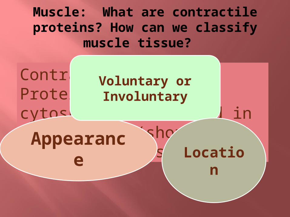
Muscle: What are contractile proteins? How can we classify
muscle tissue?
Contractile proteins: Proteins of the cytoskeleton involved in contraction (shortening) of muscle cellsAppearan
ce
Voluntary or Involuntary
Location

Appearance
Non-StriatedStriatedUniform
arrangement of contractile proteins
Can see microscopically
Stronger Contractions
Randomized pattern of contractile
proteins
Cannot see microscopically
Weaker contractions

Voluntary or Involuntary
Large degree of control
Some unconsciously (breathing)
Some contractions are intentional
Contract without conscious control
Jobs that are automatic or in conjunction with other organ systems
Voluntary Involuntary

Location
Cardiac Skeletal Smooth
Spindle or teardrop cells
Fibers not visible
Weak contractions
that last a long time
Linings of BVsTubular organs
Most involuntary
Large cells with distinct
striations
Strong directional
contractions
Attach to bones and joints that
produces body movement
Most are voluntary
Make up the heart
Striated
Connected by intercalated
disks
Involuntary

Types of Muscle Tissue: Guiding Questions
Briefly describe myogenesis. Briefly characterize the three types of
muscle cells How do contractile proteins contribute to
skeletal function? Why is the “intrinsic beat” of cardiac
muscle cells significant? Why is “peristalsis” significant? Describe the relationship between muscle
cells and muscle fibers

Briefly describe myogenesis:
Muscle develops in mesoderm cells: myogenesis
Stem cells form myoblasts Myoblasts move to other developing tissues to
form the 3 muscle types Growth factors (chemicals that act as signals
to initiate cell division & differentiation) by tissues give direction as to what type of muscle needs to form.

Briefly Characterize the 3 Types of Muscle Cells
Cardiac Skeletal Smooth• Lining of BV, digestive organs, urinary system, respiratory system•Nonstriated•Weak involuntary contractions can last for a long time•Dilation and constriction of BV and tubular structures in respiratory system•Peristalsis: laid in sheets in digestive system. Moves food & wastes through
•Provides movement•Large cells with distinct striations•Powerful contractile capabilities•One cell is composed of several myoblasts that fuse into a muscle fiber—why so many nuclei?•Each fiber stimulated by a motor nerve cell that controls several muscle fibers at once
•Form around large BV and form heart•Strong contractions •Not conscious control•Have 2 nuclei per cell•Cells are branched•Communicate thru intercalated disks•Intrinsic beat: all cardiac cells act in unison, coordinated thru intercalated disks
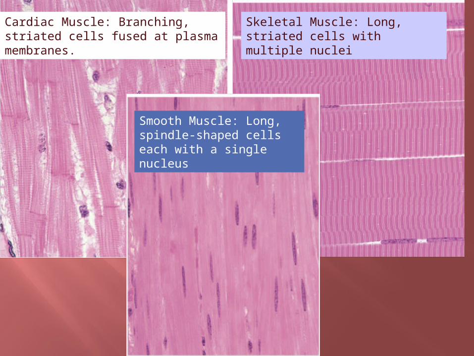
Cardiac Muscle: Branching, striated cells fused at plasma membranes.
Skeletal Muscle: Long, striated cells with multiple nuclei
Smooth Muscle: Long, spindle-shaped cells each with a single nucleus

What type of muscle cells?

Now, you should be able to answer these questions!
How do contractile proteins contribute to skeletal function?
Why is the “intrinsic beat” of cardiac muscle cells significant?
Why is “peristalsis” significant? Describe the relationship between muscle
cells and muscle fibers

Muscle Cell Structure Guiding Questions
Describe the basic structure of skeletal muscle cells
Briefly summarize the various types of fibers found in a muscle cell
Describe the relationship between myofibrils, muscle fibers, and fasciculi
Why is a sarcomere called the “contractile unit” of the muscle?

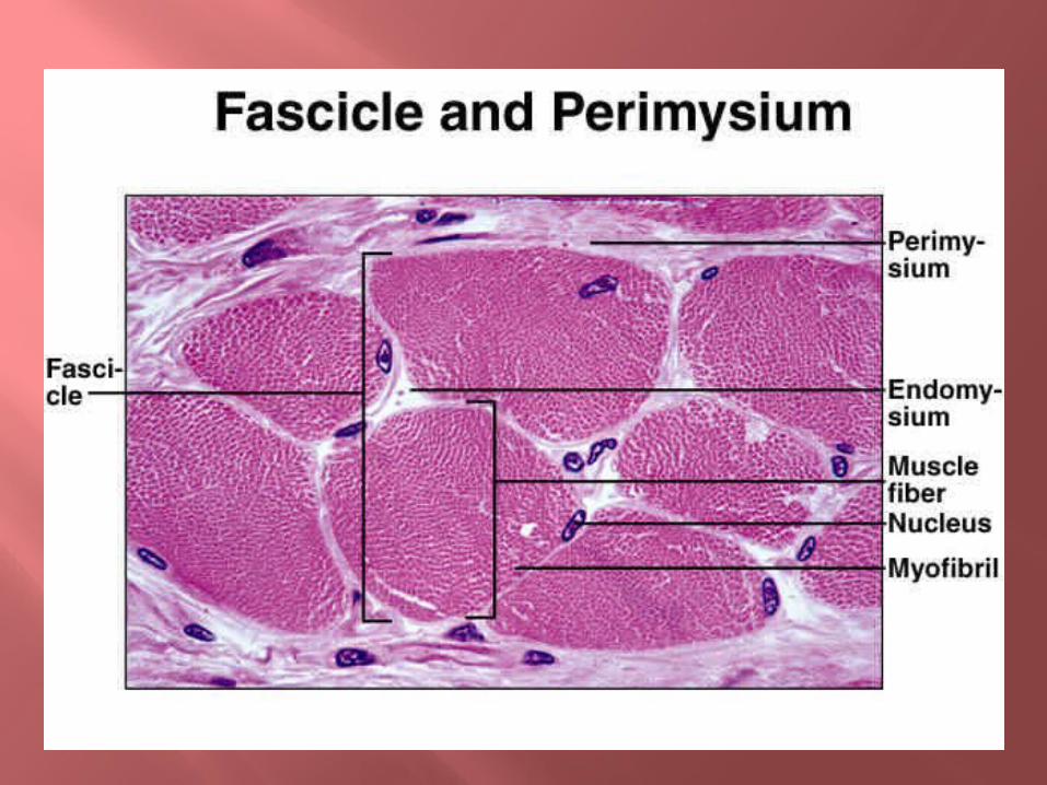
Organization of (skeletal) muscle
Skeletal muscle fibers located in muscles Entire muscle
surrounded by epimysium, a CT layer
Subdivided into fiber bundles called fascicles (fasciculi)
Fascilcles surrounded by perimysium, also CT

Membranes
Portions of perimysium extend into the endomysium Thin layer of CT that
covers each muscle fiber
Muscle fiber (bundle)= multinucleate cell

SARCOMERE
Sarcomere= basic (functional) contractile unit Separated by each other by
dark Z lines/discs Actin & myosin slide past
each other as the muscle contracts
Contraction requires Ca2+ and ATP
Actin Myosin
Sarcomere

Other Key Points
About Sarcomeres
Z-line/disc – vertical protein bands that hold sarcomere to sarcolemma.
I Bands Lighter areas of non-overlap between actin and
myosin Contain the Z-lines.
Dark Bands = A Bands Areas where some overlap occurs = “Striations” on the slide Coincide with the length of myosin myofilaments. H-zone – light area within A-band

Sarcoplasmic Reticulum Each myofibril is surrounded by network
of tubes and storage sacs (Transverse tubules and sarcoplasmic reticulum)
Releases Ca2+ ions when stimulated by motor neuron
Triggers contraction (more on this later…)

MICROSCOPIC STRUCTURE
Muscle FIBERS: grouped into bundles (fasciculi)
= 1 cell! Fibers contain myofibrils
with: ACTIN: thin myofilaments
Also contain: Tropomyosin Troponin
MYOSIN: thick myofilaments, with “swiveling” arm and head
TITIN: elastic fibers that hold myosin in place, controlling stretch of sarcomere


Another look…
Muscle Cell Function Guiding Questions
How would a muscle appear to change microscopically during a contraction?
What are the three stages of muscle contraction? What is the role of neurotransmitters during muscle
contraction? Describe the ion concentrations found inside and
outside a resting muscle cell Briefly describe the events that occur during the
muscle contraction phase. What is the role of ATP during this phase? What must occur for a muscle cell to “fully” recover
after a contraction? What occurs during “rigor mortis”?
Muscle Contraction
Sarcomeres shorten, distance between z-lines reduced
Thick and thin myofilaments overlap more during contraction
3 stages: Neural stimulation Muscle cell contraction Muscle cell relaxation

Motor Unit Stimulation of a muscle by a nerve
impulse (motor nerve) is required before a muscle can shorten
Neuromuscular junction: point of contact b/w nerve ending and the muscle fiber it innervates.
Motor unit: motor neuron + muscle cell
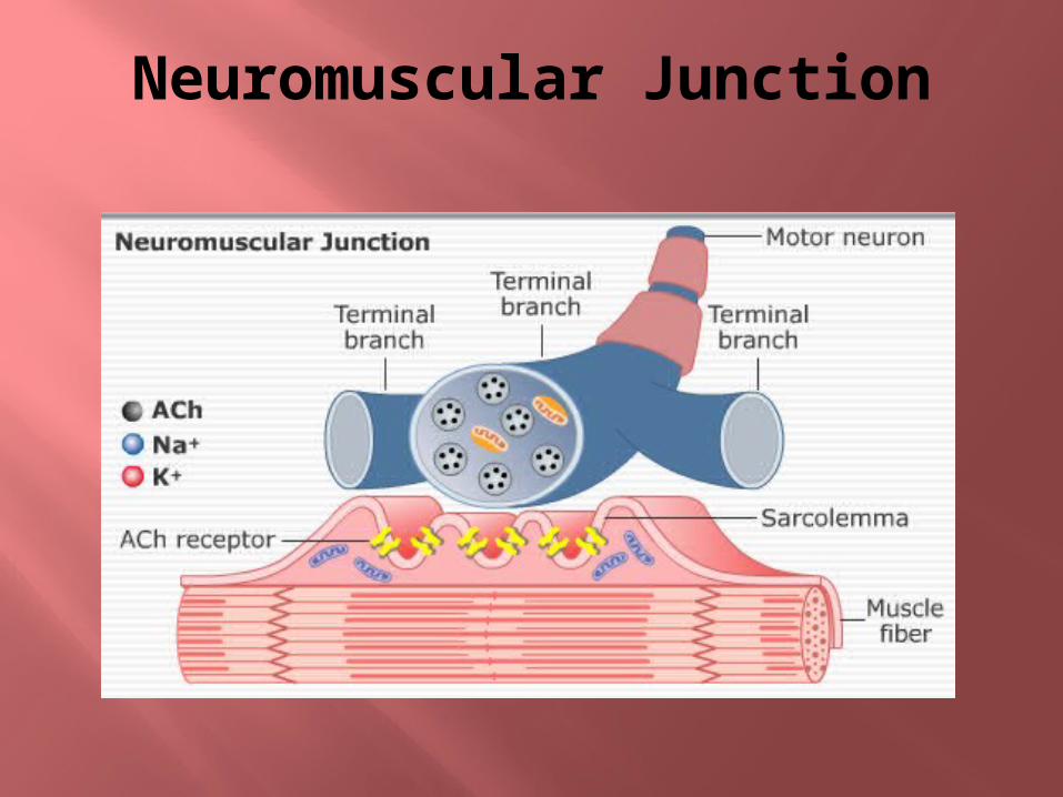
Neuromuscular Junction

Neural Stimulation
Motor neuron releases neurotransmitters to stimulate a contraction Acetylcholine (Ach) binds to receptors
located on sarcolemma Changes transport proteins found in
sarcolemma Alters transport of ions
Normally, more Sodium (Na+) ions outside muscle cell, while Potassium (K+) higher inside
Sodium/Potassium pumps maintain this unequal concentration
Excitable condition

When stimulated, ion channels open, depolarizing the cell Na+ flows in, K+ out sarcoplasmic reticulum releases stored
calcium Ca2+ travels to sarcomere, initiating muscle
contraction phase

Muscle Contraction Animation
Click here to view animation

Sliding filament theory In the absence of
Calcium ions…Troponin “hat” sits
on Tropomyosin filament These blocks access to the myosin head’s binding site on actin.
TroponinTropomyosinCa2+

When Calcium is released by the Sarcoplasmic reticulum it diffuses into the muscles binds to the troponin “hat” shifting both the troponin and tropomyosin
filament

Myosin splits ATP and undergoes a conformational change into a high-energy state.
The head of myosin binds to actin Forms a cross-bridge between the thick
and thin filaments.

The energy stored by myosin is released ADP and phosphate released from
myosin. The myosin molecule relaxes
Causes rotation of the globular head This leads to the sliding of the
filaments. This cycle continues until Ca2+ ions gone
(and stimulus stops)
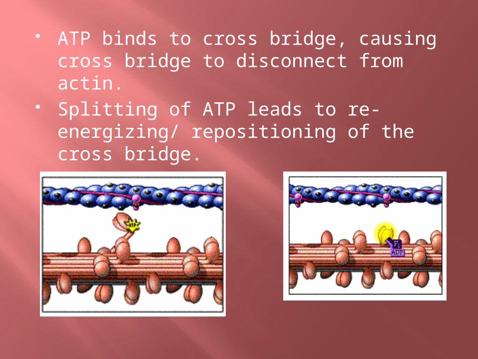
ATP binds to cross bridge, causing cross bridge to disconnect from actin.
Splitting of ATP leads to re-energizing/ repositioning of the cross bridge.

Relaxation Phase
Complete contraction of muscle cell requires several cycles of neural stimulation and contraction phases
Ca2+ ions transported back to sarcoplasmic reticulum (req. ATP)
When the calcium level decreases troponin locks tropomyosin back into the
blocking position thin filament (actin) slides back to the resting
state (when ATP binds to myosin head)

Relaxation phase occurs when no more neural stimulations are exciting the sarcolemma Na+/K+ pump returns ions to resting
state Muscle cell remains in contracted, but
pliable state Must be “stretched” back into position

Review of the Role of ATP
1. ATP transfers its energy to the myosin cross bridge, which in turn energizes the power stroke.
2. ATP disconnects the myosin cross bridge from the binding site on actin.
3. ATP fuels the pump that actively transports calcium ions back into the sarcoplasmic reticulum.

Rigor Mortis
In death… Calcium leaks out of sarcoplasmic
reticulum into sarcomere Causes muscle tension = rigor mortis Muscle cell structures start breaking down,
causing muscle to loosen (unless body becomes dehydrated)

Creatine phosphate
Stores energy in muscle cells Collects energy from ATP, stores for long
periods of time Transfers back to ATP when needed
Glycogen
Stored form of glucose Energy reserve for muscle action Continuous supply needed to produce
ATP
Myoglobin
Red pigment that stores oxygen for muscle cells “Grabs” oxygen from hemoglobin in blood High affinity for oxygen
Allows cells to produce large amounts of ATP

Musculature Guiding Questions
What determines a muscle’s morphology?
Distinguish between a muscle’s origin and insertion
Gross Skeletal Muscle Types Guiding Questions
Review the location of the various gross skeletal muscle types listed on page. 231
List, and briefly describe, the various terms that describe the muscle structures, patterns, and shapes

Parallel or Pinnate
Parallel general-purpose muscles Sheets of muscle cells that run in the same
direction Contractions for moving light loads over a
long distance Pinnate
Feather-pattern Great strength for moving heavy loads over a
short distance Strong movements for the arms and legs

Gross Muscle Cell Types/TermsMuscle group Shape Function
Deltoid Triangular Pulling power
Trapezius Trapezoid Pulling power
Rhomboideus Diamond Holding power for scapulae
Serratus Saw-toothed
Short movements of the arms, rib cage, and shoulders
Biceps 2 heads Upper arms
Triceps 3 heads Upper arms
Quadriceps 4 heads Upper legs
Size Description
Maximus Largest muscle in the group
Minimus Smallest muscle in the group
Longus Longest muscle in the group (arms and legs)
Brevis Shortest muscle in the group (arms and legs)

Skeletal Muscle Structure Guiding Questions
In words, briefly review the basic structure of a skeletal muscle
What occurs to a muscle during atrophy? Hypertrophy?

Review: Muscle Cell Structure
C
B
A
E(fluid in cells)
D(membrane)
H
G
F

A (blue line)
B (Red line)
FE
C CD
E
A (blue line)
B (Red line)
C C
G
G
EF
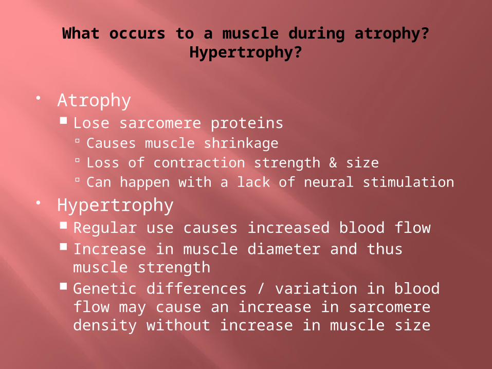
What occurs to a muscle during atrophy? Hypertrophy?
Atrophy Lose sarcomere proteins
Causes muscle shrinkage Loss of contraction strength & size Can happen with a lack of neural stimulation
Hypertrophy Regular use causes increased blood flow Increase in muscle diameter and thus muscle
strength Genetic differences / variation in blood flow
may cause an increase in sarcomere density without increase in muscle size

Skeletal Muscle Action Guiding Questions
How are “graded effects” accomplished during a muscle contraction?
Differentiate between strength and endurance.
What is an antagonistic effect? Why are these essential to normal muscle function?
List and briefly describe the various categories of muscle action.

Origin vs. Insertion
Origin – point of attachment of a muscle that remains fixed during contraction
Insertion – point of attachment of a muscle connected to movable component on other end
Shortening/contraction = moves insertion closer to origin

Threshold
All muscle fibers contract with a particular strength when threshold neural stimulation reached
How are muscles able to perform at different “powers”? Graded effects can be accomplished by:
Contracting more fascicles = more strength Muscles working together
Endurance = producing contracting and relaxing fascicle groups

Differentiate between strength and endurance
Strength = ability to do more work Endurance = longer period of work

What is an antagonistic effect? Why are these essential to normal muscle function?
One muscle opposes or resists the action of another
Weakens muscle strength Gravity can have antagonistic effect Essential!
Pulls relaxed muscles back to original strength Cartilage can do this (in ribcage during
breathing) Synergism = muscles work together
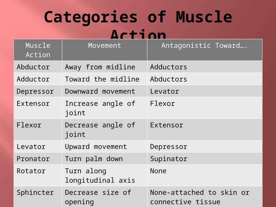
Categories of Muscle Action
Muscle Action
Movement Antagonistic Toward….
Abductor Away from midline Adductors
Adductor Toward the midline Abductors
Depressor Downward movement Levator
Extensor Increase angle of joint Flexor
Flexor Decrease angle of joint
Extensor
Levator Upward movement Depressor
Pronator Turn palm down Supinator
Rotator Turn along longitudinal axis
None
Sphincter Decrease size of opening
None-attached to skin or connective tissue
Supinator Turn palm up Pronator
Tensor Posture, make body part more rigid, tense
Many

Isotonic or Isometric?
Another way to define muscle action
Isotonic: muscle is actively shortening or lengthening. Lifting/lowering weights
Isometric: muscle remains steady in length, undergoing indistinguishable pulses of shortening and lengthening Pushing against something too
heavy to move
Pathology of Musculature Guiding Questions
Differentiate between muscle strains and sprains
Differentiate between spasms and cramps of muscles
How are rigid and flaccid paralysis different?
What causes tetanus? Review the various myopathies listed on p.
242. Make a chart/concept map to summarize the major characteristics of each.

Strain vs. Sprain
Strain Overworking
muscle Pain, swelling in:
Fascia Joints Ligaments Tendons
Due to tearing of muscle/tendon fibers Nerves
stretched/swollen
Sprain More severe Sudden/violent
stress on joint/muscle
Causes tearing: Ligament Muscle Tendon
Damage to blood vessels

Strain vs. Sprain

Spasms vs. Cramps
Spasm = involuntary, abnormal contractions of a muscle/group Many causes Often associated with pain
Cramps = painful contraction of muscle Extreme exertion can cause May develop while working in cold Poisons, bacterial infections

Rigid vs. Flaccid Paralysis
Paralysis = complete failure of muscle function Caused by muscle stiffness? Rigid Lack of muscle contraction? Flaccid
Many causes (infection, injury, degeneration)

Causes of Tetanus (aka lockjaw)
Soil bacteria (Clostridium tetani)infect nervous system
Produce poisons that block nerve signals at neuromuscular junction, cause rigid paralysis (severe muscle spasms). Can cause muscle tears/spinal fractures if severe
Do you have your tetanus “shot”?

For tomorrow…
Reading “Aging of the Musculature” Bring textbook…
Make concept map of pathology and aging sections (like the one I made for you last time)
Try to think about “groupings” you could use to make a web “Overwork” “Trauma” “Abnormal muscle control” Etc…

Aging of the Musculature Guiding Questions
What can cause cachexia? Why do muscles require a high protein
turnover? What is the role of IGF-1 in muscle
health?