, Spencer Walse, James R. Rocca, Arthur S. Edison, Single ...
6
Aaron T. Dossey, Spencer Walse, James R. Rocca, Arthur S. Edison, Single Insect NMR: A New Tool to Probe Chemical Biodiversity , (2006) ACS Chem Biol, 1, (8), 512-514.; Featured in Chem Eng News, Ivan Amato, Individual Insects Make Signature Venoms: Walking stick study hints at chemical biodiversity in these insects , Sept. 25, 2006, p. 15.; A. T. Dossey featured in ACS Chem Biol, Introducing our Authors , (2006), 1, (8), p. 473.
Transcript of , Spencer Walse, James R. Rocca, Arthur S. Edison, Single ...

Aaron T. Dossey, Spencer Walse, James R. Rocca, Arthur S. Edison, Single Insect NMR: A New Tool to Probe Chemical Biodiversity, (2006) ACS Chem Biol, 1, (8), 512-514.; Featured in Chem Eng News, Ivan Amato, Individual Insects Make Signature Venoms: Walking stick study hints at chemical biodiversity in these insects, Sept. 25, 2006, p. 15.; A. T. Dossey featured in ACS Chem Biol, Introducing our Authors
, (2006), 1, (8), p. 473.

473www.acschemicalbiology.org VOL.1 NO.8 • ACS CHEMICAL BIOLOGY
Current position: Genentech, Inc., Department of Protein Engineering and Medicinal Chemistry, research associate with Dr. Kurt Deshayes
Education: University of Wisconsin–Madison, B.S. in chemistry, 1999
Nonscientifi c interests: Bicycling the streets of San Francisco, camping and backpacking in the redwood forests, and guitar playing
As an organic chemist, a major interest of mine has been designing
and creating new organic compounds. My work at Genentech
allows me to incorporate this passion into the study of biology
that is crucial for understanding and developing therapeutics. For
example, the regulation of apoptosis, which is the current focus of
our lab. To me this paper is an exciting example of progressing from
a protein target to specifi c tight binding small molecules through
the use of rationally designed peptidomimetics. We demonstrate
this through a class of [7,5] bicyclolactame compounds that bind
to the baculoviral inhibitor of apoptosis protein (IAP) repeat (BIR)
and BIR3 domains of melanoma IAP and X-chromosome-linked IAP.
(Read Zobel’s article on p 525.)Kerry Zobel
Current position: University of Califor-nia, Berkeley, Department of Chemistry, postdoctoral research associate with Prof. Michael Marletta
Education: Massachusetts Institute of Technology, S.B. in chemistry, 1994; Mas-sachusetts Institute of Technology, Ph.D. in toxicology with Dr. Steven Tannenbaum, 2001; Harvard Medical School, M.D., 2002
Nonscientifi c interests: Most sports—defi nitely cricket!
One of my interests is the development of versatile and easily
accessible tools for addressing fundamental questions in biology.
Aptamer technology has many attributes compatible with this goal,
especially because diverse cellular processes can be targeted with
exquisite specifi city. In this paper, we have used heme-binding
aptamers to specifi cally target the well-studied heme biosynthetic
pathway in the model organism Escherichia coli, demonstrating
the applicability of this technology in modulating a small-
molecule-regulated pathway. In the long run, I am interested in
extending these approaches to nonmodel organisms that are less
amenable to traditional genetic methods for studying essential
cellular processes. (Read Niles’s article on p 515.)
Current position: University of Florida, Col-lege of Medicine, Department of Biochem-istry and Molecular Biology, postdoctoral research associate with Prof. Arthur Edison
Education: Oklahoma State University, B.S. in biochemistry and molecular biology, cum laude, 2001; University of Florida, Gainesville, Ph.D. in biochemistry and mo-lecular biology, with Prof. Art Edison, 2006
Nonscientifi c interests: Entomology, keep-ing and breeding invertebrates, comedy, playing trumpet, nature photography, travel, gardening, camping, fi shing, and hiking through the wilderness
Jacquin Niles
This project was my fi rst chance to incorporate my passion for
studying insects with my formal training in biochemistry. One
aspect that I found most fascinating was that we were able to
analyze venom from a single insect and discover unreported
components of that substance. Such a discovery opens new doors
to understanding arthropod chemistry. Indeed, only a tiny fraction
of the total chemical biodiversity that exists in insects alone has
been determined. I hope to continue exploring the large potential
for discovery that exists in these creatures. Using cutting-edge
technologies such as the microsample NMR used in our study of
phasmid insect venom, we can now begin to push the frontiers
of natural products chemistry. I am currently looking for future
work involving medicinal and natural product discovery from
invertebrates. (Read Dossey’s article on p 511.)
Published online September 15, 2006 • 10.1021/cb600387e CCC: $33.50
© 2006 by American Chemical Society
Aaron Dossey
cb600387e.indd 473cb600387e.indd 473 9/13/2006 5:52:09 PM9/13/2006 5:52:09 PM
Single-Insect NMR: A New Tool To ProbeChemical BiodiversityAaron T. Dossey†, Spencer S. Walse‡, James R. Rocca§, and Arthur S. Edison†,§,¶,*†Department of Biochemistry and Molecular Biology, University of Florida, Gainesville, Florida 32610-0245, ‡Center forMedical, Agricultural and Veterinary Entomology, USDA-ARS, Gainesville, Florida 32604, §McKnight Brain Institute,University of Florida, Gainesville, Florida 32610, and ¶National High Magnetic Field Laboratory, University of Florida,Gainesville, Florida 32610
I ndividual organisms often producenatural products in very small quantities(1). Accordingly, their isolation and
identification traditionally require largeamounts of starting material and a signifi-cant effort in sample preparation. Analyticaltechniques such as mass spectrometry(MS), capillary electrophoresis, and fluores-cence spectroscopy are now extremely sen-sitive and are being used to expedite thisprocess. The use of NMR on the other hand,has lagged behind due to large samplerequirements. Although notoriously insensi-tive, NMR is indispensable to naturalproduct identification because it providesstructural information that is not accessiblewith other techniques. Microcoil (2–6) andcryogenic (7) technology for NMR probeshas significantly reduced sample massrequirements and enhanced several naturalproduct studies (3, 8–12). We recently com-bined the advantages of small-diametersamples with cryogenic technology in a1-mm-diam NMR probe made from high-temperature superconducting (HTS) mate-rial to achieve �25� greater sensitivity thana conventional probe (13). Here we haveused this novel probe to characterize thedefensive secretions of individual walkingstick insects.
Anisomorpha buprestoides (order Phas-matodea) is common in the southeasternU.S. and is often found in pairs with thesmaller male riding on the back of thefemale (14). When threatened, it accurately
sprays a secretion at predators (14, 15). Fol-lowing the extraction of �1000 A. bupres-toides “milkings” into methylene chloride,Eisner and Meinwald (15) identified itsactive component as a cyclopentanoidmonoterpene dialdehyde that they namedanisomorphal. At about the same time,Cavill and Hinterberger (16) identified asimilar compound in ants that they nameddolichodial. Anisomorphal had lower opticalactivity than dolichodial, suggesting thatA. buprestoides secretions contained amixture of isomers or an optically activeimpurity (15). Subsequently, two related ste-reoisomers were identified from a plant inthe mint family, Teucrium marum (catthyme) (17–19). The minor isomer fromT. marum was assigned to anisomorphal(17). For clarity, we will refer to any of thestereoisomers with the covalent structure as“dolichodial-like” (Scheme 1); we willsuggest specific assignments at the end ofthis work.
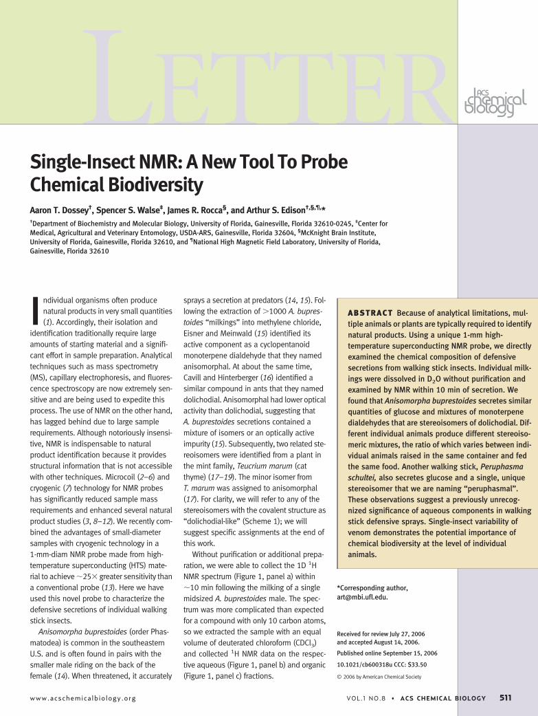
Without purification or additional prepa-ration, we were able to collect the 1D 1HNMR spectrum (Figure 1, panel a) within�10 min following the milking of a singlemidsized A. buprestoides male. The spec-trum was more complicated than expectedfor a compound with only 10 carbon atoms,so we extracted the sample with an equalvolume of deuterated chloroform (CDCl3)and collected 1H NMR data on the respec-tive aqueous (Figure 1, panel b) and organic(Figure 1, panel c) fractions.
*Corresponding author,[email protected].
Received for review July 27, 2006and accepted August 14, 2006.
Published online September 15, 2006
10.1021/cb600318u CCC: $33.50
© 2006 by American Chemical Society
ABSTRACT Because of analytical limitations, mul-tiple animals or plants are typically required to identifynatural products. Using a unique 1-mm high-temperature superconducting NMR probe, we directlyexamined the chemical composition of defensivesecretions from walking stick insects. Individual milk-ings were dissolved in D2O without purification andexamined by NMR within 10 min of secretion. Wefound that Anisomorpha buprestoides secretes similarquantities of glucose and mixtures of monoterpenedialdehydes that are stereoisomers of dolichodial. Dif-ferent individual animals produce different stereoiso-meric mixtures, the ratio of which varies between indi-vidual animals raised in the same container and fedthe same food. Another walking stick, Peruphasmaschultei, also secretes glucose and a single, uniquestereoisomer that we are naming “peruphasmal”.These observations suggest a previously unrecog-nized significance of aqueous components in walkingstick defensive sprays. Single-insect variability ofvenom demonstrates the potential importance ofchemical biodiversity at the level of individualanimals.
LETTER
www.acschemicalbiology.org VOL.1 NO.8 • ACS CHEMICAL BIOLOGY 511
We confirmed that the aqueous fractioncontains essentially pure glucose by adding0.9 �L of 50 mM D-glucose with 0.11 mM3-(trimethylsilyl) propionate-2,2,3,3-d4
(TSP) in D2O to a similarly prepared sample.Only the peaks corresponding to those inthe aqueous fraction increased in intensity(Figure 1, panel e), no additional resonanceswere detected, and the resonances ob-served within the aqueous fraction wereidentical with those of aqueous D-glucose(Figure 1, panel f). HPLC/MS of aqueousfractions supplemented with 13C6 D-glucosealso supports this conclusion (Supplemen-tary Figure 1). Using HPLC and colorimetric(20) assays, we estimate that an A. bupres-toides secretion contains between 140 and
280 mM glucose. By NMR, we find roughlyequal amounts of glucose and dolichodial-like isomers (Figure 1), but the exact ratiovaries between animals. We are unaware ofany previous reports of glucose in phasmidinsect secretions.
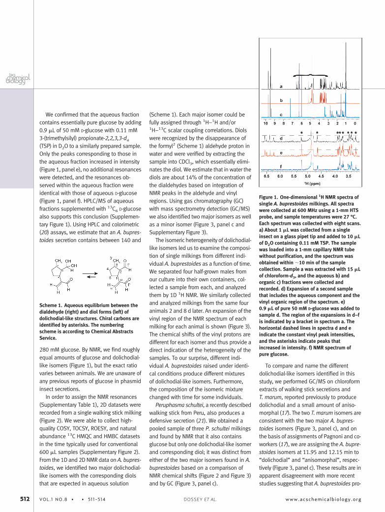
In order to assign the NMR resonances(Supplementary Table 1), 2D datasets wererecorded from a single walking stick milking(Figure 2). We were able to collect high-quality COSY, TOCSY, ROESY, and naturalabundance 13C HMQC and HMBC datasetsin the time typically used for conventional600 �L samples (Supplementary Figure 2).From the 1D and 2D NMR data on A. bupres-toides, we identified two major dolichodial-like isomers with the corresponding diolsthat are expected in aqueous solution
(Scheme 1). Each major isomer could befully assigned through 1H–1H and/or1H–13C scalar coupling correlations. Diolswere recognized by the disappearance ofthe formyl2 (Scheme 1) aldehyde proton inwater and were verified by extracting thesample into CDCl3, which essentially elimi-nates the diol. We estimate that in water thediols are about 14% of the concentration ofthe dialdehydes based on integration ofNMR peaks in the aldehyde and vinylregions. Using gas chromatography (GC)with mass spectrometry detection (GC/MS)we also identified two major isomers as wellas a minor isomer (Figure 3, panel c andSupplementary Figure 3).
The isomeric heterogeneity of dolichodial-like isomers led us to examine the composi-tion of single milkings from different indi-vidual A. buprestoides as a function of time.We separated four half-grown males fromour culture into their own containers, col-lected a sample from each, and analyzedthem by 1D 1H NMR. We similarly collectedand analyzed milkings from the same fouranimals 2 and 8 d later. An expansion of thevinyl region of the NMR spectrum of eachmilking for each animal is shown (Figure 3).The chemical shifts of the vinyl protons aredifferent for each isomer and thus provide adirect indication of the heterogeneity of thesamples. To our surprise, different indi-vidual A. buprestoides raised under identi-cal conditions produce different mixturesof dolichodial-like isomers. Furthermore,the composition of the isomeric mixturechanged with time for some individuals.
Peruphasma schultei, a recently describedwalking stick from Peru, also produces adefensive secretion (21). We obtained apooled sample of three P. schultei milkingsand found by NMR that it also containsglucose but only one dolichodial-like isomerand corresponding diol; it was distinct fromeither of the two major isomers found in A.buprestoides based on a comparison ofNMR chemical shifts (Figure 2 and Figure 3)and by GC (Figure 3, panel c).
To compare and name the differentdolichodial-like isomers identified in thisstudy, we performed GC/MS on chloroformextracts of walking stick secretions andT. marum, reported previously to producedolichodial and a small amount of aniso-morphal (17). The two T. marum isomers areconsistent with the two major A. bupres-toides isomers (Figure 3, panel c), and onthe basis of assignments of Pagnoni and co-workers (17), we are assigning the A. bupre-stoides isomers at 11.95 and 12.15 min to“dolichodial” and “anisomorphal”, respec-tively (Figure 3, panel c). These results are inapparent disagreement with more recentstudies suggesting that A. buprestoides pro-
Scheme 1. Aqueous equilibrium between thedialdehyde (right) and diol forms (left) ofdolichodial-like structures. Chiral carbons areidentified by asterisks. The numberingscheme is according to Chemical AbstractsService.
Figure 1. One-dimensional 1H NMR spectra ofsingle A. buprestoides milkings. All spectrawere collected at 600 MHz using a 1-mm HTSprobe, and sample temperatures were 27 °C.Each spectrum was collected with eight scans.a) About 1 �L was collected from a singleinsect on a glass pipet tip and added to 10 �Lof D2O containing 0.11 mM TSP. The samplewas loaded into a 1-mm capillary NMR tubewithout purification, and the spectrum wasobtained within �10 min of the samplecollection. Sample a was extracted with 15 �Lof chloroform-d3, and the aqueous b) andorganic c) fractions were collected andrecorded. d) Expansion of a second samplethat includes the aqueous component and thevinyl organic region of the spectrum. e)0.9 �L of pure 50 mM D-glucose was added tosample d. The region of the expansions in d–fis indicated by a bracket in spectrum a. Thehorizontal dashed lines in spectra d and eindicate the constant vinyl peak intensities,and the asterisks indicate peaks thatincreased in intensity. f) NMR spectrum ofpure glucose.
512 VOL.1 NO.8 • • 511-514 www.acschemicalbiology.orgDOSSEY ET AL.

duces a single dolichodial-like isomer (22).This could be due to improvement of analyti-cal methods, genetic variability, or environ-mental factors. The P. schultei and minorA. buprestoides isomers at 11.78 min(Figure 3, panel c) appear to be the sameand, we believe, are previously unreported.We are naming this isomer “peruphasmal”.
Previous studies using MS, electrophore-sis, or LC have reported individual variationin polypeptide toxins from snakes (23–25),
cone snails (26), and a variety of arthropods(27–30). To our knowledge, it has neverbefore been possible to perform a detailedmolecular study of a mixture of natural prod-ucts from an individual insect using NMR.This new capability provides the possibilityof elucidating chemical variation, such asstereochemistry, in greater detail. Threemajor findings on walking stick defensesecretions were enabled by high-sensitivityNMR (13): (i) the heterogeneity of defensivedolichodial-like stereoisomers that variesbetween A. buprestoides individuals andwith time, (ii) a new dolichodial-like isomercalled peruphasmal from P. schultei, and(iii) the identification of glucose in phasmidsecretions. The quantity of glucose suggestsa biological or chemical role in walking stickvenom that merits further investigation.
METHODSInsect Rearing and Sample Preparation. Adult
A. buprestoides were collected at night in GulfHammock, FL, during the fall of 2005. Eggs pro-duced by the insects were hatched in captivity. Theyoung phasmids were fed a diet of only variegatedLigustrum sinense purchased from a local plantnursery. We were able to collect single milkingsfrom half-grown males consisting of �1 �L of awhitish fluid by gently touching the secretory ductwith a glass pipet. To this we added 10 �L of D2Ocontaining 0.11 mM TSP as a chemical shift refer-ence to the sample.
NMR. NMR experiments were done using a 600-MHz 1-mm triple-resonance HTS cryogenic probethat was developed through collaborationbetween the University of Florida, the NationalHigh Magnetic Field Laboratory (NHMFL), andBruker Biospin (13). The total sample volume is�8 �L, and each sample was loaded into a 1-mm� 100-mm capillary NMR tube (Norell, Inc.) using a10-�L syringe with a fixed 110-mm � 30-gaugeblunt needle. The capillary tube was held in a stan-dard 10-mm spinner using a Bruker MATCH device,and the capillary–MATCH–spinner combinationwas lowered vertically into the magnet on an aircolumn as usual. The sample temperature wasregulated at 27 °C. The spectrometer was a BrukerAvance 600 with Xwin-NMR software, and all otherdata acquisition was done using standard technol-ogy. Two-dimensional datasets were processedusing NMRPipe (31) and manually assigned usingNMRView (32).
GC–Flame Ionization Detector. A Hewlett-Packard (Palo Alto, CA) 5890 series II gas chro-matograph and a flame ionization detector(GC–FID) with nitrogen make-up gas (1.5 mL/min)and helium carrier gas (1.3 mL/min) were used.Cool on-column and splitless injections (1 �L)
were at 40 and 200 °C, respectively; the detectorwas maintained at 260 °C. The oven program wasas follows: isothermal for 5 min, heating from 40to 200 °C at 11 °C/min, isothermal for 10 min,heating from 200 to 250 °C at 25 °C/min, and thenisothermal for 15 min. GlasSeal connectors(Supleco) fused three silica columns in series: aprimary deactivated column (8 cm long, 0.53 mmi.d.), an HP-1MS retention gap column (2 m long,0.25 mm i.d., df � 0.25 �m), and a J&W DB-5 ana-lytical column (30 m long, 0.25 mm i.d., df �0.25 �m).
GC/MS. A Varian 3400 gas chromatograph anda Finnigan MAT Magnum ion trap mass spectrom-eter in electron impact ionization mode (70 eV)with a filament bias of 11765mV or chemical ion-ization mode (isobutane) were employed toacquire full-scan spectra over the ranges m/z40–400 at 0.85 s per scan. Holox (Charlotte, NC)high-purity helium was used as a carrier gas(1.4 mL/min). Injection and oven conditions wereas above. Transfer-line and manifold temperatureswere 240 and 220 °C, respectively.
Acknowledgments: A sample of defensive secre-tions from P. schultei was kindly provided by O. V.Conle of Bolsterlang, Germany. We thank Drs.W. W. Brey (NHMFL) and R. S. Withers and R. E.Nast (Varian NMR) for the collaboration andsupport on the 1-mm HTS probe. Dr. P. Teal (USDALaboratory, Gainesville, FL) provided helpfulencouragement and discussions. Supported byNIH P41RR016105, the Human Frontier ScienceProgram (ASE), and the NHMFL. NMR data werecollected in the Advanced Magnetic ResonanceImaging and Spectroscopy Facility at McKnightBrain Institute of the University of Florida.
Supporting Information Available: This materialis available free of charge via the Internet.
REFERENCES1. Metcalf, R. L. (1998) Ultramicrochemistry of insect
semiochemicals, Mikrochim. Acta. 129, 167–180.
2. Olson, D. L., Peck, T. L., Webb, A. G., Magin, R. L., andSweedler, J. V. (1995) High-resolution microcoil H-1-NMR for mass-limited, nanoliter-volume samples,Science 270, 1967–1970.
3. Gronquist, M., Meinwald, J., Eisner, T., and Schroe-der, F. C. (2005) Exploring uncharted terrain in Na-ture’s structure space using capillary NMR spectros-copy: 13 Steroids from 50 fireflies, J. Am. Chem.Soc. 127, 10810–10811.
4. Peti, W., Norcross, J., Eldridge, G., and O’Neil-Johnson, M. (2004) Biomolecular NMR using a mi-crocoil NMR probe—new technique for the chemicalshift assignment of aromatic side chains in pro-teins, J. Am. Chem. Soc. 126, 5873–5878.
5. Li, Y., Logan, T. M., Edison, A. S., and Webb, A.(2003) Design of small volume HX and triple-resonance probes for improved limits of detection inprotein NMR experiments, J. Magn. Reson. 164,128–135.
Figure 2. Two-dimensional expansions ofCOSY (right panels) and ROESY (left andcenter panels) from a single milking of A.buprestoides (top) and a pooled sample fromthree P. schultei milkings (bottom). One-dimensional 1H spectra from the samesamples are shown along the top. All datawere collected at 600 MHz using the 1-mmHTS probe. The COSY experiments werecollected in �2.5 h with 8 scans and 512complex indirect data points. The ROESYexperiments were collected in �9 h with 32scans, 512 complex points, and a 400-msmixing time.
LETTER
www.acschemicalbiology.org VOL.1 NO.8 • • 511-514 513

6. Li, Y., Webb, A. G., Saha, S., Brey, W. W., Zachariah,C., and Edison, A. S. (2006) Comparison of the per-formance of round and rectangular wire in smallsolenoids for high-field NMR, Magn. Reson. Chem.44, 255–262.
7. Kovacs, H., Moskau, D., and Spraul, M. (2005) Cryo-genically cooled probes—a leap in NMR technology,Prog. Nucl. Magn. Reson. Spectrosc. 46, 131–155.
8. Rogers, E. W., and Molinski, T. F. (2005) A cytotoxiccarotenoid from the marine sponge Prianos osiros,J. Nat. Prod. 68, 450–452.
9. Wolters, A. M., Jayawickrama, D. A., and Sweedler, J.V. (2005) Comparative analysis of a neurotoxin fromCalliostoma canaliculatum by on-line capillary iso-tachophoresis 1H NMR and diffusion 1H NMR, J. Nat.Prod. 68, 162–167.
10. McPhail, K. L., France, D., Cornell-Kennon, S., andGerwick, W. H. (2004) Peyssonenynes A and B, novelenediyne oxylipins with DNA methyl transferase in-hibitory activity from the red marine alga Peyssonne-lia caulifera, J. Nat. Prod. 67, 1010–1013.
11. Russell, D. J., Hadden, C. E., Martin, G. E., Gibson,A. A., Zens, A. P., and Carolan, J. L. (2000) A compar-ison of inverse-detected heteronuclear NMR perfor-mance: conventional vs cryogenic microprobe per-formance, J. Nat. Prod. 63, 1047–1049.
12. Saman, D., Cvacka, J., Svatos, A., Bouman, E. A. P.,and Kalinova, B. (2006) Structural identification ofan anthrasteroid hydrocarbon from the sheep tickIxodes ricinus. J. Nat. Prod. DOI. 10.1021/np0680127.
13. Brey, W. W., Edison, A. S., Nast, R. E., Rocca, J. R.,Saha, S., and Withers, R. S. (2006) Design, construc-tion, and validation of a 1-mm triple-resonancehigh-temperature-superconducting probe for NMR,J. Magn. Reson. 179, 290–293.
14. Eisner, T. (1965) Defensive spray of a phasmidinsect, Science 148, 966–968.
15. Meinwald, J., Chadha, M. S., Hurst, J. J., and Eisner,T. (1962) Defense mechanisms of arthropods. 9.Anisomorphal, the secretion of a phasmid insect,Tetrahedron Lett. 29–33.
16. Cavill, G. W., and Hinterberger, H. (1961) Chemistryof ants. 5. Structure and reactions of dolichodial,Aust. J. Chem. 14, 143–149.
17. Pagnoni, U. M., Pinetti, A., Trave, R., and Garanti, L.(1976) Monoterpenes of teucrium-marum, Aust.J. Chem. 29, 1375–1381.
18. Eisner, T., Eisner, M., Aneshansley, D. J., Wu, C. L.,and Meinwald, J. (2000) Chemical defense of themint plant, Teucrium marum (Labiatae), Chemoecol-ogy 10, 211–216.
19. Ricci, D., Fraternale, D., Giamperi, L., Bucchini, A.,Epifano, F., Burini, G., and Curini, M. (2005) Chemi-cal composition, antimicrobial and antioxidant ac-tivity of the essential oil of Teucrium marum (Lami-aceae), J. Ethnopharmacol. 98, 195–200.
20. Hendrix, D. L. (1993) Rapid extraction and analysisof nonstructural carbohydrates in plant-tissues, CropSci. 33, 1306–1311.
21. Conle, O. V., and Hennemann, F. H. (2005) Studieson neotropical Phasmatodea I: A remarkable newspecies of Peruphasma Conle & Hennemann, 2002from northern Peru (Phasmatodea: Pseudophas-matidae: Pseudophasmatinae), Zootaxa 1068,59–68.
22. Eisner, T., Morgan, R. C., Attygalle, A. B., Smedley,S. R., Herath, K. B., and Meinwald, J. (1997) Defen-sive production of quinoline by a phasmid insect(Oreophoetes peruana), J. Exp. Biol. 200,2493–2500.
23. Menezes, M. C., Furtado, M. F., Travaglia-Cardoso,S. R., Camargo, A. C., and Serrano, S. M. (2006) Sex-based individual variation of snake venom pro-teome among eighteen Bothrops jararaca siblings,Toxicon 47, 304–312.
24. Francischetti, I. M., Gombarovits, M. E., Valenzuela,J. G., Carlini, C. R., and Guimaraes, J. A. (2000) In-traspecific variation in the venoms of the SouthAmerican rattlesnake (Crotalus durissus terrificus),Comp. Biochem. Physiol., Part C: Toxicol. Pharma-col. 127, 23–36.
25. Monteiro, R. Q., Yamanouye, N., Carlini, C. R., Guima-raes, J. A., Bon, C., and Zingali, R. B. (1998) Variabil-ity of bothrojaracin isoforms and other venom prin-ciples in individual jararaca (Bothrops jararaca)snakes maintained under seasonally invariant con-ditions, Toxicon 36, 153–163.
26. Jakubowski, J. A., Kelley, W. P., Sweedler, J. V., Gilly,W. F., and Schulz, J. R. (2005) Intraspecific variationof venom injected by fish-hunting Conus snails,J. Exp. Biol. 208, 2873–2883.
27. Borges, A., Garcia, C. C., Lugo, E., Alfonzo, M. J.,Jowers, M. J., and Op den Camp, H. J. (2006) Diversi-ty of long-chain toxins in Tityus zulianus and Tityusdiscrepans venoms (Scorpiones, Buthidae): molecu-lar, immunological, and mass spectral analyses,Comp. Biochem. Physiol., Part C: Toxicol. Pharma-col. 142, 240–252.
28. Lai, C. C., and Her, G. R. (2000) Analysis of phospho-lipase A2 glycosylation patterns from venom of indi-vidual bees by capillary electrophoresis/electro-spray ionization mass spectrometry using an iontrap mass spectrometer, Rapid Commun. MassSpectrom. 14, 2012–2018.
29. Pimenta, A. M., de Lima, M. E., De Marco Almeida,F., Martin-Eauclaire, M. F., and Bougis, P. E. (2003)Individual variability in Tityus serrulatus (Scorpio-nes, Buthidae) venom elicited by matrix-assistedlaser desorption/ionization time-of-flight mass spec-trometry, Rapid Commun. Mass Spectrom. 17,413–418.
30. Escoubas, P., Corzo, G., Whiteley, B. J., Celerier, M. L.,and Nakajima, T. (2002) Matrix-assisted laser de-sorption/ionization time-of-flight mass spectrome-try and high-performance liquid chromatographystudy of quantitative and qualitative variation in ta-rantula spider venoms. Rapid Commun. MassSpectrom. 16, 403–413.
31. Delaglio, F., Grzesiek, S., Vuister, G. W., Zhu, G.,Pfeifer, J., and Bax, A. (1995) Nmrpipe—a multidi-mensional spectral processing system based onUnix pipes, J. Biomol. NMR 6, 277–293.
32. Johnson, B. A., and Blevins, R. A. (1994) NMR View—a computer-program for the visualization and analy-sis of NMR data, J. Biomol. NMR 4, 603–614.
Figure 3. Isomeric variation of venom. a) Expansions of the vinyl region of NMR spectra of single milkings from individual A. buprestoides collectedon different days. Samples were dissolved in 10 �L of D2O � TSP without further purification. b) Same expansion from a mixture of three milkingsfrom P. schultei. The shoulder marked with an asterisk corresponds to one of the vinyl peaks of the diol, and the broad peak at 6.54 ppm is anoverlap of the diol and dialdehyde isomers (Scheme 1). c) GC analysis of chloroform extracts of A. buprestoides (red) and P. schultei (blue)secretions, and T. marum (green). With both cool on-column (shown) and splitless injection, all isomers ionized with comparable efficieny (FID),fragmented similarly (electron impact MS), and had identical masses of 166 Da (chemical ionization MS).
514 VOL.1 NO.8 • • 511-514 www.acschemicalbiology.orgDOSSEY ET AL.