homeEcon.menofia.edu.eg ISSN 1110-2578 Impact of maternal ...
22
Journal of Home Economics, Volume 26, Number (2), 2016 155 Journal of Home Economics Volume 26, Number (2), 2016 Journal of Home Economics http://homeEcon.menofia.edu.eg ISSN 1110-2578 Impact of maternal nutrition and socio-economic status on birth weight of babies in Port Said Governorate, Egypt Yousif Elhassaneen 1 *, Safaa Al-Wasef 2 , NaglaaFathy 2 Reham Abo-Samra 2 1 Department of Nutrition and Food Science, Faculty of Home Economics, Minoufiya University, Shebin El-Kom, 2 Department of Home Economics, Faculty of Specific Education, Port Said University, Port Said, Egypt * Corresponding author: [email protected] Abstract: Low birth weight (LBW) is a major public health problem in most developing countries including Egypt, being associated with a high incidence of neonatal mortality in these regions. There is, therefore, an urgent need to determine ways and means to prevent LBW and its consequences. This study was designed to study the impact of maternal nutrition and socio-economic status on birth weight of babies in Port Said Governorate, Egypt. Data shows that the prevalence of LBW in the stated health centers varies widely by pregnant mothers age, area, socioeconomic status (family income, parents education), the weight gain during the pregnancy etc. The mean weight gain of the pregnant mothers in this study was standard is 11 kg). Weight gain in pregnancy, maternal hemoglobin, serum iron and serum antioxidant vitamins were all found to be significant for LBW ( p< 0.05-0.001). Maternal nutritional status impacted significantly on newborn birth weight as poorly nourished mothers were observed to produce a higher percentage of LBW babies when compared to those who were better nourished. Therefore, the challenge of addressing the LBW problem therefore remains an urgent imperative for development. Keywords: Socio-demographic, hemoglobin, iron, vitamins, dietary habits Introduction Birth weight is the single most important criterion for determining the neonatal and infant survival. Low birth weight (LBW) is a sensitive indicator of the socio-economic conditions and indirectly measures the
Transcript of homeEcon.menofia.edu.eg ISSN 1110-2578 Impact of maternal ...

Journal of Home Economics, Volume 26, Number (2), 2016
155
Journal of Home Economics
Volume 26, Number (2), 2016
Journal of Home Economics
http://homeEcon.menofia.edu.eg ISSN 1110-2578
Impact of maternal nutrition and socio-economic status on
birth weight of babies in Port Said Governorate, Egypt
Yousif Elhassaneen
1*, Safaa Al-Wasef
2, NaglaaFathy
2
Reham Abo-Samra2
1Department of Nutrition and Food Science, Faculty of Home Economics, Minoufiya
University, Shebin El-Kom, 2Department of Home Economics, Faculty of Specific
Education, Port Said University, Port Said, Egypt * Corresponding author: [email protected]
Abstract: Low birth weight (LBW) is a major public health problem in most
developing countries including Egypt, being associated with a high incidence of
neonatal mortality in these regions. There is, therefore, an urgent need to
determine ways and means to prevent LBW and its consequences. This study
was designed to study the impact of maternal nutrition and socio-economic
status on birth weight of babies in Port Said Governorate, Egypt. Data shows
that the prevalence of LBW in the stated health centers varies widely by
pregnant mothers age, area, socioeconomic status (family income, parents
education), the weight gain during the pregnancy etc. The mean weight gain of
the pregnant mothers in this study was
standard is 11 kg). Weight gain in pregnancy, maternal hemoglobin, serum iron
and serum antioxidant vitamins were all found to be significant for LBW (p<
0.05-0.001). Maternal nutritional status impacted significantly on newborn birth
weight as poorly nourished mothers were observed to produce a higher
percentage of LBW babies when compared to those who were better nourished.
Therefore, the challenge of addressing the LBW problem therefore remains an
urgent imperative for development.
Keywords: Socio-demographic, hemoglobin, iron, vitamins, dietary habits
Introduction
Birth weight is the single most important criterion for determining
the neonatal and infant survival. Low birth weight (LBW) is a sensitive
indicator of the socio-economic conditions and indirectly measures the

Journal of Home Economics, Volume 26, Number (2), 2016
156
health of the mother and the child. The World Health Organization
(WHO) defined LBW as that below 2,500 g (Kramer, 1987). LBW is a
major public health problem due to it's the main cause of mortality,
morbidity and disability (Amosu and Degun, 2014). It has been shown
that the mortality range can vary 100-fold across the spectrum of birth
weight and rises continuously with decreasing weight (Wilcox, 2001).
It is generally recognized that being born with a LBW is a
disadvantage for the infant. Pre-term birth is a direct cause of 27% of the
4 million neonatal deaths that occur globally every year (Lawn et al.,
2005). Pre-term birth and SGA are also important indirect causes of
neonatal deaths. Low birth weight directly or indirectly may contribute up
to 60–80% of all neonatal deaths (Lawn et al., 2005). LBW infants are at
higher risk of early growth retardation, infectious disease, developmental
delay and death during infancy and childhood (McCormick, 1985;
Ashworth, 1998).
During the newborn period LBW/ SGA (A small for gestational
age) babies present increased risk of hypoglycemia, hypothermia,
hypercoagulability, hyperbilirubinemia, hypotension, necrotizing
enterocolitis, respiratory distress syndrome, lower Apgar scores,
umbilical artery acidosis, more intubations and complications during
delivery and approximately 20 times increased risk of neonatal death than
babies born with an appropriate for gestational age (AGA) weight
(Claussonet al., 1998; McIntire et al., 1999; Jancevskaet al., 2012). Also,
UNICEF and WHO (2004) reviewed that LBW due to restricted foetal
growth affects the person throughout life and is associated with poor
growth in childhood and a higher incidence of adult diseases, such as type
2 diabetes, hypertension and cardiovascular disease. An additional risk for
girls is having smaller babies when they become mothers. Furthermore,
Carlos and Marilia, (2013) reviewed that a clear phenomenological
association has been demonstrated by many epidemiological studies
between LBW and increased risk later in life, for many diseases such as
IR, mortality by type 2 diabetes (T2D), hypertension(HT) and ischemic
heart diseases (IHD), cardiovascular diseases (CVD), dyslipidemia,
obesity, breast and testicular cancer, end-stage renal disease, osteoporosis,
spontaneous hypothyroidism, adult asthma and hearing loss, cardiac
hypertrophy, depression, liver cirrhosis, schizophrenia, polycystic ovary
syndrome, precocious pubarche, hypospadias, cryptorchidism, low scores
Journal of Home Economics, Volume 26, Number (2), 2016
157
of alertness, mood instability, significant differences in academic and
professional achievement.
LBW is a major public health problem in most developing
countries, being associated with a high incidence of neonatal mortality in
these regions (Wilcox, 2001). In Egypt, the average prevalence of LBW is
estimated to be about 12% (UNICEF, 2004). The immediate
consequences are higher morbidity and mortality rates in the perinatal and
neonatal periods [Osrinand de L Costello, 2000]. The late consequences
may include prolonged impairment of immunological defense
mechanisms and neurological sequelae which interfere with the normal
development of the child and on a national level, are serious obstacles to
development (Davies and Stewart, 1995; Ferguson, 1998).
Many factors affect the duration of gestation and of foetal growth,
and thus, the birth weight. They relate to the infant, the mother or the
physical environment and play an important role in determining the
infant‟s birth weight and future health (WHO (2004). The multiple causes
of LBW, include early induction of labour or caesarean birth (for medical
or non-medical reasons), multiple pregnancies, infections and chronic
conditions such as diabetes and high blood pressure (Christopher and
Siobhan, 2014; WHO, 2014). Other important causes could include low
maternalnutrient intake, higher nutrient losses, and/or increased
nutritional requirements i.e. malnutrition during pregnancy (Amosu and
Degun, 2014). Maternal morbidity during pregnancy is highly prevalent
in the low socio-economic groups (Sumithra, 2009) and protective
mechanisms such as the bactericidal zinc peptide system in the amniotic
fluid (Schlievertet al., 1976), may be impaired partly because of
malnutrition during pregnancy (Naeyeet al., 1997). All of these reasons
indicated that the maternal nutrition is an important factor from a public
health point of view because it is modifiable and therefore susceptible to
public health interventions. There is, therefore, an urgent need to
determine ways and means to prevent LBW and its consequences. This
thesis was designed to study the maternal nutritional and socio-economic
factors and their role in BW of the newborn in Port Said Governorate,
Egypt.
Material and Methods
Chemicals and equipments
Standard vitamins (A, C, and E) were purchased from Sigma
Chemical Co. (St. Louis, MO). All chemicals and buffers, except

Journal of Home Economics, Volume 26, Number (2), 2016
158
stipulated, were in analytical grade and purchased from Al-Gomhoria
Company for Drugs, Chemicals and Medical Instruments, Cairo, Egypt.
Throughout this study, a SP Thermo Separation Products Liquid
Chromatography (ThermoSeparation products, San Jose, CA) was used
with a ConstaMetvic 4100 pump, a SpectraSeries AS100, Spectra System
UV 1000 UV/Visible Spectrophotometer Detector, SpectraSystem FL
3000, and a PC 1000 system software. The columns used (Alltech,
Deerfield, IL)were a Hypersil BDS-C18 (5 µm, 150 x 4.6–mm internal
diameter) for PAH; a reversed phasewaterAdsorbosil C18 (5 µmol/L, 100
mm x 4.6–mm internal diameter) for vitamin C; and normal Ultrasphere
Si (5 µmol/L, 250 mm x 4.6–mm internal diameter) for analysisof
vitamins A and E.
Study protocol
The present study was conducted in the Department of Home
Economics, Faculty of Specific Education, Port Said University, Port
Said, Egypt from January 2015 to March 2016. The research reported in
the thesis was approved by the Research Ethics Committee of Faculty of
Home Economics, Minoufiya University, Shebin El-Kom, Egypt.
Approval was granted prior to the commencement of data collection by
questionnaire. This is a cross-sectional and descriptive study design for
booked pregnant mothers who delivered in five randomly selected health
facilities in Port Said City, Port Saied Governorate, Egypt.
The subjects were selected from each health facility as they became
available. Two hundred and ninety two randomly selected pregnant
mothers attending antenatal clinics in the health facilities were booked
and advised to report regularly for antenatal care. Complete abdominal
examination, clinical profile along with height, weight,weight gain in
pregnancy, blood pressure, haematological and biochemical examinations
were carried out by the clinics staff. All the newborns were weighed and
their general physical conditions assessed immediately following
delivery. All infants weighing below 2.5 kg were recorded as low birth
weight (LBW) babies.
A field pretested interviewing questionnaire was used for data
collection which covering the following points: pregnant mother age, sex,
residence (urban or rural) and family size. Sociodemographic status of
pregnant mothers and their families, socioeconomic score, which
contained social variables including fathers' education and work (score 2-
Journal of Home Economics, Volume 26, Number (2), 2016
159
10), mothers' education and work (scores 1-10) and crowding index
(scores 1-5). The total score calculation was: Score from 19-25 means
high social class, score from 12 to 18 means middle social class and score
below 12 means low social class such as mentioned by Fahmy and El-
Sherbini (1983).
Dietary recall and the record were used for mothers who were asked
for all foods and fluids consumed while she was at home. Daily iron
intake of each infant was calculated using the "Diet Analysis Program,
1995" (Lifestyles Technologies, Inc., Northbridge Point, Valencia,
California) and was then compared with the Recommended Dietary
Allowances (RDA, 1989).
Hematological analysis
Blood samples were withdrawn from the antecubital vein into glass
centrifuge tubes containing oxalate solution (1.34%) as anticoagulant.
After centrifugation at 1500 X g for 10 minutes, plasma was withdrawn
and used for analysis of blood lipid parameters and vitamins. The
erythrocyte residue was washed with three successive portions of sodium
chloride solution (0.9%) and then hemolyzed with deionized water for 30
minutes. Hemolysate was then centrifuged at 105,000 X g for 30 minutes,
and the supernatant fractions was transferred to a clean test tube and
analyzed for antioxidant enzymes (Stroev EA, Makarova, 1989).
Serum iron (Fe) content samples were determined by the adaptation
the method mentioned by Singh et al., (1991). One hundred µl of plasma
sample were transferred into a digested glass tube and 2 ml of tri-acids
mixture (containing nitric acid: perchloric acid: sulfuric acid in the ratio
of 20: 4: 1 v/v respectively) were added to each tube. The tubes content
were digested gradually as follow, 30 min at 70 0C; 30 min at 180 0C and
30 min at 220 0C. After digestion, the mixture was cooled, dissolved in
MilliQ water, and the volume was increased to 10 ml in volumetric
beaker. After filtration in ashless filter paper, aliquots were analyzed for
Fe and Se content using of atomic absorption spectrophotometer, type
Perkin - Elmer, Model 2380.
Blood hemoglobin (Hb) concentration was determined using
cyanmethemoglobin method according to Villanova (1994). Anemia was
diagnosed when Hb concentrations below the values adjusted for age
groups (Wonkeet al., 2007). In infants from 6 months to 6 years mild,
moderate and severe anemia was diagnosed if Hb level was 10-11, 7-9.9,
Journal of Home Economics, Volume 26, Number (2), 2016
160
or below 7 g/dl, respectively (Bermejo and García-López, 2009).
The plasma ferritin concentrations and hematocrit value were assayed
using specific Kits (Al-Gomhoria Company for Drugs, Chemicals and
Medical Instruments, Cairo, Egypt) according to the methods mentioned
in Tietz, (1999). The complete blood count was done using Coulter 1660
to determine the erythrocyte indices (mean corpuscular volume [MCV],
mean corpuscular Hb [MCH], MCH concentration, and red cell diameter
width.
Determination of vitamins
All vitamins (A, C, and E) were extracted according to methods
previously detailedByEpler and Zeigler , (1993), Moeslinger et al., (1994)
and Hung et al., (1980), respectively and were analyzed by HPLC
techniques. For vitamins A and E, the chromatographic conditions were
as follows: flow rate, 1.5 mL/min; detection, UV absorption at 265 nm,
volume of injection, 20 µL; temperature, room temperature; and the
mobile phase composition was an isocratic system of isopropanol:hexane
(1:99). For vitamin C, the conditions were: flow rate, 1 mL/min;
detection, UV absorption at 254 nm, volume of injection, 20 µL;
temperature, room temperature, and mobile phase composition was an
isocratic system of 100% methanol. Retention times and absorbance ratio
against those of standards were used to identify the separated vitamins.
Quantitative determination of each vitamin was determined from its
respective peak area and corresponding response factor. The percent
recoveries of vitamins were also studied by adding each vitamin to
plasma after sample preparation and HPLC determination. Under such
chromatographic conditions, mean values(±SD) of vitamins A, C, and E
recoveries were 89.92 ±2.91, 92.01 ±3.25, and 88.11 ±2.34, respectively.
Statistical analysis Data were summarized using means, SD, range for quantitative
variables and number, and percentage for qualitative variables. Data
obtained from the study were coded, entered and statistical analyzed using
the software MINITAB 12 computer program (Minitab Inc., State
College, PA).
Journal of Home Economics, Volume 26, Number (2), 2016
161
Results and Discussion
Socio-demographic characteristics of the pregnant mothers studied
group
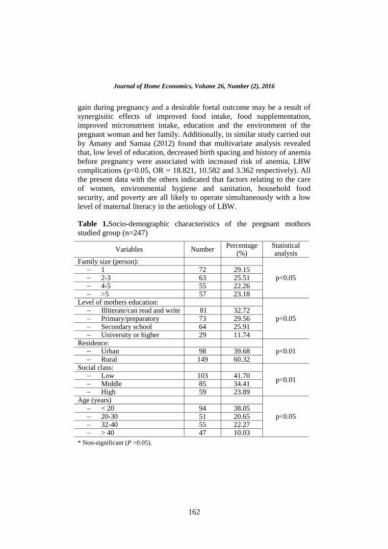
In the present study Among 292 pregnant mothers enrolled, 45
(11.47%) women were not analyzed because of unavailability of consent
for study or inadequate sample. The remaining enrolled 247 pregnant
women and their newborn babies aged 0 to 12 months with a mean age of
(29.8± 12.4 years for pregnant women) and (2.4 ± 1.8 months for the
babies) years. The sociodemographic data are shown in Table (1). Based
on the body weight, pregnant mothers were selected after borned low
birth weight (LBW) babies. A logistic regression model was used to
assess the effects of the significant explanatory variables in order to
distinguish predictors of LBW. It was found that pregnant women from
rural areas (60.32%), those from family size, 1 person (29.15%), those of
illiterate mothers (32.72%), those of low social class (41.70%) and those
of small age (38.05%) were the significant risk factors for LBW in these
women.
Studies worldwide have examined the effect of socio-economic
status (SES) indicators, including maternal education, on birth weight.
Maternal illiteracy and low SES have been shown to be major risk factors
for LBW (Mavalankaret al., 1992; Sumithra, 2009). In the developing
world, lacking proper health systems and resources, the level of maternal
education may be of prime importance in the determination of health
outcomes of mothersand their infants and children. In the Javier et
al.,(2004) study, it is reviewed that there are many known risk factors, the
most important of which are socio-economic factors, medical risks before
or during gestation and maternal lifestyles. However, although
interventions exist to prevent many of these factors before and during
pregnancy, the incidence of LBW has not decreased. Also, Joshi et al.,
(2007) reported that the main factors which were significantly associated
with LBW were maternal education, stature, age at delivery; short inter
pregnancy interval, inadequate antenatal care, and per capita income of
family. Furthermore, Arnaud and Vincent (2007) studied the relationship
between mother‟s education and birth weight and find modest but
heterogenous positive effects of maternal education on birth weight with
an increase from the baseline weight ranging from 2% to 6%. In this
direction, Sumithra (2009) reported that there is a significant protective
effect of higher maternal education (beyond high school). Optimal weight
Journal of Home Economics, Volume 26, Number (2), 2016
162
gain during pregnancy and a desirable foetal outcome may be a result of
synergisitic effects of improved food intake, food supplementation,
improved micronutrient intake, education and the environment of the
pregnant woman and her family. Additionally, in similar study carried out
by Amany and Samaa (2012) found that multivariate analysis revealed
that, low level of education, decreased birth spacing and history of anemia
before pregnancy were associated with increased risk of anemia, LBW
complications (p<0.05, OR = 18.821, 10.582 and 3.362 respectively). All
the present data with the others indicated that factors relating to the care
of women, environmental hygiene and sanitation, household food
security, and poverty are all likely to operate simultaneously with a low
level of maternal literacy in the aetiology of LBW.
Table 1.Socio-demographic characteristics of the pregnant mothors
studied group (n=247)
Variables Number Percentage
(%) Statistical analysis
Family size (person):
p<0.05 1 72 29.15 2-3 63 25.51 4-5 55 22.26 >5 57 23.18
Level of mothers education:
p<0.05 Illiterate/can read and write 81 32.72 Primary/preparatory 73 29.56 Secondary school 64 25.91 University or higher 29 11.74
Residence: p<0.01 Urban 98 39.68
Rural 149 60.32 Social class:
p<0.01 Low 103 41.70 Middle 85 34.41 High 59 23.89
Age (years)
p<0.05 < 20 94 38.05 20-30 51 20.65 32-40 55 22.27 > 40 47 10.03
* Non-significant (P >0.05).

Journal of Home Economics, Volume 26, Number (2), 2016
163
Maternal weight gain in pregnancy of the mothers studied group
The mothers‟ weights were measured before and immediately after
delivery of the baby, placenta and membranes. Primiparous mothers were
found to gain significantly less weight than multiparous mothers
(p
kg. Primiparous mothers recorded the lowest mean weight gain of 6.02
pregnancy increasing with increase in parity
as seen in Table (2). In similar studies, Jelliffe, (1966) and Amosu and
Degun (2014) found that triceps skin fold thickness of the mothers as
measured using a Harpenden caliper was expressed as a percentage of
normal values. This variable indicates the amount of adipose tissue in the
upper arm, with primiparous mothers recording lower percentage than the
multiparae.Such as shown in Table (3) the high LBW incidence recorded
in mothers with weight gain of 7 kg and below, while the lowest LBW
incidence recorded with mothers gained 9 kg and above. The relationship
between weight gain in pregnancy and newborn birth weight has been
known for several decades (Beilly and Kurkland, 1945), and recently by
Amosu and Degun (2014). Therefore, even now its importance to
pregnancy outcome is being increasingly recognized. This importance is
reflected in the recommendation that pregnant women should be
encouraged to gain at least 11kg during gestation (UN, 2004).
Table 2.Parity and maternal weight gain during pregnancy of mothers
studied group (n=247)
Parity Frequency
(n)
Mean ± SD
(kg)
Statistical
analysis
Primiparous 69 6.02± 0.96
p<0.05
Para 1 38 6.71± 0.85
Para 2 48 6.92± 1.01
Para 3 31 7.85± 0.59
≥ Para 4 61 9.39± 1.42
Table 3.Maternal weight gain in pregnancy of the mothers studied group
(n=247) Weight gain in pregnancy
(kg) Number of
mothers/newborns Statistical analysis
< 7 155
p<0.001 7.1 - 8 44 > 8.1-9 39
> 9 9

Journal of Home Economics, Volume 26, Number (2), 2016
164
Hematological and biochemical parameters of pregnant mothers and
their LBW babies of studied group
Data of the hematological findings among pregnant mothers and
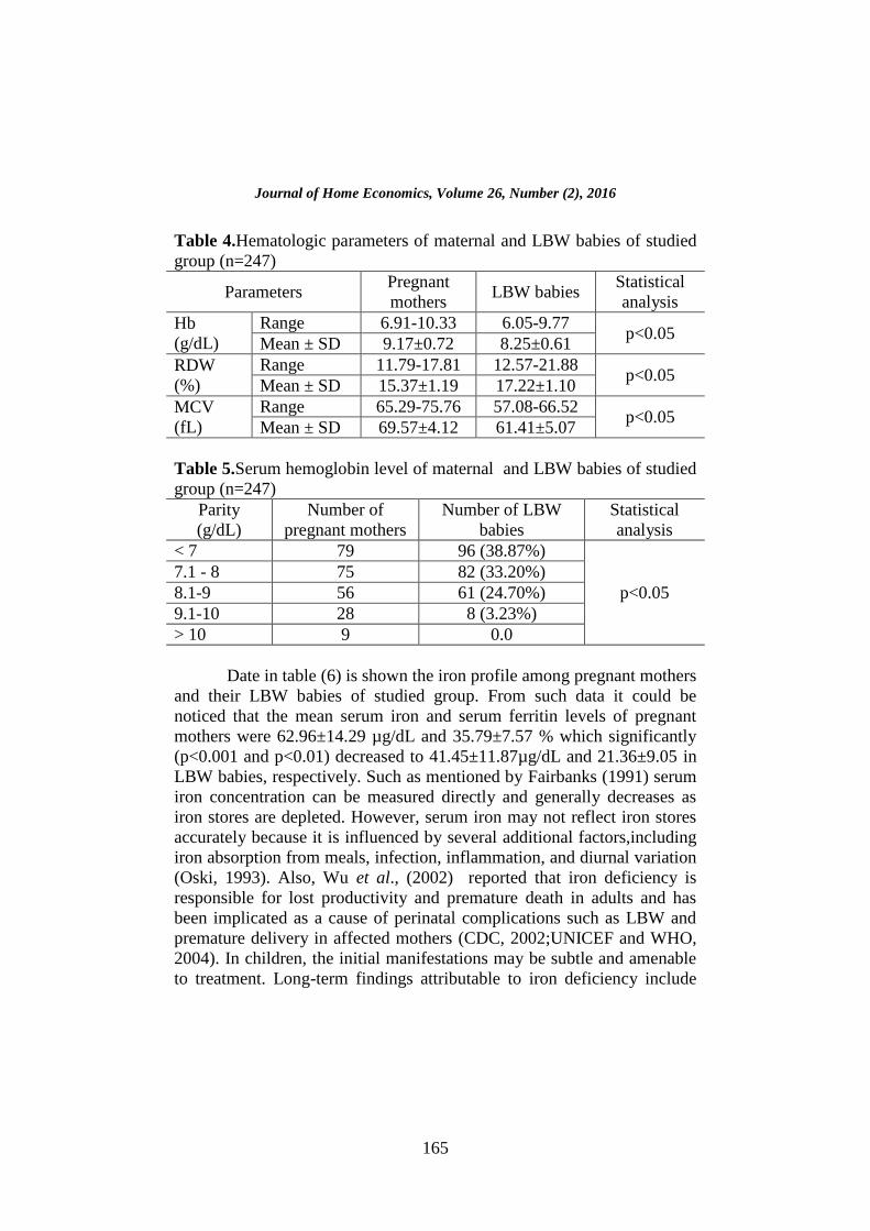
their LBW babies are shown in Tables (4-5). From such data it could be
noticed that the mean hemoglobin (Hb) level and mean corpuscular
volume (MCV) of pregnant mothers were 9.17±0.72 g/dL and 69.57±4.12
fL which significantly decreased to 8.25±0.61 g/dL (p<0.05) and
61.41±5.07fL (p<0.01) in LBW babies, respectively. The opposite
direction was observed for the red blood cell distribution width (RDW)
which recorded 15.37±1.19% in pregnant women and significantly
(p<0.01) increased to 17.22±1.10% in LBW babies. Such as metioned by
Ann et al., (2002) measurement of Hgb, the concentration of oxygen
carrying protein, is a more sensitive and direct test for anemia, a
complication concomitance with the LBW public health problem, than
others tests such measurement of hematocrit (Hct), the percentage of
whole blood that is occupied by RBCs. Anemia generally is defined as
Hgb values below the 5th percentile in a healthy reference population:
less than 11.0 g/dL for infants 6 months to 2 years of age. Hgb
measurement is inexpensive, readily available test for anemia and is used
most commonly to screen for iron deficiency. MCV, the average volume
of RBCs, is reported in automated analyses, but it also can be calculated
as the ratio of Hct to RBC count. MCV is useful for categorizing anemia
as microcytic, normocytic, and macrocytic. Also, RDW measures
variations in the size of RBCs and increases with iron deficiency. In one
study of adults, high RDW (>15%) was 71% to 100% sensitive and 50%
specific in diagnosing iron deficiency. Another study of 12-month- old
infants found that high RDW (>14%) was 100% sensitive and 82%
specific. Because of its relatively low specificity, RDW is not as useful
alone as a screening test, but it is used frequently in conjunction with
MCV to differentiate among various causes of anemia. For example,
RDW is high in iron deficiency anemia (IDA), but low in thalassemia
minor (Booth and Aukett, 1997 and Ann et al.,2002). Also, data in Table
(5) indicated that mothers whose Hb was below 7 g/dL were associated
with the highest percentage of LBW (38.87%), while with increasing
maternal Hb level, LBW incidence decreased. This is also in conformity
with results from studies by Bhatia et al., (1981), Chadhaet al., (1992)
andAmosu and Degun (2014).
Journal of Home Economics, Volume 26, Number (2), 2016
165
Table 4.Hematologic parameters of maternal and LBW babies of studied
group (n=247)
Parameters Pregnant
mothers LBW babies
Statistical
analysis
Hb
(g/dL)
Range 6.91-10.33 6.05-9.77 p<0.05
Mean ± SD 9.17±0.72 8.25±0.61
RDW
(%)
Range 11.79-17.81 12.57-21.88 p<0.05
Mean ± SD 15.37±1.19 17.22±1.10
MCV
(fL)
Range 65.29-75.76 57.08-66.52 p<0.05
Mean ± SD 69.57±4.12 61.41±5.07
Table 5.Serum hemoglobin level of maternal and LBW babies of studied
group (n=247)
Parity
(g/dL)
Number of
pregnant mothers
Number of LBW
babies
Statistical
analysis
< 7 79 96 (38.87%)
p<0.05
7.1 - 8 75 82 (33.20%)
8.1-9 56 61 (24.70%)
9.1-10 28 8 (3.23%)
> 10 9 0.0
Date in table (6) is shown the iron profile among pregnant mothers
and their LBW babies of studied group. From such data it could be
noticed that the mean serum iron and serum ferritin levels of pregnant
mothers were 62.96±14.29 µg/dL and 35.79±7.57 % which significantly
(p<0.001 and p<0.01) decreased to 41.45±11.87µg/dL and 21.36±9.05 in
LBW babies, respectively. Such as mentioned by Fairbanks (1991) serum
iron concentration can be measured directly and generally decreases as
iron stores are depleted. However, serum iron may not reflect iron stores
accurately because it is influenced by several additional factors,including
iron absorption from meals, infection, inflammation, and diurnal variation
(Oski, 1993). Also, Wu et al., (2002) reported that iron deficiency is
responsible for lost productivity and premature death in adults and has
been implicated as a cause of perinatal complications such as LBW and
premature delivery in affected mothers (CDC, 2002;UNICEF and WHO,
2004). In children, the initial manifestations may be subtle and amenable
to treatment. Long-term findings attributable to iron deficiency include
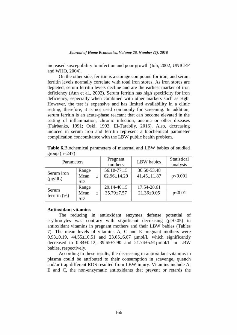
Journal of Home Economics, Volume 26, Number (2), 2016
166
increased susceptibility to infection and poor growth (Ioli, 2002, UNICEF
and WHO, 2004).
On the other side, ferritin is a storage compound for iron, and serum
ferritin levels normally correlate with total iron stores. As iron stores are
depleted, serum ferritin levels decline and are the earliest marker of iron
deficiency (Ann et al., 2002). Serum ferritin has high specificity for iron
deficiency, especially when combined with other markers such as Hgb.
However, the test is expensive and has limited availability in a clinic
setting; therefore, it is not used commonly for screening. In addition,
serum ferritin is an acute-phase reactant that can become elevated in the
setting of inflammation, chronic infection, anemia or other diseases
(Fairbanks, 1991; Oski, 1993; El-Tarabily, 2016). Also, decreasing
induced in serum iron and ferritin represent a biochemical parameter
complication concomitance with the LBW public health problem.
Table 6.Biochemical parameters of maternal and LBW babies of studied
group (n=247)
Parameters Pregnant
mothers LBW babies
Statistical
analysis
Serum iron
(µg/dL)
Range 56.10-77.15 36.50-53.48
p<0.001 Mean ±
SD
62.96±14.29 41.45±11.87
Serum
ferritin (%)
Range 29.14-40.15 17.54-28.61
p<0.01 Mean ±
SD
35.79±7.57 21.36±9.05
Antioxidant vitamins
The reducing in antioxidant enzymes defense potential of
erythrocytes was contrary with significant decreasing (p>0.05) in
antioxidant vitamins in pregnant mothers and their LBW babies (Tables
7). The mean levels of vitamins A, C and E pregnant mothers were
0.93±0.19, 44.55±10.51 and 23.05±6.07 µmol/L which significantly
decreased to 0.84±0.12, 39.65±7.90 and 21.74±5.91µmol/L in LBW
babies, respectively.
According to these results, the decreasing in antioxidant vitamins in
plasma could be attributed to their consumption in scavenge, quench
and/or trap different ROS resulted from LBW injury. Vitamins include A,
E and C, the non-enzymatic antioxidants that prevent or retards the

Journal of Home Economics, Volume 26, Number (2), 2016
167
oxidation of sensitive molecules found in the body. Vitamin E is
considered as primarily intracellular antioxidants associated with cell
membranes (krinskey, 1992). It is family of substance with different
degrees of unsaturation e.g. tocopherols and tacotrienols, and of
methylation e.g. mono, di-and trimethyanalog (Packer, 1992). α-
tocopherol is the form of vitamin E determined to be biologically most
active. Vitamin E is a potent peroxyl radical scavenger (Burton et al.,
1986) and can protect polyunsaturated fatty acids (PUFA) within
phospholipids of biological membranes and in plasma lipoproteins
(Jialalet al., 1995). β-carotene i.e. precursor of vitamin A and other
carotenoids belong to the large family of conjugated polyenes.
Carotenoids are bleached when exposed to radicals such as those that arise
during lipid peroxidation, which indicates that these pigments; must also
intercept active oxygen species. Their long, conjugated double bond
systems make them excellent substrates for radical attack (Kennedy et al.,
1991). They have antioxidant activity through its property as singlet
oxygen (1 O2) quenchers and their ability to trap peroxyl radicals
(Truscott, 1990; Stahl and Sies 1993). They are also able to inhibit free
radical reactions (PalozzaandKrinsky, (1992). Vitamin C is an important
antioxidant. Its water solubility allows it to be widely available in both the
extracellular and intracellular spaces in most biological systems (Halliwell
and Gutteridge 1990). Antioxidant roles of ascorbic acid can be'
summarized in the following: scavenge O2.- and OH
. with the formation
of the sernidehydro-ascorbate free radical that is subsequently reduced by
GSH to generate dehydroascorbate and GSSG, as most cells contain a
GSH-dependent dehydroascorbatereductase that generates ascorbate and
GSSG (Anderson et al.,1988), scavenges water-soluble peroxyl (RO2)
radicals (Frei, 1991), Scavenges thiyl and sulphenyl radicals, powerful
scavenger and quencher of single O2 in aqueous solution (Halliwell and
Gutterige 1990), “Repairs” and so prevents damage by, radicals arising by
attack of OH upon uric acid, inhibits lipid peroxidation by hemoglobin or
myoglobin H2O2 mixtures and prevents heme breakdown to release iron
ions by being preferentially oxidized by ferrylprotiens
(Halliwell&Gutteridge 1990), reduces - tocopheryl radicals in
membranes back to the lipid-soluble chain-breaking antioxidant -
tocopherol(Slater, 1984), reduces nitroxide radicals, e.g. the radicals
formed by attack of O2 or OH upon desferrioxamine (Hoffman and
Garewell 1995), it also protects plasma lipids against peroxidation induced

Journal of Home Economics, Volume 26, Number (2), 2016
168
by activated neutrophils (Frei. 1991), and protects against oxidants present
in cigarette smoke. (Hialliwell and Qutteridge 1990). The antioxidant
vitamins levels may be also important indicators of the adverse effects
caused by LBW public health problem.
Table 7.Antioxidant vitamins level of maternal and LBW babies of
studied group (n=247)
Parameters Pregnant mothers LBW babies Statistical
analysis
Vit A
(µmol/L)
Range 0.81-1.24 0.73-0.95 p<0.05
Mean ± SD 0.93±0.19 0.84±0.12
Vit C
(µmol/L)
Range 39.16-48.95 36.66-44.06 p<0.05
Mean ± SD 44.55±10.51 39.65±7.90
Vit E
(µmol/L)
Range 20.55-27.41 19.05-24.56 p<0.05
Mean ± SD 23.05±6.07 21.74±5.91
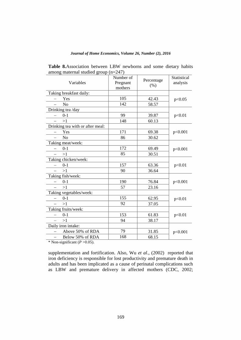
Association between LBW newborns and dietary habits among
maternal studied group
Association between LBW newborns and dietary habits among
maternal studied group are shown in Table (8). Data shows associations
between maternal feeding habits and LBW based on daily breakfast
taking (p<0.05), drinking tea (p<0.01), drinking tea with or after meal
(p<0.05), taking meat (p<0.001), chicken (p<0.01)and fish (p<0.001),
taking vegetables and fruits (p<0.01) and daily iron intake (p<0.001).
The present data are in accordance with that obtained by Amany and
Samaa (2012) and El-Tarabily, (2016). They reported that associations
between feeding habits and IDA, a complication concomitance with the
LBW public health problem, based on daily breakfast taking,
consumption of tea after meals and adequacy of iron intake. Infants who
were consuming iron containing foods below 50% of RDA of iron were
significantly associated with IDA (P = 0.027).
In similar study, Sumithra (2009) reviewed that micronutrient
deficiencies during pregnancy have been shown to have serious
implications on the developing foetus. Nearly half the pregnant women
still suffer from varying degree of anaemia, with the highest prevalence in
India, which also has the highest number of maternal deaths in the Asian
region. Of specific concern is compliance with iron supplementation,
cultural beliefs regarding diet in pregnancy, and the issue of nutrition
Journal of Home Economics, Volume 26, Number (2), 2016
169
Table 8.Association between LBW newborns and some dietary habits
among maternal studied group (n=247)
Variables
Number of
Pregnant
mothers
Percentage
(%)
Statistical
analysis
Taking breakfast daily:
p<0.05
Yes 105 42.43
No 142 58.57
Drinking tea /day
p<0.01 0-1 99 39.87
>1 148 60.13
Drinking tea with or after meal:
p<0.001 Yes 171 69.38
No 86 30.62
Taking meat/week:
p<0.001
0-1 172 69.49
>1 85 30.51
Taking chicken/week:
p<0.01 0-1 157 63.36
>1 90 36.64
Taking fish/week:
p<0.001 0-1 190 76.84
>1 57 23.16
Taking vegetables/week:
p<0.01
0-1 155 62.95
>1 92 37.05
Taking fruits/week:
p<0.01 0-1 153 61.83
>1 94 38.17
Daily iron intake:
p<0.001
Above 50% of RDA 79 31.85
Below 50% of RDA 168 68.15 * Non-significant (P >0.05).
supplementation and fortification. Also, Wu et al., (2002) reported that
iron deficiency is responsible for lost productivity and premature death in
adults and has been implicated as a cause of perinatal complications such
as LBW and premature delivery in affected mothers (CDC, 2002;
Journal of Home Economics, Volume 26, Number (2), 2016
170
UNICEF and WHO, 2004). In children, the initial manifestations may be
subtle and amenable to treatment. Long-term findings attributable to iron
deficiency include increased susceptibility to infection and poor growth
(Ioli, 2002, UNICEF and WHO, 2004).
In developed countries IDA is more commonly due to insufficient
iron intake. Iron deficiency anemia (anemia resulting from lack of
adequate iron to meet needs for red blood cell formation) affects 3%
children under 2 years of age, up to 3% of adolescent females and less
than 1% of adolescent males (Tender and Chang, 2002). Reduction of
iron deficiency and anemia in these vulnerable populations remains a
national health objective for 2010 (CDC, 2002). Women in developing
countries are always in a state of precarious iron balance during their
reproductive years. Their iron stores are not well developed because of
poor nutritional intake, recurrent infections, menstrual blood loss and
repeated pregnancies (Brabinet al., 2001; El-Tarabily, 2016).
Iron deficiency is the most commonly recognized nutritional
deficiency in both the developed and the developing world (WHO, 2014;
El-Tarabily, 2016; Elhassaneenet al., 2016). It is estimated that < 50 per
cent of women do not have adequate iron stores for pregnancy.
Requirements for absorbed iron increase during pregnancy from 0.8
mg/day in the first trimester to 7.5 mg/day in the third trimester (RDA,
1989). Average requirement during the entire gestation is approximately
4.4 mg/ day. An adequate iron balance during pregnancy implies body
iron reserves of >500 mg at conception. The physiologic iron
requirements in the second half of gestation cannot be fulfilled solely
through dietary iron (Milman, 2006). Data of the present study indicated
that IDA has been shown to be associated with LBW and preterm
delivery (See table 8). In this direction, Zhou et al., (1998) suggested that
the effect of maternal anaemia on preterm delivery was the most
detectable during the 1st trimester, before maternal plasma volume
expanded.
The mechanisms that operate by which poor iron status may affect
birth weight and preterm births are still incompletely understood. Some of
the mechanisms are hypotheses by Rasmussen (2001), Sloan et al.,
(2002), Cogswellet al., (2003) and Sumithra, (2009), El-Tarabily, (2016),
Elhassaneenet al., (2016) which could be summarized as follow: 1) iron
deficiency may affect immune function adversely and thus increase the
host susceptibility to genital tract infections, 2) iron deficiency may
Journal of Home Economics, Volume 26, Number (2), 2016
171
increase the stress hormones norepinephrine and cortisol, 3) low
haemoglobin concentrations i.e. iron deficiency may cause chronic
hypoxia, which can activate the body‟s stress response and thus increase
circulating levels of corticotrophin releasing hormone, 4) iron deficiency
may increase the oxidative stress of the placenta, blood serum and RBC's,
and 5) iron deficiency may decrease the enzymatic and non-enzymatic
antioxidant defense system in RBC's.
Conclusion
This study has established that maternal nutritional status impacted
significantly on newborn birth weight, as poorly nourished mothers were
observed to produce a higher percentage of LBW babies in comparison to
those who were better nourished. The challenge of addressing the
problem therefore remains an urgent imperative for development. Also,
antioxidant vitamins and levels of hematological and biochemical
parameters may be also important indicators of the adverse effects caused
by LBW public health problem. Finally, some of the mechanisms that
operate by which maternal malnutrition status may affect birth weight and
preterm births were proposed/explained in the present study.
References Amany, M. and Samaa, S. (2012). Prevalence and Risk Factors of Anemia
among a Sample of Pregnant Females Attending Primary Health Care
Centers in Makkah, Saudi Arabia. Pakistan Journal of Nutrition, 11 (12):
1113-1120.
Amosu, A. M. and Degun, A. M. (2014). Impact of maternal nutrition on birth
weight of babies. Biomedical Research 2014; 25(1): 75-78.
Ann, W.; Leann, L. and Henry, B. (2002). Screening for Iron Deficiency.
Pediatrics in Review, 23(5): 171-178.
Arnaud, C. and Vincent, O. (2007). Mother‟s education and birth weight.
IZA (Institute zurZukunft der Arbeit) Discussion Paper No. 2640, Bonn,
Germany.
Ashworth, A. (1998). Effects of intrauterine growth retardation on mortality and
morbidity in infants and young children.Eur J ClinNutr. 52 (Suppl 1): S34-
S42.
Beilly, J. S. and Kurkland, I. I. (1945).The relationship of maternal weight
gain and weight of newborn infants. Am.J. Obstet. Gynaecol. 50: 202-206.
Journal of Home Economics, Volume 26, Number (2), 2016
172
Bermejo, F. and García-López, S. (2009). A guide to diagnosis of iron
deficiency and iron deficiency anemia in digestive diseases.World J
Gastroenterol. 15:4638-43.
Bhatia, B. D.; Sur, A. M. and Tyagi, N. K. (1981). LBW babies in relation to
nutritional status and primipara. Indian J Paed . 27 (23): 507.
Booth, I. W. and Aukett, M. A. (1997). Iron deficiency anemia in infancy
and early childhood.Arch Dis Child. 76:549–554.
Carlos, A. N. and Marilia, B. G. (2013). Low birth weight: causes and
consequences. Negrato and Gomes Diabetology& Metabolic Syndrome
2013, 5:49
CDC. (Centers for Disease Control). (2002). MMWR Weekly: Iron deficiency-
United States, 1999-2000.
Chadha, V. K.; Bachani, D.; Chawla, S. C. and Bansal, R. D. (1992).
Nutritional status of urban poor mothers and birth weight.J ObstGynae. 46
(6): 278-292.
Christopher, J. and Siobhan, J. (2014). Low Birth Weight, Review of risk
factors and interventions: Summary Report. NHS Wales and Public Health
Wales, UK, pp.1-12.
Clausson, B.; Cnattingius, S. and Axelsson, O. (1998). Preterm and term
births of small for gestational age infants: a population-base study of risk
factors among.
Cogswell, M. E.; Parvanta, I.; Ickes, L.; Yip, R. andBrittenham, G. M.
(2003). Iron supplementation during pregnancy, anemia, and birth weight: a
randomised controlled trial. Am J ClinNutr .78 : 773-81.
Davies, P. A. and Stewart, A. L. (1995). Low Birth Weight infants:
Neurological sequence and later intelligence. Br. Med. Bull. 31, 85-91.
Elhassaneen , Y., Safaa Al-Wasef; RyeaanSayed; NaglaaFathy and Heba El-
Tarabily (2016). Prevalence of Iron-Deficiency Anemia in Infants and
Young Children (0 –6 Years of Age) of Maternal and Child Care Centers,
Port Said Governorate, Egypt. 4th International-18th Arab Conference of
Home Economics "Home Economics and Development Issues" 5-6 April,
2016, Faculty of Home Economics. Minoufiya University, Egypt. Journal
of Home Economics (Special issue), 26 (2): 73-86. [http:// homeEcon.
menofia. edu.eg] [ISSN 1110-2578].
El-Tarabily, H. (2016). The prevalence of iron deficiency anemia among
preschool children attending maternity child care (MCH) in port said
governorate. M.Sc. Thesis, Faculty of Specific Education, Port Saied
University, Port Saied, Egypt.
Epler, K. S. and Zeigler, R. G. ( 1993). Liquid chromatographic method for the
determination of carotenoids, retinoids and tocopherols in human serum and
in food. J Chromatog .619:37–48.

Journal of Home Economics, Volume 26, Number (2), 2016
173
Fahmy, S.and El-Sherbini, A. ( 1983). Determining simple parameters for
social classifications for health research. Bull High Inst Public
Health .13:95-108.
Fairbanks, V. F. (1991). Laboratory testing for iron status. HospPract. 26S:17–
24.
Ferguson, A. C. (1998). Prolonged Impairment of Cellular Immunity in
Children with intrauterine growth retardation. J. Paediatr. 93, 52-56.
Frei, B. (1991). Ascorbic acid protects lipids in human plasma and low density
lipoprotein against oxidative damage. Am. J. Clin. Nutr. 54: 1113S-8S.
Halliwell, B. and Gutteridge, J. M. (1990). The antioxidants of human
extracellular fluids. Arch. Biochem. Biophys. 280:1-8.
Heffner, J. E. and Repine, J. E. (1989). Pulmonary strategies of antioxidant
defence. Am. J. Resplr. Dis. 140:531-54.
Ioli, J. G. (2002). Anemia. In J.A. Fox (Ed.),Primary health care of infants,
children, and Adolescents(2nd ed.) St. Louis: Mosby. 471-480.
Jancevska, A.; Tasic, V.; Damcevski, N.; Danilovski, D.; Jovanovska, V.
and Gucev, Z. (2012).Children born small for gestational age (SGA).
Prilozi.33(2):47–58.
Javier, V.; Trinidad, S.; Romana, A.; Margarita, J.; Mar a, E .; David, M.
and Vicente, D. (2004). Risk factors for low birth weight: a review.
European Journal of Obstetrics & Gynecology and Reproductive Biology,
116 (1): 3–15
Jelliffe, D. B. (1966). The assessment of nutritional status of the community.
WHO Monogr. Ser. No 53. Geneva.
Joshi, H. S.; Srivastava, P. C.; Agnihotri, A. K.; Joshi, M. C.;
ChandraShalini, and Mahajan, V. (2007). Risk Factors for Low Birth
Weight (LBW) Babies and its Medico-Legal Significance. J Indian Acad
Forensic Med, 32(3) : 212-215.
Kennedy, T. A.andLiebler, D. C. (1991). Peroxyl radical oxidation of .beta.-
carotene: formation of .beta.-carotene epoxides Chem. Res-Toxicol.
4(3):290-295.
Kramer, M. S. (1987).„Determinants of Low Birth Weight: Methodological
assessment and meta-analysis‟, Bulletin of the World Health Organization,
65(5): 663–737.
Krinsky, N. 1.(1992). Mechanism of action of biological
antioxidants.Proc.Sci.Exp. Biol. Med. 200: 248.
Lawn, J. E.; Cousens, S. and Zupan, J. (2005). 4 million neonatal deaths:
when? Where? Why? Lancet. 365(9462):891–900.
Mavalankar, D. V.; Gray, R. H. and Trivedi, C. R. (1992). Risk factors for
preterm and term low birth weight in Ahmedabad, India. Int J
Epidemiol.21: 263-72.

Journal of Home Economics, Volume 26, Number (2), 2016
174
McCormick, M. C. (1985). The contribution of low birth weight to infant
mortality and childhood morbidity.New England Journal of Medicine.
312:82–90.
McIntire, D. D.; Bloom, S. L.; Casey, B. M. and Leveno, K. J. (1999). Birth
weight in a relation to morbidity and mortality among newborn infants. N
Engl J Med.340:1234–1238.
Milman, N. (2006). Iron and pregnancy - a delicate balance. Ann Hematol. 85 :
559-65.
Moeslinger, T.; Brunner, M. and Spieckermann, G. (1994).
Spectrophotometric determination of dehydroascorbic acid in biological
samples.Anal Biochem. 221:290–6.
Naeye, R. L.; Tafari, N.; Judge, D.; Gilmour, D. andMalboe, C. (1997).
Amniotic fluid infections in an African city. Pediatrics, 99: 965-972.
Oski, F. (1993). Iron deficiency in infancy and childhood. N Engl J Med.
329:190–193.
Osrin, D.and de- L. Costello, A. M. (2000). Maternal nutrition and fetal
growth: practical issues in International health. Seminars in Neonatology,
5:209-19.
Packer, L.(1992). Interaction among antioxidants in health and disease; Vit.E
and its redox cycle. Proc. Soc. Exp. Biol. Med. 200:271.
Palozza, P. and Krinsky N.I. (1992). Antioxidants effects of carotenoids in vivo
and in vitro: An overview. Methods Enzymol.1992; 213: 403-420.
Rasmussen, K. M. (2001). Is there a causal relationship between iron deficiency
or iron-deficiency anemia and weight at birth, length of gestation and
perinatal mortality? J Nutr 2001; 131 : 590S-603S.
RDA. (1989). Recommended Dietary Allowances, Food and Nutrition Board,
National Academy of Series, National Research Council, U.S.A.
Schlievert, P.; Johnson, W. and Galask, R. P. (1976): Bacterial growth
inhibition by amniotic fluid. Evidence for a zinc- peptide antibacterial
system. Am. J. Obstet .Gynaecol. 125: 900-910.
Singh, K.; Sundarro, K.; Tinkerame, J.; Kaluwin, C. and Matsuoka, T.
(1991). Lipid content fatty acid anid mineral composition of Mud Crabs
(Seyllaserrata) from Papua new Guinea. Journal of Food Composition and
Analysis, 4 (3): 276 – 280.
Sloan, N. L.; Jordan, E. andWinikoff, B. (2002). Effects of iron
supplementation on maternal hematologic status in pregnancy. Am J Public
Health 2002; 92 : 288-93.
Stahl, W. and H. Sies, (1993).Physical quenching of singlet oxygen and cis-
trans isomerization of carotenoids.Ann.N.Y. Acad. Sci. 691: 10-19.
Sumithra, M. (2009). Maternal nutrition & low birth weight - what is really
important? Indian J Med Res 130, November 2009, pp 600-608.

Journal of Home Economics, Volume 26, Number (2), 2016
175
Tietz, N . W. (1999). Textbook of clinical chemistry, Carl A. Burtis, 3rd ed.,
WB Saunders, Philadelphia.
Villanova, P. A. (1994). Reference and selected procedures for the quantitative
determination of hemoglobin in blood: approved standards. 2nd ed.,
National Committee for Clinical Laboratory Standards.
WHO.(World Health Organization), (2014). Global targets 2025. To improve
maternal, infant and young child nutrition
(www.who.int/nutrition/topics/nutrition_globaltargets2025/en/,accesd 17
October 2014).
WHO. (2004). Technical Consultation, „Towards the development of a strategy
for promoting optimal fetal growth‟, Report of a meeting (draft), World
Health Organization, Geneva.
Wilcox, A. J. (2001). „On the importance – and the unimportance – of
birthweight‟, International Journal of Epidemiology, 30 (6): 1233–1241.
Wonke, B.; Modell, M.; Marlow, T.; Khan, M. and Modell, B. (2007).
Microcytosis, iron deficiency and thalassaemia in a multi-ethnic
community: A pilot study. Scand J Clin Lab Invest. 67:87-95.
Wu. A. C.; L. Lesperance, and H. Bernstein, (2002).Screening for iron
deficiency. Pediatrics in Rev., 23: 171-177.
Zhou, L. M.; Yang, W. W.; Hua, J. Z.; Deng, C. Q.; Tao, X. and Stolzfus,
R. J. (1998). Relation of hemoglobin measured at different times in
pregnancy to preterm birth and low birth weight in Shanghai, China. Am J
Epidemiol. 148: 998-1006.
Journal of Home Economics, Volume 26, Number (2), 2016
176
تأثيز تغذية الأمهات والحالة الإجتماعية والإقتصادية على وسن الأطفال فى
محافظة بىر سعيذ
يىسف الحساويه
1، صفاء الىصيف
2، وجلاء فتحى
2ريهام أبى سمزة ،
2
1 ، ، شب١ اى لس اخغذ٠ت ع الأطعت، و١ت الالخصاد اش ، جاعت اف١ت
2 لس
١ت اخزب١ت اع١ت، جاعت بر سع١د ، بر سع١د، صزالالخصاد اش، و
الملخص العزبى:
حعد شىت اا١د الص اس أحد اشاو اصح١ت اات ف أغب ادي اا١ت
با ف١ا صز، اخ حزحبظ إرحباطا ث١ما باعدي اعا ث الأجت ف حه ابدا. ذه واج
ذه حت زاست طزق حددة ع حه اظازة ا ٠خزحب ع١ا شاو. ان احاجت ا
حأث١ز حغذ٠ت الأاث احات الإجخاع١ت الإلخصاد٠ت ع صج ادراست احا١ت عزفت د
س الأطفاي ف حافظت بر سع١د. مد أشارث اخائج اخحص ع١ا أ عدي إخشار
ف ازاوش االعت ححج ادراست ٠خأثز ع٠ا بعز الأاث أثاء زحت اا١د الص اس
-اح ااطك اخ ٠مطا )ر٠ف أ حضز( احات الإجخاع١ت الإلخصاد٠ت )دخ الأسزة
سخ اخع١( غ١زا وذه اس اىخسب لأ أثاء فخزة اح اذ سج ف ذ
و١جزا(. وا سجج 11زا )اسخ اثا اص ب عا١ا و١ج 9337ادراست
( ب١ اس اىخسب أثاء اح سخ ١جب١ اد p< 0.05-0.001علالاث ع٠ت )
احصي ع اا١د احد٠د اف١خا١اث اضادة لأوسدة باس١ز لأاث احا عدي
اغذائ١ت لأاث أثاء فخزة اح حأث١زا وب١زا ع حه اعاداث حاتوا وا . الص اس
باخا اظازة اخ ساد عدا بخعزض حه الأاث سء اخغذ٠ت الأزاض اخعمت با.
٠بم ضزرة اا١د الص اس( س٠ادة عديفإ اخحد اخث ف عاجت حه اشىت )
حت خ١ت.
-ف١خا١اثا -حد٠دا -١جب١ا -اد٠جزاف١ت الإجخاع١ت اظزفالكلمات المفتاحية:
اعاداث اغذائ١ت.