
Tissue Train Culture System - Flexcell International …® INTERNATIONAL CORPORATION 1 ABSTRACT...
8
TECH REPORT 100: Tissue Train ® Culture System A method for culture and mechanical loading of cells in a 3D matrix Authors: Albert J. Banes, Ph.D., Jie Qi, Ph.D., David S. Anderson B.S., P.E., Melissa Maloney M.S., and Ruwan Sumanasinghe Ph.D. Document: Tissue Train Tech Report, Rev 5.0 05-12-17 Culturing Cells in a Mechanically Active Environment ™ Flexcell International Corporation 2730 Tucker Street, Suite 200 Burlington, NC 27215 800-728-3714 (919) 732-1591 FAX: (919) 732-5196 www.flexcellint.com COPYRIGHT © 2009 FLEXCELL INTERNATIONAL CORPORATION
Transcript of Tissue Train Culture System - Flexcell International …® INTERNATIONAL CORPORATION 1 ABSTRACT...

TECH REPORT
100:
Tissue Train® Culture System
A method for culture and mechanical loading of cells in a 3D matrix
Authors: Albert J. Banes, Ph.D., Jie Qi, Ph.D., David S. Anderson B.S., P.E., Melissa Maloney M.S., and Ruwan Sumanasinghe Ph.D.
Document: Tissue Train Tech Report, Rev 5.0
05-12-17
Culturing Cells in a Mechanically Active Environment™ Flexcell International Corporation 2730 Tucker Street, Suite 200 Burlington, NC 27215
800-728-3714 (919) 732-1591 FAX: (919) 732-5196 www.flexcellint.com
COPYRIGHT © 2009 FLEXCELL INTERNATIONAL CORPORATION

FLEXCELL® INTERNATIONAL CORPORATION
1
ABSTRACT Flexcell®’s Tissue Train® Culture System utilizes a novel method to culture and mechanically load cells in a three-dimensional (3D) hydrogel or cell-assembled matrix. Cells and matrix attach to the mesh-like anchors at the culture well periphery of the flexible bottomed Tissue Train® culture plate (Fig. 1, arrow).
Figure 1. Tissue Train® culture plate for 3D
culture of cell-matrix constructs. (A) A Trough Loader beneath the flexible membrane. (B) An Arctangle® loading post, which is used during uniaxial tension, beneath the culture well. (C)
Anchors (arrow) for attachment of cells and gel of a linear uniaxial construct.
To cast cell-populated gels, the Tissue Train® culture plate is placed on a gasketed baseplate atop Trough Loaders™, planar faced, cylindrical loading posts bearing a central trough (Fig. 1 upper left; Fig. 2 right). Each loading post has a trough with evacuation holes. A vacuum applied to the trough bottom will deform the membrane into the conformation of the trough, providing a space for delivery of cells and hydrogel (Fig. 3). The baseplate and culture plates are then transferred to a CO2 incubator at 37 °C, where the construct is held in position in the trough until polymerization of the cell-gel construct is complete. When vacuum is released, the
construct rises into the plane of the culture well. Three milliliters of growth media can then be added to each well, and the constructs cultured using standard techniques.
Figure 2. (Left) Arctangle® loading post used
when uniaxial tension is applied; (Right) Trough Loader used during preparation of a 3D cell-gel
matrix. Mechanical load may be applied using the Flexcell® Tension System (Fig. 4). Linear or trapezoidal shaped cell-gel constructs or cell-assembled matrices may be mechanically loaded uniaxially, using special Arctangle®
Loading Stations™ (Fig. 2). Whereas, circular shaped constructs can be loaded equibiaxially, using cylindrical Loading Stations™. A user can use the FlexSoft® program to regulate the % elongation, frequency and duration of mechanical load. BACKGROUND Cells form tissues in vitro by populating 3D matrices that are both structural and functional. These matrices have their own particular structures, material composition, and biomechanical properties. As a tissue develops, cells fabricate an extracellular matrix in a given geometry according to developmental pathway cues.
A
B
C

FLEXCELL® INTERNATIONAL CORPORATION
2
Figure 3. (Top view) A cell-gel construct in a Tissue Train® well attached to the anchor stems.
(Side view) The rubber membrane deforms downward to create a space for the gel, when
vacuum is applied via the vacuum holes. Mechanical deformation, which includes inside-out as well as outside-in signaling components, regulates one or more pathways. An inside-out pathway may involve cell contraction in response to a ligand, such as a growth factor, a cytokine or a hormone. An outside-in pathway may involve matrix deformation that is transmitted to the cell via a linkage to integrins, focal adhesion complexes (mechanosensory complex) and the cytoskeleton, cell adhesion molecules, ion channels or other membrane-linked mechano-detection systems (Banes et al., 1995, 2002).
Figure 4. Flexcell Tension System used to apply regulated strain to 3D cell seeded-gel constructs.
Cells require an appropriate regimen of mechanical deformation to maintain a set point of intrinsic strain. It is well accepted that immobilization of limbs, bed rest or a reduction in the intrinsic strain level in a tissue leads to bone mineral loss, tissue atrophy, weakness and, in general, a reduction in anabolic activity and an increase in catabolic activity. Physical activity results in anabolic effects including an increase in biomechanical strength and an increase in the intrinsic strain in a tissue. These processes are cell-driven. Thus, mechanical loading alters pathways in these cells. It is also well accepted that culturing cells in a dynamic strain environment, such as application of fluid shear stress, substrate tension or compressive load, alters the shape, metabolism and other properties of cells, especially primary cells. Adding dynamic strain as well as culturing cells in a 3D environment more closely simulates a native environment than a static 2D monolayer culture method. Flexcell®’s Tissue Train® Culture System was developed to provide researchers with a method to both culture cells in a 3D matrix while applying a dynamic strain to these constructs. Investigators can use the 3D culture system as a stand-alone device to create 3D geometries for cell culture in a matrix gel or
FLEXCELL® INTERNATIONAL CORPORATION
3
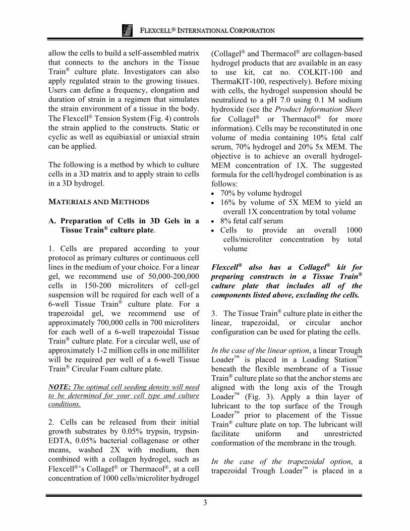
allow the cells to build a self-assembled matrix that connects to the anchors in the Tissue Train® culture plate. Investigators can also apply regulated strain to the growing tissues. Users can define a frequency, elongation and duration of strain in a regimen that simulates the strain environment of a tissue in the body. The Flexcell Tension System (Fig. 4) controls the strain applied to the constructs. Static or cyclic as well as equibiaxial or uniaxial strain can be applied. The following is a method by which to culture cells in a 3D matrix and to apply strain to cells in a 3D hydrogel. MATERIALS AND METHODS A. Preparation of Cells in 3D Gels in a
Tissue Train® culture plate. 1. Cells are prepared according to your protocol as primary cultures or continuous cell lines in the medium of your choice. For a linear gel, we recommend use of 50,000-200,000 cells in 150-200 microliters of cell-gel suspension will be required for each well of a 6-well Tissue Train® culture plate. For a trapezoidal gel, we recommend use of approximately 700,000 cells in 700 microliters for each well of a 6-well trapezoidal Tissue Train® culture plate. For a circular well, use of approximately 1-2 million cells in one milliliter will be required per well of a 6-well Tissue Train® Circular Foam culture plate. NOTE: The optimal cell seeding density will need to be determined for your cell type and culture conditions. 2. Cells can be released from their initial growth substrates by 0.05% trypsin, trypsin-EDTA, 0.05% bacterial collagenase or other means, washed 2X with medium, then combined with a collagen hydrogel, such as Flexcell’s Collagel or Thermacol, at a cell concentration of 1000 cells/microliter hydrogel
(Collagel and Thermacol are collagen-based hydrogel products that are available in an easy to use kit, cat no. COLKIT-100 and ThermaKIT-100, respectively). Before mixing with cells, the hydrogel suspension should be neutralized to a pH 7.0 using 0.1 M sodium hydroxide (see the Product Information Sheet for Collagel or Thermacol for more information). Cells may be reconstituted in one volume of media containing 10% fetal calf serum, 70% hydrogel and 20% 5x MEM. The objective is to achieve an overall hydrogel-MEM concentration of 1X. The suggested formula for the cell/hydrogel combination is as follows: 70% by volume hydrogel 16% by volume of 5X MEM to yield an
overall 1X concentration by total volume 8% fetal calf serum Cells to provide an overall 1000
cells/microliter concentration by total volume
Flexcell® also has a Collagel® kit for preparing constructs in a Tissue Train® culture plate that includes all of the components listed above, excluding the cells. 3. The Tissue Train® culture plate in either the linear, trapezoidal, or circular anchor configuration can be used for plating the cells. In the case of the linear option, a linear Trough Loader™ is placed in a Loading Station™ beneath the flexible membrane of a Tissue Train® culture plate so that the anchor stems are aligned with the long axis of the Trough Loader™ (Fig. 3). Apply a thin layer of lubricant to the top surface of the Trough Loader™ prior to placement of the Tissue Train® culture plate on top. The lubricant will facilitate uniform and unrestricted conformation of the membrane in the trough. In the case of the trapezoidal option, a trapezoidal Trough Loader™ is placed in a

FLEXCELL® INTERNATIONAL CORPORATION
4
Loading Station™ beneath the flexible membrane of a Tissue Train® culture plate so that the anchor stems are aligned with the long axis of the Trough Loader™ (Fig. 3). Apply a thin layer of lubricant to the top surface of the Trough Loader™ prior to placement of the Tissue Train® culture plate on top. The lubricant will facilitate uniform and unrestricted conformation of the membrane in the trough. In the case of the circular option, a Trough Loader™ and vacuum source are not needed. The foam annulus in the Tissue Train® Circular Foam culture plate serves as both the anchor and forms a well into which the cells and gel are cast. Skip to Step 5. 4. The Tissue Train® culture plate is placed on Flexcell®’s baseplate with gaskets. A baseplate is then connected to the Flexcell Tension System or other regulated vacuum source (NOTE: users should connect the baseplate directly to a regulated vacuum source to achieve the suggested vacuum level of -84 kPa). Vacuum is applied to the baseplate in a steady “hold” mode so that the flexible membrane is deformed and held in the void in the Trough Loader™. To supply the proper vacuum level with the FX-3000™ or FX-4000™ systems, it is recommended that a maximum of 23% elongation be used with the Tissue Train Loading Station™ (24 mm) baseplate setting. To supply the proper vacuum level with the FX-5000™ system, it is recommended that a maximum of 20% elongation be used with the Tissue Train Loading Station™ (24 mm) platform setting. These are the equivalent of -90 kPa. (See the Tissue Train User Manual for more information on loading constructs) Be sure that you allow enough vacuum tubing for your baseplate to reach from your incubator to your tissue culture hood. When using the Flexcell Tension System, use the minimal amount of tubing required, as longer tubing
decreases cyclic strain performance. It is most practical to use two sets of tubing to supply vacuum to the Tissue Train® baseplate. One set should be used for 3D gel formation and must be long enough to span the distance from vacuum source and both the cell culture hood and the incubator. The second vacuum line set can be dedicated to the cyclic strain process and should be routed through the incubator portal. Such an arrangement optimizes the cyclic strain process and minimizes potential sources of contamination. 5. For linear and trapezoidal gels, the cell and matrix protein gel fluid is pipetted into the deformed space in each Tissue Train® well. First pipette a small drop of gel at each end of the trough, under the anchor stems. Then press the anchor stems into the trough and pipette over the top of them. Finally, fill the middle of the trough with gel, moving the pipette back and forth to create a uniform strip of gel in the well (see a video on Flexcell®’s web site, www.flexcellint.com). For circular gels, pipette the cell and matrix protein gel solution into the space in the center of the foam anchor and around the inner perimeter of the annulus anchor (approximately 1.5 mm wide). Use the pipette tip to compress the edge of the foam so that the cell and matrix protein gel solution is drawn into it. This step ensures that the matrix and cells will integrate with the structure of the foam and form a secure bond to support the artificial tissue disk during mechanical loading. 6. The baseplate is placed in a 37 °C incubator. Gels are allowed to set at 37 °C. Please see your product information sheets for time needed for constructs to polymerize. After polymerization, the vacuum is released, and 3 ml of serum-containing media are added to each well. The gels should appear as a linear band of gel attached at each anchor end in the Tissue Train® well (Fig. 5 & 6) or as a circular

FLEXCELL® INTERNATIONAL CORPORATION
5
sheet. Tissue Train® culture plates are then placed in a humidified CO2 incubator at 37 °C. The user may then observe cultures or assay for differentiated functions. Cultures may be mechanically loaded (see Part B) at any time after gelation of the matrix or after cells have self-assembled their own matrix. The elongation, frequency and duration of the strain applied to the constructs are determined by the investigator for his/her specific use or parameters.
Figure 5. 3D linear tissue constructs in a Tissue
Train® culture plate.
Figure 6. A bioartificial tendon (BAT) may be constructed using the linear anchor and stem
configuration. Cells consolidate the matrix and contract the gel over time (Garvin et al., 2003).
7. Spherical-shaped cells can be observed in the gel using an inverted phase contrast microscope directly after plating (Fig. 7). By day one, the cells will begin to attach and
spread into the matrix. Next, cells will form attachments to each other and intercommunicate. By days 3-5, cells will reorganize and contract the matrix into a band in linear constructs (Fig. 6). 8. The user can monitor cell shape, organization, migration, division, gene expression, protein expression /secretion, mediator secretion DNA and protein synthesis in the gels (see Fig. 8). Flexcell® ScanFlex™ with XyFlex™ is a device that allows users to monitor cell-driven compaction of hydrogels in vitro.
Tissue TrainTM 3D Culture System: Tendon Cells in Uniaxial Strain
Day 0
Day 1No Stretch
Day 1Stretch
Day 2Stretch
Figure 7. Cells in a 3D construct are initially rounded (day 0 white circle) then attach and
spread (day 1 circle). Application of 1% elongation increases cell spreading.
B. Application of Regulated Strain to Cells
in a 3D Gel using the Tissue Train® Culture System
1. Cell-matrix constructs can be mechanically loaded in the 3D matrix by using the Flexcell Tension System to apply a regimen of controlled elongation (strain), frequency or duration with added rest periods.

FLEXCELL® INTERNATIONAL CORPORATION
6
2. The regimen parameters must be tested beforehand to ensure that the cell-seeded constructs are not broken during strain. Usually, the cell matrix constructs can be stretched at 1-3% elongation for several minutes to several hours per day without matrix failure. In the case of cardiac myocytes, the myocyte-matrix construct can maintain its beat frequency for a week or more in the matrix. 3. With Flexcell®’s FX-5000™ system, regimens can be programmed with strains ranging from 020% and frequencies from 0.01 5.0 Hz. When using a Tissue Train® culture plate with Flexcell®’s FX-3000™ or FX-4000™ systems, regimens can be
programmed with strains ranging from 023% and frequencies from 0.15.0 Hz. (NOTE: Higher frequencies will limit the system’s ability to maintain higher strain ranges). When downloading regimens, choose the Tissue Train® Loading Station™ (24 mm) baseplate/platform setting. 4. When using Tissue Train® with the FX-2000™ system, use the provided vacuum vs. strain data for Tissue Train® plates. (NOTE: The FX-2000™ will not allow vacuum levels above -40 kPa and therefore can apply a maximum of 6% strain to the Tissue Train® construct).
Actin Stain in Stretched Tendon
5
43
21
1 Hz3.5% elongation8h/d, 2 daysPrinciple Strain
Figure 8. Tendon cells stretched in a 3D gel polymerize actin and align.
REFERENCES FOR TISSUE TRAIN® Garvin J, Qi J, Maloney M, Banes AJ. A novel system for engineering bioartifical tendons and application of mechanical load. Tissue Engineering Journal 9(5):967-979, 2003. Triantafillopoulos IK, Karas SG, Bowman KF, Yang X, Evans GA, Banes AJ. Anabolic steroid and load enhance matrix remodeling and biomechanical strength of bio-artificial
tendons. Abstract 49th Annual Meeting ORS, Feb 2-5, 2003, New Orleans, LA. Banes AJ, Qi J, Maloney M, Almekinders L, Bynum D. Bioartificial tendons: a model 3D system for testing tenocyte responses to drugs, cytokines and mechanical load ex vivo. American Academy of Orthopaedic Surgeons, Accepted 12, 2002. Garvin J, Baldwin N, Banes AJ. Novel culture system for the development of a

FLEXCELL® INTERNATIONAL CORPORATION
7
bioartifical tendon. Abstract 48th Annual Meeting, ORS, Feb 10-13, 2002, Dallas, TX. Triantafillopoulos IK, Banes AJ, Bowman KF, Maloney M, Garrett WE, Karas SG. Nandrolone decanoate and load increase remodeling and strength in human supraspinatus bioartificial tendons. Am J Sports Med 32(4):934-943, 2004. Guilak F, Jones WR, Ting-Beall HP, Lee GM. The deformation behavior and mechanical properties of chondrocytes in articular cartilage. Spine 24(23); 2475-2483, 1999. Banes AJ, Tsuzaki M, Yamamoto J, Fisher T, Brigman B, Miller L. Mechanoreception at the cellular level; the detection, interpretation, and the diversity of responses to mechanical signals. Biochem. & Cell Biology 73:349-365, 1995. Banes AJ. Mechanical strain and the mammalian cell, in Physical forces and the mammalian cell. Academic Press, ed., John Frangos, pg 81-123, 1993. Qi J, Chi L, Bynum D, Banes AJ. Gap junctions in IL-1β-mediated cell survival response to strain. J Appl Physiol 110(5):1425-1431, 2011. Epub 2011 Jan 6. Qi J, Chi L, Faber J, Koller B, Banes AJ. ATP reduces gel compaction in osteoblast-populated collagen gels. J Appl Physiol 102(3):1152-60, 2007. Qi J, Chi L, Maloney M, Yang X, Bynum D, Banes AJ. Interleukin-1 increases elasticity of human bioartificial tendons. Tissue Eng 12(10):2913-2925, 2006. Qi J, Fox AM, Alexopoulos LG, Chi L, Bynum D, Guilak F, Banes AJ. IL-
1decreases the elastic modulus of human tenocytes. J Appl Physiol 101(1):189-95, 2006. Qi J, Chi L, Wang J, Sumanasinghe R, Wall M, Tsuzaki M, Banes AJ. Modulation of collagen gel compaction by extracellular ATP is MAPK and NF-B pathways dependent. Exp Cell Res 315(11):1990-2000, 2009. Epub 2009 Feb 23. FLEXCELL PATENTS Flexcell International Corporation’s Tissue Train® Culture System is protected by one or more of the following United States or International Patents, with other patents pending: US Patent 4,789.601 US Patent 4,822,741 US Patent 4,839,280 US Patent 5,122,470 US Patent 5,518,909 US Patent 5,593,891 US Patent 6,037,141 US Patent 6,048,723 US Patent 6,218,178 US Patent 6,472,202 US Patent 6,586,235 Japanese Patent 25-28174 German Patent 3855631.6 United Kingdom Patent 0,365,536 Canadian Patent 2,204,862


