Production Bile Duct- Hyperplasia Gallstones … · Production of Bile Duct-Hyperplasia and...
13
Journal of Clinical Investigation Vol. 45, No. 8, 1966 Production of Bile Duct- Hyperplasia and Gallstones by Lithocholic Acid * ROBERT H. PALMER t AND ZDENEK HRUBAN (From the Departments of Medicine and Pathology, and the Argonne Cancer Research Hospital,4 University of Chicago, Chicago, Ill.) Lithocholic acid 1 is an important metabolite of cholesterol in man and other animals. Interest in its biological properties stems from its marked toxicity. It is the most potent of the naturally oc- curring steroids that produce intense fever and in- flammation in man (2, 3) and inflammation in a number of other species (3, 4). It is also one of the most active steroid hemolysins (5). In addi- tion, its oral administration produces cirrhosis of the liver in rabbits (6) and ductular cell hyper- plasia in a variety of species, including rodents (7), reptiles (8), and primates (9). In view of its physiological occurrence, its known toxic proper- ties, and its potential relevance to human disease, we have investigated the effects of feeding large amounts of lithocholic acid to rats. The resulting bile duct hyperplasia and choledocholithiasis are described in this report. Methods The acute effects of lithocholic acid and sodium litho- cholate on Sprague-Dawley rats were investigated in * Submitted for publication August 23, 1965; accepted April 21, 1966. Presented in part at the Annual Meeting of the Ameri- can Society for Clinical Investigation, Atlantic City, N. J., May 2, 1965. A preliminary report has been published (1). Supported in part by U. S. Public Health Service re- search grant CA-05310 from the National Cancer -In- stitute. t Address requests for reprints to Dr. Robert H. Palmer, Dept. of Medicine, University of Chicago, 950 E. 59th St., Chicago, Ill. 60637. t Operated by the University of Chicago for the U. S. Atomic Energy Commission. 1 Trivial names of bile acids used in this report are as follows: lithocholic acid, 3a-hydroxy-5p-cholanoic acid; 6P-hydroxylithocholic acid, 3a,6fi-dihydroxy-5p-cholanoic acid; hyodeoxycholic acid, 3a,6a-dihydroxy-5,f-cholanoic acid; chenodeoxycholic acid, 3a,7a-dihydroxy-5p-cholanoic acid; deoxycholic acid, 3a,12a-dihydroiy-SjS-cholanoic acid; cholic acid, 3a,7a,12a-trihydroxy-5f8-cholanoic acid. experiments I and II and chronic effects in experiments III and IV. Lithocholic acid was obtained commer- cially 2; no quantitatively significant bile acid contami- nants were observed when it was analyzed by thin layer chromatography [system S15 of Eneroth (10)]. The sodium salt was prepared by neutralization of the acid with sodium hydroxide. In the first experiment, 5 male rats, average weight 372 g, were force fed 300 mg sodium lithocholate per kg body weight per day in a liquid diet (11) containing 3.6 g casein hydrolysate per kg body weight per day. The rats were fed 3 times daily at 8-hour intervals and killed with ether on the sixth day. In the second experiment, 9 males and 9 females, av- erage weight 171 g, were divided into 3 equal groups and force fed the basic liquid diet as described above. One group served as controls. A second group was fed litho- cholic acid, 300 mg per kg body weight per day, in the diet. The third group received sodium lithocholate, 300 mg per kg body weight per day, in the diet All rats were killed with ether on the twelfth day. In the third experiment, 10 male and 10 female control rats (average weights: males, 193 g; females, 157 g) were fed ad libitum an 8% protein diet 8 containing 8% casein, 78% starch, 10% vegetable oil, and 4% salt mixture U.S.P. XIV, to which had been added 1 kg Vitamin Diet Fortification Mixture2 per 100 pounds diet. A similar group of rats (average weights: males, 193 g; females, 157 g) was fed the same diet containing 1% lithocholic acid. The powdered diets were mixed with sufficient wa- ter to permit molding into conveniently sized balls. Generally a 1- to 2-weeks' supply was made up and re- frigerated. A ball was placed in each cage every 2 to 4 days, depending on spillage. At the end of the fourth month the surviving animals were lklled by decapitation. In the fourth experiment, 5 control and 15 treated rats (150 to 200 g) were force fed the liquid diet of the first experiment for 1 month and then placed on the solid diet of the third experiment ad libitum. The treated animals received sodium lithocholate, 300 mg per kg per day in the liquid diet and 1% in the solid diet. After 4 months, the surviving animals were killed. At autopsy, livers were weighed and tissue samples fixed in buffered formalin and Carnoy's fluid. Sections were stained with hematoxylin-eosin, Mallory trichrome, periodic acid Schiff, and methyl green-pyronine. 2Nutritional Biochemicals Corp., Cleveland, Ohio. 8 "Low 8% Protein," Nutritional Biochemicals Corp. 1255
Transcript of Production Bile Duct- Hyperplasia Gallstones … · Production of Bile Duct-Hyperplasia and...
Journal of Clinical InvestigationVol. 45, No. 8, 1966
Production of Bile Duct- Hyperplasia and Gallstones byLithocholic Acid *
ROBERTH. PALMERt ANDZDENEKHRUBAN(From the Departments of Medicine and Pathology, and the Argonne Cancer Research
Hospital,4 University of Chicago, Chicago, Ill.)
Lithocholic acid 1 is an important metabolite ofcholesterol in man and other animals. Interest inits biological properties stems from its markedtoxicity. It is the most potent of the naturally oc-curring steroids that produce intense fever and in-flammation in man (2, 3) and inflammation in anumber of other species (3, 4). It is also one ofthe most active steroid hemolysins (5). In addi-tion, its oral administration produces cirrhosis ofthe liver in rabbits (6) and ductular cell hyper-plasia in a variety of species, including rodents(7), reptiles (8), and primates (9). In view of itsphysiological occurrence, its known toxic proper-ties, and its potential relevance to human disease,we have investigated the effects of feeding largeamounts of lithocholic acid to rats. The resultingbile duct hyperplasia and choledocholithiasis aredescribed in this report.
Methods
The acute effects of lithocholic acid and sodium litho-cholate on Sprague-Dawley rats were investigated in
* Submitted for publication August 23, 1965; acceptedApril 21, 1966.
Presented in part at the Annual Meeting of the Ameri-can Society for Clinical Investigation, Atlantic City, N. J.,May 2, 1965. A preliminary report has been published(1).
Supported in part by U. S. Public Health Service re-search grant CA-05310 from the National Cancer -In-stitute.
t Address requests for reprints to Dr. Robert H.Palmer, Dept. of Medicine, University of Chicago, 950 E.59th St., Chicago, Ill. 60637.
t Operated by the University of Chicago for the U. S.Atomic Energy Commission.
1 Trivial names of bile acids used in this report are asfollows: lithocholic acid, 3a-hydroxy-5p-cholanoic acid;6P-hydroxylithocholic acid, 3a,6fi-dihydroxy-5p-cholanoicacid; hyodeoxycholic acid, 3a,6a-dihydroxy-5,f-cholanoicacid; chenodeoxycholic acid, 3a,7a-dihydroxy-5p-cholanoicacid; deoxycholic acid, 3a,12a-dihydroiy-SjS-cholanoicacid; cholic acid, 3a,7a,12a-trihydroxy-5f8-cholanoic acid.
experiments I and II and chronic effects in experimentsIII and IV. Lithocholic acid was obtained commer-cially 2; no quantitatively significant bile acid contami-nants were observed when it was analyzed by thin layerchromatography [system S15 of Eneroth (10)]. Thesodium salt was prepared by neutralization of the acidwith sodium hydroxide.
In the first experiment, 5 male rats, average weight372 g, were force fed 300 mg sodium lithocholate per kgbody weight per day in a liquid diet (11) containing 3.6g casein hydrolysate per kg body weight per day. Therats were fed 3 times daily at 8-hour intervals and killedwith ether on the sixth day.
In the second experiment, 9 males and 9 females, av-erage weight 171 g, were divided into 3 equal groups andforce fed the basic liquid diet as described above. Onegroup served as controls. A second group was fed litho-cholic acid, 300 mg per kg body weight per day, in thediet. The third group received sodium lithocholate, 300mg per kg body weight per day, in the diet All ratswere killed with ether on the twelfth day.
In the third experiment, 10 male and 10 female controlrats (average weights: males, 193 g; females, 157 g) werefed ad libitum an 8% protein diet 8 containing 8% casein,78% starch, 10% vegetable oil, and 4% salt mixtureU.S.P. XIV, to which had been added 1 kg Vitamin DietFortification Mixture2 per 100 pounds diet. A similargroup of rats (average weights: males, 193 g; females,157 g) was fed the same diet containing 1% lithocholicacid. The powdered diets were mixed with sufficient wa-ter to permit molding into conveniently sized balls.Generally a 1- to 2-weeks' supply was made up and re-frigerated. A ball was placed in each cage every 2 to 4days, depending on spillage. At the end of the fourthmonth the surviving animals were lklled by decapitation.
In the fourth experiment, 5 control and 15 treated rats(150 to 200 g) were force fed the liquid diet of the firstexperiment for 1 month and then placed on the solid dietof the third experiment ad libitum. The treated animalsreceived sodium lithocholate, 300 mg per kg per day inthe liquid diet and 1% in the solid diet. After 4 months,the surviving animals were killed.
At autopsy, livers were weighed and tissue samplesfixed in buffered formalin and Carnoy's fluid. Sectionswere stained with hematoxylin-eosin, Mallory trichrome,periodic acid Schiff, and methyl green-pyronine.
2Nutritional Biochemicals Corp., Cleveland, Ohio.8 "Low 8% Protein," Nutritional Biochemicals Corp.
1255

ROBERTH. PALMERAND ZDENEKHRUBAN
TABLE I
Body and liver weights of rats fed lithocholic acid for 4 months(experiment III)
No. Inital Average weight p Average liver psurviving weight gain A SD value weight A SD value
g I gFemale controls 10 157 70.4 ± 12.6 .6.00 :1 0.71
<0.05 <0.025Female treated 8 157 55.0 ± 13.2 7.16 =t 1.13
Male controls 10 193 96.9 4± 13.8 9.18 4 1.70NS <0.01
Male treated 7 193 100.1 ± 21.7 11.87 i 1.67
For electron microscopic studies, rat liver samples fromexperiment I were fixed for 1 hour in 1%osmic acid buf-fered with 2,4,6-trimethylpyridine at pH 7.4, dehydratedin graded alcohols, and embedded in methacrylate. Threeextrahepatic duct calculi from experiment III were fixedin 2% osmic acid for 24 hours, dehydrated in absolute al-cohol for 12 hours, and embedded in methacrylate. Thesections were stained with lead hydroxide (12).
Gallstone analysis. Gallstones were stored in thefreezer under nitrogen before analysis. Stones from in-dividual animals were crushed and weighed. Samples of25 mg were dried at 1100 C for 24 hours to obtain theper cent dry weight and then ashed for mineral analysis.Calcium was measured with a Perkin-Elmer atomic ab-
FIG. 1. BILIARY TRACT OF RAT FED SODIUM LITHOCHO-LATE FOR 4 MONTHS (EXPERIMENT IV). The enlargedcommon bile duct (arrow) is shown with adherent fat.Liver, L; intestine, I. X 2.6.
sorption spectrophotometer and sodium and potassium byflame spectrophotometry. A second sample of 100 mg ofcrushed stones was homogenized, in a Waring blendor,in 25 ml CHCI.: MeOH, 1: 4, and portions of the sus-pension were used for bile acid, total lipid (13), andbilirubin (14) determinations. Cholesterol, in a thirdsample of 20 mg, was measured by the Tschugaeff re-action (15).
Qualitative bile acid analysis was performed by thinlayer chromatography, with the solvent systems describedby Ginshirt, Koss, and Morianz (16) and Eneroth (10),both before and after the sample was hydrolyzed in 5 NNaOH for 24 hours at 1300 C. Total bile acids werequantitated enzymatically with 3a- and 3fi-hydroxy-steroid dehydrogenases. The method (to be reported indetail elsewhere) is similar to that used by Hurlock andTalalay (17) for the analysis of neutral steroids, andapplied by Iwata and Yamasaki (18) to bile acids.
Isotopic studies. Sodium lithocholate-24-Y4C,' with aspecific activity of 1 mc per mg, was prepared accordingto the method of Bergstr6m, Rottenberg, and Voltz (19).Animals were housed in individual metabolism cages, andurine and feces were collected separately. After an in-traperitoneal injection of approximately 4 X 101 dpm ofsodium lithocholate-24-14C in 0.5 ml of 50% ethanol, sam-ples of urine and of CHC13: MeOHextracts of feces andintestinal contents were counted in 1,4-bis-2- (5-phenyl-oxazolyl)benzene (POPOP) with a Nuclear-Chicagoliquid scintillation counter with internal quench correc-tion. Labeled bile acids in intestinal contents were chro-matographed in a solvent system consisting of 50 ml bu-tanol, 5 ml acetic acid, and 5 ml water, as described above,and radioactivity was detected with a Vanguard 885 glassplate scanner. Standard spots of glycocholic and tauro-lithocholic acids were located by spraying with phospho-molybdic acid (20). The per cent of labeled compoundsconjugated with taurine was calculated by cutting out andweighing the areas under the appropriate curves on therecorder strip.
Miscellaneous. Urinary taurine was measured by themethod of Bergeret and Chatagner (21), except that theNinhydrin reaction was performed according to Rubin-stein and Pryce (22). Serum cholesterol was determinedby Sackett's modification of Bloor's method (23).
4Nichem, Inc., Bethesda, Md.
1256

LITHOCHOLIC ACID-INDUCED BILE DUCT PROLIFERATION AND GALLSTONES
Results
Morphological changes. In the acute experi-ments, the rats appeared to be normal, and nogross pathological changes were noted at autopsyexcept that one rat in the sodium lithocholategroup of experiment II had a small soft calculus inthe distal hepatic duct and some dilatation of theduct proximally. In the chronic experiments, thetreated rats lost hair and often assumed a hunchedposture. At the time of autopsy, as shown in Ta-ble I, treated female rats had not gained weight aswell as controls, but treated male rats had gainednormally. Liver weights in treated animals ofboth sexes were significantly increased over con-trols. In both chronic experiments, however, themost striking pathological feature was the presenceof large common duct calculi in all of the treatedrats (Figures 1 and 2). The stones were usuallymultiple, yellow or green, and soft and friable, butmarked variations in color, size, and shape wereapparent.
Livers of rats fed lithocholic acid or sodiumlithocholate in both acute and chronic experimentsshowed microscopic changes that were essentiallysimilar and limited to the biliary system. The por-
p*;. '9
' k
44
4z
I@,~~~~~~FIG. 3. PORTAL AREA OF LIVER FROMA RAT FED LITHO-
CHOLIC ACID FOR 4 MONTHS(EXPERIMENT III). Bile ductproliferation and pericholangitis are present. Some ductsare distended and contain amorphous material. H and Estain; x 85.
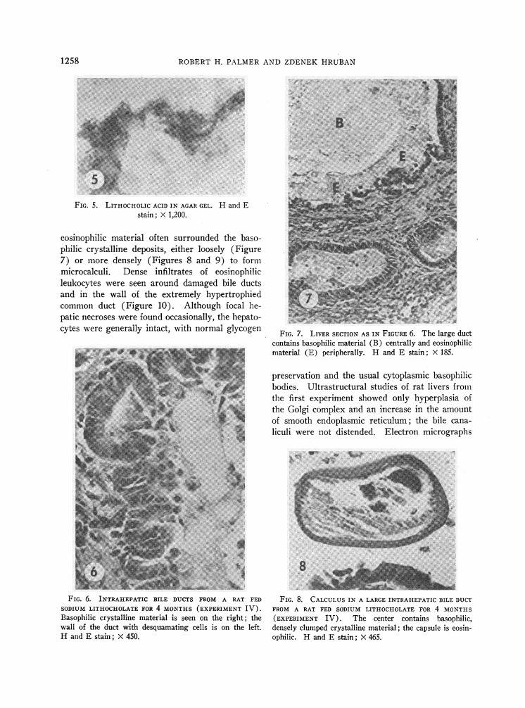
tal areas of the liver showed marked bile duct pro-liferation and variable mononuclear cell infiltration(Figure 3). Within bile ducts, basophilic crystal-line deposits (Figure 4) with staining charac-teristics similar to those of lithocholic acid em-1)edded in agar (Figure 5) were frequently ob-served. Ductal cells, some undergoing necrosis,were seen associated with eosinophilic debris in theltimina of the bile ducts (Figure 6). A similar
FIG. 2. RAT SHOWNIN FIGURE 1 AFTER OPENINGTHECOMMONDUCT. Calculi of various sizes are shown.
1257
----- ----- - ---- ---F 4lwa;
FIG. 4. CRYSTALLINE BASOPHILIC MATERIAL FROMTHE
CENTEROF A CALCULUS. H and E stain; X 1,200.
ROBERTH. PALMERAND ZDENEK
FIG. 5. LITHOCHOLIC ACID IN AGARGEL. H and Estain; X 1,200.
eosinophilic material often surrounded the baso-philic crystalline deposits, either loosely (Figure7) or more densely (Figures 8 and 9) to formmicrocalculi. Dense infiltrates of eosinophilicleukocytes were seen around damaged bile ductsand in the wall of the extremely hypertrophiedcommon duct (Figure 10). Although focal he-patic necroses were found occasionally, the hepato-cytes were generally intact, with normal glycogen
*'ii~ owsFw wy
F#--=w *fi- w~~~~~~~~~~~~~~~~~~~~~~~~~~~~~
V.- 5 9SRK
FIG. 6 INTRA EPATIC BIL S.........F....A.. R Fe
Basphiiccrysain maeil is see on. th r .:igt. the.....wall of th duc wit eqaaigcell is on th lef
H¢andE Fstain >( 450 .....................
FIG L>.6 .:. INR.E I BIL DUCT FROM A RAT FED
_pi ic crstalin maera is^ see on. th rigt h
and Estin X 50
,Y
FIG. 7. LIVER SECTION AS IN FIGURE 6. The large ductcontains basophilic material (B) centrally and eosinophilicmaterial (E) peripherally. H and E stain; X 185.
preservation and the usual cytoplasmic basophilicbodies. Ultrastructural studies of rat livers fromthe first experiment showed only hyperplasia ofthe Golgi complex and an increase in the amountof smooth endoplasmic reticulum; the bile cana-liculi were not distended. Electron micrographs
FIG. 8. CALCULUSIN A LARGE INTRAHEPATIC BILE DUCTFROM A RAT FED SODIUM LITHOCHOLATE FOR 4 MONTHS(EXPERIMENT IV). The center contains basophilic,densely clumped crystalline material; the capsule is eosin-ophilic. H and E stain; _ 465.
HRU13AN1258

LITHOCHOLIC ACID-INDUCED BILE DUCT PROLIFERATION AND GALLSTONES
FIG. 9. CALCULUS AS IN FIGURE 8. The denselypacked outer shell and the reticular inner layer are dis-cernible. H and E stain; X 1,200.
of the calculi revealed electron dense flaky materialin a lucid matrix, similar but not identical to theappearance of calcium lithocholate preparedsimilarly.
Composition of lithocholic acid-induced rat gall-stones. The calculi in experiment III had average
dry weights of 5.97 0.71 g for males and 4.141.75 g for females. Gallstones from 15 of the ratswere analyzed, with the results given in Table II.Eighty-five per cent of the dry weight was ac-
counted for by the analyzed constituents, primarilybile acids. Small amounts of nondiazo-reactingpigments, carbohydrate, and probably protein andnucleic acids were also present, but in the presence
of large amounts of bile acids, identification was
not reliable.The relative amounts of bile acids and cations
indicated that the bile acids occurred primarilyas calcium salts. The composition in mEq per g
dry weight (mean standard deviation) was as
follows: bile acids, 1.57 0.24; Ca, 1.59 0.23;Na, 0.32 0.18; K, 0.19 + 0.12; and Mg, 0.130.02.
Analysis of the bile acids in several stones bythin layer chromatography showed varying pro-
portions of free and glycine-conjugated com-
pounds; only traces of taurine-conjugated bileacids were present. Thin layer chromatography ofthe bile acid extract after alkaline hydrolysisshowed a consistent pattern from animal to ani-mal; the predominant steroids were lithocholic and3a,6f8-dihydroxy-5f3-cholanoic acids, with smalleramounts of hyodeoxycholic and cholic acids andtraces of chenodeoxycholic acid being detected.Quantitative fractionation of the bile acids in hy-drolyzed extracts of gallstones from four repre-
sentative animals is shown in Table III. Be-
FIG. 10. PORTION OF THE WALL OF THE COMMONDUCT
FROMA RAT FED LITHOCHOLIC ACID FOR 4 MONTHS(EX-PERIMENT III). Polymorphonuclear cells, predominantlyeosinophiles, infiltrate the wall. H and E stain; X 200.
tween 93 and 100% of the bile acids present inthe extract were recovered by eluting the com-
ponent spots, and there was good agreement be-tween replicate chromatographies. No bile acidswith 3,8-hydroxyl groups were detected.
Prevention of stone formation. Because of theremarkable paucity of taurine conjugates in a spe-
TABLE II
Composition of lithocholic acid-induced gallstones
Females Males
Wet weight i4 SD, g 5.05 A1.68* 7.39 40.24*Per cent water (range) 19.1 (5.5-54.6) 21.8 (9.4-34.8)
Dry weight ± SD, g 4.14a:1.75t 5.97 i 0.71tBile acids (calculated), % 71 69Ash, % 12 12
(Ca) (3.3 ) (3.0(Na) (0.77) (0.81)(Mg) (0.16) (0.15)(K) (0.07) (0.08)(P) (0.02) (0.02)
Total lipid, % 0.9 0.4(Cholesterol) (0.04) (0.04)
Bilirubin. % 0.12 0.17
*p <0.005.t <0.05.
1259
ROBERTH. PALMERAND ZDENEKHRUBAN
TABLE III
Analysis of bile acids in hydrolyzed gallstone extracts*
1 2 3 4
mjmoles % mumoles % mpmoles % mjmoles %Total bile acids 44.5 100.0 41.4 100.0 43.8 100.0 40.9 100.0
Lithocholic acid 12.2 15.7 12.3 12.313.3 29.1 15.9 38.6 10.7 26.3 11.6 29.013.6 11.3
6,B-Hydroxylithocholic acid 19.0 10.9 18.3 15.718.8 42.4 11.5 27.1 19.7 42.9 14.2 36.4
18.6 15.4Hyodeoxycholic acid 4.2 5.8 4.9 7.8
4.1 9.2 5.9 14.3 3.5 11.0 7.8 18.86.1
Cholic acid 3.6 6.5 5.4 4.65.2 11.9 4.5 14.0 4.9 11.6 6.1 13.87.0 6.4 6.4
Chenodeoxycholic acid 0 0 1.5 0 0 0 02.8 5.82.9
Total 92.6 99.8 91.8 98.0
* A 5-Ml sample of the hydrolyzed chloroform: methanol gallstone extract from each of 4 animals was assayed directlyto give the total bile acid value, designated 100%. Three other 5-Ml portions were chromatographed with phase systemS11 (10). Individual spots were eluted and assayed. The average value from 2 or 3 chromatographies was used todetermine the fractional bile acid composition.
cies normally conjugating bile acids predominantlywith taurine, we investigated the effects of proteinor taurine dietary supplements on stone formation.Four groups of 5 male and 5 female rats were
maintained on the diets listed in Table IV for 8weeks. The basic diet was the same as that usedin experiment III. As expected from the resultsof experiment III, none of the 8% protein con-
trol rats (group I) and all of the 8%o protein,lithocholic acid-treated rats (group II) developedlarge accumulations of common duct stones (av-
erage weight, 4.15 g). In contrast, in the rats re-
ceiving the protein-supplemented lithocholic aciddiet (group III), there were only 2 females witha few very small stones. In rats receiving the tau-rine-supplemented lithocholic acid diet (groupIV), there were no stones. Some rats in groups
III and IV, usually females, had thickened andslightly dilated common bile ducts, although no
obstruction was apparent. The bile was generallythick, and some precipitated sludge was observed.The extrahepatic ducts appeared to be normal in
TABLE IV
Modification of the effects of 8 weeks' treatment with lithocholic acid by dietary supplements
S-containingAverage amino acids Urinary
Group Diet weight gain in the diet taurine
g /umoles/g ,umoles/24 hours*diet
I 8%protein 44 14.3 15.9 i= 0.3
II 8%protein 30 14.3 17.6 i= 2.21 %lithocholic acid
III 27% protein 111 47.9 31.0 4= 8.41 %lithocholic acid
8%proteinIV 1% lithocholic acid 40 94.1 70.0 :i: 13.8
1 %taurine
* Mean + standard deviation.
1260

LITHOCHOLIC ACID-INDUCED BILE DUCTPROLIFERATION AND GALLSTONES
TABLE V
Effects of dietary alterations on the metabolism of lithocholic acid-24-14C injected intraperitoneally*
Labeled bile acids recoveredin intestinal contents
at autopsyIsotope
recovered Per centin urine conjugated Mean 4 standard
Group Animal Sex Day 1 with taurine deviation
dpmXlO4I 1 F 13.8 0
2 F 10.5 213 M 7.6 174 M 6.5 13
13 1 7.8II 5 F 9.3 7
6 F 10.7 227 M 6.9 98 M 8.1 6t
11 ± 6.6III 9 F 6.8 42
10 F 8.9 5711 M 5.2 1712 M 9.2 85
50 4 25IV 13 F 7.3 48
14 F 13.9 6715 M 4.0 40t16 M 5.2 96
63 ± 22
*4 X 106 dpm injected.t 13% free bile acids.t 8%free bile acids.
the remaining animals. Protein supplementationresulted in greatly increased growth, whereas tau-rine supplementation did not.
To investigate the metabolic changes associatedwith dietary prevention of stone formation, westudied several animals in each group before au-topsy. Urinary taurine excretion was determinedin 2 males and 2 females in each group, and theresults are shown in Table IV. The urinary tau-rine closely reflected the total sulfur-containingamino acid content (including taurine) of the diet.
Two different males and females in each groupwere given intraperitoneal injections of sodiumlithocholate-24-14C (4 x 10⁸ dpm) 3 days beforeautopsy. Urinary bile acid-14C excretion was de-termined, and at autopsy the small intestinal bileacid pool was analyzed for taurine-conjugatedlabeled bile acids. The results are shown in Ta-ble V. Urinary excretion of labeled bile acids wassimilar in all groups, but females excreted moreisotope in the urine on day 1 than did males(P < 0.01).
Analysis of labeled bile acids in the intestinalcontents revealed significant enhancement of tau-rine conjugation in groups III and IV (p < 0.01).
The proportion of bile acids conjugated with tau-rine rose with increasing amounts of sulfur-con-taining amino acids in the diet and correlated wellwith the observed inhibition of stone formation.
Effects of diet on bile duct hyperplasia. His-tological examination of the livers revealed no sig-nificant differences between the extent of bile ductproliferation in groups II, III, and IV, althoughthe proliferation tended to be slightly more in-tense in group II. No correlation could be madewith stone formation.
DiscussionThe production of gallstones in any species by
orally administered lithocholic acid has not beenreported previously. Rat gallstones are extremelyuncommon, presumably due to the absence of agallbladder in this species, and the usual methodsof producing gallstones experimentally in otherspecies have not been effective in rats. The con-sistent production of common duct gallstones inrats by lithocholic acid, as described in this re-port, represents an important new toxic effectof this steroid and provides a new experimentalmodel for the study of gallstone formation, In
1261

ROBERTH. PALMERAND ZDENEKHRUBAN
addition, the direct participation of one of thebile acids endogenous to man and other species inthe production of biliary calculi has important im-plications for current concepts of the pathogenesisof cholelithiasis.
The pathogenesis of the gallstones induced inthe present studies can be inferred from histologi-cal and chemical data. An early step in stoneformation appears to be the precipitation of thecalcium salts of free and glycine-conjugated litho-cholic acid and its 6,8-hydroxy derivative in thesmall bile ducts. These steroids are less solublethan most common bile acids, and since calciumsalts of bile acids are less soluble than sodiumsalts, the precipitation of these particular com-pounds is not surprising. In rabbits fed choles-tanol, an analogous precipitation of calcium gly-coallodeoxycholate also leads to stone formation(24, 25). The composition of the stones resemblesthat of the naturally occurring pig gallstones,which consist mainly of lithocholic and 3,8,6a-dihydroxy-58-cholanoic acids (26). Cellular de-bris, resulting from the intense desquamative andcytotoxic effects of lithocholic acid, apparently fa-cilitates stone formation by helping to bind pre-cipitated bile salts into microcalculi (Figures 6to 8). Stones formed in this manner probablyenlarge by accretion, and any stasis developingsecondary to partial obstruction would undoubtedlyenhance this process. Finally, microbial modifica-tion of biliary constituents may be reflected in thestone composition. Variations in the occurrence,type, or extent of biliary infection in these experi-ments probably accounted for the different pig-ments and proportions of free and conjugated bileacids formed and incorporated into these stones.Similarly, through effects on local pH and on con-jugate hydrolysis, bacterial infection may havepromoted further stone formation.
The prevention of lithocholic acid-induced gall-stones by dietary supplements of protein or taurineis an interesting finding. Taurine is normallyused preferentially for bile acid conjugation by therat (27), and the supply of taurine is known tobe rate limiting in this reaction (28). The lowlevel of taurine conjugation in rats receiving the8%o protein diet (groups I and II) suggests thatthis diet is deficient in the sulfur-containing aminoacids (cystine and methionine) that are prerequi-sites for taurine formation. A deficiency of this
type in the 8% protein diet is also suggested bythe fact that rats gained much more weight on the27% protein-supplemented diet (group III) thanon the 8% protein diet supplemented with taurine(group IV), and it has been shown previouslythat taurine cannot replace the growth requirementfor cystine and methionine (29). Therefore, theability of lithocholic acid to induce stone formationin animals on the low protein diet is probably re-lated to the lack of sufficient sulfur-containingamino acids in this diet. Increasing the supply ofactual or potential dietary taurine leads to in-creased availability (Table IV) and increasedutilization of this amino acid for bile acid conju-gation (Table V) and results in a marked de-crease in stone formation. It would be of inter-est to know whether taurine conjugation of allo-deoxycholic acid could prevent cholestanol-in-duced gallstones in rabbits and whether taurineadministration or a high protein diet might be ofbenefit in some cases of human cholelithiasis.
These studies, demonstrating that lithocholicacid-induced cholelithiasis can be inhibited by en-hancing bile acid conjugation with taurine but notglycine, again emphasize the importance of thespecific conjugating substance in determining thephysiological or pathological activity of bile acids.The exact mechanism by which taurine conjuga-tion inhibits stone formation is not clear, but pre-sumably the strongly polar sulfate group of taurineresults in a more polar bile acid conjugate with alow pK. This conjugate, while still relatively in-soluble, is considerably more soluble than glycine-conjugated or free lithocholate. Wehave previ-ously reported that the pyrogenic activity of litho-cholic acid in man is abolished by conjugation ofthe steroid with taurine but not with glycine (3).Isselbacher and his co-workers have describeddifferences between the effects of taurine- and gly-cine-conjugated bile salts on glucose and fat me-tabolism in the small intestinal mucosa (30, 31).Other substances, such as sulfate (26) and orni-thine (32), which may be found conjugated withbile acids, might also therefore be expected to af-fect their physiological or pathological properties.Ornithine-conjugated bile acids, with polaritycharacteristics intermediate between those of tau-rine- and glycine-conjugated bile acids, have beendescribed by Peric-Golia and Jones (33) in thebile of certain patients with cholelithiasis. In
1262
LITHOCHOLIC ACID-INDUCED BILE DUCT PROLIFERATION AND GALLSTONES
man, lithocholic acid occurs to a significant ex-
tent as a conjugated compound more polar thaneither taurolithocholic or glycocholic acids (34).The nature of this conjugating substance is un-
known, but in view of the toxic effects of litho-cholic acid, investigations on the composition andproporties of this conjugate will be of considerableinterest.
The urinary excretion of 14C-labeled bile acidswas similar in animals on different diets, with or
without stones, but females in all groups con-
sistently excreted significantly more isotopethan did males. This sex difference could re-
sult from a difference in the bile acid pool sizes,from a difference in the renal tubular reabsorptionof bile acids (35), or from an inhibitory effect ofestrogens on biliary bile acid excretion analogousto their effect on sulfobromophthalein excretion,which has been studied extensively by Kappas andcollaborators (36, 37). A sex difference in abilityto excrete toxic bile acids into the bile might be ofimportance in considering the increased severityof liver disease in females, particularly duringpregnancy.
In addition to gallstone formation, a second andequally important effect of lithocholic acid inthese experiments was the production of markedbile duct proliferation and hyperplasia of the com-
mon duct mucosa. Liver damage induced by litho-cholic acid was first observed by Holsti in 1956(6). He described cirrhosis of the liver in rab-bits fed whole or dessicated hog bile (38) and ina series of experiments showed that this cirrhosiscould be produced by a bile acid extract of hogbile (39), by lithocholic acid (6), and by glyco-lithocholic or chenodeoxycholic acids (40). Stolkhas confirmed this effect of lithocholic acid in rep-
tiles (8). Bile ductal and ductular cell hyper-plasia in chickens fed lithocholic acid has also beenreported by Hunt, Leveille, and Sauberlich (41).Eyssen, Vandeputte, and Evrard (42) found thatlithocholic acid, taurolithocholic acid, and 3-keto-cholanic acids could all produce these changes inchickens, but that chenodeoxycholic acid could not.
Similar changes have been observed in guinea pigs,hamsters, and monkeys (7, 9) and to a lesser ex-
tent in rats and mice (7, 42). The apparent re-
sistance of rats to these effects of lithocholic acidis of interest and could have been related in part
to the presence of enzyme systems in rat and mouse
livers capable of catalyzing the extensive hy-droxylation of lithocholic acid at the 6a, 6,8, and 7apositions (43) and thus presumably inactivatingit. Such changes have been shown to decreasethe hemolytic and cytotoxic activity of lithocholicacid (3-5). Therefore, large doses of lithocholicacid were used in these experiments, and extensivebile duct proliferation was noted in all animals,both on the low (8%) and normal (27%o) pro-tein diets.
The pathological changes produced by lithocholicacid in rats were slightly different from those seenin chickens and rabbits. Marked bile duct pro-liferation was present, but sheet-like or finger-likeareas of proliferating ductular cells, the "ductularcell reaction" (41) seen in other species, were notas prominent. Instead, large numbers of infiltra-ting eosinophilic leukocytes were seen in portalareas with damaged bile ducts and in the walls ofthe large hepatic and common bile ducts. Thesecells have not been described as part of the re-sponse to lithocholic acid in other species, andtheir significance is entirely speculative.
The ductular reaction apparently occurred with-out significant mechanical obstruction. It wasseen in acute experiments without demonstrableextrahepatic stone formation [increased incorpora-tion of tritiated thymidine occurs within 24 hours(44)] ; there was extensive epithelial prolifera-tion in the common duct down to the point whereit enters the duodenum (presumably below anyobstruction); and electron micrographs failed toshow canalicular dilatation. The relation betweenbiliary tract obstruction and cellular proliferationwas investigated by Jacoby in the guinea pig (45).Ligation of the common duct produced an increasein mitotic activity of all cells in the gallbladder,starting with the epithelial cells. Ligation of thecystic duct and distention of the gallbladder withparaffin did not result in increased mitotic ac-tivity, indicating that increased pressure was notthe sole stimulus for proliferation. Fry and Staf-feldt (46) have shown that deoxycholate-fed micehave a greatly increased cell turnover, in both thegallbladder and the small intestine, as determinedby the uptake of tritiated thymidine, but ductalproliferation was not observed. These studies areconsistent with the idea, suggested by Hunt,Leveille, and Sauberlich (7), that excretion oflithocholic acid or a metabolite may be a specific
1263
ROBERTH. PALMERAND ZDENEKHRUBAN
stimulus to ductal or ductular cell hyperplasia.The similarity between the early and late lesionsin our rats, together with the absence of cirrhosisin the chronic experiments, is consistent with thehypothesis that the lesions are directly related tothe continuing stimulus of some such agent. Fur-thermore, when lithocholic acid is removed fromthe diets of rabbits (38) and chickens (41), thelesions regress. Alternatively, the proliferationmay be simply a nonspecific response to the cyto-toxic effects of lithocholic acid. Unpublishedstudies indicate that cellular proliferation inducedby lithocholic acid can be largely prevented by thesimultaneous feeding of cholic acid and cholesterol,even in the presence of significant stone forma-tion. In contrast, as reported here, taurine sup-plementation suppressed stone formation but notductal proliferation, suggesting that ductal hyper-plasia and gallstone formation occur by differentmechanisms.
The liver and biliary tract changes seen in ratsafter lithocholic acid administration were remark-ably similar to those produced by vitamin A defi-ciency (47, 48). Hamre observed extensive bileduct proliferation in several of his vitamin A defi-cient rats (48), and in a high percentage of ani-mals he found epithelial cells sloughing off in thesmall bile ducts and accumulating behind the am-pulla to form large calculi. He also noted eosin-ophilic infiltration in the wall of the common ductaround the stones. The similarity between thesefindings and those in lithocholic acid-fed rats isstriking; however, specific signs of vitamin A de-ficiency were not observed in our rats, and furtherexperiments have shown that vitamin A adminis-tration does not inhibit lithocholic acid-inducedstone formation despite the presence of greatlyincreased liver and serum levels of vitamin A.
Lithocholic acid-induced cholelithiasis and bileduct proliferation in rats have relevance to hu-man disease because of the important role of litho-cholic acid in human bile acid metabolism. Litho-cholic acid is formed in the intestine as a resultof bacterial dehydroxylation of chenodeoxycholicacid at carbon 7. The transformation is an effi-cient one, in view of the paucity of C7 hydroxy-lated bile acids in feces, and a reasonable esti-mate for the daily production of lithocholic acidin humans would be 100 to 400 mg. In some in-dividuals, lithocholic acid may undergo further
metabolic transformations, mainly to the 3/3-hy-droxy isomer, isolithocholic acid (34), and it isnow generally agreed that these two monohydroxy-cholanic acids form a large (25 to 50%o) fractionof the total fecal bile acids (49-51). The extentof lithocholic acid metabolism, which is highly vari-able, may well be of great importance in deter-mining the amount of lithocholic acid absorbedfrom the intestine; its major metabolite, isolitho-cholic acid, apparently is not well absorbed,and exceedingly small amounts are present in bile(34). Lithocholic acid is absorbed, however, andhas been tentatively described in human serum byMihaesco and Fauvert (52) and Sandberg, Sjov-all, Sjovall, and Turner (53). It has been iso-lated and identified in normal and pathologicalsera by Carey and Williams (54), and we havedetermined its concentration in several instances.5It is excreted in the bile, but due to technical diffi-culties in its estimation, there are very few reliablequantitative data. Wootton and Wiggins (55)examined the bile from 10 patients and found thatlithocholic acid comprised 5%o of the total bileacids in one patient; recovery data were not givenfor their method. Based on an estimated dailybiliary excretion of 20 to 30 g of bile acids (56),this would correspond to a daily biliary excretionof lithocholic acid in excess of 1,000 mg in that pa-tient. [Six milligrams of lithocholate injected in-tramuscularly or intravenously is sufficient to pro-duce intense fever and local inflammation in hu-mans (3).] Lithocholic acid has also been de-scribed in bile in similar proportions by Hauton,Greusard, Perrot, and Sarles (57) and by Kuksis(51), but the problems of extraction, hydrolysis,and recovery of lithocholic acid are such (34, 54)that these figures may be regarded as minimal.Rosenfeld has isolated 91 mg of lithocholic acid asthe methyl ester from 41 ml of human gallbladderbile (58), a figure close to 5%o of the total esti-mated pool size. More meaningful data, with re-spect to gallstone formation, would relate to theconcentration of lithocholic acid relative to itssolubility in bile; unfortunately these are notavailable, but lithocholic acid and its taurine andglycine conjugates are all highly insoluble in aque-ous media, even at neutral pH. Lithocholic acid
5 The levels of lithocholic acid in 3 jaundiced patientswere 0.13, 0.45, and 0.73 ,mole per 100 ml, or 0.49, 1.70,and 2.75 ,ug per ml.
1264

LITHOCHOLIC ACID-INDUCED BILE DUCTPROLIFERATION AND GALLSTONES
has been reported among free bile acids isolatedfrom human gallstones (59, 60), and recently ithas been shown to produce cholestasis after itsintravenous infusion in rats (61), an activity ofimportance with respect to both bile duct prolifera-tion and gallstone formation. The presence ofthis highly insoluble and inflammatory substancein human bile and gallstones strongly suggeststhat serious consideration be given to its possiblerole in human as well as animal cholelithiasis.
Finally, lithocholic acid must be regarded as anendogenous compound potentially capable of pro-ducing bile duct proliferation in human liver dis-ease. It has the capacity to produce this lesion inall species studied to date, from reptiles to mam-mals, and its daily production in humans is of thesame order of magnitude as that required to pro-duce bile duct proliferation in rabbits and chick-ens. Particular consideration should be given toits possible role in diseases such as ulcerativecolitis, in which protein deficiency frequently co-exists with a loss of mucosal integrity and largeamounts of lithocholic acid might be absorbedand presented to an unusually susceptible liver.
Summary
The oral administration of 1%o lithocholic acidin a low protein diet consistently produced bileduct proliferation and common duct gallstones inrats. The proliferation was fully developed after5 days of lithocholic acid administration, did notincrease with long term feeding, and was inde-pendent of gallstone formation. The gallstonesresulted from precipitation of the calcium salts offree and glycine-conjugated lithocholic acid andits 6,8-hydroxy derivative. The stones could beprevented by increasing the dietary content ofsulfur-containing amino acids and thus enhancingbile acid conjugation with taurine. The consistentproduction of choledocholithiasis in rats, as de-scribed in these studies, provides a new experi-mental method for studying gallstone formation.
Acknowledgments
Drs. Edward A. Doisy and S. L. Hsia generouslyprovided several bile acid standards. We thank Dr.Myer Lubran and Mrs. Merry Bolt for technical as-sistance, Mrs. V. Shahrakhizadeh for help in the prepa-ration of histological sections, Mr. Owen Mayer for
photography, and Dr. Attallab Kappas for helpful ad-vice and support.
References1. Palmer, R. H. Gallstones produced experimentally
by lithocholic acid in rats. Science 1965, 148, 1339.2. Palmer, R. H., and A. Kappas. Fever-producing
action of steroids. Med. Clin. N. Amer. 1963,47, 101.
3. Palmer, R. H., P. B. Glickman, and A. Kappas.Pyrogenic and inflammatory properties of certainbile acids in man. J. dlin. Invest. 1962, 41, 1573.
4. Palmer, R. H. Inflammatory effects of pyrogenicsteroids in animals. Proc. Soc. exp. Biol. (N. Y.)1965, 119, 108.
5. Palmer, R. H. Haemolytic effects of steroids.Nature (Lond.) 1964, 201, 1134.
6. Holsti, P. Cirrhosis of the liver induced in rabbitsby gastric instillation of 3-monohydroxycholanicacid. Nature (Lond.) 1960, 186, 250.
7. Hunt, R. D., G. A. Leveille, and H. E. Sauberlich.Dietary bile acids and lipid metabolism. III. Ef-fects of lithocholic acid in mammalian species.Proc. Soc. exp. Biol. (N. Y.) 1964, 115, 277.
8. Stolk, A. Induction of hepatic cirrhosis in Iguanaiguana by 3-monohydroxycholanic acid treatment.Experientia (Basel) 1960, 16, 507.
9. Hunt, R. D. Proliferation of bile ductules (theductular cell reaction) induced by lithocholic acid.Fed. Proc. 1965, 24, 431.
10. Eneroth, P. Thin-layer chromatography of bile acids.J. Lipid Res. 1963, 4, 11.
11. Wissler, R. W., L. F. Frazier, K. H. Soules, P.Barker, and E. C. Bristow III. The acute effectsof betas thienylalanine in the adult male albino rat.Observations on nitrogen balance, antibody for-mation, and tumor growth. Arch. Path. 1956, 62,62.
12. Karnovsky, M. J. Simple methods for "stainingwith lead" at high pH in electron microscopy. J.biophys. biochem. Cytol. 1961, 11, 729.
13. Swahn, B. Studies on blood lipids. Scand. J. clin.Lab. Invest. 1953, 5 (suppl. 9).
14. Malloy, H. T., and K. A. Evelyn. The determinationof bilirubin with the photoelectric colorimeter.J. biol. Chem. 1937, 119, 481.
15. Hanel, H. K., and H. Dam. Determination of smallamounts of total cholesterol by the Tschugaeffreaction with a note on the determination oflathosterol. Acta chem. scand. 1955, 9, 677.
16. Ginshirt, H., F. W. Koss, and K. Morianz. Unter-suchung zur quantitativen Auswertung der Dunn-sichtschromatographie. Arzneimittel-Forsch. 1960,10, 943.
17. Hurlock, B., and P. Talalay. Enzymatic estimationof steroids in human urine. Endocrinology 1958,62, 201.
1265

ROBERTH. PALMERAND ZDENEKHRUBAN
18. Iwata, T., and K. Yamasaki. Enzymatic determina-tion and thin-layer chromatography of bile acidsin blood. J. Biochem. 1964, 56, 424.
19. Bergstrom, S., M. Rottenberg, and J. Voltz. Thepreparation of some carboxyllabelled bile acids.Acta chem. scand. 1953, 7, 481.
20. Hofmann, A. F. Separation of 1- and 2-mono-glycerides by thin-layer adsorption chromatographyon hydroxyl-apatite. J. Lipid Res. 1962, 3, 391.
21. Bergeret, B., and F. Chatagner. Influence d'unecarence en vitamine Be sur la teneur en acidestauro-conjugues et glyco-conjugues de la bile durat. Biochim. biophys. Acta (Amst.) 1956, 22,273.
22. Rubinstein, H. M., and J. D. Pryce. The colorimetricestimation of alpha-amino nitrogen in tissue fluids.J. clin. Path. 1959, 12, 80.
23. Sackett, G. E. Modification of Bloor's method forthe determination of cholesterol in whole bloodor blood serum. J. biol. Chem. 1925, 64, 203.
24. Bevans, M., and E. H. Mosbach. Biological studiesof dihydrocholesterol; production of biliary con-crements and inflammatory lesions of the biliarytract in rabbits. Arch. Path. 1956, 62, 112.
25. Hofmann, A. F., and E. H. Mosbach. Identifica-tion of allodeoxycholic acid as the major com-ponent of gallstones induced in the rabbit by 5a-cholestan-3p8-ol. J. biol. Chem. 1964, 239, 2813.
26. Haslewood, G. A. D. Recent developments in ourknowledge of bile salts. Physiol. Rev. 1955, 35,178.
27. Norman, A. On the conjugation of bile acids in therat. Acta physiol. scand. 1954, 32, 1.
28. Bergstrom, S., and U. Gloor. Metabolism of bileacids in liver slices and homogenates (abstract).Acta chem. scand. 1954, 8, 1109.
29. Lewis, G. T., and H. B. Lewis. The metabolism ofsulfur. XI. Can taurine replace cystine in the dietof the young white rat? J. biol. Chem. 1926, 69,589.
30. Dawson, A. M., and K. J. Isselbacher. Studies onlipid metabolism in the small intestine with ob-servations on the role of bile salts. J. clin. Invest.1960, 39, 730.
31. Holt, P. R., H. A. Haessler, and K. J. Isselbacher.Effects of bile salts on glucose metabolism byslices of hamster small intestine. J. clin. Invest.1963, 42, 777.
32. Peric-Golia, L., and R. S. Jones. Ornithocholanicacids-abnormal conjugates of bile acids. Proc.Soc. exp. Biol. (N. Y.) 1962, 110, 327.
33. Peric-Golia, L., and R. S. Jones. Ornithocholanicacids and cholelithiasis in man. Science 1963, 142,245.
34. Norman, A., and R. H. Palmer. Metabolites oflithocholic acid-24-C14 in human bile and feces.J. Lab. clin. Med. 1964, 63, 986.
35. Weiner, I. M., J. E. Glasser, and L. Lack. Renalexcretion of bile acids: taurocholic, glycocholic,and cholic acids. Amer. J. Physiol. 1964, 207, 964.
36. Mueller, M. N., and A. Kappas. Estrogen pharma-cology. I. The influence of estradiol and estriolon hepatic disposal of sulfobromophthalein (BSP)in man. J. clin. Invest. 1964, 43, 1905.
37. Gallagher, T. F., Jr., M. N. Mueller, and A. Kappas.Studies on the mechanism and structural specificityof the estrogen effect on BSP metabolism. Trans.Ass. Amer. Phycns 1965, 78, 187.
38. Holsti, P. Experimental cirrhosis of the liver inrabbits induced by gastric instillation of dessicatedwhole bile. Acta path. microbiol. scand. 1956(suppl. 113), 1.
39. Holsti, P. Zirrhogene Einwirkung von gallensaurenVerbindungen. Naturwissenschaften 1958, 45, 165.
40. Holsti, P. Bile acids as a cause of liver injury.Cirrhogenic effect of chenodeoxycholic acid inrabbits. Acta path. microbiol. scand. 1962, 54,479.
41. Hunt, R. D., G. A. Leveille, and H. E. Sauberlich.Dietary bile acids and lipid metabolism. II. Theductular cell reaction induced by lithocholic acid.Proc. Soc. exp. Biol. (N. Y.) 1963, 113, 139.
42. Eyssen, H., M. Vandeputte, and E. Evrard. Effectof various dietary bile acids on nutrient absorptionand on liver size in chicks. Arch. int. Phar-macodyn. 1965, 158, 292.
43. Thomas, P. J., S. L. Hsia, J. T. Matschiner, E. A.Doisy, Jr., W. H. Elliott, S. A. Thayer, and E. A.Doisy. Bile acids. XIX. Metabolism of lithocholicacid-24-"C in the rat. J. biol. Chem. 1964, 239,102.
44. Palmer, R. H., and R. J. M. Fry. Unpublished ob-servations.
45. Jacoby, F. Cell interactions and environmentalfactors in Mitogenesis, H. S. Ducoff and C. F.Ehret, Eds. Chicago, University of Chicago Press,1959, p. 55.
46. Fry, R. J. M., and E. Staffeldt. Effect of a dietcontaining sodium deoxycholate on the intestinalmucosa of the mouse. Nature (Lond.) 1964, 203,1396.
47. Fujimaki, Y. Formation of urinary and bile-ductcalculi in animals fed on experimental rations.Progress of the Science of Nutrition in Japan1926, 369.
48. Hamre, C. J. Dilatation of the bile ducts and intra-hepatic lesions with obstructive jaundice in ratsfed diets deficient in vitamin A. Amer. J. med.Sci. 1950, 220, 183.
49. Rosenfeld, R. S., and L. Hellman. Excretion ofsteroid acids in man. Arch. Biochem. 1962, 97,406.
50. Danielsson, H., P. Eneroth, K. Hellstr6m, S. Lind-stedt, and J. Sjovall. On the turnover and ex-cretory products of cholic and chenodeoxycholicacid in man. J. biol. Chem. 1963, 238, 2299.
51. Kuksis, A. Gas-liquid chromatography of bile acids.J. Amer. Oil Chemists' Soc. 1965, 42, 276.
52. Mihaesco, E., and R. Fauvert. Analyse qualitativedes acides biliares libres dans de sang par la chro-
1266

LITHOCHOLIC ACID-INDUCED BILE DUCT PROLIFERATION AND GALLSTONES
matographie sur couches minces. Rev. frany Ptud.clin. biol. 1964, 9, 893.
53. Sandberg, D. H., J. Sj6vall, K. Sjdvall, and D. A.Turner. Measurement of human serum bile acidsby gas-liquid chromatography. J. Lipid Res. 1965,6, 182.
54. Carey, J. B., Jr., and G. Williams. Lithocholic acidin human-blood serum. Science 1965, 150, 620.
55. Wootton, I. D. P., and H. S. Wiggins. Studies inthe bile acids. 2. The non-ketonic acids of humanbile. Biochem. J. 1953, 55, 292.
56. Bergstr6m, S., H. Danielsson, and B. Samuelson.Formation and metabolism of bile acids in LipidMetabolism, K. Bloch, Ed. New York, JohnWiley, 1960.
57. Hauton, J. C., C. Greusard, M. J. Perrot, and H.Sarles. Dosage des acides biliares de la bile parchromatographie avec gradient de polarite. Bull.Soc. Chim. biol. (Paris) 1962, 44, 1153.
58. Rosenfeld, R. Personal communication.59. Jones, R. S., H. Socic, and F. Hirayama. Free bile
acids in gall stones and bile of man (abstract).Fed. Proc. 1965, 24, 167.
60. Jones, R. S., -H. Socic, and F. Hirayama. Choleli-thiasis in the American Indian; cholesterol, freeand conjugated bile acids in gallbladder bile andgallstones. Personal communication.
61. Javitt, N. B. An experimental model for the studyof cholestasis (abstract). Gastroenterology 1966,50, 394.
1267