Myopathy - SVNIRTARsvnirtar.nic.in/sites/default/files/resourcebook/20._Myopathy-_Sujatha.pdf ·...
83
417 Myopathy SUJATA MAHARATHI, DEMONSTRATOR, PHYSIOTHERAPY In medicine ,a myopathy is a muscular disease \ in which the muscle fibers do not function for any one of many reasons, resulting in muscular weakness. "Myopathy" simply means muscle disease (myo- "muscle", pathy-―suffering"). This meaning implies that the primary defect is within the muscle, as opposed to the nerves ("neuropathies" or "neurogenic" disorders) or elsewhere (e.g., the brain & neuromuscular junction.). .). Myopathies are a heterogeneous group of conditions with diverse etiologies. They usually affect muscle without involving the nervous system or any disorder of the neuromuscular junction. Muscle cramps, stiffness, and spasm can also be associated with myopathy. Muscular disease can be classified as neuromuscular or musculoskeletal in nature. Some conditions, such as myositis, can be considered both neuromuscular and musculoskeletal. Abnormalities of muscle cell structure and metabolism lead to various patterns of weakness and dysfunction. In some cases, the pathology extends to involve cardiac muscle fibers, resulting in a hypertrophic or dilated cardiomyopathy. Myopathies may be divided into two main categories: inherited and acquired. The temporal course, the pattern of muscle weakness, and the absence or presence of a family history of myopathy help distinguish between the two types Acquired Myopathies & Inherited myopathies Acquired myopathies: Inflammatory Myopathy Dermatomyositis and polymyositis
Transcript of Myopathy - SVNIRTARsvnirtar.nic.in/sites/default/files/resourcebook/20._Myopathy-_Sujatha.pdf ·...
417
Myopathy
SUJATA MAHARATHI, DEMONSTRATOR, PHYSIOTHERAPY
In medicine ,a myopathy is a muscular disease\ in which the muscle fibers do not function for
any one of many reasons, resulting in muscular weakness. "Myopathy" simply means muscle
disease (myo- "muscle", pathy-―suffering"). This meaning implies that the primary defect is
within the muscle, as opposed to the nerves ("neuropathies" or "neurogenic" disorders) or
elsewhere (e.g., the brain & neuromuscular junction.). .). Myopathies are a heterogeneous group
of conditions with diverse etiologies. They usually affect muscle without involving the nervous
system or any disorder of the neuromuscular junction.
Muscle cramps, stiffness, and spasm can also be associated with myopathy.
Muscular disease can be classified as neuromuscular or musculoskeletal in nature. Some
conditions, such as myositis, can be considered both neuromuscular and musculoskeletal.
Abnormalities of muscle cell structure and metabolism lead to various patterns of weakness and
dysfunction. In some cases, the pathology extends to involve cardiac muscle fibers, resulting in a
hypertrophic or dilated cardiomyopathy.
Myopathies may be divided into two main categories: inherited and acquired. The temporal
course, the pattern of muscle weakness, and the absence or presence of a family history of
myopathy help distinguish between the two types
Acquired Myopathies & Inherited myopathies
Acquired myopathies:
Inflammatory Myopathy
Dermatomyositis and polymyositis
418
Primary polymyositis (idiopathic adult).
Dermatomyositis (idiopathic adult).
Childhood dermatomyositis (or myositis with necrotizing vasculitis).
Polymyositis associated with connective tissue disorder.
Polymyositis or dermatomyositis associated with neoplasia
Inclusion body myositis
Infection
Viral infections (HIV, influenza virus, Epstein-Barr virus)
Bacterial polymyositis (Staphylococcus aureus and streptococci are common organisms)
Spirochete (Lyme disease)
Parasitic infections such as trichinosis
Toxic Myopathy
Medications causing myopathy
o Steroids
o Cholesterol-lowering medications: statins, fibrates, niacin, and ezetimibe
o Propofol
o Amiodarone
o Colchicine
o Chloroquine
o Antivirals and protease inhibitors
o Omeprazole
o Tryptophan
Toxins
419
o Alcohol
o Toluene
Myopathy Associated with Systemic Diseases
Endocrine disorders
o Thyroid
o Parathyroid
o Pituitary or adrenal dysfunction
o Diabetes mellitus
o Cushing’s diease
Systemic inflammatory diseases
o Systemic lupus erythematosus
o Rheumatoid arthritis
o Scleroderma
o Sjögren's syndrome
o Mixed connective disease
o Sarcoidosis
Electrolyte imbalance
o Potassium or magnesium abnormalities
o Hypophosphatemia
Critical illness myopathy
o Nondepolarizing neuromuscular blocking agents
o Steroids
Amyloid myopathy
o Primary amyloidosis
o Familial amyloidosis (TTR mutation)
Inherited Myopathies
Muscular Dystrophy
420
Dystrophinopathy (Duchene muscular dystrophy, Becker muscular dystrophy)
Myotonic dystrophy 1 and 2
Facioscapulohumeral muscular dystrophy
Limb girdle muscular dystrophy
Emery-Dreifuss muscular dystrophy
Rare forms of muscular dystrophy including:
Distal muscular dystrophy.
Oculopharyngeal muscular dystrophy.
Congenital muscular dystrophy (CMD) - caused by genetic mutations and
generally autosomal recessive disorders:
Extracellular matrix protein defects:
Laminin-alpha 2 deficiency.
Ulrich’s CMD.
Integrin alpha 7 deficiencies.
Glycosyltransferases:
Walker-Warburg syndrome.
Muscle-eye-brain (MEB) disease.
Fukuyama CMD - quite common in Japan (7-12 per
100,000).
CMD with laminin deficiency (two types).
CMD with mental retardation.
Proteins of the endoplasmic reticulum:
Rigid spine syndrome.
Congenital myopathies - these are rare (unknown incidence) conditions, in which gene
defects lead to muscle protein defects:
o Nemaline rod myopathy.
o Central core disease.
421
o Centronuclear myopathy.
o Minimulticore myopathy.
o Type 1 fiber predominance.
Metabolic myopathies:
o Hereditary muscle disorders caused by enzymatic defects (usually considered to be
inborn errors of metabolism affecting the three major pathways of ATP supply)
and relatively rare (much less common than the muscular dystrophies):
Glycogen storage diseases:
Pompe's disease - acid maltase deficiency (prevalence 1 in
40,000).[9]
McArdle's disease - (prevalence 1 in 100,000).
Other forms.
Lipid storage disease:
Carnitine palmitoyl transferase deficiency - (relative deficiency
identified in as many as 1 in 150 patients).
Myopathic carnitine deficiency.
Disorders of purine nucleotide metabolism (affects replenishment of ATP).
Mitochondrial disorders.
Mitochondrial Myopathy
Myoclonic epilepsy and ragged red fibers (MERRF)
Mitochondrial myopathy, lactic acidosis, and strokes (MELAS)
Mitochondrial neurogastrointestinal encephalomyopathy (MNGIE)
Progressive external ophthalmoplegia (PEO)
422
Presentation &clinical course
Type of myopathy clinical presentation
Muscular Dystrophies early onset, chronic & progressive
Metabolic myopathies occasionally precipitated acutely, may be progressive,
fixed or recurrent. .
Congenital myopathies Chronic, slowly progressive
Systemic myopathy late onset, acute or sub acute
Endocrine myopathies Adult onset, acute or sub acute
Inflammatory &toxic Onset in any age, acute or sub acute
General signs and symptoms of myopathy include the following:
Symmetric proximal muscle weakness is typical.
Pelvic muscles are more affected than the proximal muscles.
Malaise, fatigue, cramps, stiffness and, exertional fatigue, impaired function in ADL are
common symptoms.
Difficulty rising from a chair, climbing stairs, changing a light bulb, or washing and
combing their hair (weakness of proximal muscles).
Weakness of distal muscles: Weak grasp, handwriting problems, and walking difficulties,
(e.g., flapping gait).
Metabolic myopathies present difficulty with exercise
423
Dark colored urine (suggests myoglobinuria) and/or fever after intense exercise in
metabolic myopathy associated with Rhabdomyolysis
Absence of sensory complaints or paresthesias; however, deep tendon reflexes (DTRs)
may be diminished/absent in hypokalemic paralysis
Very late findings: Atrophy and hyporeflexia (early presence usually implicates
neuropathies)
Normal level of consciousness
Gottron papules in dermatomyositis: Pink-to-violaceous scaly areas over knuckles,
elbows, and knees
Atypical distributions of weakness ininclusion body myositis, an inflammatory myopathy
seen typically in older men that manifests with weakness in the finger flexors and
quadriceps.
The acuity of symptom onset may aid in the diagnosis, as follows:
Weakness progressing over hours: Possible toxic etiology or one of episodic paralyses
Weakness developing over days: May be an acute dermatomyositis or Rhabdomyolysis
Symptom development over a period of weeks: May be polymyositis, steroid myopathy,
or myopathy resulting from endocrine causes (e.g., hyperthyroidism, hypothyroidism)
424
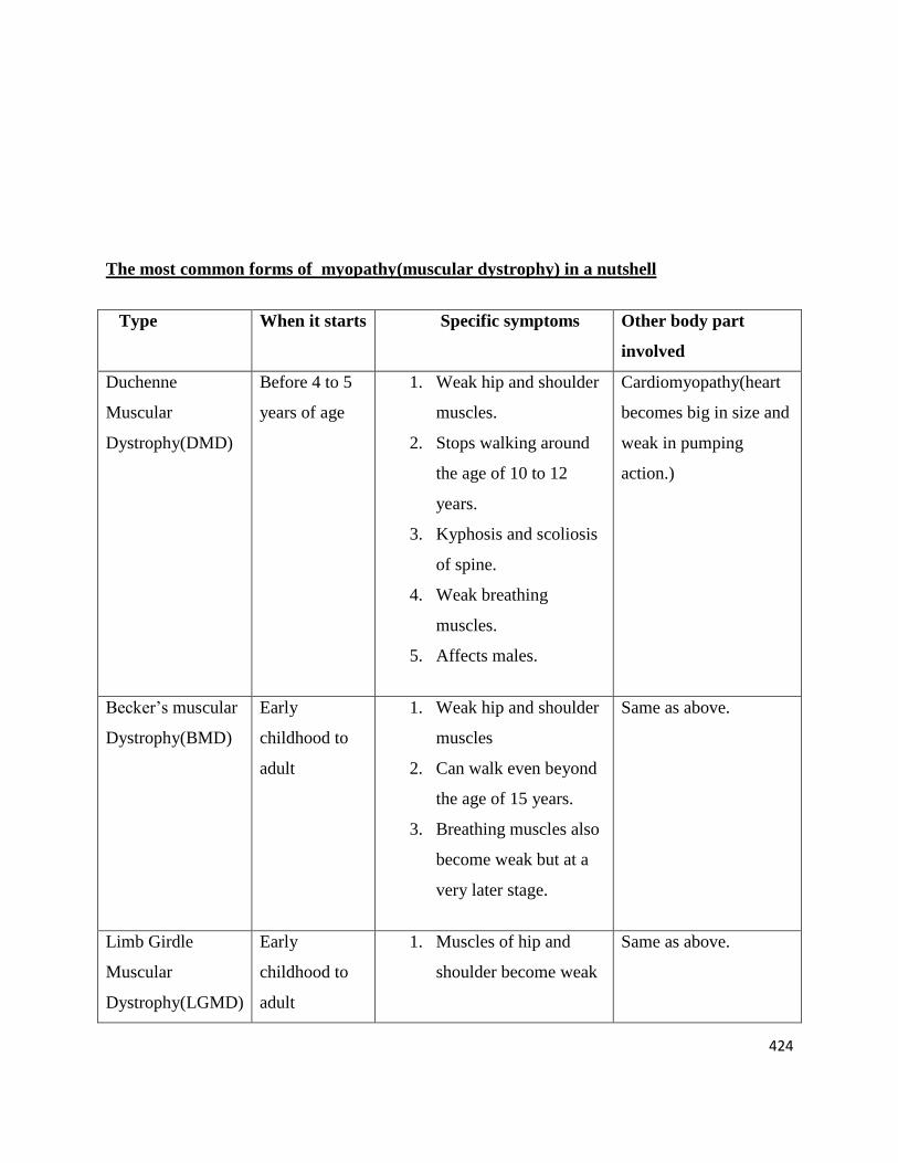
The most common forms of myopathy(muscular dystrophy) in a nutshell
Type When it starts Specific symptoms Other body part
involved
Duchenne
Muscular
Dystrophy(DMD)
Before 4 to 5
years of age
1. Weak hip and shoulder
muscles.
2. Stops walking around
the age of 10 to 12
years.
3. Kyphosis and scoliosis
of spine.
4. Weak breathing
muscles.
5. Affects males.
Cardiomyopathy(heart
becomes big in size and
weak in pumping
action.)
Becker’s muscular
Dystrophy(BMD)
Early
childhood to
adult
1. Weak hip and shoulder
muscles
2. Can walk even beyond
the age of 15 years.
3. Breathing muscles also
become weak but at a
very later stage.
Same as above.
Limb Girdle
Muscular
Dystrophy(LGMD)
Early
childhood to
adult
1. Muscles of hip and
shoulder become weak
Same as above.

425
but slowly.
Less common forms of myopathy(muscular dystrophy )in a nutshell
Type When it
Starts?
Specific symptoms Other body part involved
4.Fascioscapulo
Humeral Dystrophy(FSHD)
Before the
age of 19
to 20
1. Weakness of shoulder, face
and upper arm muscles but
slowly.
None
5.Congenital Muscular
Dystrophy
At birth or
within
First few
months
1.Low tone or floppy
2.Contractures
3. Delayed milestones.
4. Weak breathing muscles.
Mentally retarded and
problems with the eyes.
6.Myotonic Dystrophy Starts
between
11and 20
years of
age
1. Weakness of shoulder, face
and upper arm muscles but
slowly.
Mental retardation,
cataracts.
Reduced size of testicles,
heart problems.
426
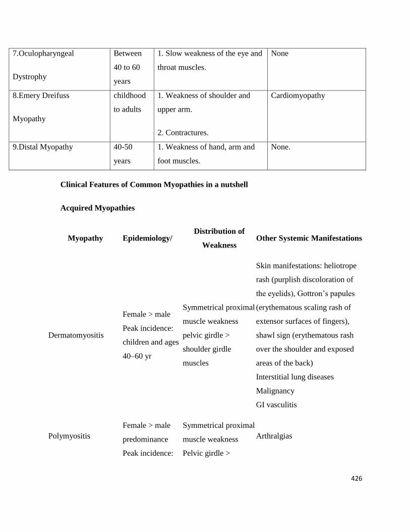
7.Oculopharyngeal
Dystrophy
Between
40 to 60
years
1. Slow weakness of the eye and
throat muscles.
None
8.Emery Dreifuss
Myopathy
childhood
to adults
1. Weakness of shoulder and
upper arm.
2. Contractures.
Cardiomyopathy
9.Distal Myopathy 40-50
years
1. Weakness of hand, arm and
foot muscles.
None.
Clinical Features of Common Myopathies in a nutshell
Acquired Myopathies
Myopathy Epidemiology/ Distribution of
Weakness Other Systemic Manifestations
Dermatomyositis
Female > male
Peak incidence:
children and ages
40–60 yr
Symmetrical proximal
muscle weakness
pelvic girdle >
shoulder girdle
muscles
Skin manifestations: heliotrope
rash (purplish discoloration of
the eyelids), Gottron’s papules
(erythematous scaling rash of
extensor surfaces of fingers),
shawl sign (erythematous rash
over the shoulder and exposed
areas of the back)
Interstitial lung diseases
Malignancy
GI vasculitis
Polymyositis
Female > male
predominance
Peak incidence:
Symmetrical proximal
muscle weakness
Pelvic girdle >
Arthralgias
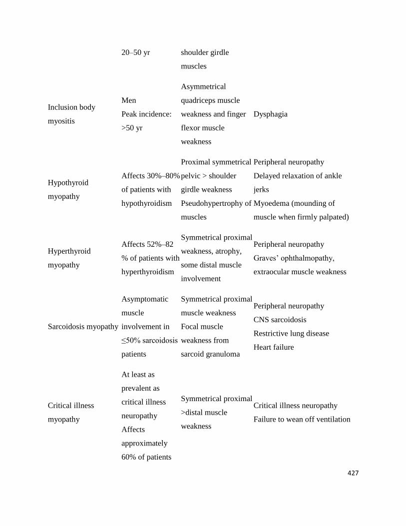
427
20–50 yr shoulder girdle
muscles
Inclusion body
myositis
Men
Peak incidence:
>50 yr
Asymmetrical
quadriceps muscle
weakness and finger
flexor muscle
weakness
Dysphagia
Hypothyroid
myopathy
Affects 30%–80%
of patients with
hypothyroidism
Proximal symmetrical
pelvic > shoulder
girdle weakness
Pseudohypertrophy of
muscles
Peripheral neuropathy
Delayed relaxation of ankle
jerks
Myoedema (mounding of
muscle when firmly palpated)
Hyperthyroid
myopathy
Affects 52%–82
% of patients with
hyperthyroidism
Symmetrical proximal
weakness, atrophy,
some distal muscle
involvement
Peripheral neuropathy
Graves’ ophthalmopathy,
extraocular muscle weakness
Sarcoidosis myopathy
Asymptomatic
muscle
involvement in
≤50% sarcoidosis
patients
Symmetrical proximal
muscle weakness
Focal muscle
weakness from
sarcoid granuloma
Peripheral neuropathy
CNS sarcoidosis
Restrictive lung disease
Heart failure
Critical illness
myopathy
At least as
prevalent as
critical illness
neuropathy
Affects
approximately
60% of patients
Symmetrical proximal
>distal muscle
weakness
Critical illness neuropathy
Failure to wean off ventilation
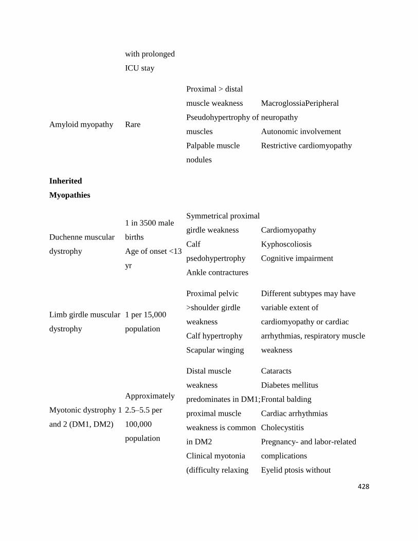
428
with prolonged
ICU stay
Amyloid myopathy Rare
Proximal > distal
muscle weakness
Pseudohypertrophy of
muscles
Palpable muscle
nodules
MacroglossiaPeripheral
neuropathy
Autonomic involvement
Restrictive cardiomyopathy
Inherited
Myopathies
Duchenne muscular
dystrophy
1 in 3500 male
births
Age of onset <13
yr
Symmetrical proximal
girdle weakness
Calf
psedohypertrophy
Ankle contractures
Cardiomyopathy
Kyphoscoliosis
Cognitive impairment
Limb girdle muscular
dystrophy
1 per 15,000
population
Proximal pelvic
>shoulder girdle
weakness
Calf hypertrophy
Scapular winging
Different subtypes may have
variable extent of
cardiomyopathy or cardiac
arrhythmias, respiratory muscle
weakness
Myotonic dystrophy 1
and 2 (DM1, DM2)
Approximately
2.5–5.5 per
100,000
population
Distal muscle
weakness
predominates in DM1;
proximal muscle
weakness is common
in DM2
Clinical myotonia
(difficulty relaxing
Cataracts
Diabetes mellitus
Frontal balding
Cardiac arrhythmias
Cholecystitis
Pregnancy- and labor-related
complications
Eyelid ptosis without
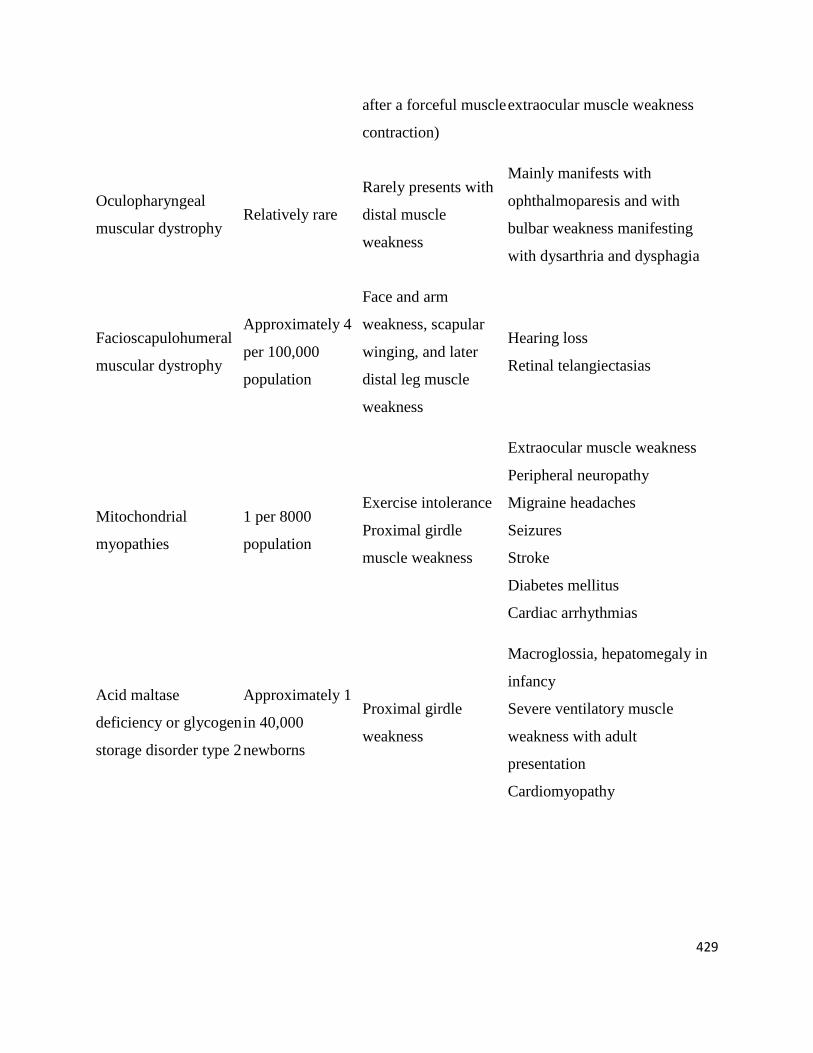
429
after a forceful muscle
contraction)
extraocular muscle weakness
Oculopharyngeal
muscular dystrophy Relatively rare
Rarely presents with
distal muscle
weakness
Mainly manifests with
ophthalmoparesis and with
bulbar weakness manifesting
with dysarthria and dysphagia
Facioscapulohumeral
muscular dystrophy
Approximately 4
per 100,000
population
Face and arm
weakness, scapular
winging, and later
distal leg muscle
weakness
Hearing loss
Retinal telangiectasias
Mitochondrial
myopathies
1 per 8000
population
Exercise intolerance
Proximal girdle
muscle weakness
Extraocular muscle weakness
Peripheral neuropathy
Migraine headaches
Seizures
Stroke
Diabetes mellitus
Cardiac arrhythmias
Acid maltase
deficiency or glycogen
storage disorder type 2
Approximately 1
in 40,000
newborns
Proximal girdle
weakness
Macroglossia, hepatomegaly in
infancy
Severe ventilatory muscle
weakness with adult
presentation
Cardiomyopathy
430
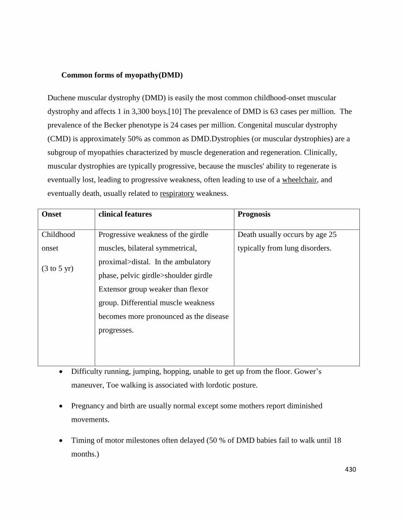
Common forms of myopathy(DMD)
Duchene muscular dystrophy (DMD) is easily the most common childhood-onset muscular
dystrophy and affects 1 in 3,300 boys.[10] The prevalence of DMD is 63 cases per million. The
prevalence of the Becker phenotype is 24 cases per million. Congenital muscular dystrophy
(CMD) is approximately 50% as common as DMD.Dystrophies (or muscular dystrophies) are a
subgroup of myopathies characterized by muscle degeneration and regeneration. Clinically,
muscular dystrophies are typically progressive, because the muscles' ability to regenerate is
eventually lost, leading to progressive weakness, often leading to use of a wheelchair, and
eventually death, usually related to respiratory weakness.
Onset clinical features Prognosis
Childhood
onset
(3 to 5 yr)
Progressive weakness of the girdle
muscles, bilateral symmetrical,
proximal>distal. In the ambulatory
phase, pelvic girdle>shoulder girdle
Extensor group weaker than flexor
group. Differential muscle weakness
becomes more pronounced as the disease
progresses.
Death usually occurs by age 25
typically from lung disorders.
Difficulty running, jumping, hopping, unable to get up from the floor. Gower’s
maneuver, Toe walking is associated with lordotic posture.
Pregnancy and birth are usually normal except some mothers report diminished
movements.
Timing of motor milestones often delayed (50 % of DMD babies fail to walk until 18
months.)
431
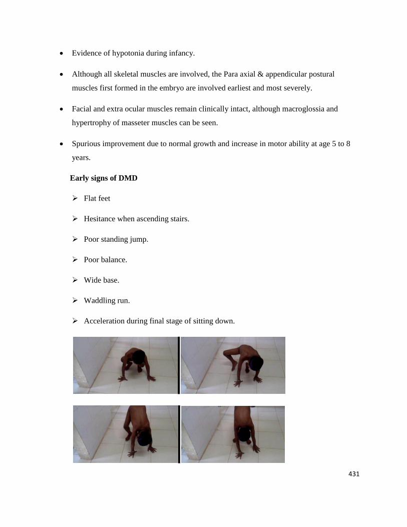
Evidence of hypotonia during infancy.
Although all skeletal muscles are involved, the Para axial & appendicular postural
muscles first formed in the embryo are involved earliest and most severely.
Facial and extra ocular muscles remain clinically intact, although macroglossia and
hypertrophy of masseter muscles can be seen.
Spurious improvement due to normal growth and increase in motor ability at age 5 to 8
years.
Early signs of DMD
Flat feet
Hesitance when ascending stairs.
Poor standing jump.
Poor balance.
Wide base.
Waddling run.
Acceleration during final stage of sitting down.
432
Later signs of DMD
Waddling gait(Hip abductor weakness)
Lordosis(Hip extensor weakness accompanied by hip flexion contracture leading to
increased lumbar lordosis)
Frequent fall.
Difficulty ascending stairs(more difficulty descending than ascending, the knees
loaded with up to 7 times body wt when descending than ascending)
Positive Gower’s sign.(tripod sign)
Weak neck flexion.
Exercise cramping may occur.
Deep tendon reflexes are depressed as muscles weaken. They tend to cease on the
dominant side first. The non dominant Achilles reflex is usually the last to disappear;
this is because the heel cord is the strongest tendon in the body.
True muscle hypertrophy and later pseudo hypertrophy (substitution of fat and areolar
tissue for muscle) seen in calves (occasionally in deltoids, triceps, serratus anterior
and vastus lateralis muscle) Involved muscles have a doughy consistency on
palpation. The specific gravity of this muscle is less than normal because of
replacement by adipose and fibrous tissue.
Sensation unaffected.
Weakness accentuated by immobilization. Loss of ambulation between 9-12 years of
age. Death (pulmonary or cardiac) in late teens.
Significant number of patients is intellectually impaired.
433
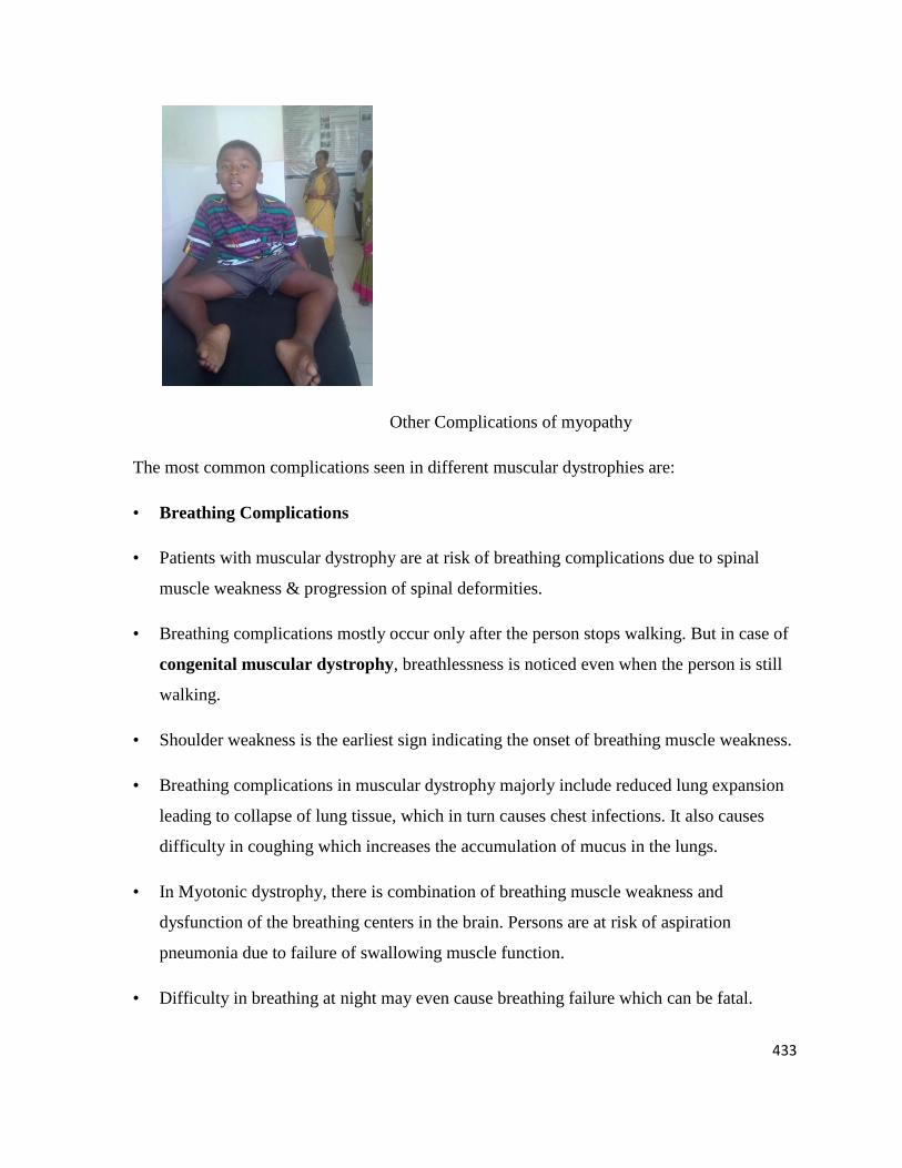
Other Complications of myopathy
The most common complications seen in different muscular dystrophies are:
• Breathing Complications
• Patients with muscular dystrophy are at risk of breathing complications due to spinal
muscle weakness & progression of spinal deformities.
• Breathing complications mostly occur only after the person stops walking. But in case of
congenital muscular dystrophy, breathlessness is noticed even when the person is still
walking.
• Shoulder weakness is the earliest sign indicating the onset of breathing muscle weakness.
• Breathing complications in muscular dystrophy majorly include reduced lung expansion
leading to collapse of lung tissue, which in turn causes chest infections. It also causes
difficulty in coughing which increases the accumulation of mucus in the lungs.
• In Myotonic dystrophy, there is combination of breathing muscle weakness and
dysfunction of the breathing centers in the brain. Persons are at risk of aspiration
pneumonia due to failure of swallowing muscle function.
• Difficulty in breathing at night may even cause breathing failure which can be fatal.
434
• In later stages of muscular dystrophy, the person may have to be put on a ventilator.
• Breathing complications are the major cause of death in 90% of Duchenne muscular
dystrophy patients.
2. Heart Complications
• Involvement of the heart is very common in muscular dystrophy patients.
• Weakness of the heart muscle and replacement of muscle tissue with connective tissue or
fat, results in complications of the heart.
• These complications are generally progressive leading to ECG abnormalities and poor
ability of the heart to pump blood, which may be life threatening.
• Breathing problems & spine deformities may also affect the functioning of the heart.
• Approximately 70% of boys with Becker’s muscular dystrophy have cardiac involvement
by age 20.
• Heart problems in Becker’s muscular dystrophy are worse than in Duchenne muscular
dystrophy patients. Myotonic dystrophy type 1 has more than one system affected with
prominent heart problems leading to an increased incidence of sudden death.
3. Psychosocial complications:
• People with muscular dystrophy may experience psychological & social difficulties due
to their limited ability to participate in many activities when their friends are doing well.
• They may feel helpless as they become dependent on others.
• Social isolation or withdrawal, emotional disturbances like anger, depression, anxiety &
reduced self-esteem are some of the psychosocial issues.
• Stopping schooling (due to difficulties in carrying the child and moving in the school
building) has a huge impact on the psychosocial functioning of kids.
435
• Associated conditions such as ADHD, learning difficulties or autism spectrum disorders
should be identified early to reduce psychological issues.
• A fear about the future may always worry them.
4. Obesity:
• People with muscular dystrophy often are overweight due to lack of physical activity.
• It adds strain to weak muscles due to which the person can approach non walking stage
faster.
5. SLEEP DISTURABNCE
• As muscle weakness increases, the person with Muscular dystrophy will not be able to
change his position on his own. Therefore the patient's & his caregivers sleep would be
disturbed throughout the night.
• Breathing difficulties also may keep the person awake.
6. OSTEOPOROSIS:
• Osteoporosis is the thinning of the bone in which they lose calcium and become soft and
brittle.
• These soft bones are more prone to fractures.
• Risk of osteoporosis increase with age and loss of walking as the bones are not subjected
to normal weight bearing.
• Fractures in the walking phase lead long periods of bed rest which in turn could result in
loss of walking.
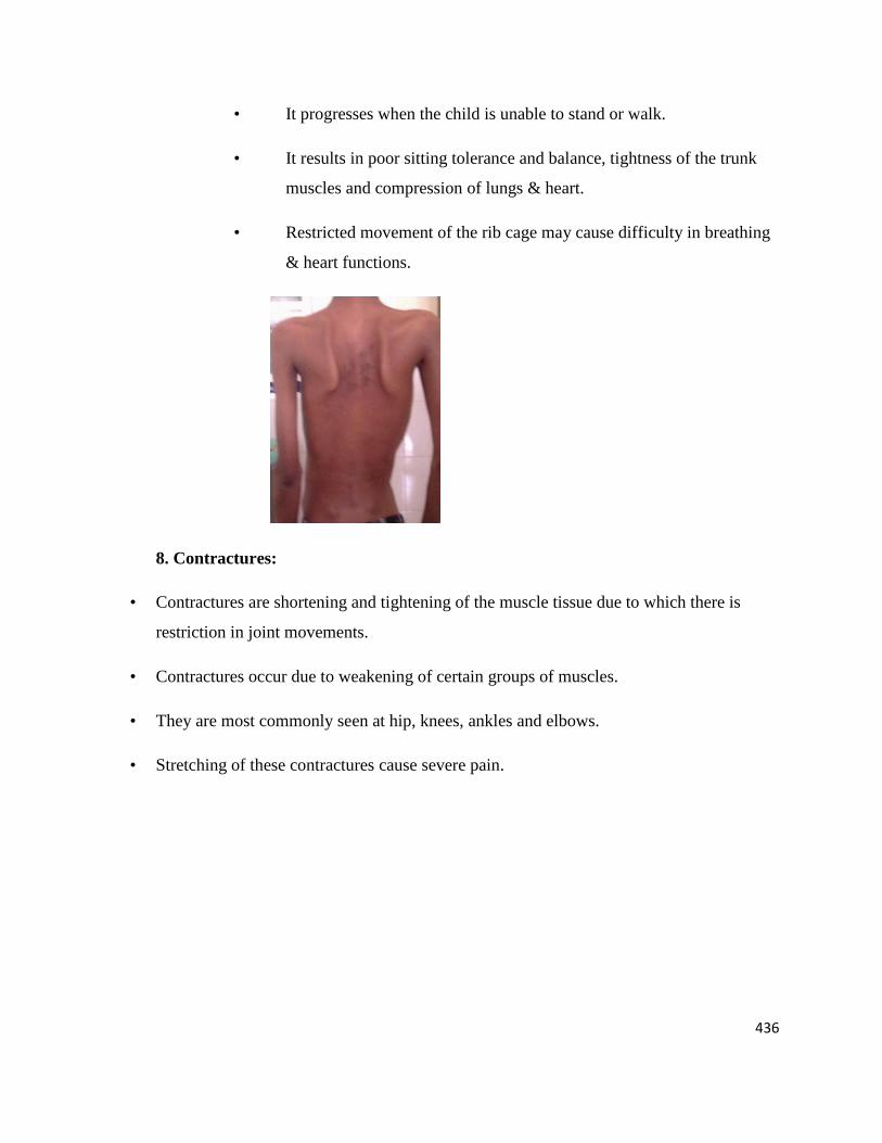
7. SCOLIOSIS
• Scoliosis is twisting in the spine (S shaped curvature) caused due to
weakness in trunk muscles.
436
• It progresses when the child is unable to stand or walk.
• It results in poor sitting tolerance and balance, tightness of the trunk
muscles and compression of lungs & heart.
• Restricted movement of the rib cage may cause difficulty in breathing
& heart functions.
8. Contractures:
• Contractures are shortening and tightening of the muscle tissue due to which there is
restriction in joint movements.
• Contractures occur due to weakening of certain groups of muscles.
• They are most commonly seen at hip, knees, ankles and elbows.
• Stretching of these contractures cause severe pain.
437
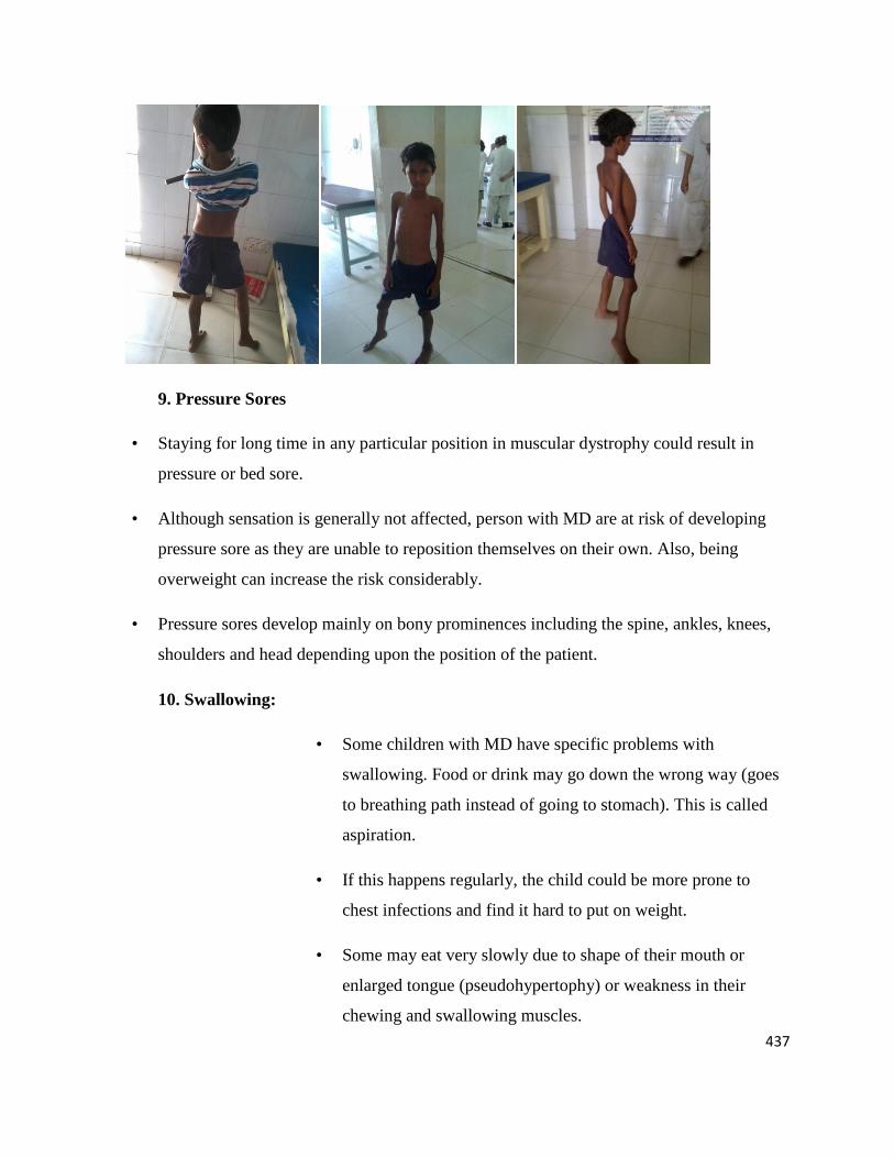
9. Pressure Sores
• Staying for long time in any particular position in muscular dystrophy could result in
pressure or bed sore.
• Although sensation is generally not affected, person with MD are at risk of developing
pressure sore as they are unable to reposition themselves on their own. Also, being
overweight can increase the risk considerably.
• Pressure sores develop mainly on bony prominences including the spine, ankles, knees,
shoulders and head depending upon the position of the patient.
10. Swallowing:
• Some children with MD have specific problems with
swallowing. Food or drink may go down the wrong way (goes
to breathing path instead of going to stomach). This is called
aspiration.
• If this happens regularly, the child could be more prone to
chest infections and find it hard to put on weight.
• Some may eat very slowly due to shape of their mouth or
enlarged tongue (pseudohypertophy) or weakness in their
chewing and swallowing muscles.
438
Also children may have weakness in their arms and upper limbs due to which they are
unable to feed themselves
11. Complications due to prolonged steroid treatment include:
• Cataracts
• Cushingoid features(moon face)
• Obesity
• Short stature
• Constipation
• Hypertension
• Delayed puberty
• Behavioral changes (irritability, hyperactivity)
• Occasionally slight increase in body hair.
Laboratory diagnosis
a)Typical EMG
b) Typical biopsy
c) Markedly increased CPK (at least 10 times normal in early stages, though
reduced later when walking ceases). Increased urinary excretion of 3-methylhistadine.
e) No dysphagia or sphincter difficulty.
Becker’s muscular Dystrophy
439
1. X-linked. This variant constitutes 10% of DMD.
2. Later onset and patients are ambulatory into third decade with longer life expectancy
than DMD.
3. Similar proximal distribution of muscle weakness as in DMD but asymmetrical;
usually maintains neck flexor strength. May present with pes cavus, unusual hypertrophy (thenar
eminence), or patellar subluxation secondary to quadriceps weakness.
a) Triceps power often greater than biceps.
b) May develop ambulatory scoliosis because of asymmetrical Para spinal muscle
weakness.
4. Cardiac involvement less than in DMD.
5. Occasionally linked with deuteranopia (color blindness)
6. Mental retardation uncommon.
7. Laboratory diagnosis:
a) Increased CPK
b) Mixed pattern on EMG
c) Biopsy somewhat different than Duchenne Dystrophy (muscle fibers usually not
rounded and hyaline fibers rare).
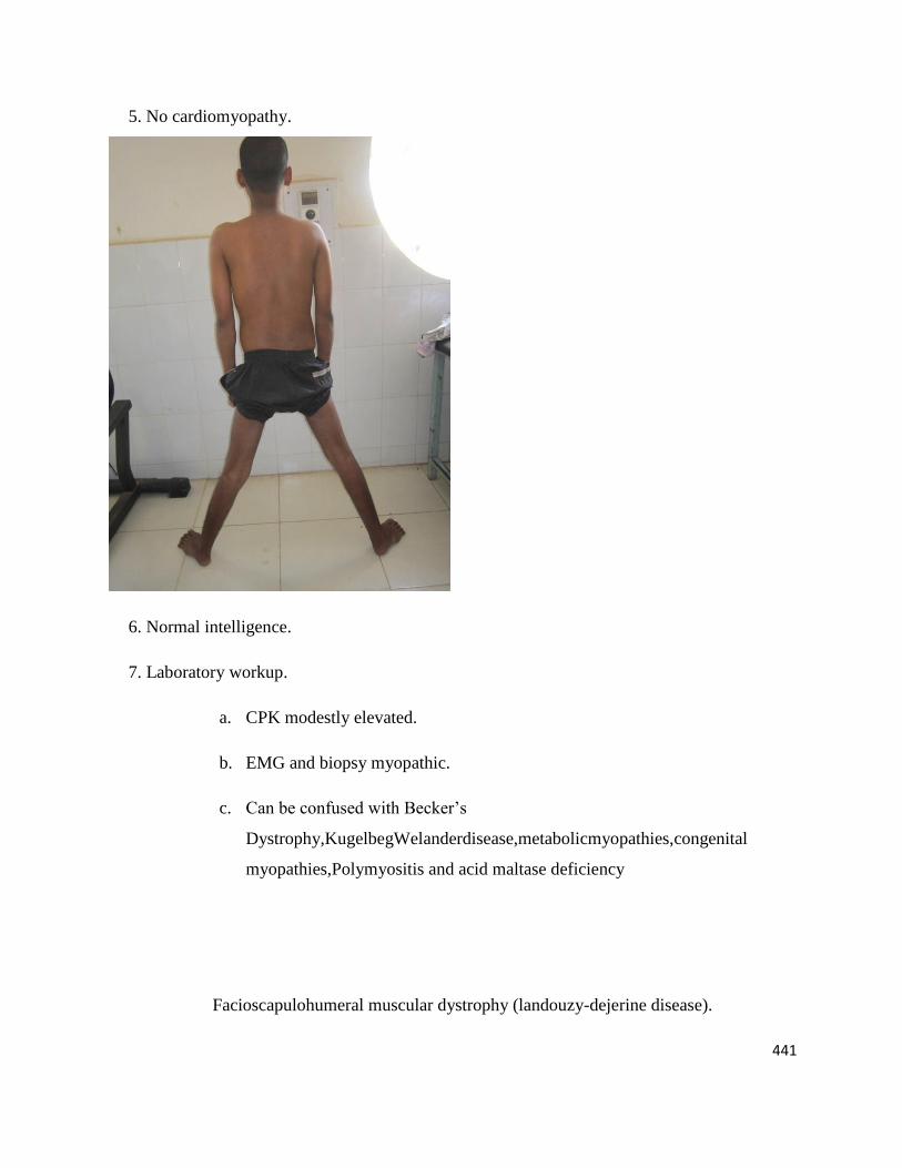
LIMB GIRDLE DYSTROPHY
1. Autosomal recessive (many sporadic); consanguineous mating increases incidence (for e.g.,
first cousins, have one- eighth of their genes in common).
440
2. Onset usually in second or third decade.
3. Life expectancy reduced but variable.
4. - Usually pelvic girdle weakness is more than shoulder girdle with variable progression.
Prognosis is better in patients who manifest shoulder weakness first.
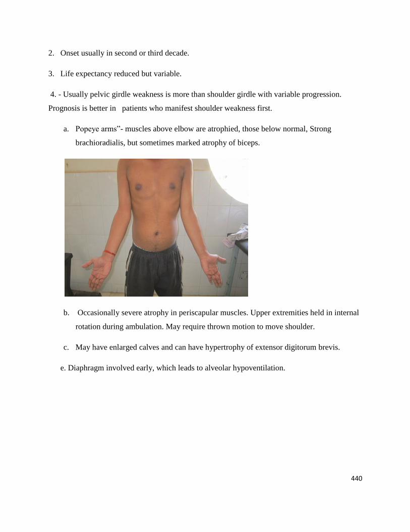
a. Popeye arms‖- muscles above elbow are atrophied, those below normal, Strong
brachioradialis, but sometimes marked atrophy of biceps.
b. Occasionally severe atrophy in periscapular muscles. Upper extremities held in internal
rotation during ambulation. May require thrown motion to move shoulder.
c. May have enlarged calves and can have hypertrophy of extensor digitorum brevis.
e. Diaphragm involved early, which leads to alveolar hypoventilation.
441
5. No cardiomyopathy.
6. Normal intelligence.
7. Laboratory workup.
a. CPK modestly elevated.
b. EMG and biopsy myopathic.
c. Can be confused with Becker’s
Dystrophy,KugelbegWelanderdisease,metabolicmyopathies,congenital
myopathies,Polymyositis and acid maltase deficiency
Facioscapulohumeral muscular dystrophy (landouzy-dejerine disease).
442
Autosomal dominant—with variable expressivity (from patient to patient within a given
pedigree, as well as from family to family).one parent always has at least sub clinical
disease
Onset usually in adolescence or early adult life with slow progression and normal life
expectancy. But Childhood form is a more malign disease which runs a more rapidly
disabling course. (May show inflammatory response in muscle biopsy). May be
stationary for periods of time.
a) Weakness of face, shoulder girdle (particularly muscles of scapular
fixation), and lower fibers of trapezius is often affected with high riding
scapula. Deltoids often preserved.
b) Brachioradialis, dorsiflexor weakness of wrists & fingers produces ―Praying
mantis‖ posture.
Positive bell’s sign, accentuation of lateral lip ―dimples,‖ transverse smile,
inability to whistle, inability to wrinkle forehead or puff cheeks, ―tapir‖
mouth (lip eversion & protrusion).
Lower limb usually affected 10-15 years after onset. Distal leg weakness of
tibialis anterior and toe extensors causes foot drop and slapping gait. Back
extensors, quadriceps, & tensor fascia often exempt.
a) Pelvic girdle weakness with increased lumbar lordosis. Occasionally cauda
equina syndrome with leg paresthesias secondary to sway back may occur
Involvement usually asymmetrical in distribution and degree..
Cardiac involvement & intellectual impairment not characteristic.
Lab workup.
a) CPK variably & slightly increased. Pyruvate kinase can be elevated (even
with CPK normal).
443
b) EMG myopathic but can show some neuropathic elements.
c) Biopsy myopathic with an occasional inflammatory finding. Some cases show
type 1 fibre predominance.
Scapulo peroneal muscular dystrophy (FSH minus the F)
X-linked disease can often be characterised as Emery-Dreifuss
muscular dystrophy
Insidious onset in childhood and progression is slow without loss of
ambulation.
May be myopathic or neuropathic (in which case EKG abnormalities are
observed) & hereditary pattern is variable.
Muscle weakness mainly confined to scapular and peroneal groups of
muscles.
Achilles tendon & elbow flexion contractures as well as inability to fully
flex the neck &spine.
By mid adulthood, atrial conduction defects occur that can cause sudden
death.
The syndrome of Facioscapulohumeral muscle weakness & wasting can be seen
in such diverse conditions as myotubular myopathy, central core disease,
nemaline myopathy, myasthenia gravis, polymyositis, adult acid maltase
deficiency, & spinal muscular dystrophy.
444
Ocular (Oculopharyngeal) myopathy
Common in French Canadian families near Quebec & Spanish-American families
in the south western united states. Regarded as a mitochondrial myopathy..
Usually autosomal dominant, onset in third to fourth decade, disease is
Progressive with frequently asymmetrical distribution; women affected more than
men.
a) Ptosis followed by external ophthalmoplegia with dysphasia.
b) Facial &upper limb muscles (late) may be involved.
c) CPK normal to slightly increased, EMG myopathic, ragged red fibbers on
biopsy.
Oculo-cranio-somatic neuromuscular disease.
a) Onset in first decade.
b) Progressive external ophthalmoplegia plus:
Retinitis pigmentosa& optic atrophy.
Cardiomyopathy & heart block.
Cerebellar ataxia & spasticity.
Deafness.
Mental retardation.
Skeletal deformities.
Limb myopathy.
Peripheral neuropathy.
Pharyngeal weakness.
c) CPK slightly elevated, EMG myopathic, ragged red fibres on biopsy.
445
Distal Myopathy
Inherited as a dominant character.
Usually begins between 40-60 years of age.
Affects small muscles of hands & peripheral leg muscles spreading slowly proximally.
Comparatively benign condition found mostly in large Swedish kindreds.
Morphologically specific myopathies
(So called as benign or congenital myopathies)
Major ―structural‖ types—histological diagnosis.
a) Central core disease(in which the nuclei are abnormally found in the centre of the
muscle fibres).
b) Nemaline Myopathy (rod diseases) –childhood & adult forms.
c) Myotubular Myopathy.
d) Mitochondrial Myopathy (which are due to defects in mitochondria, which provide a
critical source of energy for the muscle).
e) Minimulticore myopathy (characterised by multiple small cores or areas of disruption
in the muscle fibres.)
f) Congenital fibre type disproportion
Similar clinical pictures for most types such as:
a) Genetically determined (often autosomal dominant) but many sporadic cases.
b) Hypotonia after birth. (floppy infants) or later development of muscle weakness.
c) Delayed motor milestones.
d) Commonly slowly progressive generalised muscle weakness more marked
proximally; facial or extra ocular muscle weakness may be present.
Occasionally rapid progression of disease
446
e) Can overlap with some of metabolic myopathies.
f) Dysmorphic features.
High arched palate.
Long-facies-dolichocephalic head.
Pectus carinatum or excavatum.
Absence of a single muscle (Myotubular Myopathy).
Long tapering fingers.
g) Skeletal abnormalities:
i. Congenital dislocation of hip, particularly in central core disease, Responds
poorly to closed reduction because of muscle weakness, usually requires
operative stabilization
ii. Pescavus.
iii. Scoliosis.
Lab diagnosis
i. CPK usually normal.
ii. EMG normal or myopathic.
iii. Biopsy reveals specific identifying histological abnormalities as seen by
light or electron microscopy & in many cases type l fibre predominance.
Congenital muscular dystrophy
Hypotonia at birth (with facial involvement).
Motor milestones late but disease generally non progressive
Progressive muscular contracture.
Usually no cardiomyopathy or intellectual impairment.
447
CPK modestly elevated; EMG myopathic; biopsy shows advanced dystrophic changes with
early & extensive endomysial fibrosis.
Several kindred with dysplastic brain abnormalities & dystrophic involvement of skeletal
muscles have been reported in Japan` (Fukuyama). These have markedly elevated CPK.
Requires early aggressive orthopaedic attention.
Metabolic myopathies
Myopathies of varying degree, secondary to biochemical defects of muscle metabolism.
May be progressive, fixed or recurrent, difficulty with exercise.
Fluctuating muscle power with exercise induced weakness, cramps, & sometimes
myoglobinuria.
Diagnosis is made on the basis of clinical findings (e.g., an unusual craving for salt can
be associated with one of the mitochondrial myopathies),
Enzyme assays, elevated CPK, ECG changes when heart is involved, typical histological
findings (i.e., ragged red fibers in mitochondrial disease).
EMG changes in some of conditions, special histochemical staining to identify specific
metabolites, or lack of particular enzymes.
I. Glycogenosis the seven recognized type muscles disease associated with enzyme
deficiency in the carbohydrate metabolism of muscles are outlined in table. The site of
action (liver, heart, muscle, etc) of the deficient enzyme determines the format of the
disease. Myopathy is present in types ll, lll, V & Vll. In types V & Vll, myopathic
symptoms are the major manifestations.
II. Abnormalities of lipids metabolism. Myopathies are characterised by neutral lipid
accumulation in muscle due to mitochondrial metabolic defects. Carnitine (synthesized in
the liver & transported via the blood stream) facilitates the passage of free fatty acids
448
across the mitochondrial membrane. Diagnosis is by muscle biopsy (vacuolar [lipid]
myopathy).
A. Carnitine deficiency.
1. Systemic: metabolic acidosis, encephalopathy, Hepatomegaly, cardiomyopathy
2. Muscle: progressive proximal Myopathy.
3. Secondary to genetic defects of intermediary metabolism or other conditions
(e.g.cytochrome oxidase deficiency, glutaric aciduria, chronic renal failure treated by
haemodialysis, cirrhosis with cachexia, renal fanconi syndrome).
4. Can be treated with diet.(Low in long chain fatty acids), steroid & oral L-Carnitine.
B. Carnitine palmityl transferase deficiency.post exertional or fasting pain with
myoglobinuria, followed by weakness, tenderness or muscle swelling. Treat with
high carbohydrate foods or medium chain fatty acids supplements.
lll. Periodic paralysis.
Inherited as an Autosomal dominant trait having tendency towards remission &
relapse.
The primary defect for the many of the periodic paralysis is believed to be an
increased membrane conductance to sodium ions resulting in membrane hypo
excitability.
Classified in to hypokalemic, normokalemic, & hypokalemic, depending on the
level of venous blood potassium during an episode. This different form may
occur at different times in the same patient (biphasic periodic paralysis).
Clinical features overlaps. Episodic limb weakness usually occurring after
exercise, typically spares muscles of respiration & disappear in an
unpredictable manner. Clinical myotonia may be present.
Lab diagnosis

449
Provocative testing with potassium may be helpful in making the diagnosis.
Hypokalemic variety can often be induced by glucose & insulin, heavy
exercise followed by rest, excessive alcohol, cold, trauma or stress. CPK can be
raised during attacks. EMG changes may be present. Changes are apparent on
biopsy if the specimen is taken during attacks.,
Treatment
Hypokalemic variety can be treated with oral potassium, carbonic anhydride
inhibitors, acetazolamide, sodium restriction, and high carbohydrate diet.. The
norm kalmia type is best relieved with sodium infusion. Carbonic anhydride
inhibitors may also help.
Glycogenoses
Type Enzyme Deficiency Eponymous or
other names
Skeletal
muscles
affected
Clinical
features
Other tissues
affected
l Glucose-6-
phosphatase
Von Glerke’s
disease
No
ll α-1,4-glucosidase(acid
maltase)
Pompe’s disease Yes a) Severe form:
Generalized;
resembles
infantile spinal
muscular
atrophy
b) Mild form;
resembles limb
Heart, nervous
system, kidney,
leukocytes
? Heart
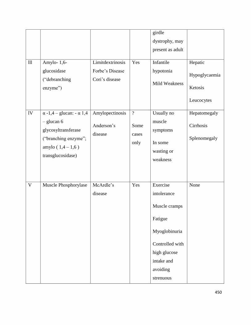
450
girdle
dystrophy, may
present as adult
lll Amylo- 1,6-
glucosidase
(―debranching
enzyme‖)
Limitdextrinosis
Forbe’s Disease
Cori’s disease
Yes Infantile
hypotonia
Mild Weakness
Hepatic
Hypoglycaemia
Ketosis
Leucocytes
lV α -1,4 – glucan: - α 1,4
– glucan 6
glycosyltransferase
(―branching enzyme‖;
amylo ( 1,4 – 1,6 )
transglucosidase)
Amylopectinosis
Anderson’s
disease
?
Some
cases
only
Usually no
muscle
symptoms
In some
wasting or
weakness
Hepatomegaly
Cirrhosis
Splenomegaly
V Muscle Phosphorylase McArdle’s
disease
Yes Exercise
intolerance
Muscle cramps
Fatigue
Myoglobinuria
Controlled with
high glucose
intake and
avoiding
strenuous
None
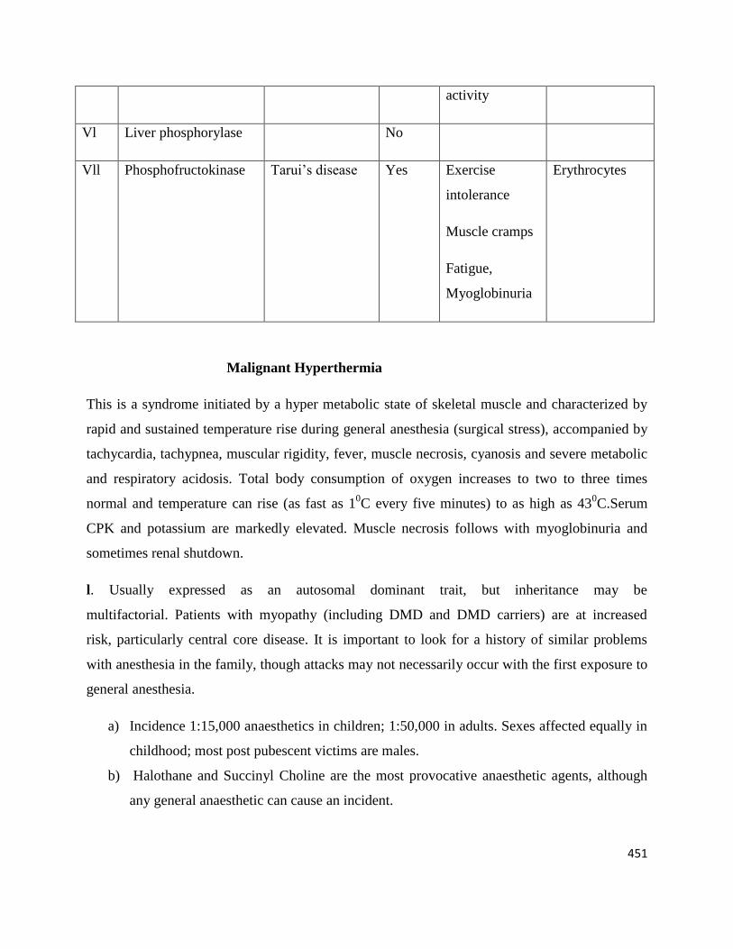
451
activity
Vl Liver phosphorylase No
Vll Phosphofructokinase Tarui’s disease Yes Exercise
intolerance
Muscle cramps
Fatigue,
Myoglobinuria
Erythrocytes
Malignant Hyperthermia
This is a syndrome initiated by a hyper metabolic state of skeletal muscle and characterized by
rapid and sustained temperature rise during general anesthesia (surgical stress), accompanied by
tachycardia, tachypnea, muscular rigidity, fever, muscle necrosis, cyanosis and severe metabolic
and respiratory acidosis. Total body consumption of oxygen increases to two to three times
normal and temperature can rise (as fast as 10C every five minutes) to as high as 43
0C.Serum
CPK and potassium are markedly elevated. Muscle necrosis follows with myoglobinuria and
sometimes renal shutdown.
l. Usually expressed as an autosomal dominant trait, but inheritance may be
multifactorial. Patients with myopathy (including DMD and DMD carriers) are at increased
risk, particularly central core disease. It is important to look for a history of similar problems
with anesthesia in the family, though attacks may not necessarily occur with the first exposure to
general anesthesia.
a) Incidence 1:15,000 anaesthetics in children; 1:50,000 in adults. Sexes affected equally in
childhood; most post pubescent victims are males.
b) Halothane and Succinyl Choline are the most provocative anaesthetic agents, although
any general anaesthetic can cause an incident.
452
c) Patients may have large muscle mass (they tend to be muscular and athletic) and a history
of cramping (especially nocturnal, which tends to stop by the third decade), exercise
intolerance in hot weather, and stress associated acrocyanosis. Musculoskeletal
abnormalities such as Ptosis, clubfoot, scoliosis, pectus carinatum, and hernia are
common.
d) The marked elevation of temperature is believed to be secondary to high calcium
concentration in the myoplasm
ll. Lab diagnosis
a) Elevated CPK may be found during work up.
b) Muscle biopsy shows abnormal in vitro sensitivity to halothane, succinyl choline,
or caffeine, which increases calcium efflux into the cell.
lll. Treatment
If Local or regional anaesthesia is not feasible, general anaesthesia can be
accomplished with nitrous oxide, narcotics, barbiturates, ketamine or doperidol.
a) Stop anaesthesia and administer 100% O2.
b) Cool the patient externally and internally (Gastric, rectal, peritoneal lavage) to
combat hyperthermia.
c) Insert bladder and CVP catheters.
Administer:
1. Intravenous procaine or procainaminde( Avoidlidocaine ) to combat rigidity.
These drugs decrease intracellular calcium transport.
2. Intravenous steroids.
3. Bicarbonate to control metabolic acidosis.
4. Glucose and insulin infusion to treat hyperkalemia.
453
5. Dantrolene sodium ( IV – 2.5 – 10 mg per kg ) may avert an attack. Action:
excitation – contraction uncoupling by decreasing release of calcium from
sarcoplasmic reticulum.
6. Preanasthetic oral dantrolene loading ( 4 – 8 mg/kg for 2 days with final dose 2
hours before anaesthesia ) may avert or lessen the severity of an episode.
d) Monitor for renal failure after recovery
Myotonic Disorder
This is a group of conditions (Usually hereditary) having in common clinical
myotonia(delay of muscular relaxation after contraction). This is best seen in the
clenched fist or in the orbicularis oculi muscles. Myotonia in all myotonic
diseases is often aggravated by cold. It can be improved ( fatigues ) by repetitive
activities, but sometimes this increases its severity ( myotonia paradoxica ).
Myotonia can be elicited by percussion. This is usually demonstrated in the
tongue or thenar muscles.
EMG is characterised by high frequency repetitive discharges that initially
increase in frequency and amplitude then rapidly diminish (Dive-bomber effect).
Myotonia is of muscular origin and independent of motor nerve activity. The
repetitive activity persists even though the motor nerve is sectioned or the neuro
muscular junction is blocked with curare. It is believed to be a membrane defect
(Hyper excitability) related to one abnormality of chloride (Myotonia congenita)
or calcium (dystrophia Myotonica) conductance.
It can be induced by drugs (i. e., Clofibrate), may appear as a remote effect of
lung carcinoma, maybe found in thyroid dysfunction or adult acid maltase
deficiency. The major forms of myotonic disease are
1. Myotonia congenita ( Thomsen’s disease )
A. Generalised non progressive muscular hypertrophy with muscle stiffness and
weakness, relieved by exercise, occurring in two forms.
454
i. Autosomal dominant; Mild nonprogressive myotonia diagnosed in
infancy.
ii. Autosomal recessive; later onset with subsequent distal atrophy and
weakness.
B. EMG myotonic; CPK slightly elevated: biopsy fibre hypertrophy. Increased
creatine tolerance.
C. May present with complaints of garbled speech after eating iced foods (
associated with tongue myotonia induced by cold )
2. Dystrophia myotonica ( Steinert’s disease )
A. Autosomal dominant multisystem disorder (linked with the secretor gene)
with poor congruence in affected family members, the commonest form of
which usually becomes apparent in early adulthood. Expression is variable,
and the disease is characterised by
i. Stellate cataracts and retinal alterations.
ii. Gonadal atrophy.
A) Impotence in males, chronic abortion in females.
iii. Faulty tolerance to carbohydrate (diabetic glucose tolerance curve), Defective
insulin metabolism.
1. Mild ― diabetes ‖ is common to many muscle diseases.
iv. Frontal and parietal alopecia(loss of hair) in males.
v. Thyroid dysfunction.
vi. Cardiac conduction defects sometimes requiring demand pacemakers. Stokes –
Adams attacks are common.
vii. Impaired pulmonary function. Pickwickian syndrome. Alveolar hypoventilation (
night sweats, nightmares )
viii. Progressive psychosocial deterioration with fall off of higher intellectual
functions. Paranoid tendency. ―belle indifference‖, ― whining dependence‖ and
depression ( which may respond to tricyclic antidepressant treatment )
ix. Cerebral ventricular dilatation.
455
x. Skull abnormalities, including hyperostosis crania. Decrease in sella turnica size,
prognathism, hyperostosis frontalis interna, and enlargement of the paranasal
sinuses.
xi. Lugubrious facies with ptosis. Thin, haggard, expressionless face. Transverse smile,
hollow temples and cheeks. Sternocleidomastoid( particularly clavicular head )
weakening, leading to swan neck.
xii. Temporal muscle wasting; myotonic lid lag.
xiii. Distal muscle weakness, especially in the forearms and tibialis anterior muscles.
Patient may trip because of weakness, and in attempting to regain balance, provoke
a myotonic response that causes a fall. It is the weakness (dystrophy), not the
myotonia that troubles these patients the most.
xiv. Percussion and effort myotonia.
a. Hand ( slowness in grip release )
b. Tongue ( dimpling on percussion)
c. Thenar eminence ( contracture on percussion )
d. Spasm of globe elevators (after forced eyelid closure with
sudden release).
NOTE: Myotonia persists after nerve section, block, or curarization. It is increased by cold,
fatigue or sudden stress. It tends to lessen and sometimes disappear in the later stages of the
disease as muscular weakness advances.
i.Dysphagia ( late ) because of pharyngeal myotonia and dysarthria
secondary to tongue myotonia.
ii. Smooth muscle disorders of lower GI tract ( megacolon ).
iii. Increased incidence of gall bladder disease.
iv. Sometimes a high frequency hearing defect.
v. Neuronal heterotopias.
vi. Nasal speech ( because of pharyngeal muscle weakness )
vii. CPK may be slightly elevated: EMG myotonic: Nerve conduction (
Motor and sensory ) may be slowed; muscle biopsy – many internal
456
nuclei appearing in long chains on longitudinal section, sarcoplasmic
masses, ring fibres, selective type 1 fiber atrophy.
viii. Lab tests
a) Low igG level ( hypercatabolized ) : low urinary creatinine;
may have low serum uric acid
b) ECG abnormalities
c) Low basal metabolic rate.
Treatment of myotonia.
1. Phenytoin, 100mg t.i.d, decreases sodium influx during membrane excitation.
2. Acetazolamide, 125 – 500mg/day, promotes kaluresis rendering muscle more
resistant to depolarisation.
3. Quinine, grains 5 t.i.d, stabilizes membrane
4. Procainamide, 0.5 to 1 g q.i.d, stabilizes muscle membrane but may impair
cardiac cinduction and can cause a lupus like syndrome.
5. Steroids.
A. Congenital ( neonatal ) dystrophia myotonica
1. Severe generalized hypotonia at birth.
2. Facial diplegia with sucking and breathing difficulty.
3. Bilateral talipes early and vigorous orthopaedic attention.
4. Frequent hydramnios in mother.
5. Mental retardation
6. Electrical and mechanical myotonia observed later.
7. Characteristic inverted V configuration to upper lip ( Shark mouth )
8. Dismaturation almost always inherited from myotonic mother.
9. Muscle biopsy – maturational arrest at various foetal developmental stages.
457
3. Paramyotonia congenita ( VanEulenberg )
a. Autosomal dominant condition manifest at birth by mild
myotonia of face and hands, aggravated by cold, with tendency
to muscle hypertrophy. Maybe due to temperature dependent
anomaly in sodium conductance.
b. Patient muscle stiffness may be increased or reduced by
exercise
c. May suffer episodes of flaccid weakness similar to the periodic
paralysis.
d. Lid lag may be elicited ( also found in myotonia congenita and
in hyperthyroid myopathy ).
NOTE: Clinical myotonia as an ancillary sign maybe found in Schwartz Jampel syndrome. It can
also be seen in myxedma, hypokaelemic paralysis, and after treatment with a variety of drugs
interfering with muscle membrane lipid metabolism. Electrical myotonia maybe noted in acid
maltase deficiency and denervation.
Endocrine Myopathies
Myopathy can accompany a variety of endocrine disorders. These usually occur in adults. The
onset of weakness is insidious. Proximal muscles are predominantly affected. CPK is elevated,
creatine urea is present, and the EMG shows myopathic characteristics. However, there may be
indications of neuropathic causation. Management of the primary disorder consists of organ
specific treatment.
1. Thyroid myopathies.
A. Hyperthyroidism
I. Bulbar and EOM can be involved in severe thyrotoxicosis ( this may be
myasthenic ). Lid lag may be seen.
B. Hypothyroidism
I. Muscle spasm and cramps can occur.
II. Delayed relaxation of ankle reflex
458
III. May find electrical myotonia
2. Parathyroid disorder
A. Hypoparathyroidism
I. Tetany with carpopedal spasm.
B. Hyperparathyroidism and osteomalacia
I. Proximal weakness.
II. Muscle tenderness and aching.
III. EMG signs of denervation.
3. Pitutary and adrenal disorder
A. Hypoadrenalism( Addison’s Disease ).
I. Fatiguablility and muscle cramping
II. Proximal wasting
III. Periodic episodes of hypokaelemic weakness
B. Hyperadrenalism ( Cushing’s Syndrome )
I. Proximal weakness and wasting of insidious onset
4. Steroid myopthay.
I. This non specific proximal muscle weakness is seen with the administration of
steroids, specially the halogenated compounds ( triamcinolone, dexamethasone ).
May be due in part to epinephrine suppression, which blocks phoshporylase
activation. Can prove difficult to diagnose, especially in the case of an
inflammatory myopathy under treatment with glucocorticoids.
II. Lab diagnosis
A. CPK maybe normal
B. Increased creatineureas ( in presence of normal or low serum enzyme level )
C. EMG – short duration polyphasic motor unit action potentials.
D. Type 2 atrophy on biopsy (versus inflammatory necrosis in polymyositis ).
E. Weakness (usually in large antigravity postural muscles of lower extremities) may be
dose related and develop rapidly.
F. Muscles often tender to palpation.
G. Other signs of hypersteroidism commonly present
459
i. Moon facies
ii. Increase in adipose tissue
iii. Acne vulgaris, diabetes.
iv. Osteoporosis (with vertebral compression fracture).
v. Hyper tension, psychiatric disorders.
H. Improvement with discontinuation of steroid medication.
5. Acromegaly
A. Early muscle hypertrophy followed by late proximal weakness
6. Hyperaldosteronism
A. Periodic attacks of hypokalemic weakness.
Nutritional and drug induced Mypoathies
2. Protein deprivation
3. Osteomalacia
4. Alcoholic myopathy
A. Acute
i. After heavy drinking bout
ii. Sudden onset of muscle pain with swelling and weakness,
typically in the large appendicular postural muscles. CPK
markedly increase ( even modest alcoholic intake causes some
increase in serum CPK.
iii. Muscle can be swollen and tender.
iv. Necrotizing myopathy ( selective type II atrophy with excess
lipid and glycogen in fibres suggesting a metabolic pathway
inhibition ) resulting in:
a) Myoglobinuria
b) Renal failure which requires supportive treatment.
B. Chronic
460
i. Slowly progressive limb-girdle weakness. Most of the effects
of chronic alcoholism are due to vitamin deficiencies,
particularly of vitamin B1.
5. Other drugs that can cause neuromuscular pathology
A. Vincristine – weakness.
B. Diazacholesterol – myotonia
C. Clofibrate –Cramping
D. Penicillamine – inflammatory myopathy.
E. Diuretics, licorice, purgatives – hypokalemic paralysis.
F. Chloroquine, heroine – subacute painless myopathy.
G. Cimetidine, lithium – mild weakness.
Inflammatory Myopathies
These disorders are thought to be due to a viral or autoimmune mechanism.
Characterized by symmetrical proximal muscle weakness often accompanied by
muscular pain and tenderness.
They occur more frequently in blacks.
Dysphagia is sometimes present.
Involvement of facial or EOM is rare.
Pseudohypertrophy occasionally occurs.
The deep tendon reflexes maybe absent, normal, hyperactive.
The heart can be involved.
Muscle atrophy with contracture and calcinosis are seen late in the course of the
disease.
CPK is raised as well as ESR and WBC ( 50% of the cases ).
Elevated ANA, positive latex test ( 50% ), and increase in serum gamma
globulins alpha 2, and IgM have also been reported.
EMG shows polyphasic, brief, small motor action potentials with spontaneous
fibrillations and positive sharp waves, insertional irritability, and bizarre high
frequency repetitive discharges.
461
The muscle pathology includes inflammatory cellular infiltrates ( principally
lymphocytes ), segmental necrosis, de and re generation ( necrosis, vacuolization
and phagocytosis ) and increase of endomysial connective tissue.
Perifascicular atrophy of both type l&ll fibres is present. This pattern is probably
related to ischaemic myopathy of fibres adjacent to perimysial collagenous
septae and is more common in dermatomyositis. All of these biopsy findings are
usually scattered.
Polymyositis is the diagnostic label given to a non-hereditary inflammatory
myopathy.
Where a skin rash is present, the term dermatomyositis is used.
These conditions can present acutely or run a subacute, relapsing, or chronic
course.
There are both childhood and adult forms.
A bimodal age distribution between ages 5 and 15 and then between 50 & 60
years, has been reported.
Polymyositis is as common as scleroderma, and half as common as systemic
lupus erythematosus. Its incidence is 5 – 8 new cases per million people each
year.
Inflammatory myopathy in childhood can be confused with DMD.
1. Idiopathic polymyosistis.
A. Pain and stiffness more marked in upper limbs, weaknesses in
lower, but one third of patients present with a non muscular first
symptom. May also mimic DMD almost completely.
B. Cervical flexors involved early.
C. Dysphasia, dysphonia, arthralgia, and Raynaud’s phenomenon may
occur. Arthritis, usually of the hands, may occur in the patients
with chronic disease. Instability of the thumb and the
interphalangeal joint may require fusion.
D. Systemic symptoms ( fever, weight loss, lethargy ) are common
E. Cardiac involvement has been reported.
462
2. Idiopathic dermatomyositis.
A. Female : male ratio 2:1
B. Less common than polymyositisin adults
C. More malign disease than polymyositis.
D. Myopathy similar to polymyositis but rash present.
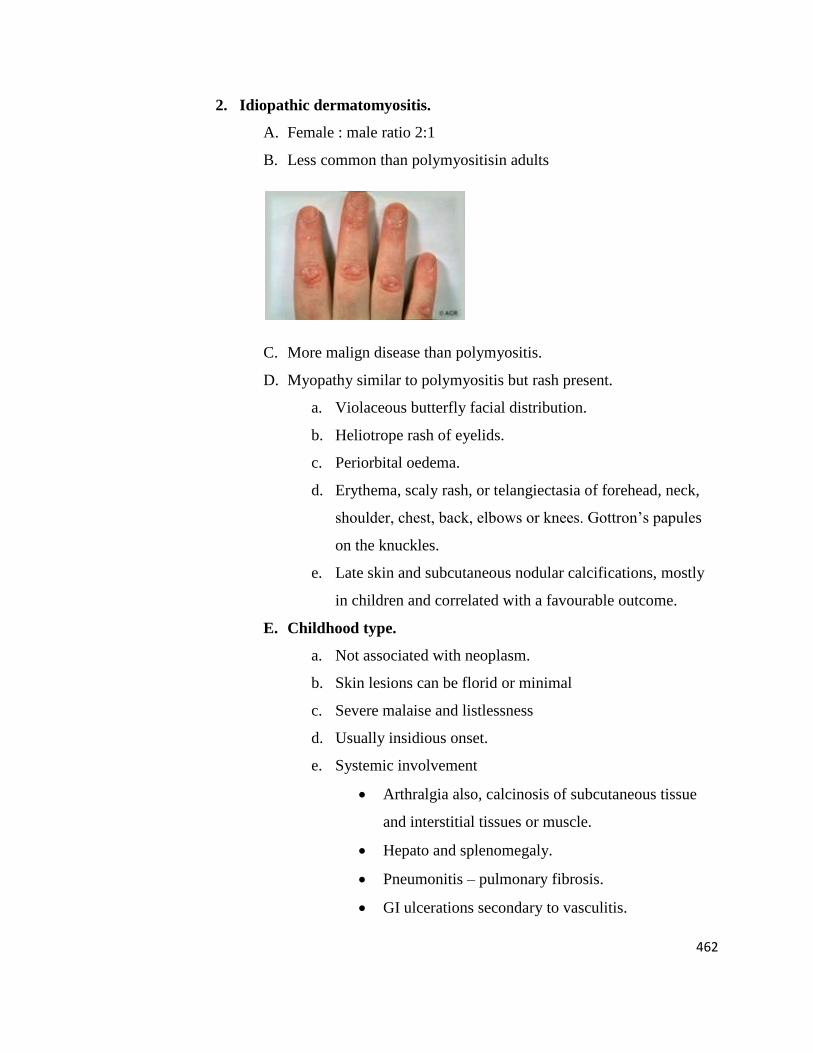
a. Violaceous butterfly facial distribution.
b. Heliotrope rash of eyelids.
c. Periorbital oedema.
d. Erythema, scaly rash, or telangiectasia of forehead, neck,
shoulder, chest, back, elbows or knees. Gottron’s papules
on the knuckles.
e. Late skin and subcutaneous nodular calcifications, mostly
in children and correlated with a favourable outcome.
E. Childhood type.
a. Not associated with neoplasm.
b. Skin lesions can be florid or minimal
c. Severe malaise and listlessness
d. Usually insidious onset.
e. Systemic involvement
Arthralgia also, calcinosis of subcutaneous tissue
and interstitial tissues or muscle.
Hepato and splenomegaly.
Pneumonitis – pulmonary fibrosis.
GI ulcerations secondary to vasculitis.

463
Cardiac involvement.
Renal involvement
Necrozingvasculitis and other angiopathic features.
Muscular contracture
f. C-reactive protein normal ( elevated with infection ), CPK
maybe normal.
3. Polymyositis and collagen vascular disease. Both dermatomyositis and
polymyositis may complicate other connective tissue disorder ( ― overlap
syndrome ― ). Either maybe found in association with:
a. Rheumatoid arthritis.
b. Systemic lupus erythematosus.
c. Scleroderma.
d. PeriarthritisNodosa.
. Inflammatory myositis associated with neoplasm.
A. Many autoimmune diseases are associated with an increased incidence of neoplasia.
Dermatomyositis with an onset after 40 years of age (particularly in a male) is often
accompanied by a malignant disease.
B. All types of malignancy may occur. Carcinoma of lungs, breast, ovary, uterus,
prostate, and stomach are most frequent.
C. In most cases, manifestations of inflammatory myopathy precede those of the tumor.
D. Treatment of the neoplasm may have favorable effect on associated muscle and skin
lesions.
V. Miscellaneous diseases that can cause secondary inflammatory myopathy.
Polymyalgia rheumatica.
Eosinophilic fasciitis
Trichinosis
Sarcoid
Cyscticercosis
464
Treatment
A. Steroids
1. High doses for three months
a. Avoid fluorinated steroid (dexamethasone and triamcinolone) as they more
frequently induce steroid myopathy.
b. Adults: 50-100 mg/kg/day.
c. Children: 1-2 mg/kg/day
2. Schedule
a. Initial daily dose
b. Can switch to alternate day dosage two to four weeks after initiating treatment
c. Observe patient closely for complications.
i. Acute complications
1) Edema
2) Weight gain
3) Hypertension
4) Diabetes
5) GI hemorrhage
ii. Chronic complications
1) Cataracts and ocular hypertension
2) Infection and poor wound healing
3) Psychosis
4) Osteoporosis ( Fractures )
5) Delayed growth
6) Myopathy
7) Cushinoid features
a) Moon facies
b) Central obesity
c) Buffalo hump
d) Facial hirsutism
e) Abdominal and thigh striae
465
8) Spontaneous tendon ruptures
a) Acne and thinning of skin
d. Necessary adjuncts to steroid therapy include:
i. Low sodium, high potassium intake.
ii. Antacids.
iii. High-protein, low carbohydrate diet.
e. Can reduce dose when clinical response has occurred.
1) Reduce very slowly.
2) Titrate treatment with serial CPK determination, but remember that CPK
normalization or increase may precede clinical remission or exacerbation by at
least several weeks. EMG can also be used to monitor disease activity
(fibrillation potentials indicate active disease)
3) Increase dose slightly or return to daily dosage if symptoms worsen or CPK
increases.
f. Recovery from dermatomyositis or polymyositis is slow ( although spontaneous
remissions can occur ) and, although some patients recover completely ( the
overall survival rate of both treated and untreated patients is 80% after five years,
although treatment seems to improve strength and lessen discomfort ), minimal
supportive steroid treatment maybe necessary for years in others. However, in
children, tapering of the dose can usually begin earlier ( persistence of skin rash is
not indicative of active disease ), and steroid treatment can often be discontinued
within three to six months.
B. Immunosuppressive drugs
1. Initiate when no response to three to six months of steroid treatment.
These drugs have teratogenic effects.
a) Methotrexate, 15-30mg/kg/day, may cause severe ulceration stomatitis
and leukopenia
b) Azathioprine, 3mg/kg/day, can cause drug fever.
c) Cyclophosphamide, 2mg/kg/day, may produce hemorrhage cystitis.
466
2. Plasmapheresis has been used as an adjunct to immunosuppressive
therapy.
C. Other measures
1. Physical therapy to prevent or treat contractures
2. Night splints as indicated
3. Topical steroids and Burow’s solution soaks for skin lesions.
4. Diphosphonates for calcinosis in dermatomyositis.
D. Factors decreasing survivorship ( most deaths occur in first two years after the
diagnosis ).
1. Age greater than 50 years.
2. Black race
3. Extreme weakness ( Dysphasia )
4. Pneumonitis
5. Neoplasm
6. Associated collagen disease
MISCELLANEOUS
I. Prader-willi syndrome ( H3O syndrome-hypotonia, hypomentia, hypogonadism, obesity
). Chromosome 15 breakage/translocation has been demonstrated in several cases.
A. Patients present with typical appearance of fair hair, blue eyes, high forehead,
small, almond-shaped eyes.
B. Diabetes develops in adolescence.
467
C. CPK, AMG and biopsy are all normal.
D. Higher incidence in males.
E. Marked hypotonia at birth-floppy infant.
F. Compulsive eating beginning at early childhood.
II. Arthrogryposis ( curved joints )
A. Symptom-complex, not a specific diagnostic entity.
B. Characterized by multiple joint contractures secondary to immobility of limbs in
utero.
C. Maybe myopathic or neuropathic.
D. Must differentiate from congenital muscular dystrophy and spinal muscular
atrophy.
E. Joint rigidity is secondary to fibrous ankylosis.
F. Fixed deformities can be surgically improved by aggressive soft tissue release.
III. Stiff man syndrome
A. Sustained repetitive activity of muscle fibers affecting both sexes, usually in adult
life.
1. Maybe produced by tightness of neck and chest muscles.
2. Results in uncontrollable contractions, mostly of musculature of the limb
girdles, but any and all voluntary muscles maybe involved.
3. Dyspnea, dysphagia, and facial grimacing may occur.
4. The limbs are held in rigid distorted position and the spasms are painful.
5. Stiffness disappears during sleep ( EEG shows less REM sleep than normal )
and general anesthesia, and maybe elicited by a variety of stimuli ( active or
passive movement, emotional stress ).
B. Physical examination reveals occasional hyperreflexia and extensor plantar
response.
C. EMG shows a sustained interference pattern but is otherwise normal.
D. Muscle spasms are abolished by curare peripheral nerve block and spinal
anesthesia.
E. Proposed etiology.

468
1. Gamma system overactivity.
2. Lack of inhibitory feedback to anterior horn cells.
3. Catecholaminergic- GABA system imbalance.
F. Treatment- high dose diazepam ( upto 300 mg/day ) or baclofen.
NOTE: Neuromyotonia ( continuous muscle fiber activity ) is yet another condition of an
abnormal muscle activity. It is characterized by myokymia secondary to brief tetanic
contractions of muscle fibers. This can be diagnosed by EMG. It may be simply annoying or
severe enough to cause rigidity and deformity. Pathology is apparently in the peripheral nerve.
Treatment is with diphenyldantoin or carbamazepine.
IV. Inclusion body myositis.
A. Progressive painless limb girdle weakness.
B. Normal mildly elevated CPK.
C. Unresponsive to steroids or immunosuppressive drugs.
D. Biopsy.
1. Myopathic changes.
2. Mononuclear inflammatory infiltrates.
3. Vacuoles lined with basophilic granules in muscle fibres.
Myopathy may attend a variety of other diseases such as collagen vascular disorders ( LE,
rheumatoid arthritis, polyarteritis nodosa ), sarcoid, carcinoma, Marfan’s syndrome.
Differential diagnosis of systemic myopathy based on age of onset
Myopathies presenting at birth:- None as systemic causes; mainly hereditary
Myopathies presenting in childhood:-
Inflammatory myopathy – dermatomyositis, polymyositis (rarely)
Infectious myopathies
Endocrine and metabolic disorders – hypokalemia, hypocalcemia, hypercalcemia
469
Myopathies presenting in adulthood
Inflammatory myopathy – polymyositis, dermatomyositis, inclusion body myositis, viral (HIV)
Infectious myopathies
Endocrine myopathies – thyroid, parathyroid, adrenal, pituitary disorders
Toxic myopathies – alcohol, corticosteroids, narcotics, colchicines, chloroquine
Critical illness myopathy
Metabolic myopathies
Para neoplastic myopathy
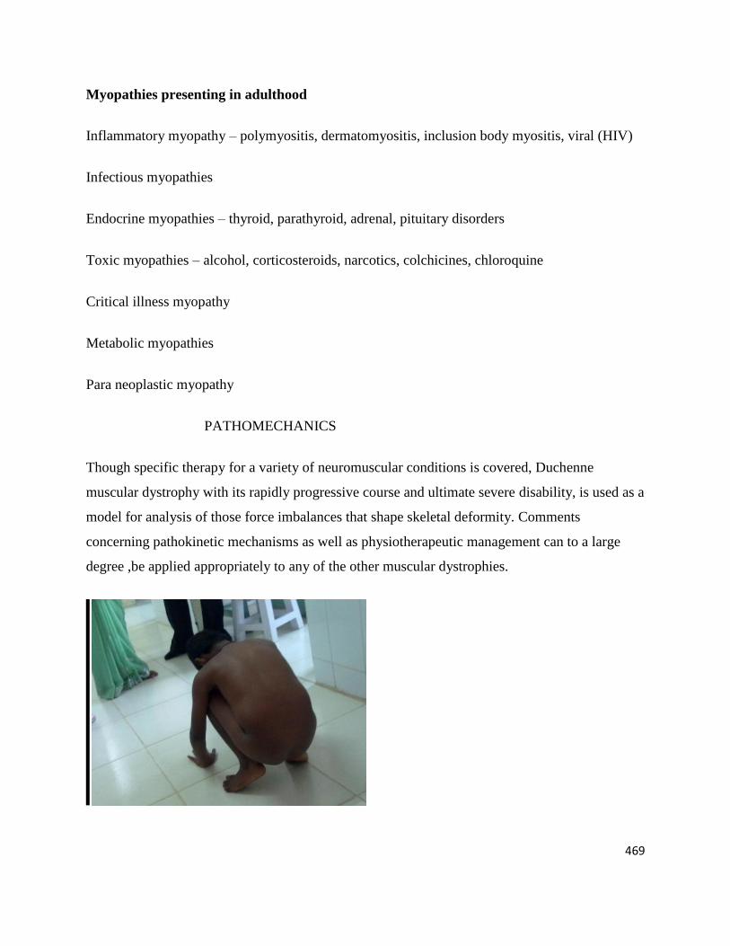
PATHOMECHANICS
Though specific therapy for a variety of neuromuscular conditions is covered, Duchenne
muscular dystrophy with its rapidly progressive course and ultimate severe disability, is used as a
model for analysis of those force imbalances that shape skeletal deformity. Comments
concerning pathokinetic mechanisms as well as physiotherapeutic management can to a large
degree ,be applied appropriately to any of the other muscular dystrophies.
470
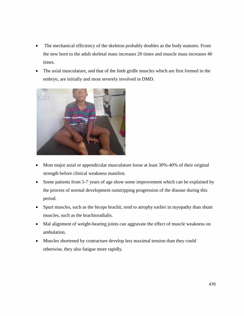
The mechanical efficiency of the skeleton probably doubles as the body matures. From
the new born to the adult skeletal mass increases 20 times and muscle mass increases 40
times.
The axial musculature, and that of the limb girdle muscles which are first formed in the
embryo, are initially and most severely involved in DMD.
Most major axial or appendicular musculature loose at least 30%-40% of their original
strength before clinical weakness manifest.
Some patients from 5-7 years of age show some improvement which can be explained by
the process of normal development outstripping progression of the disease during this
period.
Spurt muscles, such as the biceps brachii, tend to atrophy earlier in myopathy than shunt
muscles, such as the brachioradialis.
Mal alignment of weight-bearing joints can aggravate the effect of muscle weakness on
ambulation.
Muscles shortened by contracture develop less maximal tension than they could
otherwise, they also fatigue more rapidly.
471
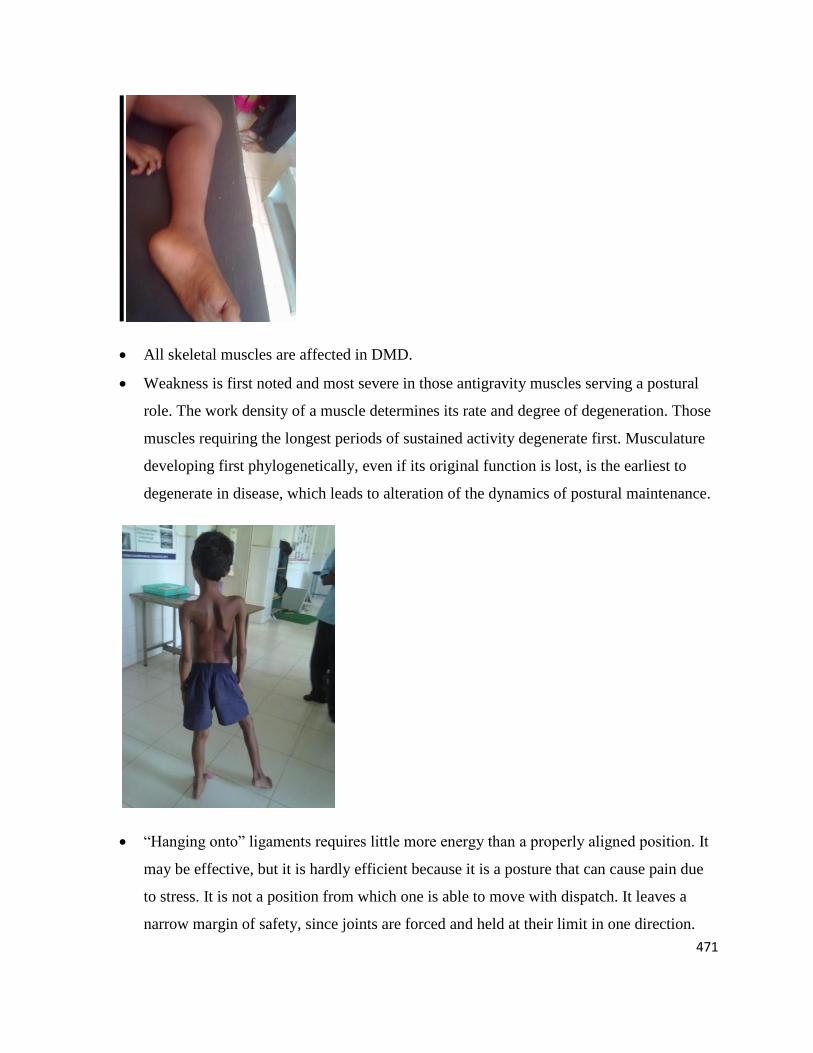
All skeletal muscles are affected in DMD.
Weakness is first noted and most severe in those antigravity muscles serving a postural
role. The work density of a muscle determines its rate and degree of degeneration. Those
muscles requiring the longest periods of sustained activity degenerate first. Musculature
developing first phylogenetically, even if its original function is lost, is the earliest to
degenerate in disease, which leads to alteration of the dynamics of postural maintenance.
―Hanging onto‖ ligaments requires little more energy than a properly aligned position. It
may be effective, but it is hardly efficient because it is a posture that can cause pain due
to stress. It is not a position from which one is able to move with dispatch. It leaves a
narrow margin of safety, since joints are forced and held at their limit in one direction.
472
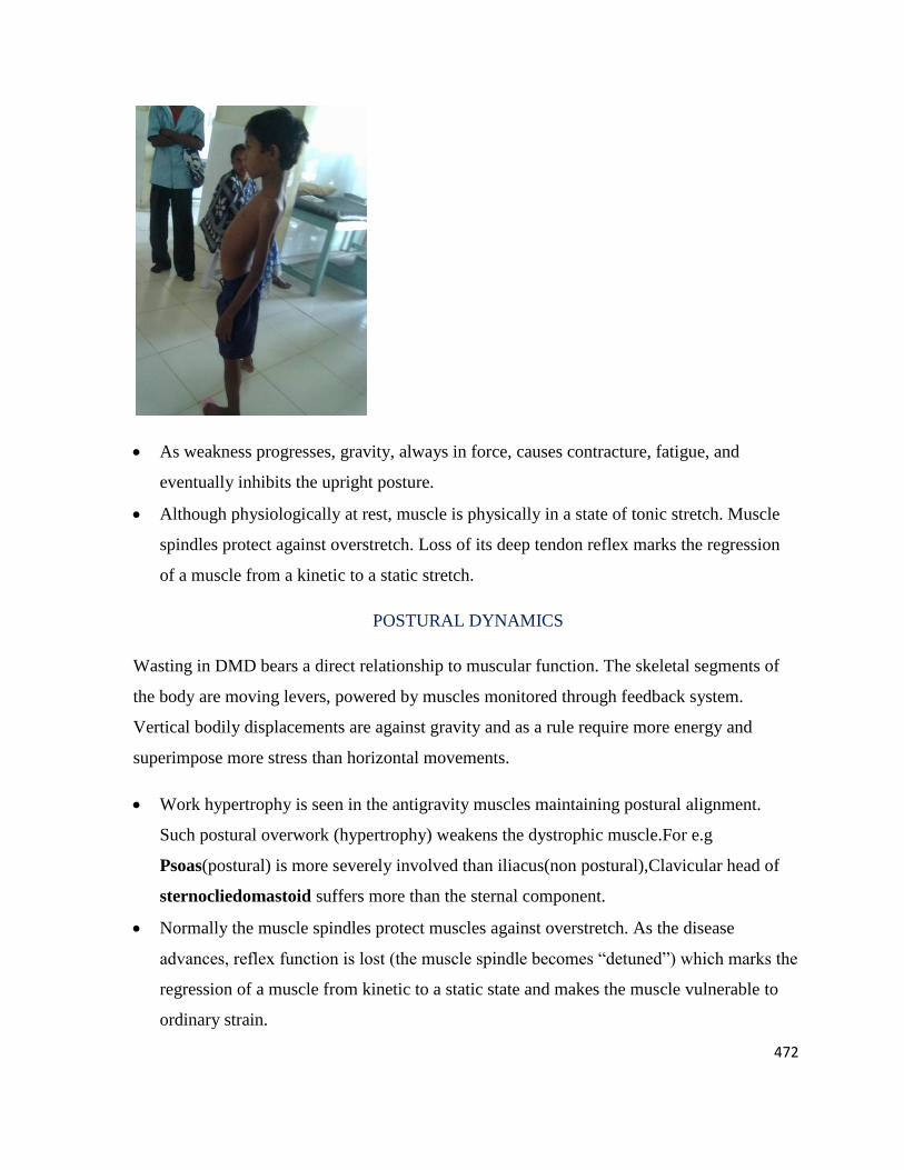
As weakness progresses, gravity, always in force, causes contracture, fatigue, and
eventually inhibits the upright posture.
Although physiologically at rest, muscle is physically in a state of tonic stretch. Muscle
spindles protect against overstretch. Loss of its deep tendon reflex marks the regression
of a muscle from a kinetic to a static stretch.
POSTURAL DYNAMICS
Wasting in DMD bears a direct relationship to muscular function. The skeletal segments of
the body are moving levers, powered by muscles monitored through feedback system.
Vertical bodily displacements are against gravity and as a rule require more energy and
superimpose more stress than horizontal movements.
Work hypertrophy is seen in the antigravity muscles maintaining postural alignment.
Such postural overwork (hypertrophy) weakens the dystrophic muscle.For e.g
Psoas(postural) is more severely involved than iliacus(non postural),Clavicular head of
sternocliedomastoid suffers more than the sternal component.
Normally the muscle spindles protect muscles against overstretch. As the disease
advances, reflex function is lost (the muscle spindle becomes ―detuned‖) which marks the
regression of a muscle from kinetic to a static state and makes the muscle vulnerable to
ordinary strain.
473
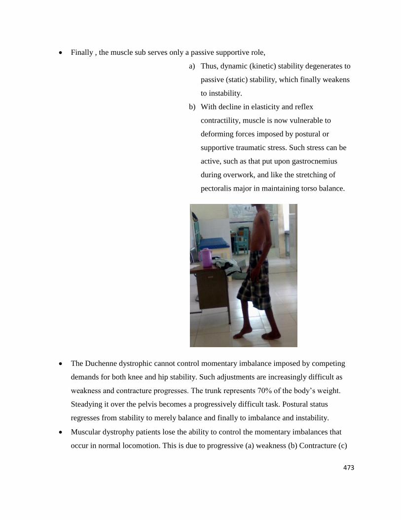
Finally , the muscle sub serves only a passive supportive role,
a) Thus, dynamic (kinetic) stability degenerates to
passive (static) stability, which finally weakens
to instability.
b) With decline in elasticity and reflex
contractility, muscle is now vulnerable to
deforming forces imposed by postural or
supportive traumatic stress. Such stress can be
active, such as that put upon gastrocnemius
during overwork, and like the stretching of
pectoralis major in maintaining torso balance.
The Duchenne dystrophic cannot control momentary imbalance imposed by competing
demands for both knee and hip stability. Such adjustments are increasingly difficult as
weakness and contracture progresses. The trunk represents 70% of the body’s weight.
Steadying it over the pelvis becomes a progressively difficult task. Postural status
regresses from stability to merely balance and finally to imbalance and instability.
Muscular dystrophy patients lose the ability to control the momentary imbalances that
occur in normal locomotion. This is due to progressive (a) weakness (b) Contracture (c)
474
and loss of muscle spindle proprioceptive function, resulting in an attempt to preserve as
minimal a level of energy expenditure as possible through exaggerations of motion at
unaffected, or less affected body levels.
Three processes contribute to the deformities of muscular dystrophy-
a) Muscle weakness
b) Muscle imbalance
c) Specific muscle contractures secondary to gravity and
compensatory postural habitus. Often contracture is
asymmetrical and less in dominant limb because of
relative increased activity.
BIOKINETICS- Clinical Correlation
In DMD ,hip flexors, TFL, and triceps surae develop ambulation limiting contractures.
Contractures are seldom symmetrical and seem to be greater on the non dominant side.
Quadriceps insufficiency is the key factor in gait deterioration.
The earliest postural change in the lower extremities is an increase in lumbar lordosis
secondary to gluteus maximus weakness. Hip extension at this time is largely performed
by the hamstrings muscle.
Some changes in joint lever systems occur. For instance, the hip is a first class lever with
force exerted by the abductors over the fulcrum of the articulated femoral head to balance
body weight. As hip abductors weaken, hip hikers (quadratuslumborum) are called upon
to elevate the hip during swing, thus creating a third class lever, where power is
sacrificed for a wider arc of movement.
475
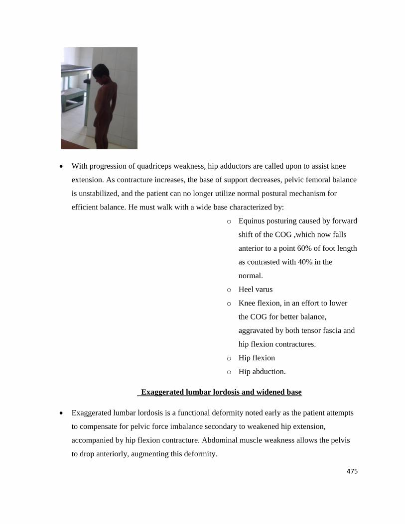
With progression of quadriceps weakness, hip adductors are called upon to assist knee
extension. As contracture increases, the base of support decreases, pelvic femoral balance
is unstabilized, and the patient can no longer utilize normal postural mechanism for
efficient balance. He must walk with a wide base characterized by:
o Equinus posturing caused by forward
shift of the COG ,which now falls
anterior to a point 60% of foot length
as contrasted with 40% in the
normal.
o Heel varus
o Knee flexion, in an effort to lower
the COG for better balance,
aggravated by both tensor fascia and
hip flexion contractures.
o Hip flexion
o Hip abduction.
Exaggerated lumbar lordosis and widened base
Exaggerated lumbar lordosis is a functional deformity noted early as the patient attempts
to compensate for pelvic force imbalance secondary to weakened hip extension,
accompanied by hip flexion contracture. Abdominal muscle weakness allows the pelvis
to drop anteriorly, augmenting this deformity.
476
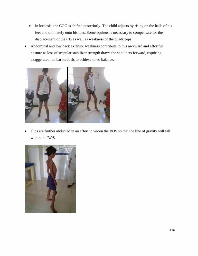
In lordosis, the COG is shifted posteriorly. The child adjusts by rising on the balls of his
feet and ultimately onto his toes. Some equinus is necessary to compensate for the
displacement of the CG as well as weakness of the quadriceps.
Abdominal and low back extensor weakness contribute to this awkward and effortful
posture as loss of scapular stabilizer strength draws the shoulders forward, requiring
exaggerated lumbar lordosis to achieve torso balance.
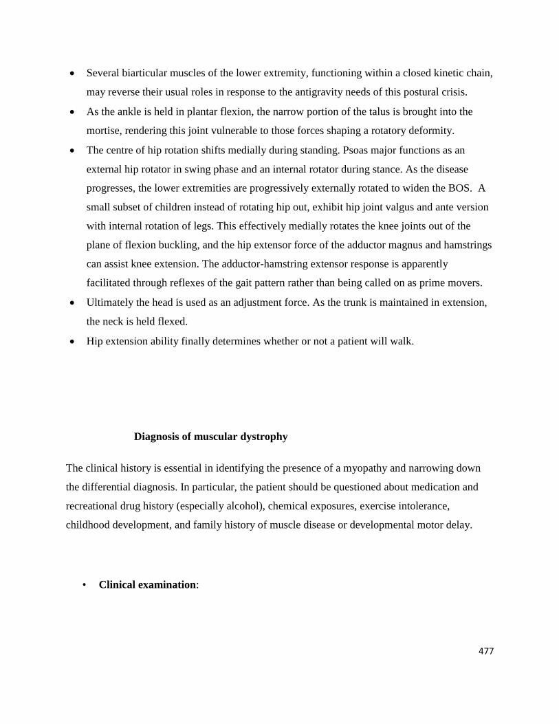
Hips are further abducted in an effort to widen the BOS so that the line of gravity will fall
within the BOS.
477
Several biarticular muscles of the lower extremity, functioning within a closed kinetic chain,
may reverse their usual roles in response to the antigravity needs of this postural crisis.
As the ankle is held in plantar flexion, the narrow portion of the talus is brought into the
mortise, rendering this joint vulnerable to those forces shaping a rotatory deformity.
The centre of hip rotation shifts medially during standing. Psoas major functions as an
external hip rotator in swing phase and an internal rotator during stance. As the disease
progresses, the lower extremities are progressively externally rotated to widen the BOS. A
small subset of children instead of rotating hip out, exhibit hip joint valgus and ante version
with internal rotation of legs. This effectively medially rotates the knee joints out of the
plane of flexion buckling, and the hip extensor force of the adductor magnus and hamstrings
can assist knee extension. The adductor-hamstring extensor response is apparently
facilitated through reflexes of the gait pattern rather than being called on as prime movers.
Ultimately the head is used as an adjustment force. As the trunk is maintained in extension,
the neck is held flexed.
Hip extension ability finally determines whether or not a patient will walk.
Diagnosis of muscular dystrophy
The clinical history is essential in identifying the presence of a myopathy and narrowing down
the differential diagnosis. In particular, the patient should be questioned about medication and
recreational drug history (especially alcohol), chemical exposures, exercise intolerance,
childhood development, and family history of muscle disease or developmental motor delay.
• Clinical examination:

478
Done by doctor/physician. Child will be asked to run, jump and climb stairs and after this he
will be asked to sit or get up from floor. If he puts his hands on the knees and pushes himself up,
it is a sign of muscular dystrophy known as Gower’s sign.
Laboratory Testing
The following laboratory tests may be used to evaluate patients with myopathies:
Blood test:
Blood sample is taken and checked for CPK level (Creatine PhosphoKinase is a chemical
substance released in blood by a damaged muscle. Usually in muscular dystrophy, the level is
10 times more than the normal.This testing, which can indicate muscle damage, includes
elevations in aldolase, lactate dehydrogenase (LDH), and liver function enzymes. A
screening panel of laboratory tests may also be obtained to rule out more common causes of
myopathy, which are listed in Box 2. In cases suspected to be a primary inflammatory
myopathy, specific autoantibodies can be considered to determine the prognosis and rule out
associated conditions. For example, the presence of anti-Jo antibody in dermatomyositis
predicts a superimposed interstitial lung disease. In addition, these patients should also be
evaluated for an underlying systemic autoimmune disease with an extensive autoimmune
panel and angiotensin-converting enzyme (ACE) levels. In myopathies that are accompanied
by polyneuropathy, renal involvement, and a restrictive cardiomyopathy, immunofixation
electrophoresis studies in the serum and urine should be considered to rule out the possibility
of amyloid disease.
Normal CPK levels:
Males: 38 – 174 units/L
Female: 96 – 140 units/L
Genetic testing
479
Genetic testing is available for some inherited myopathies. These are listed in Table 2. .
Genetic testing for DMD/BMD is commonly available in India. For LGMD or FSHD it is
available in very few centers. It is important for several reasons to have the genetic
confirmation of the diagnosis as it will help to determine if the patient is eligible for any
clinical trials and also helps the family to take decision regarding prenatal diagnosis and
future planning of pregnancies.
Carrier analysis: In few cases, the mother is the carrier of the faulty genes which means
that the mother must have passed the faulty gene or disease to the child. However, the
mother is not always a carrier. It could be a fresh mutation. If mother is found to be the
carrier, genetic counseling should be advised for other female relatives of the child.
Muscle biopsy
Under anesthesia, a small piece of muscle is taken from the bulkiest muscle and
examined under a microscope. Histopathologic examination of muscle may be helpful
in determining the specific type of muscle disease, especially in patients with a
suspected inflammatory or infectious myopathy. Selecting the optimal muscle to biopsy
is very important because factors such as severe weakness and technical artifacts can
hamper an accurate histologic diagnosis. The ideal muscle that should be sampled is
one that is clinically involved but still antigravity in strength, because more-severe
weakness can lead to unhelpful, nonspecific findings of fibrosis. Also avoid muscles
that have been examined by an EMG should be avoided because the needle portion of
the electrical study might have caused local damage, which can result in spurious
findings. Common biopsy sites include the biceps and deltoid muscles in the upper
extremity and the quadriceps and gastrocnemius muscles in the lower extremity.
The electromyogram (EMG)
EMG is an electrical study of the nerves and muscles that plays an important role in
confirming the presence, duration, and severity of a myopathy. A thin-needle is inserted
through the muscle to be tested and electrical activity is studied. This also can give a
480
clue whether the muscle is damaged or not.The study can also disclose special findings
such as myotonic potentials. This is the electrical equivalent of clinical myotonia,
which is manifested as impaired relaxation of muscles after forceful contraction; for
example, patients cannot release objects from their grip. Myotonic potentials have the
characteristic sound of a dive bomb on EMG and can help point toward the diagnosis of
myotonic dystrophy when found in the appropriate muscles.Although integral in the
evaluation of a myopathy, the EMG can be normal in mild myopathies, steroid
myopathies, and a number of metabolic myopathies. Therefore, it is important to
remember that a normal EMG does not exclude the presence of a myopathy.
Ischemic Forearm Test
A traditional test used in the evaluation of a suspected metabolic myopathy is the ischemic
forearm test. This is performed by obtaining baseline serum ammonia and lactate levels taken
from the forearm. The patient then exercises that arm for 1 minute, after which repeat serum
lactate and blood ammonia levels are measured. This is repeated at several intervals (1, 2, 5,
and 10 minutes). In normal muscle, the resultant ischemia causes a 3- to 5-fold rise in lactate
levels. In contrast, patients with glycogen storage disorders demonstrate no change in lactate
levels after exercise.
MRI or Magnetic Resonance Imaging:
MRI is a painless and non surgical procedure which is done to examine the muscle quality, size
or any abnormalities in the size. It monitors the fatty replacement of the muscle tissue as well as
progression of the disease.
Box 2 Laboratory Evaluation for Suspected Myopathy
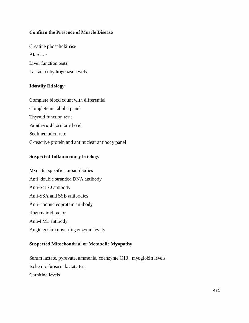
481
Confirm the Presence of Muscle Disease
Creatine phosphokinase
Aldolase
Liver function tests
Lactate dehydrogenase levels
Identify Etiology
Complete blood count with differential
Complete metabolic panel
Thyroid function tests
Parathyroid hormone level
Sedimentation rate
C-reactive protein and antinuclear antibody panel
Suspected Inflammatory Etiology
Myositis-specific autoantibodies
Anti–double stranded DNA antibody
Anti-Scl 70 antibody
Anti-SSA and SSB antibodies
Anti-ribonucleoprotein antibody
Rheumatoid factor
Anti-PM1 antibody
Angiotensin-converting enzyme levels
Suspected Mitochondrial or Metabolic Myopathy
Serum lactate, pyruvate, ammonia, coenzyme Q10 , myoglobin levels
Ischemic forearm lactate test
Carnitine levels
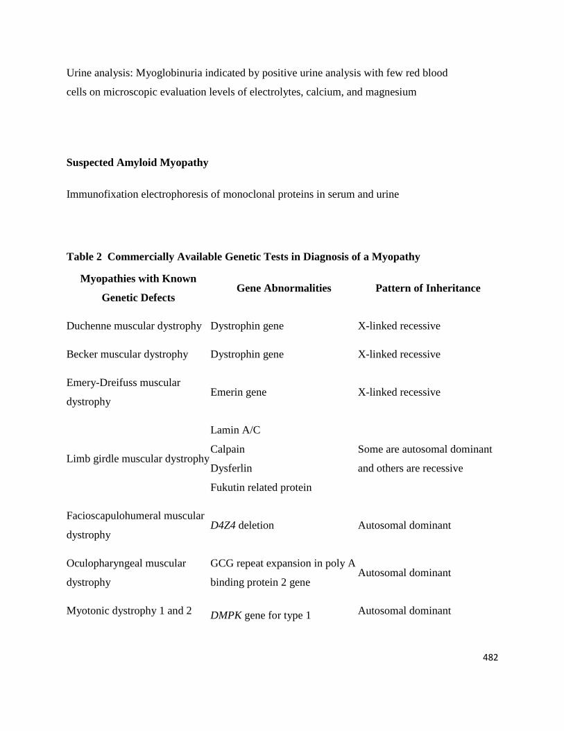
482
Urine analysis: Myoglobinuria indicated by positive urine analysis with few red blood
cells on microscopic evaluation levels of electrolytes, calcium, and magnesium
Suspected Amyloid Myopathy
Immunofixation electrophoresis of monoclonal proteins in serum and urine
Table 2 Commercially Available Genetic Tests in Diagnosis of a Myopathy
Myopathies with Known
Genetic Defects Gene Abnormalities Pattern of Inheritance
Duchenne muscular dystrophy Dystrophin gene X-linked recessive
Becker muscular dystrophy Dystrophin gene X-linked recessive
Emery-Dreifuss muscular
dystrophy Emerin gene X-linked recessive
Limb girdle muscular dystrophy
Lamin A/C
Calpain
Dysferlin
Fukutin related protein
Some are autosomal dominant
and others are recessive
Facioscapulohumeral muscular
dystrophy D4Z4 deletion Autosomal dominant
Oculopharyngeal muscular
dystrophy
GCG repeat expansion in poly A
binding protein 2 gene Autosomal dominant
Myotonic dystrophy 1 and 2 DMPK gene for type 1 Autosomal dominant
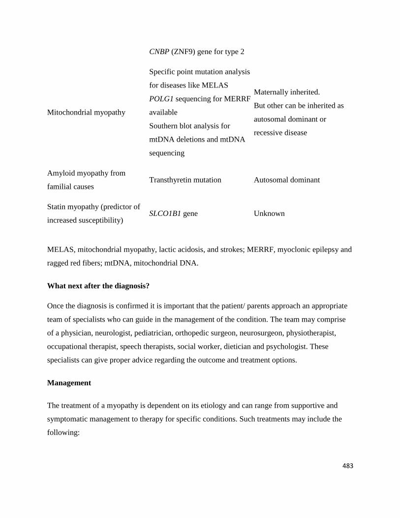
483
CNBP (ZNF9) gene for type 2
Mitochondrial myopathy
Specific point mutation analysis
for diseases like MELAS
POLG1 sequencing for MERRF
available
Southern blot analysis for
mtDNA deletions and mtDNA
sequencing
Maternally inherited.
But other can be inherited as
autosomal dominant or
recessive disease
Amyloid myopathy from
familial causes Transthyretin mutation Autosomal dominant
Statin myopathy (predictor of
increased susceptibility) SLCO1B1 gene Unknown
MELAS, mitochondrial myopathy, lactic acidosis, and strokes; MERRF, myoclonic epilepsy and
ragged red fibers; mtDNA, mitochondrial DNA.
What next after the diagnosis?
Once the diagnosis is confirmed it is important that the patient/ parents approach an appropriate
team of specialists who can guide in the management of the condition. The team may comprise
of a physician, neurologist, pediatrician, orthopedic surgeon, neurosurgeon, physiotherapist,
occupational therapist, speech therapists, social worker, dietician and psychologist. These
specialists can give proper advice regarding the outcome and treatment options.
Management
The treatment of a myopathy is dependent on its etiology and can range from supportive and
symptomatic management to therapy for specific conditions. Such treatments may include the
following:
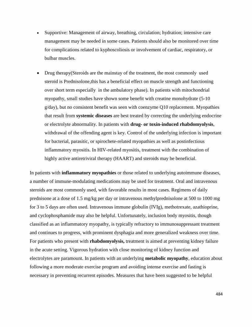
484
Supportive: Management of airway, breathing, circulation; hydration; intensive care
management may be needed in some cases. Patients should also be monitored over time
for complications related to kyphoscoliosis or involvement of cardiac, respiratory, or
bulbar muscles.
Drug therapy(Steroids are the mainstay of the treatment, the most commonly used
steroid is Prednisolone,this has a beneficial effect on muscle strength and functioning
over short term especially in the ambulatory phase). In patients with mitochondrial
myopathy, small studies have shown some benefit with creatine monohydrate (5-10
g/day), but no consistent benefit was seen with coenzyme Q10 replacement. Myopathies
that result from systemic diseases are best treated by correcting the underlying endocrine
or electrolyte abnormality. In patients with drug- or toxin-induced rhabdomyolysis,
withdrawal of the offending agent is key. Control of the underlying infection is important
for bacterial, parasitic, or spirochete-related myopathies as well as postinfectious
inflammatory myositis. In HIV-related myositis, treatment with the combination of
highly active antiretriviral therapy (HAART) and steroids may be beneficial.
In patients with inflammatory myopathies or those related to underlying autoimmune diseases,
a number of immune-modulating medications may be used for treatment. Oral and intravenous
steroids are most commonly used, with favorable results in most cases. Regimens of daily
prednisone at a dose of 1.5 mg/kg per day or intravenous methylprednisolone at 500 to 1000 mg
for 3 to 5 days are often used. Intravenous immune globulin (IVIg), methotrexate, azathioprine,
and cyclophosphamide may also be helpful. Unfortunately, inclusion body myositis, though
classified as an inflammatory myopathy, is typically refractory to immunosuppressant treatment
and continues to progress, with prominent dysphagia and more generalized weakness over time.
For patients who present with rhabdomyolysis, treatment is aimed at preventing kidney failure
in the acute setting. Vigorous hydration with close monitoring of kidney function and
electrolytes are paramount. In patients with an underlying metabolic myopathy, education about
following a more moderate exercise program and avoiding intense exercise and fasting is
necessary in preventing recurrent episodes. Measures that have been suggested to be helpful
485
include sucrose loading before exercise in some glycogen storage disorders and a low-fat, high-
carbohydrate diet in patients with lipid storage disorders.
Additives or nutritional supplements(Vitamin E ,coenzyme Q ,omega 3 fatty acid,etc
have general protective effects, act as antioxidants,have effect on strengthening the cell
wall.studies show that they support formation of new muscle cells to repair the injured
muscle.However evidence is not clear on its protective effects in muscular dystrophy.
Physical therapy
Assistive devices and alternative techniques.(Equipment to help in eating ,drinking
,grooming, home modifications, orthoses and night splints make the persons more
independent.
Bracing
Surgery, Tendon release surgery - for example, to prolong the ability to walk.
Stem cell Therapy (The usefulness of this therapy has been proved in terms of functional
improvements and slowing down the disease progression).
Prognosis
This depends on the specific diagnosis. The primary disorders are incurable
conditions with varied prognosis. Secondary myopathy may be corrected by treating the
underlying cause.
Prevention
Genetic counselling is, in some of the most common myopathies such as Duchenne muscular
dystrophy (DMD), the only intervention that can prevent disease. In general:
Give genetic counselling early.
Test early for carrier status where appropriate.
Consider prenatal diagnostic testing where appropriate.
Advances in molecular genetics may help in the future.
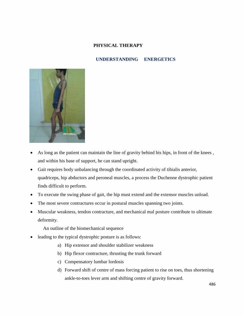
486
PHYSICAL THERAPY
UNDERSTANDING ENERGETICS
As long as the patient can maintain the line of gravity behind his hips, in front of the knees ,
and within his base of support, he can stand upright.
Gait requires body unbalancing through the coordinated activity of tibialis anterior,
quadriceps, hip abductors and peroneal muscles, a process the Duchenne dystrophic patient
finds difficult to perform.
To execute the swing phase of gait, the hip must extend and the extensor muscles unload.
The most severe contractures occur in postural muscles spanning two joints.
Muscular weakness, tendon contracture, and mechanical mal posture contribute to ultimate
deformity.
An outline of the biomechanical sequence
leading to the typical dystrophic posture is as follows:
a) Hip extensor and shoulder stabilizer weakness
b) Hip flexor contracture, thrusting the trunk forward
c) Compensatory lumbar lordosis
d) Forward shift of centre of mass forcing patient to rise on toes, thus shortening
ankle-to-toes lever arm and shifting centre of gravity forward.
487
e) Hip abduction to increase equilibrium by widening body’s BOS.
f) Secondary TFL contractures.
g) External tibial torsion; thus, ankle and knee axes are no longer in the same
plane.
h) Ankle varus.
i) Mild knee flexion to lower centre of mass and gain balance.
j) Progressive quadriceps weakness requiring to lock knees for stability.
k) With foot fixed in equinus and knee flexed, gastrocnemius and soleus can now
act to extend knee.
l) True overwork exercise hypertrophy of calves.
Generalized weakness makes it increasingly difficult to attain alignment stability when the
trunk is balanced over unstable lower extremities.
Propriceptive abilities are also affected.
A slight weight gain or a period of several days of bed rest can decrease strength enough to
impair ambulation. In its later stages DMD is a brittle disease, and the transition from
moderately to severely impaired ambulation may be abrupt.
Because of individual differences in weakness, contracture, weight, and motivation, age
alone is a poor index of disease progression.
The more disabled the patient, the more determinants of gait are lost, the more energy is
required for ambulation, and the less efficient is the gait.
PHYSICAL THERAPY - ASSESSMENT
THE roles that the physical therapist plays in the management of the patient with muscular
dystrophy are several.
The therapist monitors the assessment of specific muscle weakness, imbalance and contracture.
ASSESSMENT
Strength and range of motion: shoulders, hips, knees.
Contractures: hip flexors, TFL, and heel cord
488
Function: rising stairs, walking, etc.
Muscle testing grades key muscles as to strength and range of motion of activated joints
against gravity
The patient’s ability to perform a number of standard tasks is assessed, measuring time
required and observing method employed to complete the task. Subtle alterations in method
as well as slight changes in time needed for any given task objectively reflect changes in
strength.
The patient can be rated on a functional 10 point –step scale.
a) Stage 1, walks and climbs stairs without assistance(it
takes 15 times as much energy to ascend a flight of
ordinary stairs as to walk a level distance equal to the
vertical height of the stairs).
b) Stage2, walks and climbs stairs easily with aid of
railing
c) Stage3 walks and climbs stairs slowly with aid of
railing.
d) Stage4 walks but cannot climb stairs.
e) Stage5, walk unassisted but cannot climb chair or get
out of chair.
f) Stage6, walks only with assistance or only with braces
g) Sage7, in wheelchair, sits erect and can roll chair and
perform bed and wheelchair activities of daily living.
h) Stage8, in wheelchair, sits erect but is unable to
perform bed and chair activities without resistance.
i) Stage9, in wheelchair, sits erect only with support, and
able to do only minimal activities of daily living.
j) Stage10, in bed, cannot perform activities of daily
living without resistance.
489
The kinetic sequence of the above scale provides a treatment format for the physical therapy
program.
a) In stages 1-3, the patient is ambulating independent. Passive stretch of early
lower extremity contracture may be necessary by stage3. Functional activities of
daily living and ambulation are sufficient active exercise for stages 1-3.
b) Between stages 5-6, lower extremity surgery is indicated because contracture
leads to increased difficulty with anti gravity activities. Where contracture is
minimal or absent, orthotic modifications or bracing alone may be sufficient to
augment weakened knee extension and keep the patient ambulating. With fair
quadriceps strength, the knee flexion moment can be decreased by an anteriorly
places cushion(SACH) heel, or converted to an extension moment with an
equinus(floor reaction) AFO
c) Ambulant patients in stage 5 and 6 should not avoid activity and use wheelchair,
except when absolutely necessary and then for only brief periods of time.
d) Stage 7 should not be skipped as the patient is thus condemned to stage 10
immediately.
e) When stage 7 is reached, routine conditioning exercises are prescribed to retard
disuse atrophy and maintain independence in wheelchair activities. Prophylactic
treatment of scoliosis is also initiated at this time as well as a full program of
respiratory therapy.
f) Obesity is usually a problem after stage 6.
g) The closer a patient is to stage 10, the more assistive devices he requires.
Explanation of the testing procedures
1. Run 25 feet
Both feet off the ground at one time.
2. Walk 25 feet
a. Independent – no assistance from mechanical aids
b. Short leg braces- test items 3, 4 and 5 with braces also.
c. Long leg braces – test items 3, 4, and5 with braces also.

490
d. Crutches, walker or cane.
Stair measurements: Depth-11", height -7", railing height- 36"
3. Climb 8 stairs
a. Independent- foot over foot- no assistance such as pushing on knee. Using
rail
b. Independent- one step at a time
c. Hands on knees- assistance from rail
d. Railing- one hand
e. Railing and hand on knee- patient pulls on rail with one hand, pushes on
knee with the other.
f. Railing- two hands, some patients may use two rails
4. Walk down 8 stairs
a. Independent- No assistance from rail. May go foot over foot or one step at
a time
b. Railing- One hand , often used for safety but not for support
c. Railing- Two hands, or rail and therapists hand for safety.
5. Rise from chair
Care must be taken to seat the patient in a chair which places his feet flat on the
floor and his knees in 90°flexion. This is particularly important with children as a
higher chair would give them mechanical advantage.
a. Independent- rising without pushing on chair or knees, arms folded across
chest or extended.
b. Gower’s sign- includes pressure on knees or pressure on seat of chair or
both.
c. Turn to side, and then push up- patient turns sideways in chair to sit on
one hip, with feet on floor pushes with arms to 90° hip flexion and pushes
off to upright position or climbs up chair to upright position.
491
d. Pull up with aid of table-patient takes support from table with hips flexed
while extending knees. Once knees are locked, patient extends hip by
pushing up on table.
6. Roll over from supine to prone
Contractures may inhibit this activity.
7. Roll over from prone to supine
Contractures may inhibit this activity.
8. Get to all four position from prone
Push up to hands and knees
9. Sit up from supine
a. Independent-no aids other than having ankles held down by therapist
which is within the range of normal; must achieve sitting balance.
b. Pull up- patient holds on to leg or clothing to pull him up.
c. Pull and push on elbow- begins to pull up, then pushes on at least one
elbow.
d. Turn to side, and then push up-patient will roll to side, and then push up
with both arms to achieve sitting balance.
e. To hands and knees and then sit up-roll to prone, to hands and knees, and
then to side sit or other sitting balance.
10. Rise up from floor from prone position
a. Independent- no assistance required
b. Gower’s sign- can bring himself to his feet but must push on knees to
assume erect posture.
c. Chair to standing-patient pulls himself to feet with aid of chair, then
pushes on chair to achieve upright position
d. Chair to sitting, then to standing- patient pulls himself to sitting position in
chair, then pushes himself to upright position, using chair. Patient should
be placed on edge of table with thighs fully supported and knees flexed to
90°.
11. Sitting balance

492
a. Stable
b. Unstable- Patient sits erect but cannot recover balance if it is lost. Or,
patient is unable to maintain his balance without support of his arms.
12. Vital capacity
Measured sitting, taking highest of two readings. Take weight and height into
consideration.
13. Height
14. Weight
15. Hand grip
Measured with hand grip dynamometer taking highest of two readings for left and
right hands. Keep arms at side during measuring.
PHYSICAL THERAPY TREATMENT
Physical therapy includes stretching
(preventing and correcting contractures), muscular strengthening and gait
training, thus increasing efficiency in the functional activities of daily living,
including ADL and transfer.
Strengthening deforming muscles by prompting the patient to fight the
passive force by an active effort of his own.
Disability from weakness must be distinguished from that caused by
contracture. The essential tenodesis effect of contracure in the face of
severe loss of muscular support should be monitored by the therapist.
Initially such compensating mechanism help maintain proper body
alignment. Eventually they contribute to ambulatory loss.
Stretching of tight muscles and prevention of contractures
Overenthusiastic stretching of contractures should be avoided because it produces pain ad
stimulates the stretch reflex.
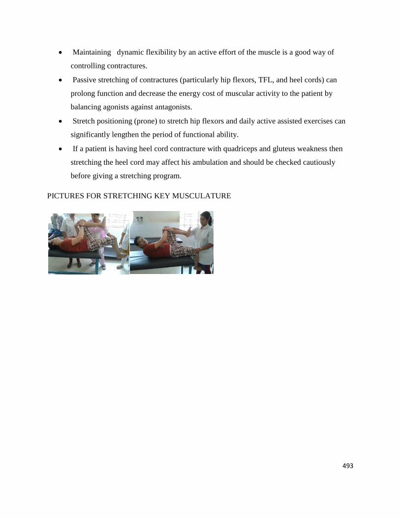
493
Maintaining dynamic flexibility by an active effort of the muscle is a good way of
controlling contractures.
Passive stretching of contractures (particularly hip flexors, TFL, and heel cords) can
prolong function and decrease the energy cost of muscular activity to the patient by
balancing agonists against antagonists.
Stretch positioning (prone) to stretch hip flexors and daily active assisted exercises can
significantly lengthen the period of functional ability.
If a patient is having heel cord contracture with quadriceps and gluteus weakness then
stretching the heel cord may affect his ambulation and should be checked cautiously
before giving a stretching program.
PICTURES FOR STRETCHING KEY MUSCULATURE
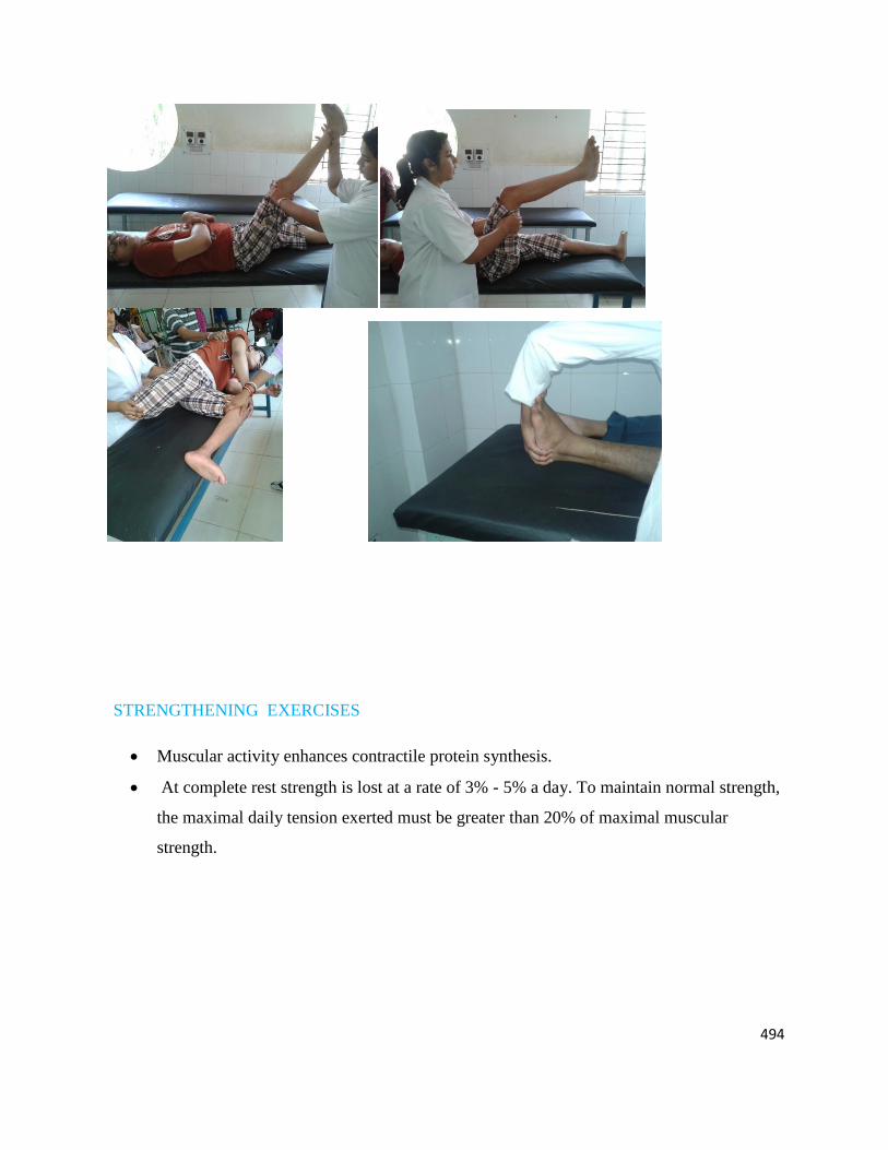
494
STRENGTHENING EXERCISES
Muscular activity enhances contractile protein synthesis.
At complete rest strength is lost at a rate of 3% - 5% a day. To maintain normal strength,
the maximal daily tension exerted must be greater than 20% of maximal muscular
strength.
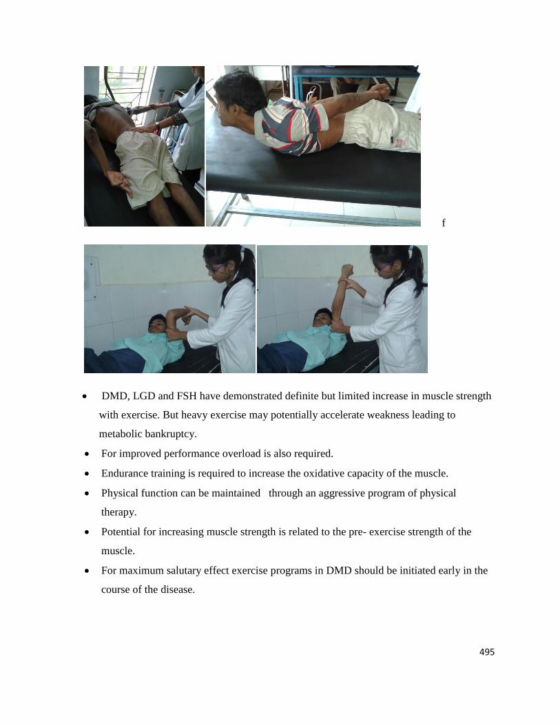
495
f
DMD, LGD and FSH have demonstrated definite but limited increase in muscle strength
with exercise. But heavy exercise may potentially accelerate weakness leading to
metabolic bankruptcy.
For improved performance overload is also required.
Endurance training is required to increase the oxidative capacity of the muscle.
Physical function can be maintained through an aggressive program of physical
therapy.
Potential for increasing muscle strength is related to the pre- exercise strength of the
muscle.
For maximum salutary effect exercise programs in DMD should be initiated early in the
course of the disease.
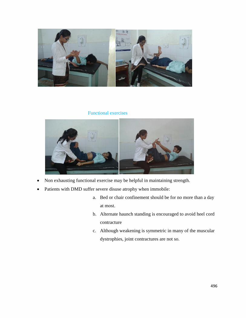
496
Functional exercises
Non exhausting functional exercise may be helpful in maintaining strength.
Patients with DMD suffer severe disuse atrophy when immobile:
a. Bed or chair confinement should be for no more than a day
at most.
b. Alternate haunch standing is encouraged to avoid heel cord
contracture
c. Although weakening is symmetric in many of the muscular
dystrophies, joint contractures are not so.
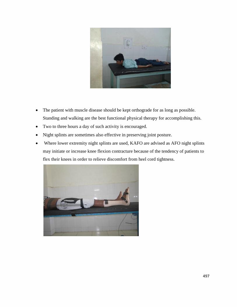
497
The patient with muscle disease should be kept orthograde for as long as possible.
Standing and walking are the best functional physical therapy for accomplishing this.
Two to three hours a day of such activity is encouraged.
Night splints are sometimes also effective in preserving joint posture.
Where lower extremity night splints are used, KAFO are advised as AFO night splints
may initiate or increase knee flexion contracture because of the tendency of patients to
flex their knees in order to relieve discomfort from heel cord tightness.
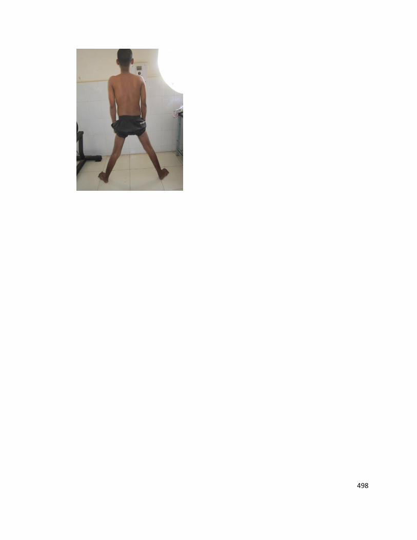
498
499
Summary
Myopathy refers to skeletal and cardiac muscle dysfunction from various inherited,
metabolic, inflammatory, infectious, or toxic etiologies.
Patients typically present with proximal muscle weakness of legs more than arms, with
no sensory involvement.
Age of presentation, duration of illness, and distribution of weakness are helpful in
determining the classification and etiology of myopathy.
Serologic testing, electromyography, muscle biopsy, and genetic testing are helpful tools
in identifying the presence of myopathy and determining the etiology.
Management is largely supportive for an inherited myopathy. In acquired myopathies,
treatment is targeted toward the underlying cause.
Treatment of statin myopathies is dependent on creatine phosphokinase levels and degree
of muscle symptoms. Consider lower doses when initiating statin therapy.


![Polymyositis-DermatomyositisandInterstitialLungDiseasein ...downloads.hindawi.com/journals/crirh/2019/4914631.pdf · with polymyositis based on Bohan’s criteria [1]. Further-more,shehadexertionaldyspnea,andtheambulatorySpO](https://static.fdocuments.net/doc/165x107/5ec17b508bf9e30bb42ca8c5/polymyositis-dermatomyositisandinterstitiallungdiseasein-with-polymyositis-based.jpg)