Intracranial hemorrhage and neonatal autoimmune ... · Intracranial hemorrhage and neonatal...
16
September 2017 Intracranial hemorrhage and neonatal autoimmune thrombocytopenia: a rare and unpredictable event SWISS SOCIETY OF NEONATOLOGY
Transcript of Intracranial hemorrhage and neonatal autoimmune ... · Intracranial hemorrhage and neonatal...
September 2017
Intracranial hemorrhage
and neonatal autoimmune
thrombocytopenia: a rare and
unpredictable event
SWISS SOCIETY OF NEONATOLOGY
Becocci A, Fluss J, Felice-Civitillo C, Laurent M, Boehlen F,
De Luca R, Neonatology and Intensive Care Unit (BA, FCC,
DLR), Pediatric Neurology Unit, Pediatric Subspecialties
Service (FJ), Pediatric Radiology Unit (LM), Division of
Angiology and Hemostasis (BF), University Hospitals of
Geneva
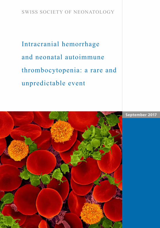
Title figure:
Human blood (source: https:/ /www.nigms.nih.gov/
education/ life-magnified/Pages/1b4_kunkel-human-
blood.aspx)
© Swiss Society of Neonatology, Thomas M Berger, Webmaster
Fetal and neonatal immune thrombocytopenia
are caused by maternal IgG crossing the placenta
and destroying fetal platelets. Two main forms are
de scribed. The autoimmune condition is related to
maternal immune thrombocytopenia (ITP), while the
alloimmune form, commonly named fetal and neo-
natal alloimmune thrombocytopenia (FNAIT), is due to
transplacental passage of specific antibodies against
fetal platelets exhibiting antigens inherited from the
father. The incidence of fetal and neonatal intra-
cranial hemorrhage in those two conditions differs
widely, with 10 – 30% in FNAIT and 0 – 2.9% in the
auto immune form (1, 2 – 4). We here describe the case
of a male newborn infant with early onset of severe
autoimmune thrombocytopenia complicated by symp-
tomatic neonatal intracranial hemorrhage.
INTRODUCTION
3
CASE REPORT
4
This Caucasian boy was born by spontaneous vaginal
delivery to a 29-year-old G1/P1 at 39 1/7 weeks of
gestation. The mother had been affected by ITP since
the age of 7 years, requiring treatment with oral
steroids and intravenous immunoglobulin (IVIG)
admini stration during adolescence.
Pregnancy had been uneventful and no anomalies had
been found on prenatal ultrasounds. The mother’s
platelet counts were between 30 and 60 G/L during
the first trimester of pregnancy. At 18 weeks of gesta-
tion, she received a short course of oral prednisone the-
rapy before amniocentesis because of a platelet count of
35 G/L. Her platelet count progressively increased
during pregnancy and no further treatment was
required. At the time of delivery, her platelet count
was 135 G/L.
Labor and spontaneous vaginal delivery were
un eventful. Birth weight was 3050 g (P10 – 25), length
48 cm (P5) and head circumference 35 cm (P25 – 50).
The neonate adapted well to extrauterine life (Apgar
scores 9, 10 and 10 at 1, 5 and 10 minutes, respecti-
vely) and clinical examination at birth was normal.
There was no risk of infection. A complete cord blood
count revealed severe thrombocytopenia (5 G/L) that
was confirmed on a venous blood sample. The baby
was promptly admitted to the neonatology unit and
received an immunoglobulin infusion (400 mg/ kg) and
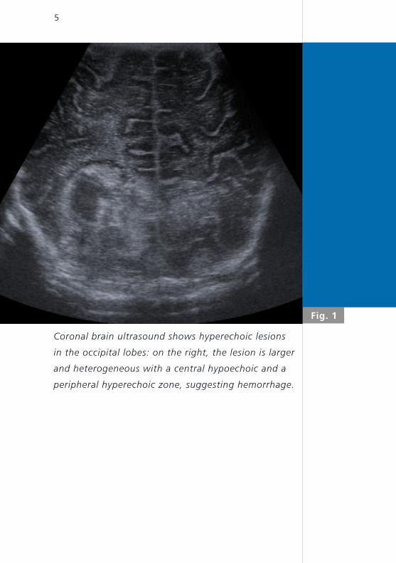
Fig. 1
5
Coronal brain ultrasound shows hyperechoic lesions
in the occipital lobes: on the right, the lesion is larger
and heterogeneous with a central hypoechoic and a
peripheral hyperechoic zone, suggesting hemorrhage.
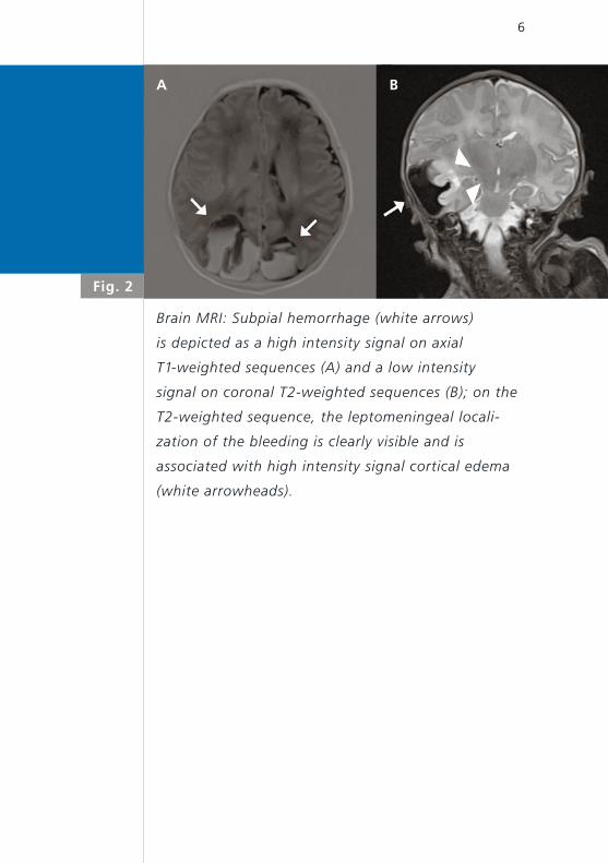
Fig. 2
6
Brain MRI: Subpial hemorrhage (white arrows)
is depicted as a high intensity signal on axial
T1-weighted sequences (A) and a low intensity
signal on coronal T2-weighted sequences (B); on the
T2-weighted sequence, the leptomeningeal locali-
zation of the bleeding is clearly visible and is
associated with high intensity signal cortical edema
(white arrowheads).
A B
7
a platelet transfusion (15 ml/kg) from donor apheresis.
The platelet count was at 10 G/ l and 35 G/ l 12 and 24
hours after the platelet transfusion, respectively.
At 24 hours of life, he developed apnea and perio-
dic breathing requiring low-flow oxygen therapy and
short bag-mask ventilation. There was no clinical or
laboratory evidence of infection. Brain ultrasound
performed on the 2nd day of life showed a large
heteroge neous right-sided temporo-parieto-occipital
lesion and a small left-sided occipital echogenic lesion,
suspicious of intracranial hemorrhages predominantly
within the right hemisphere (Fig. 1). Brain CT scan
confirmed the hemorrhagic nature of the lesions and
brain MRI was able to better depict the hemorrhagic
site, which was mainly extraparenchymal and within
the lepto meningeal space, suggestive therefore of a
subpial hematoma. The T2-weighted sequences sho-
wed swelling in the adjacent cortex (Fig. 2). There
were no anomalies in the basal nuclei.
Bedside EEG monitoring revealed multifocal electrical
seizures starting from the left occipital lobe and oral
phenobarbital treatment was started. Other possible
causes of neonatal thrombocytopenia (e.g. infections,
hemangioma or renal vein thrombosis) were excluded.
Given the severity of the intracranial hemorrhage and
the mild maternal thrombocytopenia during preg-
nancy, evaluation for possible alloimmune thrombo-
8
cytopenia was also performed. A search for maternal
antibodies (autoantibodies and alloantibodies with a
cross-match between maternal serum and paternal
platelets) was performed and completed by platelet
genotyping of the baby and both parents. There was
only one antigenic incompatibility between maternal
and baby platelets in the HPA-15 antigenic system
without anti-HPA-15b antibodies detectable in mater-
nal serum. This finding reasonably excluded the
diagnosis of alloimmune thrombocytopenia.
During the first 10 days of life, the patient received
a total 6 of platelet transfusions and 6 intravenous
infusions of immunoglobulins. The child was dischar-
ged on day of life 17 with a normal neurological exa-
mination and a platelet count of 70 G/ l. Phenobar-
bital was stopped before discharge. Platelet counts
progressively increased and were within the normal
range 8 weeks after birth (above 150 G/ l). At the last
follow-up at 12 months of life, the child exhibited nor-
mal psychomotor development and an unremarkable
neurological status.
9
DISCUSSIONNeonatal thrombocytopenia can be classified based
on several different aspects: platelet size, mode of
acquisition (congenital or acquired), age of onset
(early: < 72 hours or late: ≥ 72 hours), gestational age,
or by pathological mechanisms (allo- and autoimmune
platelet destruction are two of the most important
mechanisms).
FNAIT occurs when the mother forms antiplatelet
IgG-class antibodies against paternal platelet antigens
expressed either on fetal platelets that have entered
the maternal circulation or on the fetal trophoblast.
These antibodies can cross the placenta and destroy
fetal platelets that express a paternal antigen on their
surface. In Caucasians, the most frequently involved
antigen in severe FNAIT is the human platelet antigen
(HPA)-1a (75 – 80%) (5); in this situation, mothers with
HPA-1bb genotype develop anti-HPA-1a anti bodies.
Other commonly involved antigens are HPA-5b, HPA-
15b and HPA-3a (accounting for 15% of cases) but
other rare antigens can also be involved (< 5% of
cases). The incidence of FNAIT has been estimated
at 1/800 to 1/1000 live births (6). Clinical findings in
affected newborns are dependent on the severity of
thrombocytopenia: petechiae, bruising and intracere-
bral bleeding are the most frequent manifestations.
The presence of antiplatelet alloantibodies in maternal
serum is required to confirm the diagnosis.
ITP occurs in approximately 1/1000 pregnant women
10
and accounts for 3 to 5% of pregnancy-associated
thrombocytopenias. Maternal IgG autoantibodies
react with both maternal and fetal platelets leading
to fetal or neonatal autoimmune thrombocytope-
nia. Large prospective studies have shown that the
incidence of severe neonatal autoimmune thrombocy-
topenia (defined as a platelet count < 50 G/L) varies
from 5% to 20% and the incidence of thrombocy-
topenia less than 20 G/L varies from 1% to 5% (1, 7).
About 1% of neonates born from mothers with ITP will
have significant bleeding complications (7).
The major risk in case of severe neonatal thrombocy-
topenia is intracranial hemorrhage (ICH). This risk is
greater in alloimmune disease where an ICH incidence
of 10 – 30% has been reported. The incidence of ICH
in autoimmune thrombocytopenia is much less com-
mon (0 – 2.9 %) (1, 2 – 4).
Not only the incidence of ICH differs between the two
forms, but the also the timing and the hemorrhagic
pattern. According to Govaert et al. (8), the typical
cerebral lesion in alloimmune thrombocytopenia is
more often a superficial hemorrhage usually affecting
the temporal lobe. It is typically a subpial hemorrhage
becoming a subarachnoid hematoma by extending
towards the surface. If the hemorrhage enlarges
towards deeper structures, reaching the ventricle,
an intraventricular hemorrhage may occur. A signi-
ficant proportion of these ICHs take place in utero,
11
often associated with permanent sequelae (1 – 3). In
contrast, fetal diagnosis of ICH in the setting of auto-
immune thrombocytopenia is unusual and the majority
of cases of ICH occur after birth (9). Intraventricular
hemorrhage is frequent (10). In the majority of reported
cases, the prognosis is poor: in 22 cases, Koyoma et
al. identified five stillbirths, six deaths after live birth,
four children with psychomotor impairments, and only
four children without sequelae. For three children, the
prognosis was unknown (9).
CONCLUSION
12
Severe neonatal thrombocytopenia is a rare compli-
cation of maternal autoimmune thrombocytopenia
and is unfortunately not reliably predicted by mater-
nal characteristics such as platelet count during preg-
nancy or delivery, presence of detectable antiplate-
let antibodies, past medical history of autoimmune
thrombocytopenia or corticosteroid therapy (9, 11).
Neonatal cerebral hemorrhage due to autoimmune
thrombocytopenia is much less common than in the
alloimmune form, but potentially more serious. It also
tends to occur after birth and clinicians must be aware
of its timing.
From the experience with the presented case and the
literature review, platelet count should be tested at
birth in all babies born from mothers with throm-
bocytopenia during pregnancy, especially in case
of a known history of autoimmune thrombocytope-
nia, independent of the maternal platelet counts
during pregnancy or at delivery. If below the normal
range at birth, it should be closely monitored, as the
platelet count may fall during the first 3–5 days of life.
Following current recommendations, IVIG and plate-
let transfusion should be administered if the platelet
count is below 30 G/L. In case of bleeding, the treat-
ment should be administered regardless of the platelet
count, accompanied by platelet transfusion (12 – 16).
Because of the high rate of intracranial hemorrhage
in newborns affected by thrombocytopenia (platelet
count < 50 G/L), radiological investigations should be
13
done as soon as possible after delivery, even if the
index of suspicion is low (ultrasound, and MRI in case
of abnormal ultrasound findings) (16). Prompt diagno-
sis, followed by timely and correct treatment, as well
as close multidisciplinary follow-up are essential to
ensure the best possible outcome.
REFERENCES 1. Bussel JB. Immune thrombocytopenia in pregnancy: auto-
immune and alloimmune. J Reprod Immunol 1997;37:35 – 61
(Abstract)
2. Bonacossa IA, Jocelyn LJ. Alloimmune thrombocytopenia of
the newborn: neurodevelopmental sequelae. Am J Perinatol
1996;13:211 – 215 (Abstract)
3. Knight M, Pierce M, Allen D, et al. The incidence and outcomes
of fetomaternal alloimmune thrombocytopenia:
a UK national study using three data sources. Br J Haematol
2011;152:460 – 468 (Abstract)
4. Fujimura K, Harada Y, Fujimoto T, et al. Nationwide study of
idiopathic thrombocytopenic purpura in pregnant women
and the clinical influence on neonates. Int J Hematol
2002;75:426 – 433 (Abstract)
5. Peterson JA, McFarland JG, Curtis BR, Aster RH. Neonatal
alloimmune thrombocytopenia: pathogenesis, diagnosis and
management. Br J Haematol. 2013;161:3 – 14 (Abstract)
6. Dale ST, Coleman LT. Neonatal alloimmune thrombocytopenia:
antenatal and postnatal imaging findings in the pediatric brain.
Am J Neuroradiol 2002;23:1457 – 1465 (Abstract)
7. Kelton JG. Idiopathic thrombocytopenic purpura complicating
pregnancy. Blood Rev 2002;16:43 – 46 (Abstract)
8. Govaert P, Bridger J, Wigglesworth J. Nature of the brain lesion
in fetal allo-immune thrombocytopenia. Dev Med Child Neurol
1995;37:485 – 495 (Abstract)
9. Koyama S, Tomimatsu T, Kanagawa T, Kumasawa K, Tsutsui T,
Kimura T. Reliable predictors of neonatal immune thrombocy-
topenia in pregnant women with idiopathic thrombocytopenic
purpura. Am J Hematol 2012;87:15 – 21 (Abstract)
14
10. Koyama S, Tomimatsu T, Sawada K, et al. Prenatal diagnosis
of fetal intracranial hemorrhage in pregnancy complica-
ted by idiopathic thrombocytopenic purpura. Prenat Diagn
2010;30:489 – 491 (no abstract available)
11. Payne SD, Resnik R, Moore TR, Hedriana HL, Kelly TF. Maternal
characteristics and risk of severe neonatal thrombocytope-
nia and intracranial hemorrhage in pregnancies complicated
by autoimmune thrombocytopenia. Am J Obstet Gynecol
1997;177:149 – 155 (Abstract)
12. Chakravorty S, Roberts I. How I manage neonatal throm-
bocytopenia. British Journal of Haematology. Br J Haematol
2012;156:155 – 162 (Abstract)
13. Roberts I, Murray NA. Neonatal thrombocytopenia: causes
and management. Arch Dis Child Fetal Neonatal Ed
2003;88:F359-F364 (Abstract)
14. Cremer M, Sallmon H, Kling PJ, Bührer C, Dame C. Thrombo-
cytopenia and platelet transfusion in the neonate. Semin Fetal
Neonatal Med 2016;21:10 – 18 (Abstract)
15. Carr R, Kelly AM, Williamson LM. Neonatal Thrombocytopenia
and platelet transfusion – A UK perspective. Neonatology
2015;107:1 – 7 (Abstract)
16. Sola MC, Del Vecchio A, Rimsza LM. Evaluation and treatment
of thrombocytopenia in the neonatal intensive care unit. Clin
Perinatol 2000;27:655 – 679 (Abstract)
15

SUPPORTED BY
CONTACT
Swiss Society of Neonatology
www.neonet.ch
con
cep
t &
des
ign
by
mes
ch.c
h