Insoluble electrospun membranes for analyte pre ... · the membranes were saturated with 10% EG...
10
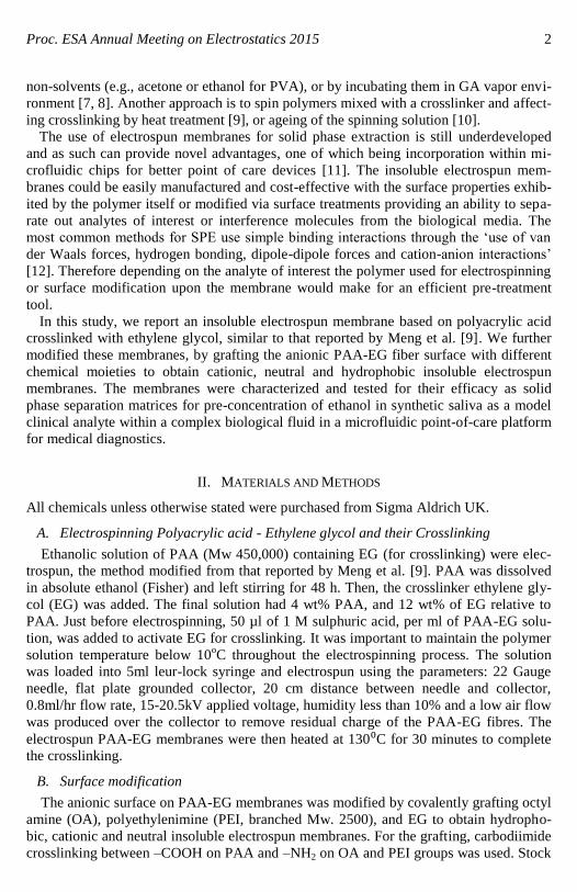
Proc. ESA Annual Meeting on Electrostatics 2015 1 Insoluble electrospun membranes for analyte pre-concentration in saliva Shavini Wijesuriya, Krishna Burugapalli, Ruth Mackay, Wamadeva Balachandran Dept. of Mechanical, Aerospace & Civil Eng. and Dept. of Electronic & Computer Eng. Brunel University phone: (44) 189-526-6926 e-mail: [email protected] Abstract— Insoluble electrospun membrane based on poly acrylic acid (PAA) crosslinked with ethylene glycol (EG) was prepared. The surface of the crosslinked PAA fibers was then grafted with polyethylenimine (PEI), octyl amine (OA) and EG using carbodiimide or ther- mal crosslinking to obtain insoluble membranes having anionic, cationic, hydrophobic and neutral characteristics for filtration of biomolecules from saliva. The resulting surface chem- istries were confirmed by their respective ATR-FTIR spectra. Differences in fiber morpholo- gy and diameters were observed between the membranes. All the membrane variants were tested for their efficacy as solid phase extraction (SPE) matrices in the pre-concentration of ethanol in synthetic saliva, using a microfluidic chip. Gas chromatography assays showed no statistical difference for ethanol in eluents through each of the tested insoluble electrospun membrane variants versus their corresponding controls. Further studies would include the testing of these membranes in the pre-concentration of clinical biomolecules, including drugs of abuse (e.g., cocaine, THC), nutrients (e.g., glucose), proteins (e.g., albumin, HbA1C) and nucleic acids for sample preparation within point of care devices. I. INTRODUCTION Electrospinning produces nanoscale fibrous membranes having large specific surface area (1 to 35 m 2 /g), excellent interconnectivity of pores, potential to incorporate active chemistry and functionality at nanoscale for use in multiple fields [1]. For example, they are used in air and water filters, electronics and sensors, drug therapy, tissue engineering and regenerative medicine [2-6]. However, a niche area for electrospun membranes that is still in its infancy is the fabrication of insoluble electrospun membranes that do not degrade within water and other solvent systems. For electrospinning, usually, high molecular weight polymers that are soluble in some solvent (water or organic) is required. Essentially, their polymer solubility can limit the scope for applications. This is especially important in the case of filtration and solid- phase extraction/separation membranes, wherein the membranes are required to be insol- uble and non-degradable. Electrospun membranes are made insoluble by crosslinking. They are usually prepared by electrospinning polymer solutions, and crosslinking affect- ed after electrospinning. Protein-based polymers, polyamines and PVA are crosslinked by glutaraldehyde (GA), either by immersing them in GA solutions in their corresponding
Transcript of Insoluble electrospun membranes for analyte pre ... · the membranes were saturated with 10% EG...
Proc. ESA Annual Meeting on Electrostatics 2015 1
Insoluble electrospun membranes for
analyte pre-concentration in saliva
Shavini Wijesuriya, Krishna Burugapalli, Ruth Mackay, Wamadeva Balachandran
Dept. of Mechanical, Aerospace & Civil Eng. and Dept. of Electronic & Computer Eng.
Brunel University
phone: (44) 189-526-6926
e-mail: [email protected]
Abstract— Insoluble electrospun membrane based on poly acrylic acid (PAA) crosslinked
with ethylene glycol (EG) was prepared. The surface of the crosslinked PAA fibers was then
grafted with polyethylenimine (PEI), octyl amine (OA) and EG using carbodiimide or ther-
mal crosslinking to obtain insoluble membranes having anionic, cationic, hydrophobic and
neutral characteristics for filtration of biomolecules from saliva. The resulting surface chem-
istries were confirmed by their respective ATR-FTIR spectra. Differences in fiber morpholo-
gy and diameters were observed between the membranes. All the membrane variants were
tested for their efficacy as solid phase extraction (SPE) matrices in the pre-concentration of
ethanol in synthetic saliva, using a microfluidic chip. Gas chromatography assays showed no
statistical difference for ethanol in eluents through each of the tested insoluble electrospun
membrane variants versus their corresponding controls. Further studies would include the
testing of these membranes in the pre-concentration of clinical biomolecules, including drugs
of abuse (e.g., cocaine, THC), nutrients (e.g., glucose), proteins (e.g., albumin, HbA1C) and
nucleic acids for sample preparation within point of care devices.
I. INTRODUCTION
Electrospinning produces nanoscale fibrous membranes having large specific surface
area (1 to 35 m2/g), excellent interconnectivity of pores, potential to incorporate active
chemistry and functionality at nanoscale for use in multiple fields [1]. For example, they
are used in air and water filters, electronics and sensors, drug therapy, tissue engineering
and regenerative medicine [2-6]. However, a niche area for electrospun membranes that
is still in its infancy is the fabrication of insoluble electrospun membranes that do not
degrade within water and other solvent systems.
For electrospinning, usually, high molecular weight polymers that are soluble in some
solvent (water or organic) is required. Essentially, their polymer solubility can limit the
scope for applications. This is especially important in the case of filtration and solid-
phase extraction/separation membranes, wherein the membranes are required to be insol-
uble and non-degradable. Electrospun membranes are made insoluble by crosslinking.
They are usually prepared by electrospinning polymer solutions, and crosslinking affect-
ed after electrospinning. Protein-based polymers, polyamines and PVA are crosslinked by
glutaraldehyde (GA), either by immersing them in GA solutions in their corresponding
Proc. ESA Annual Meeting on Electrostatics 2015 2
non-solvents (e.g., acetone or ethanol for PVA), or by incubating them in GA vapor envi-
ronment [7, 8]. Another approach is to spin polymers mixed with a crosslinker and affect-
ing crosslinking by heat treatment [9], or ageing of the spinning solution [10].
The use of electrospun membranes for solid phase extraction is still underdeveloped
and as such can provide novel advantages, one of which being incorporation within mi-
crofluidic chips for better point of care devices [11]. The insoluble electrospun mem-
branes could be easily manufactured and cost-effective with the surface properties exhib-
ited by the polymer itself or modified via surface treatments providing an ability to sepa-
rate out analytes of interest or interference molecules from the biological media. The
most common methods for SPE use simple binding interactions through the ‘use of van
der Waals forces, hydrogen bonding, dipole-dipole forces and cation-anion interactions’
[12]. Therefore depending on the analyte of interest the polymer used for electrospinning
or surface modification upon the membrane would make for an efficient pre-treatment
tool.
In this study, we report an insoluble electrospun membrane based on polyacrylic acid
crosslinked with ethylene glycol, similar to that reported by Meng et al. [9]. We further
modified these membranes, by grafting the anionic PAA-EG fiber surface with different
chemical moieties to obtain cationic, neutral and hydrophobic insoluble electrospun
membranes. The membranes were characterized and tested for their efficacy as solid
phase separation matrices for pre-concentration of ethanol in synthetic saliva as a model
clinical analyte within a complex biological fluid in a microfluidic point-of-care platform
for medical diagnostics.
II. MATERIALS AND METHODS
All chemicals unless otherwise stated were purchased from Sigma Aldrich UK.
A. Electrospinning Polyacrylic acid - Ethylene glycol and their Crosslinking
Ethanolic solution of PAA (Mw 450,000) containing EG (for crosslinking) were elec-
trospun, the method modified from that reported by Meng et al. [9]. PAA was dissolved
in absolute ethanol (Fisher) and left stirring for 48 h. Then, the crosslinker ethylene gly-
col (EG) was added. The final solution had 4 wt% PAA, and 12 wt% of EG relative to
PAA. Just before electrospinning, 50 µl of 1 M sulphuric acid, per ml of PAA-EG solu-
tion, was added to activate EG for crosslinking. It was important to maintain the polymer
solution temperature below 10oC throughout the electrospinning process. The solution
was loaded into 5ml leur-lock syringe and electrospun using the parameters: 22 Gauge
needle, flat plate grounded collector, 20 cm distance between needle and collector,
0.8ml/hr flow rate, 15-20.5kV applied voltage, humidity less than 10% and a low air flow
was produced over the collector to remove residual charge of the PAA-EG fibres. The
electrospun PAA-EG membranes were then heated at 130⁰C for 30 minutes to complete
the crosslinking.
B. Surface modification
The anionic surface on PAA-EG membranes was modified by covalently grafting octyl
amine (OA), polyethylenimine (PEI, branched Mw. 2500), and EG to obtain hydropho-
bic, cationic and neutral insoluble electrospun membranes. For the grafting, carbodiimide
crosslinking between –COOH on PAA and –NH2 on OA and PEI groups was used. Stock
Proc. ESA Annual Meeting on Electrostatics 2015 3
solutions of N-(3-dimethylaminopropyl)-N’-ethylcarbodiimide) (EDC) and N-
hydroxysuccinimide (NHS) in 0.05M 2-morpholinoethane sulfonic acid (MES) solution
were prepared and pH adjusted to 5.3 using 1N NaOH and/or 1N HCl. PAA membranes
(8mm discs) were also equilibrated in MES buffer (pH 5.3) containing OA (0.1M) or PEI
(0.1 wt%). All handling of EDC solutions was done on ice [13]. EDC and NHS were
added to the PAA membranes in MES buffer such that the final solution concentration of
each of EDC and NHS was 0.1M. The resulting solution was incubated on ice for 1 h
before transfer to room temp, wherein they were left overnight for the crosslinking reac-
tion to complete on a shaker at 150rpm. Following crosslinking the samples were washed
in DI water, and stored at 4⁰C until further use. For grafting EG on PAA-EG membranes,
the membranes were saturated with 10% EG solution in DI water, and heated at 130oC
for 30 min to obtain neutral surfaced insoluble electrospun membranes.
C. Characterization of Membrane Properties
The surface chemistry of the different membrane variants was confirmed by recording
their ATR-FTIR spectra using a Perkin Elmer Spectrum One Fourier Transform Infrared
(FTIR) spectrometer equipped with a Specac Golden Gate ATR accessory. Each spec-
trum, acquired in transmittance mode, was an average of 128 scans at a resolution of 4
cm−1
.
The electrospun membranes were also sputter coated for 30 s with gold using an
AGAR high resolution sputter-coater and observed using a JEOL JCM-6000 NeoScope
Benchtop SEM for morphology.
The fiber diameters were measured on SEM images using Image J software. A mini-
mum of 60 measurements were made on five different SEM images, each representing a
non-overlapping random field of view for each electrospun membrane configuration.
D. Microfluidic Chip Fabrication
Microfluidic molds were designed using Solidworks CAD software and saved as STL
files. The molds were created using 3D printing techniques with an Objet 30 Pro (Stata-
sys, US) using jetted photopolymer deposition. QSil 218 PDMS (ACC Silicones, UK)
was mixed at a ratio of 1:10 for 2 minutes and then degassed using a centrifuge for about
4 minutes. The molds were cleaned by rinsing with iso-propyl alcohol and then DI water
to remove contaminants; they were then dried using nitrogen. The mold was placed into a
custom made stainless steel frame which was cleaned in the same manner. After degas-
sing the PDMS it is poured gently into the mold and left in the oven at a set temperature
of 45° C for 4 hours. The cured PDMS was removed from the mold; a handheld corona
treater, BD20-AC (Electro-Technic Products Inc., US), was used to bond the PDMS to a
75x50mm glass slide (Sigma Aldrich, UK). The treated surfaces were then pressed to-
gether and placed in an oven at 54° C for 8 hours. The microfluidic device incorporates
sample inlets 3mm long, 250µm high with 1mm diameter, 10mm entrance port, 500µm
wide followed by a 8mm diameter collection outlet open chamber of 2mm depth and total
volume 250µl.
E. Testing of Efficacy of the Insoluble Electrospun Membranes in the Pre-
Concentration of Ethanol in Synthetic Saliva
The composition of synthetic saliva used in this study was (mg/L): potassium chloride:
1360, bovine mucin: 1300; potassium hydrogen phosphate: 950; sodium chloride: 860;

Proc. ESA Annual Meeting on Electrostatics 2015 4
sodium azide: 500; sodium hydrogen carbonate: 440; potassium thiocyanate: 250; calci-
um chloride: 210; urea: 180; magnesium chloride: 60; Brij 30: 100; with ultrapure
(>18MΩ) deionized water as the solvent.
Three 8 mm discs of each membrane variant were stacked in each chamber of the mi-
crofluidic cartridge and held down with a nitrile o-ring. The 2 ml of test solution (GC
standard ethanol solution of concentrations 1, 0.5, 0.1, 0.05 or 0.01% in synthetic saliva
or ultrapure DI water) was loaded into 10ml leur-lock syringe (Fisher, UK) and connect-
ed to the inlet of the membrane incorporated chamber on the microfluidic cartridge using
PTFE tubing (RS Components, UK). Using a syringe pump, the test solution in the sy-
ringe was pumped at a flow rate of 10ml/hr and 1ml of eluent solution was collected from
the open chamber outlet. The remaining test solution in the syringe was used as the con-
trol.
For the assay of ethanol in the different solutions was done using an Agilent 6890N
gas chromatograph equipped with an FID and a TCH detector (in series). Manual injec-
tions of 1 µl were split 22:1 in the split injection port. Injector and detector temperatures
were 250oC. The column was a 30 m x 0.25 mm HP Innowax column with highly polar
polyethylene glycol (PEG) stationary phase thickness of 0.25 µm (max temp: 260oC).
The oven program was 50oC for 2 min, ramp 1 of 15
oC/min to 80
oC, ramp 2 of 40
oC/min
to 120oC, hold at 120
oC for 2.0 min; a total run time of 7 min. The hydrogen carrier gas
has a flow rate of 1.6 ml/min through the column and a head pressure of 40 kPa and a
total flow rate of 50 ml/min.
Ethanol calibration standards of concentrations of 0.0001, 0.00025, 0.0005, 0.00075,
0.001, 0.0025, 0.005, 0.0075, 0.01, 0.025, 0.075, 0.1, 0.25, 0.5, 0.75, and 1 wt % were
prepared using stock solutions of: 10, 1, 0.1, 0.01 and 0.001 wt % ethanol in DI water and
in synthetic saliva. The calibration standards and test samples were assayed for ethanol
concentrations using the above gas chromatography method.
F. Statistical Analysis
Statistical analyses were carried out using statistical software (SPSS v.20). Statistical
variances between groups were determined by one-way analysis of variance (ANOVA).
Tukey’s test was used for post hoc evaluation of differences between groups. A p value
of <0.05 was considered to be statistically significant. Unless otherwise mentioned, all
data presented are expressed as mean ± standard error of mean.
III. RESULTS AND DISCUSSION
Making electrospun membranes insoluble has significant potential for their application
as solid phase separation matrices for filtration and chromatography. In this study, we
report one such membrane and its tailorable surface chemistry.
A. Fabrication of insoluble electrospun membranes based on polyacyrlic acid and
ethylene glycol
PAA and EG were dissolved in ethanol at 88:12 weight ratio, at a solution concentration
of 4 wt%. This solution had a pH and conductivity of 3.26 and 1 μS respectively (Table
1). To make this solution electrospinnable, we added 50μl of 1M sulfuric acid, bringing
its pH and conductivity to 1.28 and 950 μS respectively. At this pH the carboxylic acid

Proc. ESA Annual Meeting on Electrostatics 2015 5
groups on PAA would be charged (–COO-) and its 950 μS conductivity at 4wt% solution
concentration was appropriate for polymer solution to overcome surface tension to form
the tailor cone and the electrospinning jet as reported by Meng et al. [9]. However, to
replicate Meng et al.’s method in our laboratory, we had to modify two key electrospin-
ning parameters – one, a smaller spinneret needle gauze size of 22 and second, a much
lower ambient temperature <10oC compared to the room temperature. If spun at room
temperature, the fibers did not deposit on the collector and instead formed cobweb like
structure in the form of a cone extending from the tip of the spinneret needle to the col-
lector in mid-air. This we think could be due to rapid evaporation of the solvent (ethanol)
before the electrospinning jet reaches the collector plate. Electrospinning with a cold
polymer solution helped in overcoming this problem [14].
The crosslinking of PAA and EG was effected after electrospinning by heating the as-
spun membranes at 130oC for 30 min. The completion of crosslinking was ascertained
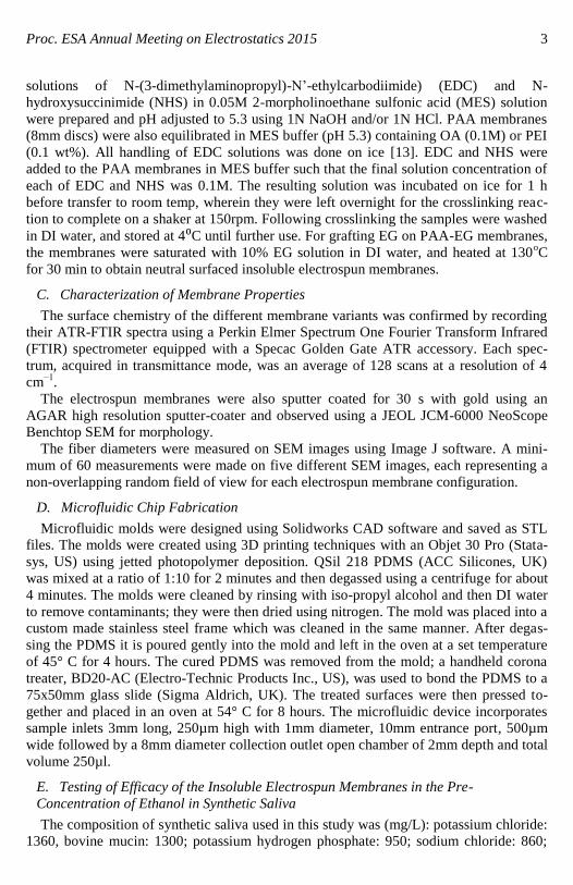
using ATR-FTIR. The FTIR spectra for the raw materials PAA and EG, and their cross-
linked electrospun forms are shown in Fig. 1. EG showed its typical O–H stretching, C–
H asymmetric and symmetric stretching peaks at 3294 cm-1
, 2936 cm-1
and 2874 cm-1
respectively. Further, the C–O stretching vibrations were observed as a doublet at 1083
cm-1
and 1033 cm-1
; while its CH2 rocking and C–C stretching peaks were observed at
881 cm-1
and 860 cm-1
respectively. The FTIR spectrum for PAA was characterized by a
strong carbonyl peak of its –COOH functional groups at 1695 cm-1
, broad –OH peak of
the –COOH group from 2900 cm-1
to 3300 cm-1
, overlapping with the –CH2 peak at 2934
cm-1
. The completion of the crosslinking reaction between PAA and its crosslinker EG,
is evident from the complete absence of –OH peak and the prominent –CH2 peaks of EG
in the PAA-EG spectrum; the appearance of peaks at 872 and 1042 for the C-C and CH2
bonds corresponding to EG; and the presence of the carbonyl –OH (2900 cm-1
to 3300
cm-1
) and C=O (1698 cm-1
), as well as the –CH2 (2924 cm-1
) peaks of PAA.
B. Surface Modification of insoluble electrospun PAA-EG membranes
Both EDC-NHS and ester formation between –COOH and –OH groups, based crosslink-
ing reactions are zero-length crosslinking mechanisms, wherein desired side chain moie-
ties are covalently grafted on to the PAA-EG membranes. Reactions were carried out
between –COOH groups on PAA-EG membranes and –NH2 on OA and PEI using EDC-
NHS crosslinking chemistry to obtain hydrophobic and cationic membranes. Similarly,
ester reaction between –COOH PAA-EG and –OH groups on EG was used to obtain
neutral surfaced membranes. Again, the completion of the crosslinking addition of side
chains on PAA-EG membranes was ascertained using FTIR spectra (Fig. 2a-c).
TABLE 1: CONDUCTIVITY AND PH OF PAA SOLUTIONS FOR ELECTROSPINNING
Conductivity (μS) pH
4% PAA (wt/v) in Ethanol 1 3.05
4% PAA-EG in Ethanol 1 3.26
4% PAA-EG in Ethanol + H2SO4 950 1.28
Proc. ESA Annual Meeting on Electrostatics 2015 6
Fig. 1. ATR-FTIR spectra for PAA and EG (as supplied), and the insoluble electrospun PAA-EG membrane.
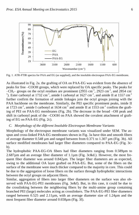
As illustrated in Fig. 2a. the grafting of OA on PAA-EG was evident from the absence of
peaks for free –COOH groups, which were replaced by OA specific peaks. The peaks for
–CH2– groups on the octyl residues are prominent (2955 cm-1
, 2923 cm-1
, and 2854 cm-
1). Ester carbonyl at 1732 cm
-1, amide I carbonyl at 1627 cm
-1, and amide II at 1557 cm
-1
further confirm the formation of amide linkages join the octyl groups joining with the
PAA backbone on the membrane. Similarly, the PEI specific prominent peaks, imide II
at 1723 cm-1
, amide I carbonyl at 1634 cm-1
and amide II at 1553 cm-1
confirm the graft-
ing of PEI on PAA-EG membranes (Fig. 2b). The decrease in the broad –OH peak and
shift in carbonyl peak of the –COOH on PAA showed the covalent attachment of graft-
ing of EG on PAA-EG (Fig. 2c).
C. Morphology of the different Insoluble Electrospun Membrane Variants
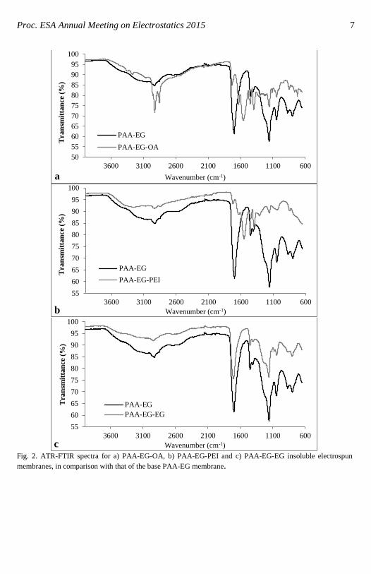
Morphology of the electrospun membrane variants was visualized under SEM. The as-
spun and cross-linked PAA-EG membranes shown in Fig. 3a have thin and smooth fibers
of average diameter 0.548 µm and ranged between from 0.371 to 1.307 µm (Fig. 3b). All
surface modified membranes had larger fiber diameters compared to PAA-EG (Fig. 3c-
h).
The hydrophobic PAA-EG-OA fibers had fiber diameters ranging from 0.589µm to
2.687µm and an average fiber diameter of 1.1µm (Fig. 3c&d.). However, the most fre-
quent fiber diameter was around 0.842µm. The larger fiber diameters are as expected,
owing to the additional OA layer grafted on PAA-EG. But, some of the fibers on the
surface of the membrane were much thicker compared to the majority in core. This could
be due to the aggregation of loose fibers on the surface through hydrophobic interactions
between the octyl groups on adjacent fibers.
Similar, aggregation of fibers into larger fiber diameters on the surface was also ob-
served with PAA-EG-PEI membranes (Fig. 3e&f). But, in this case, it could be due to
the crosslinking between the neighboring fibers by the multi-amine group containing
branched PEI (large) molecules acting as crosslinkers. The PAA-EG-PEI fiber diameters
ranged between 0.525 and 2.11µm, with an average diameter size of 1.24µm and the
most frequent fiber diameter around 0.658µm (Fig. 3f).
19
29
39
49
59
69
79
89
99
600110016002100260031003600
Tra
nsm
itta
nce (
%)
Wavenumber (cm-1)
EG
PAA
PAA-EG
Proc. ESA Annual Meeting on Electrostatics 2015 7
Fig. 2. ATR-FTIR spectra for a) PAA-EG-OA, b) PAA-EG-PEI and c) PAA-EG-EG insoluble electrospun
membranes, in comparison with that of the base PAA-EG membrane.
50
55
60
65
70
75
80
85
90
95
100
600110016002100260031003600
Tra
nsm
itta
nce (
%)
Wavenumber (cm-1) a
PAA-EG
PAA-EG-OA
55
60
65
70
75
80
85
90
95
100
600110016002100260031003600
Tra
nsm
itta
nce (
%)
Wavenumber (cm-1) b
PAA-EG
PAA-EG-PEI
55
60
65
70
75
80
85
90
95
100
600110016002100260031003600
Tra
nsm
itta
nce
(%
)
Wavenumber (cm-1) c
PAA-EG
PAA-EG-EG
Proc. ESA Annual Meeting on Electrostatics 2015 8
a b
c d
e f
g h Fig 3. Morphology and fiber diameter distribution histograms respectively for the different electrospun mem-
branes: a & b) PAA-EG, c & d) PAA-EG-OA, e & f) PAA-EG-PEI, and g & h) PAA-EG-EG.
The PAA-EG-EG fibers showed a different kind of fiber aggregation at the membrane
surface compared to both PAA-EG-OA and PAA-EG-PEI. EG being a very short bifunc-
tional crosslinker can only crosslink closely apposed fibers, as evident from the clear
outline of the neighboring parallel fibers joined together (Fig. 3g). The larger size of
branched PEI and longer chains of OA entangled through hydrophobic interactions, we
Proc. ESA Annual Meeting on Electrostatics 2015 9
think obscures the outline of neighboring fibers making them appear as thicker single
fibers (Fig 3c&e). The covalent grafting the small EG molecules on PAA-EG fibers had
the smallest increase in fiber diameter among the three surface modifications as evi-
denced by the smallest increase in the minimum fiber diameter of 0.389µm compared to
0.525µm and 0.589µm respectively for PAA-EG-PEI and PAA-EG-OA membranes. The
fiber diameters for PEI-EG-EG ranged from 0.389µm to 77.865µm, with average fiber
diameter of 7.682µm and the most frequent fiber diameter around 1.201µm. The larger
fiber diameters for PEI-EG-EG are due to the aggregated fibers skewing the results.
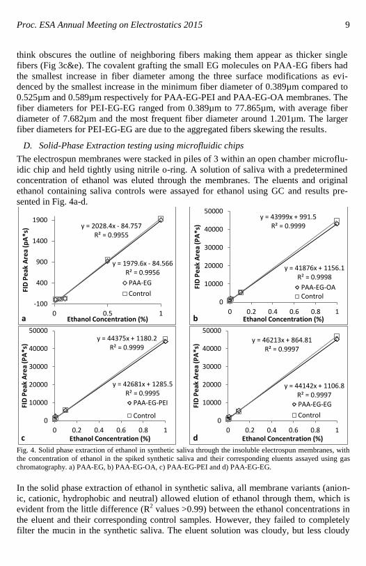
D. Solid-Phase Extraction testing using microfluidic chips
The electrospun membranes were stacked in piles of 3 within an open chamber microflu-
idic chip and held tightly using nitrile o-ring. A solution of saliva with a predetermined
concentration of ethanol was eluted through the membranes. The eluents and original
ethanol containing saliva controls were assayed for ethanol using GC and results pre-
sented in Fig. 4a-d.
Fig. 4. Solid phase extraction of ethanol in synthetic saliva through the insoluble electrospun membranes, with the concentration of ethanol in the spiked synthetic saliva and their corresponding eluents assayed using gas
chromatography. a) PAA-EG, b) PAA-EG-OA, c) PAA-EG-PEI and d) PAA-EG-EG.
In the solid phase extraction of ethanol in synthetic saliva, all membrane variants (anion-
ic, cationic, hydrophobic and neutral) allowed elution of ethanol through them, which is
evident from the little difference (R2 values >0.99) between the ethanol concentrations in
the eluent and their corresponding control samples. However, they failed to completely
filter the mucin in the synthetic saliva. The eluent solution was cloudy, but less cloudy
y = 1979.6x - 84.566 R² = 0.9956
y = 2028.4x - 84.757 R² = 0.9955
-100
400
900
1400
1900
0 0.5 1
FID
Pe
ak A
rea
(pA
*s)
Ethanol Concentration (%) a
PAA-EG
Control
y = 41876x + 1156.1 R² = 0.9998
y = 43999x + 991.5 R² = 0.9999
0
10000
20000
30000
40000
50000
0 0.2 0.4 0.6 0.8 1
FID
Pe
ak A
rea
(PA
*s)
Ethanol Concentration (%) b
PAA-EG-OAControl
y = 42681x + 1285.5 R² = 0.9995
y = 44375x + 1180.2 R² = 0.9999
0
10000
20000
30000
40000
50000
0 0.2 0.4 0.6 0.8 1
FID
Pe
ak A
rea
(PA
*s)
Ethanol Concentration (%) c
PAA-EG-PEI
Control
y = 44142x + 1106.8 R² = 0.9997
y = 46213x + 864.81 R² = 0.9997
0
10000
20000
30000
40000
50000
0 0.2 0.4 0.6 0.8 1
FID
Pe
ak A
rea
(PA
*s)
Ethanol Concentration (%) d
PAA-EG-EG
Control
Proc. ESA Annual Meeting on Electrostatics 2015 10
compared to the controls. Increasing the quantity of the membrane used for the solid
phase extraction could potentially increase their efficiency in filtering the mucin in saliva.
IV. CONCLUSION
Insoluble PAA-EG membranes were successfully electrospun and characterized to pro-
vide anionic, cationic, neutral and hydrophobic membrane surface properties. The mem-
branes exhibited varying fibre morphologies with all post-treatment modifications effec-
tively being incorporated onto the membrane as shown in the FTIR results. The mem-
brane morphology also altered with addition of altering surface chemistries as shown in
the SEM images as well as fibre diameter sizes and range, with the modifications caus-
ing an increase in range of fibre diameters as well as size. The microfluidic SPE extrac-
tion of ethanol from saliva also showed promising results with the GC results showing
>0.99 R2 linearity compared with the standard curve for ethanol in synthetic saliva.
Overall, the results were promising, but can be improved upon to gain further insight into
the relationship of the membrane and separation through increase in the quantity of the
membrane used for the solid phase extraction to potentially increase their efficiency in
filtering the mucin in saliva.
REFERENCES
[1] R. S. Barhate and S. Ramakrishna, “Nanofibrous filtering media: Filtration problems and solutions from tiny materials” Journal of Membrane Science, 2007, 296(1-2). pp. 1-8.
[2] D. Li and Y. N. Xia, “Electrospinning of Nanofibers: Reinventing the Wheel?”, Adv. Mater. 2004, 16(14),
pp 1151-1170. [3] G. Greiner and J. H. Wendorff, “Electrospinning: a fascinating method for the preparation of ultrathin
fibers” Angew. Chem. Int. Ed. 2007, 46(30), pp 5670-5703.
[4] S. Ramakrishna, K. Fujihara, W.-E. Teo, T. Yong, Z Ma, and R. Ramaseshan, “Electrospun nanofibers: solving global issues” Materials Today, 2006, 9(3), pp. 40-50.
[5] T. J. Sill and H. A. von Recum, “Electrospinning: Applications in drug delivery and tissue engineering”
Biomaterials, 2008, 29(13), pp. 1989-2006. [6] B. Ding, M. Wang, X. Wang, J. Yu and G. Sun, “Electrospun nanomaterials for ultrasensitive sensors”,
Materials Today, 2010, 13(11), pp. 16-27.
[7] C. Tang, C. D. Saquing, J. R. Harding, and S. A. Khan, “In situ cross-linking of electrospun poly(vinyl alcohol) nanofibers” Macromolecules 2010, 43, pp. 630–637.
[8] N. Wang, K Burugapalli, S Wijesuriya, M. Y. Far, W. Song, F. Moussy, Y. Zheng, Y. Ma, Z. Wu and K.
Li, “Electrospun polyurethane-core and gelatin-shell coaxial fibre coatings for miniature implantable bio-sensors” Biofabrication, 2014, 6, 015002 pp. 1-18.
[9] L. Meng, W. Klinkajon, P. Khasuwan, S. Harkin, P. Supaphol and G. E. Wnek, “Electrospun crosslinked
poly(acrylic acid) fiber constructs: towards a synthetic model of the cortical layer of nerve” Polym. Int. 2015, 64(1), pp. 42-48.
[10] G. W. Selling, K. K. Woods and A. Biswas, “Electrospun zein fibers using glyoxal as the crosslinking
reagent” J Appl. Polym. Sci. 2012, 123(5), 2651-2661. [11] S. Chigome and N. Torto, “Electrospun nanofiber-based solid-phase extraction”, Trends Anal. Chem.,
2012, 38, pp. 21-31.
[12] A. Żwir-Ferenc and M. Biziuk, “Solid Phase Extraction Technique – Trends , Opportunities and Applications” Polish J Environ. Studies, 2006, 15(5), pp. 677–690.
[13] J. C. Y. Chan, K. Burugapalli, H. Naik, J. L. Kelly, and A. Pandit, “Amine functionalization of cholecyst-
derived extracellular matrix with generation 1 PAMAM dendrimer” Biomacromolecules, 2008, 9(2), pp. 528-536.
[14] J. J. Barron, and C. Ashton, “The Effect of Temperature on Conductivity Measurement”. Regecon, 2010, pp. 1–5. http://www.camlabworld.com/originalimages/sitefiles/Tech_papers/TempCondMeas.pdf, last
Accesssed 16 May 2015.