Indications and techniques of biliary drainage for acute...
13
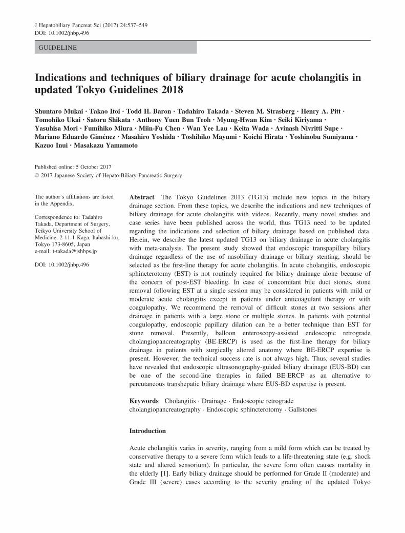
Indications and techniques of biliary drainage for acute cholangitis in updated Tokyo Guidelines 2018 Shuntaro Mukai Takao Itoi Todd H. Baron Tadahiro Takada Steven M. Strasberg Henry A. Pitt Tomohiko Ukai Satoru Shikata Anthony Yuen Bun Teoh Myung-Hwan Kim Seiki Kiriyama Yasuhisa Mori Fumihiko Miura Miin-Fu Chen Wan Yee Lau Keita Wada Avinash Nivritti Supe Mariano Eduardo Gim enez Masahiro Yoshida Toshihiko Mayumi Koichi Hirata Yoshinobu Sumiyama Kazuo Inui Masakazu Yamamoto Published online: 5 October 2017 © 2017 Japanese Society of Hepato-Biliary-Pancreatic Surgery The author’s affiliations are listed in the Appendix. Correspondence to: Tadahiro Takada, Department of Surgery, Teikyo University School of Medicine, 2-11-1 Kaga, Itabashi-ku, Tokyo 173-8605, Japan e-mail: [email protected] DOI: 10.1002/jhbp.496 Abstract The Tokyo Guidelines 2013 (TG13) include new topics in the biliary drainage section. From these topics, we describe the indications and new techniques of biliary drainage for acute cholangitis with videos. Recently, many novel studies and case series have been published across the world, thus TG13 need to be updated regarding the indications and selection of biliary drainage based on published data. Herein, we describe the latest updated TG13 on biliary drainage in acute cholangitis with meta-analysis. The present study showed that endoscopic transpapillary biliary drainage regardless of the use of nasobiliary drainage or biliary stenting, should be selected as the first-line therapy for acute cholangitis. In acute cholangitis, endoscopic sphincterotomy (EST) is not routinely required for biliary drainage alone because of the concern of post-EST bleeding. In case of concomitant bile duct stones, stone removal following EST at a single session may be considered in patients with mild or moderate acute cholangitis except in patients under anticoagulant therapy or with coagulopathy. We recommend the removal of difficult stones at two sessions after drainage in patients with a large stone or multiple stones. In patients with potential coagulopathy, endoscopic papillary dilation can be a better technique than EST for stone removal. Presently, balloon enteroscopy-assisted endoscopic retrograde cholangiopancreatography (BE-ERCP) is used as the first-line therapy for biliary drainage in patients with surgically altered anatomy where BE-ERCP expertise is present. However, the technical success rate is not always high. Thus, several studies have revealed that endoscopic ultrasonography-guided biliary drainage (EUS-BD) can be one of the second-line therapies in failed BE-ERCP as an alternative to percutaneous transhepatic biliary drainage where EUS-BD expertise is present. Keywords Cholangitis Drainage Endoscopic retrograde cholangiopancreatography Endoscopic sphincterotomy Gallstones Introduction Acute cholangitis varies in severity, ranging from a mild form which can be treated by conservative therapy to a severe form which leads to a life-threatening state (e.g. shock state and altered sensorium). In particular, the severe form often causes mortality in the elderly [1]. Early biliary drainage should be performed for Grade II (moderate) and Grade III (severe) cases according to the severity grading of the updated Tokyo J Hepatobiliary Pancreat Sci (2017) 24:537–549 DOI: 10.1002/jhbp.496 GUIDELINE
Transcript of Indications and techniques of biliary drainage for acute...
Indications and techniques of biliary drainage for acute cholangitis inupdated Tokyo Guidelines 2018
Shuntaro Mukai � Takao Itoi � Todd H. Baron � Tadahiro Takada � Steven M. Strasberg � Henry A. Pitt �
Tomohiko Ukai � Satoru Shikata � Anthony Yuen Bun Teoh � Myung-Hwan Kim � Seiki Kiriyama �
Yasuhisa Mori � Fumihiko Miura � Miin-Fu Chen � Wan Yee Lau � Keita Wada � Avinash Nivritti Supe �
Mariano Eduardo Gim�enez � Masahiro Yoshida � Toshihiko Mayumi � Koichi Hirata � Yoshinobu Sumiyama �
Kazuo Inui � Masakazu Yamamoto
Published online: 5 October 2017
© 2017 Japanese Society of Hepato-Biliary-Pancreatic Surgery
The author’s affiliations are listedin the Appendix.
Correspondence to: TadahiroTakada, Department of Surgery,Teikyo University School ofMedicine, 2-11-1 Kaga, Itabashi-ku,Tokyo 173-8605, Japane-mail: [email protected]
DOI: 10.1002/jhbp.496
Abstract The Tokyo Guidelines 2013 (TG13) include new topics in the biliarydrainage section. From these topics, we describe the indications and new techniques ofbiliary drainage for acute cholangitis with videos. Recently, many novel studies andcase series have been published across the world, thus TG13 need to be updatedregarding the indications and selection of biliary drainage based on published data.Herein, we describe the latest updated TG13 on biliary drainage in acute cholangitiswith meta-analysis. The present study showed that endoscopic transpapillary biliarydrainage regardless of the use of nasobiliary drainage or biliary stenting, should beselected as the first-line therapy for acute cholangitis. In acute cholangitis, endoscopicsphincterotomy (EST) is not routinely required for biliary drainage alone because ofthe concern of post-EST bleeding. In case of concomitant bile duct stones, stoneremoval following EST at a single session may be considered in patients with mild ormoderate acute cholangitis except in patients under anticoagulant therapy or withcoagulopathy. We recommend the removal of difficult stones at two sessions afterdrainage in patients with a large stone or multiple stones. In patients with potentialcoagulopathy, endoscopic papillary dilation can be a better technique than EST forstone removal. Presently, balloon enteroscopy-assisted endoscopic retrogradecholangiopancreatography (BE-ERCP) is used as the first-line therapy for biliarydrainage in patients with surgically altered anatomy where BE-ERCP expertise ispresent. However, the technical success rate is not always high. Thus, several studieshave revealed that endoscopic ultrasonography-guided biliary drainage (EUS-BD) canbe one of the second-line therapies in failed BE-ERCP as an alternative topercutaneous transhepatic biliary drainage where EUS-BD expertise is present.
Keywords Cholangitis � Drainage � Endoscopic retrogradecholangiopancreatography � Endoscopic sphincterotomy � Gallstones
Introduction
Acute cholangitis varies in severity, ranging from a mild form which can be treated byconservative therapy to a severe form which leads to a life-threatening state (e.g. shockstate and altered sensorium). In particular, the severe form often causes mortality inthe elderly [1]. Early biliary drainage should be performed for Grade II (moderate) andGrade III (severe) cases according to the severity grading of the updated Tokyo
J Hepatobiliary Pancreat Sci (2017) 24:537–549DOI: 10.1002/jhbp.496
GUIDELINE
Guidelines of 2013 (TG13) [2–4]. Biliary drainage, whichis the most essential therapy for acute cholangitis, is tradi-tionally divided into three types: (1) surgical, (2) percuta-neous transhepatic, and (3) endoscopic transpapillarydrainage. Of these therapies, surgical intervention causesthe highest mortality rate [1]. Recently, mortality due toacute cholangitis has decreased owing to the developmentof percutaneous transhepatic cholangial drainage (PTCD)[5] and endoscopic transpapillary biliary drainage [6, 7].Nevertheless, acute cholangitis can still be fatal unless itis treated early and properly.
The Tokyo Guidelines of 2007 (TG07) was the firstglobal guidelines in which fundamental biliary drainagetechniques for acute cholangitis were described [8]. Sub-sequently, TG07 was revised to TG13, which include theindications and procedures of newly developed biliarydrainage techniques such as endoscopic ultrasonography-guided biliary drainage (EUS-BD) and balloon entero-scope-assisted bile duct drainage in patients with surgi-cally altered anatomy [9]. As several reports of thesenewly developed biliary drainage techniques or the meth-ods and timing of stone removal after or simultaneouslywith drainage have been published, TG13 needs to beupdated. Thus, the Tokyo Guidelines Revision Committeewas assembled and the committee discussed six argumentpoints on biliary drainage for acute cholangitis as men-tioned below. In this article, we describe the latest drai-nage techniques for acute cholangitis and the treatmentmethods for stone removal in the updated Tokyo Guideli-nes 2018 (TG18).
Indications and techniques of biliary drainage
In the updated TG18, biliary drainage is recommended foracute cholangitis regardless of the degree of severityexcept in some cases of mild acute cholangitis in whichantibiotics and general supportive care are effective [10].
Q1. What is the most preferable biliary drainage foracute cholangitis? (Surgical vs. endoscopic transpapillaryvs. EUS-guided vs. percutaneous transhepatic biliarydrainage?)
We recommend endoscopic transpapillary biliarydrainage for acute cholangitis (recommendation 1,level B).
*Refer to Q6 in acute cholangitis patients with sur-gically altered anatomy.
Endoscopic transpapillary biliary drainage should beconsidered as the first-line drainage procedure because ofits less invasiveness and lower risk of adverse events thanother drainage techniques despite the risk of post-endoscopic retrograde cholangiopancreatography (ERCP)
pancreatitis [11–14]. The internal drainage by endoscopictranspapillary biliary drainage produces less pain after theprocedures than the external drainage by percutaneoustranshepatic biliary drainage (PTBD), also known as per-cutaneous transhepatic cholangial drainage (PTCD) [15].PTCD places more burden on patients owing to cosmeticproblems, skin inflammation, or bile leakage, compromis-ing the patient’s quality of life. A single treatment sessionfor a bile duct stone is possible with the endoscopictranspapillary approach, making the hospitalization dura-tion shorter. However, in patients with an inaccessiblepapilla due to upper gastrointestinal tract obstruction, orwhen skilled pancreaticobiliary endoscopists are not avail-able in the institution, PTCD is a useful alternative drai-nage procedure [5, 16]. Furthermore, PTCD can be usedas a salvage therapy when conventional endoscopictranspapillary drainage has failed owing to difficult selec-tive biliary cannulation. Recently, EUS-BD has beendeveloped and reported as a novel useful alternative drai-nage technique when standard endoscopic transpapillarydrainage has failed [17, 18].
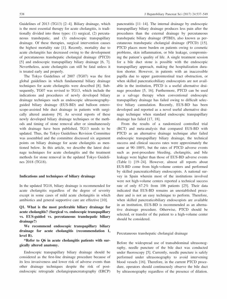
From the results of a randomized controlled trial(RCT) and meta-analysis that compared EUS-BD withPTCD as an alternative drainage technique after failedendoscopic transpapillary biliary drainage, the technicalsuccess and clinical success rates were approximately thesame at 90–100%, but the rates of PTCD adverse eventssuch as post-procedure bleeding, cholangitis, and bileleakage were higher than those of EUS-BD adverse events(Table 1) [19–24]. However, almost all reports aboutEUS-BD come from high-volume centers and performedby skilled pancreaticobiliary endoscopists. A national sur-vey in Spain wherein most of the institutions involvedwere not high-volume centers reported a technical successrate of only 67.2% from 106 patients [25]. Their dataindicated that EUS-BD remains an unestablished proce-dure and is not an easy technique to perform. Therefore,when skilled pancreaticobiliary endoscopists are availablein an institution, EUS-BD is recommended as an alterna-tive drainage procedure. Otherwise, PTCD should beselected, or transfer of the patient to a high-volume centershould be considered.
Percutaneous transhepatic cholangial drainage
Before the widespread use of transabdominal ultrasonog-raphy, needle puncture of the bile duct was conductedunder fluoroscopy [5]. Currently, needle puncture is safelyperformed under ultrasonography to avoid interveningblood vessels [16]. Therefore, in the current PTCD proce-dure, operators should continuously observe the bile ductby ultrasonography regardless of the presence of dilation.
538 J Hepatobiliary Pancreat Sci (2017) 24:537–549
PTCD is performed as previously described [5]. In brief,ultrasonography-guided transhepatic puncture of the intra-hepatic bile duct is initially performed using an 18-G to22-G needle. After confirming the backflow of bile, aguidewire is advanced into the bile duct. Finally, a 7-Fr to10-Fr catheter is placed in the bile duct under fluoroscopiccontrol over the guidewire. Puncture using a small-gauge(22-G) needle is safer in patients without biliary dilationthan in patients with biliary dilation. According to theQuality Improvement Guidelines developed by Americanradiologists, the success rates of drainage are 86% inpatients with biliary dilation and 63% in patients withoutbiliary dilation [16].
Surgical drainage
Open drainage for decompression of the bile duct is per-formed as a surgical intervention. When surgical drainagein critically ill patients with bile duct stones is performed,prolonged operations should be avoided and simple proce-dures, such as T-tube placement without choledocholitho-tomy, are recommended [26]. At present, surgicaldrainage is extremely rare because of the widespread useof endoscopic drainage or PTCD for acute cholangitistherapy.
Endoscopic transpapillary biliary drainage
Endoscopic transpapillary biliary drainage has become thegold standard technique for acute cholangitis, regardlessof a benign or malignant pathology, because it is a mini-mally invasive drainage method [6]. Endoscopic transpap-illary biliary drainage is divided into two types:endoscopic nasobiliary drainage (ENBD) for external
drainage and endoscopic biliary stenting (EBS) for inter-nal drainage. Basically, both types of endoscopic biliarydrainage can be performed in all forms of acute cholangi-tis. In the case of biliary drainage for acute cholangitistherapy, a precise endoscopic technique is mandatorybecause long and unsuccessful procedures may lead toserious complications in critically ill patients. Therefore,endoscopists who perform endoscopic transpapillary bil-iary drainage should acquire knowledge and skills ofselective biliary cannulation techniques including the dou-ble guidewire, pancreatic guidewire, and precut techniques[27].
Q2. Which procedure should be used for endoscopictranspapillary biliary drainage? ENBD or EBS?
We suggest that either ENBD or EBS may be consid-ered for biliary drainage according to the patient’s back-ground and preference (recommendation 1, level A).
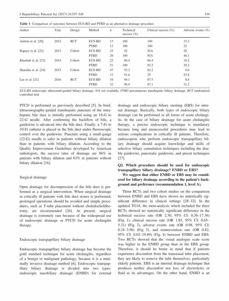
Three RCTs and two cohort studies on the comparisonbetween ENBD and EBS have shown no statistically sig-nificant difference in clinical settings [28–32]. In theupdated TG18, the meta-analysis which included the threeRCTs showed no statistically significant difference in thetechnical success rate (OR 2.50, 95% CI: 0.36–17.36)(Fig. 1), clinical success rate (OR 1.85, 95% CI: 0.65–5.31) (Fig. 2), adverse events rate (OR 0.98, 95% CI:0.24–3.96) (Fig. 3), and reintervention rate (OR 0.82,95% CI: 0.03–19.89) (Fig. 4) between ENBD and EBS.Two RCTs showed that the visual analogue scale scorewas higher in the ENBD group than in the EBS group.Therefore, it should be borne in mind that if patientsexperience discomfort from the transnasal tube placement,they are likely to remove the tube themselves, particularlyelderly patients. EBS is an internal drainage technique thatproduces neither discomfort nor loss of electrolytes orfluid as its advantages. On the other hand, ENBD is an
Table 1 Comparison of outcomes between EUS-BD and PTBD as an alternative drainage procedure
Author Year Design Method n Technicalsuccess (%)
Clinical success (%) Adverse events (%)
Artifon et al. [20] 2012 RCT EUS-BD 13 100 100 15.3
PTBD 12 100 100 25
Bapaye et al. [22] 2013 Cohort EUS-BD 25 92 N/A 20
PTBD 26 100 N/A 46.1
Khashab et al. [23] 2014 Cohort EUS-BD 22 86.4 86.4 18.2
PTBD 51 100 92.2 39.2
Sharaiha et al. [24] 2015 Cohort EUS-BD 47 93.3 62.2 6.6
PTBD 13 91.6 25 53.8
Lee et al. [21] 2016 RCT EUS-BD 34 94.1 87.5 8.8
PTBD 32 96.9 87.1 31.2
EUS-BD endoscopic ultrasound-guided biliary drainage, N/A not available, PTBD percutaneous transhepatic biliary drainage, RCT randomizedcontrolled trial
J Hepatobiliary Pancreat Sci (2017) 24:537–549 539
external drainage technique that allows monitoring orwashing of the bile via the transnasal tube, particularly ifthe bile is very purulent. Two retrospective studies haveshown that the stent occlusion from EBS was more fre-quent than the stent occlusion from ENBD in patientswith hilar cholangiocarcinoma; thus, ENBD may be moresuitable in patients with cholangitis due to hilar biliaryobstruction [33, 34].
Therefore, the balance between the advantages and dis-advantages of each drainage procedure is approximatelyequal. In the updated TG18, we suggest that either ENBDor EBS may be considered for biliary drainage by proce-dure according to the cause of the cholangitis, bile prop-erty, and patient’s preference.
Endoscopic nasobiliary drainage
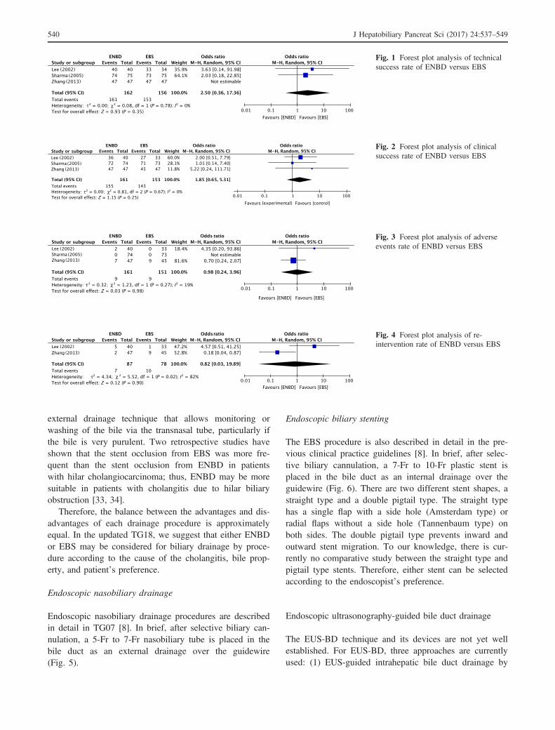
Endoscopic nasobiliary drainage procedures are describedin detail in TG07 [8]. In brief, after selective biliary can-nulation, a 5-Fr to 7-Fr nasobiliary tube is placed in thebile duct as an external drainage over the guidewire(Fig. 5).
Endoscopic biliary stenting
The EBS procedure is also described in detail in the pre-vious clinical practice guidelines [8]. In brief, after selec-tive biliary cannulation, a 7-Fr to 10-Fr plastic stent isplaced in the bile duct as an internal drainage over theguidewire (Fig. 6). There are two different stent shapes, astraight type and a double pigtail type. The straight typehas a single flap with a side hole (Amsterdam type) orradial flaps without a side hole (Tannenbaum type) onboth sides. The double pigtail type prevents inward andoutward stent migration. To our knowledge, there is cur-rently no comparative study between the straight type andpigtail type stents. Therefore, either stent can be selectedaccording to the endoscopist’s preference.
Endoscopic ultrasonography-guided bile duct drainage
The EUS-BD technique and its devices are not yet wellestablished. For EUS-BD, three approaches are currentlyused: (1) EUS-guided intrahepatic bile duct drainage by
Fig. 1 Forest plot analysis of technicalsuccess rate of ENBD versus EBS
Fig. 2 Forest plot analysis of clinicalsuccess rate of ENBD versus EBS
Fig. 3 Forest plot analysis of adverseevents rate of ENBD versus EBS
Fig. 4 Forest plot analysis of re-intervention rate of ENBD versus EBS
540 J Hepatobiliary Pancreat Sci (2017) 24:537–549
the transgastric or transjejunal approach; (2) EUS-guidedextrahepatic bile duct drainage by the transduodenal ortransgastric approach; and (3) EUS-guided antegradestenting. The choice of the drainage method and drainageroute depends on the presence of a gastric outlet obstruc-tion and the stricture site of the bile duct [35]. Severalpublished data on EUS-guided intrahepatic bile duct drai-nage and extrahepatic bile duct drainage have shown ahigh technical success rate of 95%, with 93–100% (inten-tion-to-treat) clinical response rates [16–18, 36–44]. It hasbeen reported that the technical success rate of EUS-guided antegrade stenting (77%) is inferior to those ofEUS-guided intrahepatic bile duct drainage and extrahep-atic bile duct drainage owing to the difficulty of guidewirepassage and stent delivery system insertion across the
strictures; however, EUS-guided antegrade stenting is aninteresting option because of the theoretical physiologicalbile flow in patients with inaccessible ampulla [45]. Themost serious issue during and after EUS-BD is the devel-opment of adverse events such as peritonitis. In fact, pre-vious studies have reported early adverse event rates of10–30%, although the severities of most of these adverseevents were mild or moderate [46, 47]. As the procedureis not yet established, EUS-BD should be performed byendoscopists who are skilled in both EUS and ERCP.
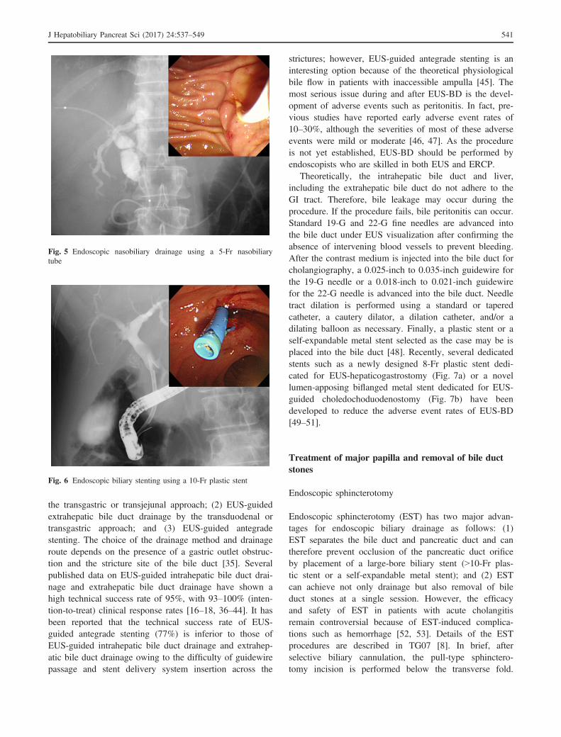
Theoretically, the intrahepatic bile duct and liver,including the extrahepatic bile duct do not adhere to theGI tract. Therefore, bile leakage may occur during theprocedure. If the procedure fails, bile peritonitis can occur.Standard 19-G and 22-G fine needles are advanced intothe bile duct under EUS visualization after confirming theabsence of intervening blood vessels to prevent bleeding.After the contrast medium is injected into the bile duct forcholangiography, a 0.025-inch to 0.035-inch guidewire forthe 19-G needle or a 0.018-inch to 0.021-inch guidewirefor the 22-G needle is advanced into the bile duct. Needletract dilation is performed using a standard or taperedcatheter, a cautery dilator, a dilation catheter, and/or adilating balloon as necessary. Finally, a plastic stent or aself-expandable metal stent selected as the case may be isplaced into the bile duct [48]. Recently, several dedicatedstents such as a newly designed 8-Fr plastic stent dedi-cated for EUS-hepaticogastrostomy (Fig. 7a) or a novellumen-apposing biflanged metal stent dedicated for EUS-guided choledochoduodenostomy (Fig. 7b) have beendeveloped to reduce the adverse event rates of EUS-BD[49–51].
Treatment of major papilla and removal of bile ductstones
Endoscopic sphincterotomy
Endoscopic sphincterotomy (EST) has two major advan-tages for endoscopic biliary drainage as follows: (1)EST separates the bile duct and pancreatic duct and cantherefore prevent occlusion of the pancreatic duct orificeby placement of a large-bore biliary stent (>10-Fr plas-tic stent or a self-expandable metal stent); and (2) ESTcan achieve not only drainage but also removal of bileduct stones at a single session. However, the efficacyand safety of EST in patients with acute cholangitisremain controversial because of EST-induced complica-tions such as hemorrhage [52, 53]. Details of the ESTprocedures are described in TG07 [8]. In brief, afterselective biliary cannulation, the pull-type sphinctero-tomy incision is performed below the transverse fold.
Fig. 5 Endoscopic nasobiliary drainage using a 5-Fr nasobiliarytube
Fig. 6 Endoscopic biliary stenting using a 10-Fr plastic stent
J Hepatobiliary Pancreat Sci (2017) 24:537–549 541
The push-type sphincterotome is used for EST inpatients with Billroth II gastrectomy or Roux-en-Y anas-tomosis. When the transverse fold is not present, thesuperior margin of the papilla bulge is used as a land-mark to determine the length of the sphincterotome. Anelectrosurgical generator with a controlled cutting sys-tem is used for EST.
Q3. Is endoscopic sphincterotomy necessary in endo-scopic transpapillary biliary drainage?
We suggest that the addition of EST may not berequired in endoscopic transpapillary biliary drainage(recommendation 2, level A).
*Refer to Q4 if stone removal at a single session isconsidered.
From the results of previous RCTs and cohort studieswhich included patients who had plastic stent (<7-Fr) ornasobiliary tube placement, the prevention of post-ERCPpancreatitis by the addition of EST to biliary drainagewas not observed [54–58]. Therefore, it is considered thatthe addition of EST is not required in acute cholangitisbecause this induces complications (e.g. hemorrhage).Acute cholangitis is one of the risk factors of post-ESThemorrhage. In particular, the use of EST in patients withsevere cholangitis complicated by coagulopathy or theadministration of antithrombotic agents should beavoided.
Endoscopic papillary balloon dilation
Endoscopic papillary balloon dilation (EPBD) is usuallyused instead of EST for the removal of bile duct stones[59]. To date, there has been apparently no comparativestudy on the use of EPBD during biliary drainage to treatacute cholangitis due to bile duct stones. A previous
systematic review has revealed that EPBD is less success-ful for stone removal, requires higher rates of mechanicallithotripsy, and carries a higher risk of pancreatitis; how-ever, it also has statistically significant lower rates ofbleeding [60]. Thus, EPBD appears to be useful for thetreatment of patients with coagulopathy and acute cholan-gitis caused by a small stone. Theoretically, as EPBDaims to preserve the function of the sphincter of Oddi,EPBD alone without biliary drainage is contraindicatedfor the therapy of acute cholangitis. In addition, EPBDshould be avoided in patients with biliary pancreatitis.After selective biliary cannulation, a small balloon with adiameter of up to 10 mm, depending on the diameters ofthe bile duct and stone, is advanced into the bile ductacross the papilla. Then, the sphincter of Oddi is graduallydilated by inflation of the balloon until the balloon waistdisappears. The bile duct stone is then cleared using abasket catheter and a balloon catheter.
Endoscopic papillary large balloon dilation
Endoscopic sphincterotomy or EPBD has been used asthe gold standard therapy for the removal of bile ductstones. However, in patients with large bile duct stones,endoscopic mechanical lithotripsy (EML) may berequired as an additional procedure [61, 62]. Since 2003,endoscopic papillary large balloon dilation (EPLBD) withor without EST has been developed to facilitate theremoval of large or difficult bile duct stones, in whichthe size of the large balloons used ranged from 12 to20 mm [63, 64]. Stone removal by EPLBD has beenshown to have a high success rate, and this might reducethe need for EML and the procedure time withoutincreasing the risk of severe pancreatitis or bile duct per-foration [65–70]. The EPLBD procedure is as follows.
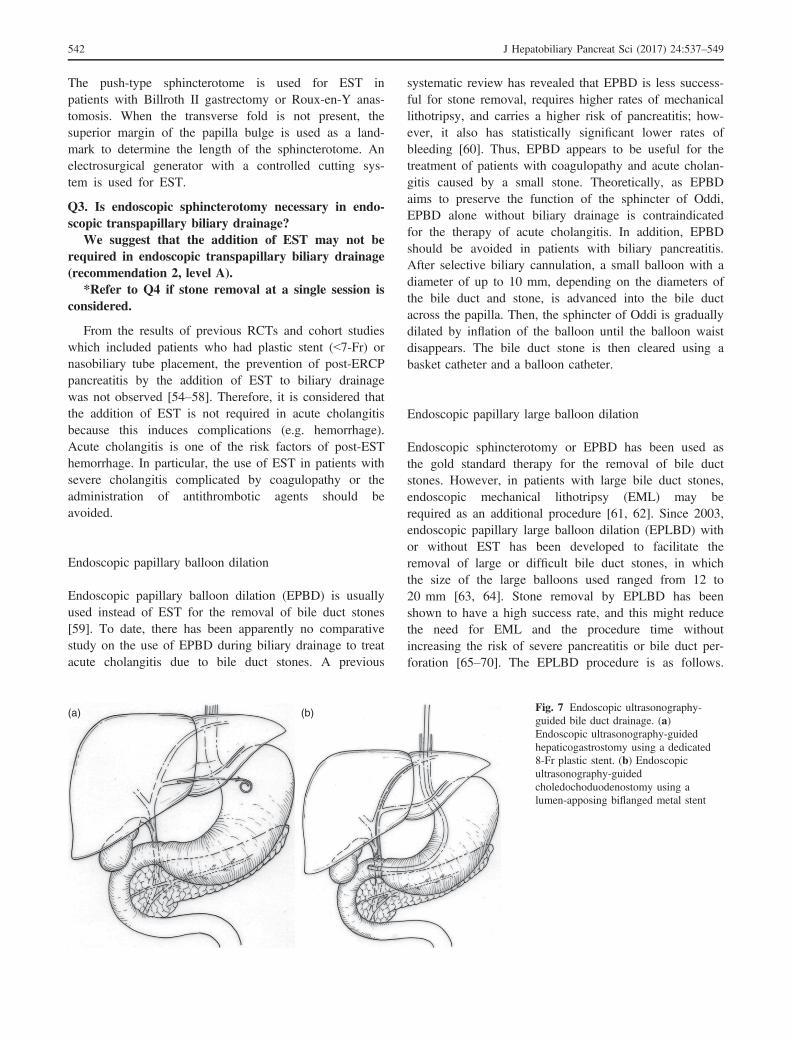
(a) (b) Fig. 7 Endoscopic ultrasonography-guided bile duct drainage. (a)Endoscopic ultrasonography-guidedhepaticogastrostomy using a dedicated8-Fr plastic stent. (b) Endoscopicultrasonography-guidedcholedochoduodenostomy using alumen-apposing biflanged metal stent
542 J Hepatobiliary Pancreat Sci (2017) 24:537–549
Large balloon dilation is performed using standard dilat-ing balloons immediately after EST. The balloon catheteris passed over a guidewire and positioned across thepapilla. The balloon is then gradually inflated with a con-trast medium under endoscopic and fluoroscopic guidanceuntil an adequate size is achieved to allow stone removalwithout lithotripsy regardless of the disappearance of theballoon waist. The stones are then removed from the bileduct using mechanical lithotripsy, a basket catheter, or aretrieval balloon.
Q4. Which is the preferred procedure for acutecholangitis associated with choledocholithiasis? Stoneremoval at a single session or at two sessions?
We suggest that bile duct stone removal followingEST at a single session may be considered in patientswith mild or moderate acute cholangitis (recommenda-tion 2, level C).
Bile duct stone is the most frequent cause of cholangi-tis [71]. Previously, two-session treatment has been rec-ommended for acute cholangitis with bile duct stone,which involves biliary drainage as an initial treatment andendoscopic stone removal after improvement of cholangi-tis [72]. This recommendation is based on previousreports that additional stone removal by EST at a singlesession increased the incidence of hemorrhagic complica-tions [52, 53]. However, these reports included cases ofsevere cholangitis accompanied by coagulopathy. Impor-tantly, the use of EST in patients with severe cholangitiscomplicated by coagulopathy should be avoided becauseof the risk of hemorrhagic complication. These findingsmay not always be applicable to mild-to-moderate cholan-gitis without coagulopathy. One multicenter prospectivestudy and two retrospective studies of mild-to-moderateacute cholangitis associated with bile duct stone haveshown that the cholangitis cure rate and the hemorrhagiccomplication rate of single-session treatment are equiva-lent to those of two-session treatment [73–75]. Stoneremoval at a single session can shorten the hospital stay,thereby reducing medical cost and the patient’s burden.Recently, EPLBD with or without EST has been per-formed as a useful technique for the removal of a largestone or multiple stones. A previous RCT has revealedthat EPLBD after EST at a single session in patients withacute cholangitis carries risks or may produce severeadverse events such as hemorrhage or perforation becauseof a possibly friable bile duct from inflammation andincreased blood flow around the bile duct [76]. Althoughfurther evaluation including a randomized controlledstudy is required, the updated TG suggest that bile ductstone removal following EST at a single session may beconsidered in patients with mild or moderate acutecholangitis except in patients under anticoagulant therapy
or with coagulopathy. However, the updated TG18 rec-ommend bile duct stone removal at two sessions afterdrainage in patients with a large stone or multiple stonesif EPLBD is required.
Q5. What is the best approach to patients with acutecholangitis who have coagulopathy or are receivingantithrombotic agents? Biliary stenting, EPBD orEST?
We recommend biliary stenting in patients withacute cholangitis who have coagulopathy (recommen-dation 1, level D).
We suggest that the approach to patients with acutecholangitis who are receiving antithrombotic agentsmust be selected according to the risks of bleeding andthromboembolism.
In the clinical guidelines for the handling ofantithrombotic agents from the European Society ofGastrointestinal Endoscopy, American Society for Gas-trointestinal Endoscopy and Japan GastroenterologicalEndoscopy Society, the original treatment by EST isconsidered as a high-risk procedure for bleeding[77–79]. Therefore, the bleeding risk of EST may evenbe higher in patients with acute cholangitis who havecoagulopathy or who are receiving antithromboticagents because cholangitis has been shown to be a riskfactor of bleeding after EST [80–82]. PTCD has ahigh risk of bleeding when performed in the liverbecause of the abundance of blood vessels. Thus,PTCD should be avoided in patients with acutecholangitis who have coagulopathy or are receivingantithrombotic agents [83, 84]. Therefore, it is recom-mended that ENBD or EBS be initially performed inpatients with acute cholangitis who have coagulopathyor are receiving antithrombotic agents as these arelow-risk procedures for bleeding. After improvementsof the coagulopathy and cholangitis, bile duct stonetreatment should be performed and antithromboticagents should be discontinued.
On the other hand, the easy discontinuation ofantithrombotic agents to avoid the risk of bleeding pre-sents the risk of causing thromboembolism, which is aneven more serious rebound phenomenon than bleeding[85]. From the results of cohort and case-control studiesindicating that the bleeding risk of EST are not signifi-cantly different between patients with and without theadministration of aspirin (an antiplatelet agent), the clin-ical guidelines for handling antithrombotic agents sug-gests that EST without discontinuing aspirin can beperformed if patients have a high risk of thromboem-bolism [76–78, 80, 82, 86, 87]. However, as bleedingincreases in high-risk procedures when a thienopyridinederivative (an antiplatelet agent) is administered, the
J Hepatobiliary Pancreat Sci (2017) 24:537–549 543
clinical guidelines recommend its discontinuation 5–7 days before performing high-risk procedures for bleed-ing [88]. If patients have a high risk of developingthromboembolism, it is recommended that aspirin orcilostazol be substituted for the thienopyridine derivativein consultation with the prescribing doctor. The admin-istration of anticoagulant agents is one of the risk fac-tors of bleeding after EST, thus heparin substitution isrecommended [89]. Regarding the treatment of themajor papilla for the removal of bile duct stones, EPBDis an alternative technique. A meta-analysis has shownthat the rate of bleeding as an adverse event of EPBDwas significantly less than that of EST, thus EPBD isclassified as a low-risk procedure for bleeding [90]. Inpatients with potential coagulopathy such as those withchronic liver cirrhosis or those with difficulty in discon-tinuing antithrombotic agents, EPBD can be a bettertechnique for the treatment of the major papilla. There-fore, the timing or the method of treatment either byEST or EPBD should be decided by evaluating thebleeding and thromboembolism risks of individualcases.
Treatment of cholangitis in patients with surgicallyaltered anatomy
Q6. What is the best approach to patients with acutecholangitis and surgically altered anatomy?
We recommend balloon enteroscopy-assisted ERCP(BE-ERCP) for patients with acute cholangitis and sur-gically altered anatomy when skilled pancreaticobiliaryendoscopists are available in the institution (recom-mendation 2, level C).
An RCT of patients with failed ERCP showed anadverse event rate of 25%–31% for PTCD and 8%–15%for EUS-BD. On the other hand, a systematic review ofBE-ERCP in patients with surgically altered anatomyshowed an adverse event rate of 3.4%. Therefore, BE-ERCP is clearly a less invasive and safer procedure thanPTCD or EUS-BD [91–108]. Furthermore, as BE-ERCPenables treatment of the causes of cholangitis such as bileduct stone or anastomotic stricture at a single session, itappears that BE-ERCP is useful if the procedure is techni-cally successful. However, BE-ERCP is technicallychallenging, time-consuming, and requires specializedequipment. Therefore, the updated TG18 recommend BE-ERCP for patients with acute cholangitis and surgicallyaltered anatomy if the procedure is performed by experi-enced endoscopists who are skilled in both balloon entero-scopy and ERCP. If the endoscopist is not proficient atthis technique or if BE-ERCP is difficult even though it is
performed by an experienced endoscopist, PTCD andEUS-BD may be considered as alternative methods, orreferral to a highly specialized institution may beconsidered.
Balloon enteroscopy-assisted bile duct drainage
Endoscopic retrograde cholangiopancreatography inpatients with surgically altered anatomy can be challeng-ing. Roux-en-Y anastomosis has been thought to precludeendoscopic access for ERCP because of the extensivelengths of the afferent limbs that must be traversed toreach the major papilla or hepaticojejunostomy site.Recently, single-balloon enteroscopy (SBE) and double-balloon enteroscopy (DBE) have enabled successful ERCPin patients with surgically altered anatomy. Several inves-tigators have reported various success rates (40–95%) withadverse events rates below 5% (Table 2). However, sinceballoon enteroscopy may be unsuccessful and time-con-suming, its indication should be cautiously decided.Although the ideal operators are those who are skilled inboth balloon enteroscopy and ERCP, in some institutions,GI endoscopists advance an endoscope to the papilla oranastomotic site and then pancreaticobiliary endoscopistsperform ERCP. Therefore, if the operators are not good atballoon enteroscopy, therapy using this technique shouldbe avoided. The SBE and DBE systems consist of a videoenteroscope, a sliding tube with a balloon, and a ballooncontroller. The DBE system has a balloon at the endo-scope tip in addition to the overtube balloon. When along-type balloon enteroscope with an effective length of200 cm and an inner channel diameter of 2.8 mm wasused, long wire accessories and long devices wererequired. To overcome these limitations, a short-type bal-loon enteroscope with an effective length of 152 cm andan inner channel diameter of 3.2 mm has recently beendeveloped [94, 98]. When a short-type balloon entero-scope was used, conventional ERCP instruments could beused.
Endoscopes are usually advanced to the papilla oranastomotic site using pushing and pulling techniques. Aninjection catheter and a tapered catheter are used for initialcannulation. In patients with Roux-en-Y reconstruction,selective biliary cannulation is occasionally challengingbecause of the difficulty in obtaining a favorable view ofthe papilla. In such cases, a technique in which the endo-scope is advanced at the inferior duodenal angle andmoved in the retroflex position is useful for obtaining abetter view of the papilla [106]. When selective biliarycannulation is not possible, a needle knife is used for pre-cutting. After a successful biliary cannulation, guidewiresare inserted into the bile duct. Finally, a nasobiliary
544 J Hepatobiliary Pancreat Sci (2017) 24:537–549
drainage catheter, a plastic stent, or a self-expandablemetallic stent is placed into the bile duct for biliarydecompression. In cases requiring EST, the sphinctero-tome and needle knife are advanced into the bile ductover or alongside the guidewire. In cases of EPBD or
EPLBD, a balloon dilation catheter is used for the papillaor hepaticojejunostomy site. A basket catheter, a retrievalballoon catheter, and a mechanical lithotripter are used forstone removal.
Endoscopic ultrasonography-guided bile duct drainage
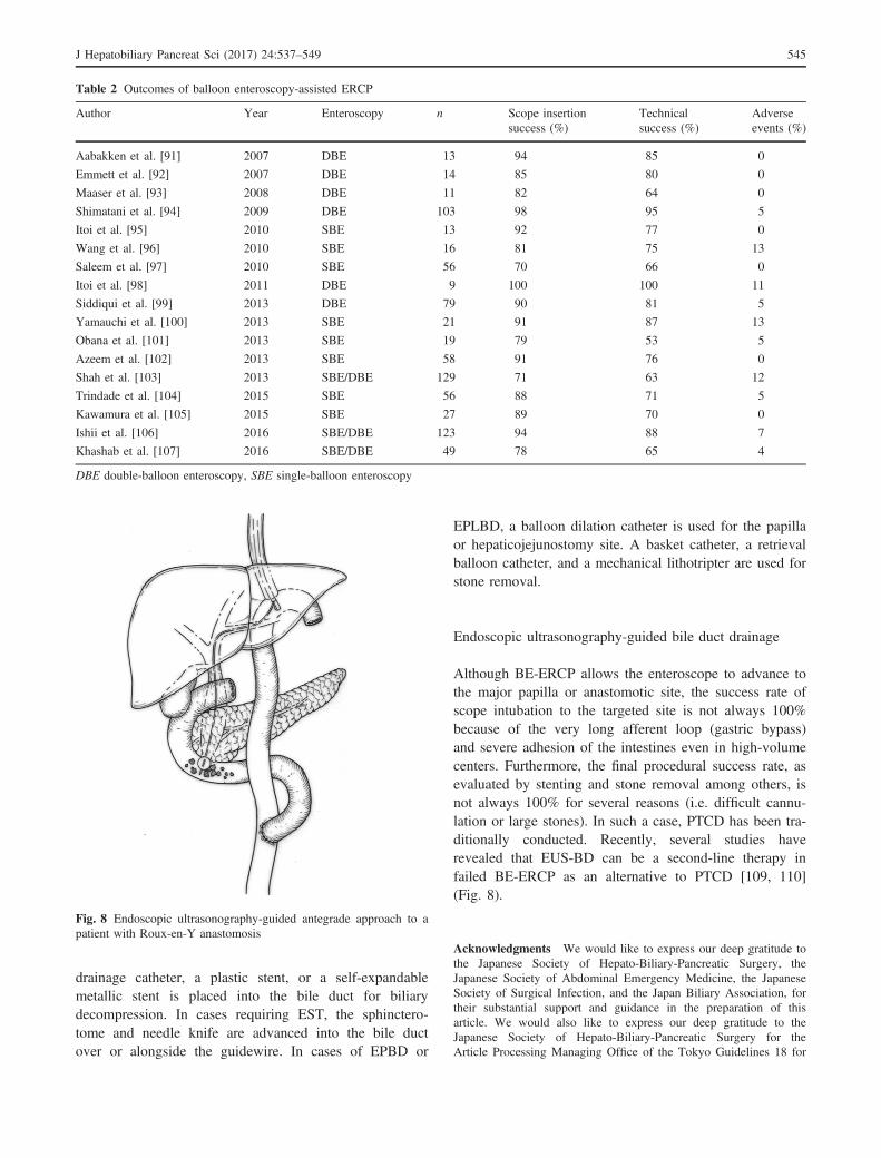
Although BE-ERCP allows the enteroscope to advance tothe major papilla or anastomotic site, the success rate ofscope intubation to the targeted site is not always 100%because of the very long afferent loop (gastric bypass)and severe adhesion of the intestines even in high-volumecenters. Furthermore, the final procedural success rate, asevaluated by stenting and stone removal among others, isnot always 100% for several reasons (i.e. difficult cannu-lation or large stones). In such a case, PTCD has been tra-ditionally conducted. Recently, several studies haverevealed that EUS-BD can be a second-line therapy infailed BE-ERCP as an alternative to PTCD [109, 110](Fig. 8).
Acknowledgments We would like to express our deep gratitude tothe Japanese Society of Hepato-Biliary-Pancreatic Surgery, theJapanese Society of Abdominal Emergency Medicine, the JapaneseSociety of Surgical Infection, and the Japan Biliary Association, fortheir substantial support and guidance in the preparation of thisarticle. We would also like to express our deep gratitude to theJapanese Society of Hepato-Biliary-Pancreatic Surgery for theArticle Processing Managing Office of the Tokyo Guidelines 18 for
Table 2 Outcomes of balloon enteroscopy-assisted ERCP
Author Year Enteroscopy n Scope insertionsuccess (%)
Technicalsuccess (%)
Adverseevents (%)
Aabakken et al. [91] 2007 DBE 13 94 85 0
Emmett et al. [92] 2007 DBE 14 85 80 0
Maaser et al. [93] 2008 DBE 11 82 64 0
Shimatani et al. [94] 2009 DBE 103 98 95 5
Itoi et al. [95] 2010 SBE 13 92 77 0
Wang et al. [96] 2010 SBE 16 81 75 13
Saleem et al. [97] 2010 SBE 56 70 66 0
Itoi et al. [98] 2011 DBE 9 100 100 11
Siddiqui et al. [99] 2013 DBE 79 90 81 5
Yamauchi et al. [100] 2013 SBE 21 91 87 13
Obana et al. [101] 2013 SBE 19 79 53 5
Azeem et al. [102] 2013 SBE 58 91 76 0
Shah et al. [103] 2013 SBE/DBE 129 71 63 12
Trindade et al. [104] 2015 SBE 56 88 71 5
Kawamura et al. [105] 2015 SBE 27 89 70 0
Ishii et al. [106] 2016 SBE/DBE 123 94 88 7
Khashab et al. [107] 2016 SBE/DBE 49 78 65 4
DBE double-balloon enteroscopy, SBE single-balloon enteroscopy
Fig. 8 Endoscopic ultrasonography-guided antegrade approach to apatient with Roux-en-Y anastomosis
J Hepatobiliary Pancreat Sci (2017) 24:537–549 545
preparing this publication. We appreciate all secretariats of theJapanese Society of Hepato-Biliary-Pancreatic Surgery for theirtechnical support.
Conflict of interest Anthony Yuen Bun Teoh has receivedconsultant fees from Boston Scientific Corporation, USA, CookMedical, USA, and Taewoong Medical, Korea.
Appendix: author’s affiliations
Shuntaro Mukai and Takao Itoi, Department of Gastroen-terology and Hepatology, Tokyo Medical UniversityHospital, Tokyo, Japan; Todd H. Baron, Division of Gas-troenterology and Hepatology, University of North Caro-lina at Chapel Hill, Chapel Hill, NC, USA; TadahiroTakada, Fumihiko Miura and Keita Wada, Department ofSurgery, Teikyo University School of Medicine, Tokyo,Japan; Steven M. Strasberg, Section of HPB Surgery,Washington University in St. Louis, St. Louis, MO, USA;Henry A. Pitt, Lewis Katz School of Medicine at TempleUniversity, Philadelphia, PA, USA; Tomohiko Ukai,Department of Family Medicine, Mie Prefectural IchishiHospital, Mie, Japan; Satoru Shikata, Director, Mie Pre-fectural Ichishi Hospital, Mie, Japan; Anthony Yuen BunTeoh, Department of Surgery, The Chinese University ofHong Kong, Shatin, Hong Kong; Myung-Hwan Kim,Department of Gastroenterology, University of Ulsan Col-lege of Medicine, Seoul, Korea; Seiki Kiriyama, Depart-ment of Gastroenterology, Ogaki Municipal Hospital,Gifu, Japan; Yasuhisa Mori, Department of Surgery andOncology, Graduate School of Medical Sciences, KyushuUniversity, Fukuoka, Japan; Miin-Fu Chen, Division ofGeneral Surgery, Linkou Chang Gung Memorial Hospital,Taoyuan, Taiwan; Wan Yee Lau, Faculty of Medicine,The Chinese University of Hong Kong, Shatin, HongKong; Avinash Nivritti Supe, Department of Surgical Gas-troenterology, Seth G S Medical College and K E MHospital, Mumbai, India; Mariano Eduardo Gim�enez,Chair of General Surgery and Minimal Invasive Surgery“Taquini”, University of Buenos Aires, Argentina DAI-CIM Foundation, Buenos Aires, Argentina; MasahiroYoshida, Department of Hemodialysis and Surgery, Ichi-kawa Hospital, International University of Health andWelfare, Chiba, Department of EBM and Guidelines,Japan Council for Quality Health Care, Tokyo, Japan;Toshihiko Mayumi, Department of Emergency Medicine,School of Medicine, University of Occupational and Envi-ronmental Health, Fukuoka, Japan; Koichi Hirata, Depart-ment of Surgery, JR Sapporo Hospital, Hokkaido, Japan;Yoshinobu Sumiyama, Director, Toho University, Tokyo,Japan; Kazuo Inui, Department of Gastroenterology, Sec-ond Teaching Hospital, Fujita Health University, Aichi,Japan; Masakazu Yamamoto, Department of Surgery,
Institute of Gastroenterology, Tokyo Women’s MedicalUniversity, Tokyo, Japan.
References
1. Kimura Y, Takada T, Strasberg SM, Pitt HA, Gouma DJ,Garden OJ, et al. TG13 current terminology, etiology, andepidemiology of acute cholangitis and cholecystitis. J Hepato-biliary Pancreat Sci. 2013;20:8–23.
2. Kiriyama S, Takada T, Hwang TL, Akazawa K, Miura F,Gomi H, et al. Clinical application and verification of theTG13 diagnostic and severity grading criteria for acute cholan-gitis: an international multicenter observational study. J Hepa-tobiliary Pancreat Sci. 2017;24:329–37.
3. Gomi H, Takada T, Hwang TL, Akazawa K, Mori R, Endo I,et al. Updated comprehensive epidemiology, microbiology,and outcomes among patients with acute cholangitis. J Hepato-biliary Pancreat Sci. 2017;24:310–8.
4. Kiriyama S, Takada T, Strasberg SM, Solomkin JS, MayumiT, Pitt HA, et al. TG13 guidelines for diagnosis and severitygrading of acute cholangitis (with videos). J HepatobiliaryPancreat Sci. 2013;20:24–34.
5. Takada T, Hanyu F, Kobayashi S, Uchida Y. Percutaneoustranshepatic cholangial drainage: direct approach under fluoro-scopic control. J Surg Oncol. 1976;8:83–97.
6. Nagai N, Toki F, Oi I, Suzuki H, Kozu T. Continuous endo-scopic pancreatocholedochal catheterization. GastrointestEndosc. 1976;23:78–81.
7. Sohendra N, Reynders-Frederix V. Palliative bile duct drai-nage. A new endoscopic method of introducing a transpapil-lary drain. Endoscopy. 1980;12:8–11.
8. Tsuyuguchi T, Takada T, Kawarada Y, Nimura Y, Wada K,Nagino M, et al. Techniques of biliary drainage for acutecholangitis: Tokyo Guidelines. J Hepatobiliary Pancreat Surg.2007;14:35–45.
9. Itoi T, Tsuyuguchi T, Takada T, Strasberg SM, Pitt HA, KimMH, et al. TG13 indications and techniques for biliary drai-nage in acute cholangitis (with videos). J Hepatobiliary Pan-creat Sci. 2013;20:71–80.
10. Gomi H, Solomkin JS, Takada T, Strasberg SM, Pitt HA,Yoshida M, et al. TG13 antimicrobial therapy for acutecholangitis and cholecystitis. J Hepatobiliary Pancreat Sci.2013;20:60–70.
11. Lai EC, Mok FP, Tan ES, Lo CM, Fan ST, You KT, et al.Endoscopic biliary drainage for severe acute cholangitis. NEngl J Med. 1992;24:1582–6.
12. Leung JW, Chung SC, Sung JJ, Banez VP, Li AK. Urgentendoscopic drainage for acute suppurative cholangitis. Lancet.1989;10:1307–9.
13. Boender J, Nix GA, de Ridder MA, Dees J, Schutte HE, vanBuuren HF, et al. Endoscopic sphincterotomy and biliary drai-nage in patients with cholangitis due to common bile ductstones. Am J Gastroenterol. 1995;90:233–8.
14. Lau JY, Chung SC, Leung JW, Ling TK, Yung MY, Li AK.Endoscopic drainage aborts endotoxaemia in acute cholangitis.Br J Surg. 1996;83:181–4.
15. Umeda J, Itoi T. Current status of preoperative biliary drai-nage. J Gastroenterol. 2015;50:940–54.
16. Saad WE, Wallace MJ, Wojak JC, Kundu S, Cardella JF.Quality improvement guidelines for percutaneous transhepaticcholangiography, biliary drainage, and percutaneous cholecys-tostomy. J Vasc Interv Radiol. 2010;21:789–95.
17. Giovannini M, Moutardier V, Pesenti C, Bories E, Lelong B,Delpero JR. Endoscopic ultrasound-guided bilioduodenal
546 J Hepatobiliary Pancreat Sci (2017) 24:537–549
anastomosis: a new technique for biliary drainage. Endoscopy.2001;33:898–900.
18. Itoi T, Itokawa F, Sofuni A, Kurihara T, Tsuchiya T, Ishii K,et al. Endoscopic ultrasound-guided choledochoduodenostomyin patients with failed endoscopic retrograde cholangiopancre-atography. World J Gastroenterol. 2008;14:6078–82.
19. Sharaiha RZ, Khan MA, Kamal F, Tyberg A, Tombazzi CR,Ali B, et al. Efficacy and safety of EUS-guided biliary drai-nage in comparison with percutaneous biliary drainage whenERCP fails: a systematic review and meta-analysis. Gastroin-test Endosc. 2017;85:904–14.
20. Artifon EL, Aparicio D, Paione JB, Lo SK, Bordini A, RabelloC, et al. Biliary drainage in patients with unresectable, malig-nant obstruction where ERCP fails: endoscopic ultrasonogra-phy-guided choledochoduodenostomy versus percutaneousdrainage. J Clin Gastroenterol. 2012;46:768–74.
21. Lee TH, Choi JH, Park do H, Song TJ, Kim DU, Palk WH,et al. Similar efficacies of endoscopic ultrasound-guided trans-mural and percutaneous drainage for malignant distal biliaryobstruction. Clin Gastroenterol Hepatol. 2016;14:1011–9.
22. Bapaye A, Dubale N, Aher A. Comparison of endosonogra-phy-guided vs. percutaneous biliary stenting when papilla isinaccessible for ERCP. United European. Gastroenterol J.2013;1:285–93.
23. Khashab MA, Valeshabad AK, Afghani E, Singh VK, Kumb-hari V, Messallam A, et al. A comparative evaluation of EUS-guided biliary drainage and percutaneous drainage in patientswith distal malignant biliary obstruction and failed ERCP. DigDis Sci. 2015;60:557–65.
24. Sharaiha RZ, Kumta NA, Desai AP, DeFilippis EM, Gabr M,Sarkisian AM, et al. Endoscopic ultrasound-guided biliary drai-nage versus percutaneous transhepatic biliary drainage: predictorsof successful outcome in patients who fail endoscopic retrogradecholangiopancreatography. Surg Endosc. 2016;30:5500–5.
25. Vila JJ, P�erez-Miranda M, Vazquez-Sequeiros E, Abadia MA,P�erez-Mill�an A, Gonz�alez-Huix F, et al. Initial experience withEUS-guided cholangiopancreatography for biliary and pancre-atic duct drainage: a Spanish national survey. GastrointestEndosc. 2012;76:1133–41.
26. Saltzstein EC, Peacock JB, Mercer LC. Early operation foracute biliary tract stone disease. Surgery. 1983;94:704–8.
27. Mukai S, Itoi T. Selective biliary cannulation techniques forendoscopic retrograde cholangiopancreatography proceduresand prevention of post-endoscopic retrograde cholangiopancre-atography pancreatitis. Expert Rev Gastroenterol Hepatol.2016;10:709–22.
28. Lee DW, Chan AC, Lam YH, Ng EK, Lau JY, Law BK, et al.Biliary decompression by nasobiliary catheter or biliary stentin acute suppurative cholangitis: a prospective randomizedtrial. Gastrointest Endosc. 2002;56:361–5.
29. Sharma BC, Kumar R, Agarwal N, Sarin SK. Endoscopicbiliary drainage by nasobiliary drain or by stent placementin patients with acute cholangitis. Endoscopy. 2005;37:439–43.
30. Zhang RL, Cheng L, Cai XB, Zhao H, Zhu FC, Wan XJ.Comparison of the safety and effectiveness of endoscopic bil-iary decompression by nasobiliary catheter and plastic stentplacement in acute obstructive cholangitis. Swiss Med Wkly.2013;143:w13823.
31. Park SY, Park CH, Cho SB, Yoon KW, Lee WS, Kim HS,et al. The safety and effectiveness of endoscopic biliarydecompression by plastic stent placement in acute suppurativecholangitis compared with nasobiliary drainage. GastrointestEndosc. 2008;68:1076–80.
32. Otani K, Ueki T, Matsumura K, Maruo T, Minoda R, OtsukaY, et al. Comparison between endoscopic biliary stenting and
nasobiliary drainage in patients with acute cholangitis due tocholedocholithiasis: is endoscopic biliary stenting useful?Hepatogastroenterology. 2015;62:558–63.
33. Kawakami H, Kuwatani M, Onodera M, Haba S, Eto K, EhiraN, et al. Endoscopic nasobiliary drainage is the most suitablepreoperative biliary drainage method in the management ofpatients with hilar cholangiocarcinoma. J Gastroenterol.2011;46:242–8.
34. Kawakubo K, Kawakami H, Kuwatani M, Haba S, Kudo T,Taya YA, et al. Lower incidence of complications in endo-scopic nasobiliary drainage for hilar cholangiocarcinoma.World J Gastrointest Endosc. 2016;10:385–90.
35. Mukai S, Itoi T. How should we use endoscopic ultrasonogra-phy-guided biliary drainage techniques separately? EndoscUltrasound. 2016;5:65–8.
36. Burmester E, Niehaus J, Leineweber T, Huetteroth T. EUS-cholangio-drainage of the bile duct: report of 4 cases. Gas-trointest Endosc. 2003;57:246–51.
37. Giovannini M, Dotti M, Bories E, Moutardier V, Pesenti C,Danisi C, et al. Hepaticogastrostomy by echo-endoscopy as apalliative treatment in a patient with metastatic biliary obstruc-tion. Endoscopy. 2003;35:1076–8.
38. Kahaleh M, Hernandez AJ, Tokar J, Adams RB, Shami VM,Yeaton P. Interventional EUS-guided cholangiography: evalua-tion of a technique in evolution. Gastrointest Endosc.2006;64:52–9.
39. Artifon EL, Chaves DM, Ishioka S, Souza TF, Matuguma SE,Sakai P. Echoguided hepatico-gastrostomy: a case report. Clin-ics (Sao Paulo). 2007;62:799–802.
40. Bories E, Pesenti C, Caillol F, Lopes C, Giovannini M. Trans-gastric endoscopic ultrasonography-guided biliary drainage:results of a pilot study. Endoscopy. 2007;39:287–91.
41. Park DH, Koo JE, Oh J, Lee YH, Moon SH, Lee SS, et al.EUS-guided biliary drainage with one-step placement of afully covered metal stent for malignant biliary obstruction: aprospective feasibility study. Am J Gastroenterol. 2009;104:2168–74.
42. Horaguchi J, Fujita N, Noda Y, Kobayashi G, Ito K, Obana T,et al. Endosonography-guided biliary drainage in cases withdifficult transpapillary endoscopic biliary drainage. DigEndosc. 2009;21:239–44.
43. Yamao K, Bhatia V, Mizuno N, Sawaki A, Ishikawa H, TajikaM, et al. EUS-guided choledochoduodenostomy for palliativebiliary drainage in patients with malignant biliary obstruction:results of long-term follow-up. Endoscopy. 2008;40:340–2.
44. Binmoeller KF, Nguyen-Tang T. Endoscopic ultrasound-guided anterograde cholangiopancreatography. J HepatobiliaryPancreat Sci. 2011;18:319–31.
45. Iwashita T, Doi S, Yasuda I. Endoscopic ultrasound-guidedbiliary drainage: a review. Clin J Gastroenterol. 2014;7:94–102.
46. Park DH, Jang JW, Lee SS, Seo DW, Lee SK, Kim MH.EUS-guided biliary drainage with transluminal stenting afterfailed ERCP: predictors of adverse events and long-termresults. Gastrointest Endosc. 2011;74:1276–84.
47. Khashab MA, Levy MJ, Itoi T, Artifon EL. EUS-guided bil-iary drainage. Gastrointest Endosc. 2015;82:993–1001.
48. Itoi T, Isayama H, Sofuni A, Itokawa F, Kurihara T, TsuchiyaT, et al. Stent selection and tips of placement technique ofEUS-guided biliary drainage: trans-duodenal and trans-gastricstenting. J Hepatobiliary Pancreat Sci. 2011;18:664–7.
49. Glessing BR, Mallery S, Freeman ML, Newcomb MD, ArainMA. EUS-guided choledochoduodenostomy with a lumen-apposing metal stent before duodenal stent placement formalignant biliary and duodenal obstruction. GastrointestEndosc. 2015;81:1019–20.
J Hepatobiliary Pancreat Sci (2017) 24:537–549 547
50. Mukai S, Itoi T, Tsuchiya T, Tanaka R, Tonozuka R. EUS-guided intrahepatic bile duct stone extraction via choledo-choduodenostomy created by a lumen-apposing metal stent.Gastrointest Endosc. 2016;83:832–3.
51. Park DH, Lee TH, Paik WH, Choi JH, Song TJ, Lee SS, et al.Feasibility and safety of a novel dedicated device for one-stepEUS-guided biliary drainage: a randomized trial. J Gastroen-terol Hepatol. 2015;30:1461–6.
52. Sugiyama M, Atomi Y. The benefits of endoscopic nasobiliarydrainage without sphincterotomy for acute cholangitis. Am JGastroenterol. 1998;93:2065–8.
53. Hui CK, Lai KC, Yuen MF, Ng M, Chan CK, Hu W, et al.Does the addition of endoscopic sphincterotomy to stent inser-tion improve drainage of the bile duct in acute suppurativecholangitis? Gastrointest Endosc. 2003;58:500–4.
54. Giorgio PD, Luca LD. Comparison of treatment outcomesbetween biliary plastic stent placements with and withoutendoscopic sphincterotomy for inoperable malignant commonbile duct obstruction. World J Gastroenterol. 2004;10:1212–4.
55. Zhang RL, Zhao H, Dai YM, Zhu F, Li L, Li BW, et al.Endoscopic nasobiliary drainage with sphincterotomy in acuteobstructive cholangitis: a prospective randomized controlledtrial. J Dig Dis. 2014;15:78–84.
56. Margulies C, Siqueira ES, Silverman WB, Lin XS, Martin JA,Rabinovitz M, et al. The effect of endoscopic sphincterotomyon acute and chronic complications of biliary endoprostheses.Gastrointest Endosc. 1999;49:716–9.
57. Wilcox CM, Kim H, Ramesh J, Trevino J, Varadarajulu S.Biliary sphincterotomy is not required for bile duct stent place-ment. Dig Endosc. 2014;26:87–92.
58. Sofi AA, Nawras A, Alaradi OH, Alastal Y, Khan MA. Doesendoscopic sphincterotomy reduce the risk of post-endoscopicretrograde cholangiopancreatography pancreatitis after biliarystenting? A systematic review and meta-analysis. Dig Endosc.2016;28:394–404.
59. Komatsu Y, Kawabe T, Toda N, Ohashi M, Isayama M,Tateishi K, et al. Endoscopic papillary balloon dilation for themanagement of common bile duct stones: experience of 226cases. Endoscopy. 1998;30:12–7.
60. Weinberg BM, Shindy W, Lo S. Endoscopic balloon sphincterdilation (sphincteroplasty) versus sphincterotomy for commonbile duct stones. Cochrane Database Syst Rev. 2006;CD004890.
61. Higuchi T, Kon Y. Endoscopic mechanical lithotripsy for the treat-ment of common bile duct stone. Experience with the improveddouble sheath basket catheter. Endoscopy. 1987;19:216–7.
62. Moriai T, Hasegawa T, Fuzita M, Kimura A, Tani T, MakinoI. Successful removal of massive intragastric gallstones byendoscopic electrohydraulic lithotripsy and mechanical litho-tripsy. Am J Gastroenterol. 1991;86:627–9.
63. Ersoz G, Tekesin O, Ozutemiz AO, Gunsar F. Biliary sphinc-terotomy plus dilation with a large balloon for bile duct stonesthat are difficult to extract. Gastrointest Endosc. 2003;57:156–9.
64. Jeong S, Ki SH, Lee DH, Lee JI, Lee JW, Kwon KS, et al.Endoscopic large-balloon sphincteroplasty without precedingsphincterotomy for the removal of large bile duct stones: a pre-liminary study. Gastrointest Endosc. 2009;70:915–22.
65. Minami A, Hirose S, Nomoto T, Hayakawa S. Small sphinc-terotomy combined with papillary dilation with large balloonpermits retrieval of large stones without mechanical lithotripsy.World J Gastroenterol. 2007;13:2179–82.
66. Heo JH, Kang DH, Jung HJ, Kwon DS, An JK, Kim BS,et al. Endoscopic sphincterotomy plus large-balloon dilationversus endoscopic sphincterotomy for removal of bile-ductstones. Gastrointest Endosc. 2007;66:720–6.
67. Itoi T, Itokawa F, Sofuni A, Kurihara T, Tsuchiya T, Ishii K,et al. Endoscopic sphincterotomy combined with large balloondilation can reduce the procedure time and fluoroscopy time forof large bile duct stones. Am J Gastroenterol. 2009;104:560–5.
68. Draganov PV, Evans W, Fazel A, Forsmark CE. Large sizeballoon dilation of the ampulla after biliary sphincterotomycan facilitate endoscopic extraction of difficult bile duct stones.J Clin Gastroenterol. 2009;43:782–6.
69. Teoh AY, Cheung FK, Hu B, Pan YM, Lai LH, Chiu PW,et al. Randomized trial of endoscopic sphincterotomy with bal-loon dilation versus endoscopic sphincterotomy alone forremoval of bile duct stones. Gastroenterology. 2013;144:341–5.
70. Tonozuka R, Itoi T, Sofuni A, Itokawa F, Kurihara T, Tsu-chiya T, et al. Efficacy and safety of endoscopic papillarylarge balloon dilation for large bile duct stones in elderlypatients. Dig Dis Sci. 2014;59:2299–307.
71. Gigot JF, Leese T, Dereme T, Coutinho J, Castaing D, Bis-muth H. Acute cholangitis. Multivariate analysis of risk fac-tors. Ann Surg. 1989;209:435–8.
72. Miura F, Takada T, Strasberg SM, Solomkin JS, Pitt HA,Gouma DJ, et al. TG13 flowchart for the management of acutecholangitis and cholecystitis. J Hepatobiliary Pancreat Sci.2013;20:47–54.
73. Jang SE, Park SW, Lee BS, Shin CM, Lee SH, Kim JW, et al.Management for CBD stone-related mild to moderate acutecholangitis: urgent versus elective ERCP. Dig Dis Sci.2013;58:2082–7.
74. Eto K, Kawakami H, Haba S, Yamato H, Okuda T, Yane K,et al. Single-stage endoscopic treatment for mild to moderateacute cholangitis associated with choledocholithiasis: a multi-center, non-randomized, open-label and exploratory clinicaltrial. J Hepatobiliary Pancreat Sci. 2015;22:825–30.
75. Ito T, Sai JK, Okubo H, Saito H, Ishii S, Kanazawa R, et al.Safety of immediate endoscopic sphincterotomy in acute sup-purative cholangitis caused by choledocholithiasis. World JGastrointest Endosc. 2016;8:180–5.
76. Lee JC, Moon JH, Choi HJ, Kim DC, Choi MH, Lee TH,et al. Delayed endoscopic papillary large balloon dilation aftersphincterotomy for removing large bile duct stones in patientswith acute cholangitis. Dig Dis Sci. 2014;59:1302–6.
77. Fujimoto K, Fujishiro M, Kato M, Higuchi K, Iwakiri R,Sakamoto C, et al. Guidelines for gastroenterological endo-scopy in patients undergoing antithrombotic treatment. DigEndosc. 2014;26:1–14.
78. ASGE Standards of Practice Committee; Acosta RD, AbrahamNS, Chandrasekhara V, Chathadi KV, Early DS, et al. Themanagement of antithrombotic agents for patients undergoingGI endoscopy. Gastrointest Endosc. 2016;83:3–16.
79. Veitch AM, Vanbiervliet G, Gershlick AH, Boustiere C,Baglin TP, Smith LA, et al. Endoscopy in patients on antipla-telet or anticoagulant therapy, including direct oral anticoagu-lants: British Society of Gastroenterology (BSG) and EuropeanSociety of Gastrointestinal Endoscopy (ESGE) guidelines. Gut.2016;65:374–89.
80. Kim KO, Kim TN, Kim SB, Lee JY. Characteristics ofdelayed hemorrhage after endoscopic sphincterotomy. J Gas-troenterol Hepatol. 2010;25:532–8.
81. Freeman ML, Nelson DB, Sherman S, Haber GB, HermanME, Dorsher PJ, et al. Complications of endoscopic biliarysphincterotomy. N Engl J Med. 1996;335:909–18.
82. Hamada T, Yasunaga H, Nakai Y, Isayama H, Matsui H, Hori-guchi H, et al. Bleeding after endoscopic sphincterotomy orpapillary balloon dilation among users of antithromboticagents. Endoscopy. 2015;47:997–1004.
83. Patel IJ, Davidson JC, Nikolic B, Salazar GM, SchwartzbergMS, Walker TG, et al. Consensus guidelines for periprocedural
548 J Hepatobiliary Pancreat Sci (2017) 24:537–549
management of coagulation status and hemostasis risk in per-cutaneous image-guided interventions. J Vasc Interv Radiol.2012;23:727–36.
84. Hamada T, Yasunaga H, Nakai Y, Isayama H, Horiguchi H,Fushimi K, et al. Severe bleeding after percutaneous transhep-atic drainage of the biliary system: effect of antithromboticagents-analysis of 34606 cases from a Japanese nationwideadministrative database. Radiology. 2015;274:605–13.
85. Palareti G, Legnani C, Guazzaloca G, Frascaro M, Grauso F,De Rosa F, et al. Activation of blood coagulation after abruptof stepwise withdrawal of oral anticoagulants-a prospectivestudy. Thromb Haemost. 1994;72:222–66.
86. Hussain N, Alsulaiman R, Burtin P, Toubouti Y, Rahme E,Boivin JF, et al. The safety of endoscopic sphincterotomy inpatients receiving antiplatelet agents: a case-control study. Ali-ment Pharmacol Ther. 2007;25:579–84.
87. Lee MG, Kim J, Lee SH, Lee BS, Lee SJ, Lee YS, et al.Effect of sustained use of platelet aggregation inhibitors onpost-endoscopic sphincterotomy bleeding. Dig Endosc. 2014;26:737–44.
88. Singh M, Mehta N, Murthy UK, Kaul V, Arif A, Newman N.Postpolypectomy bleeding in patients undergoing colonoscopyon uninterrupted clopidogrel therapy. Gastrointest Endosc.2010;71:998–1005.
89. Rabenstein T, Schneider HT, Bulling D, Nicklas M, KatalinicA, Hahn EG, et al. Analysis of the risk factors associated withendoscopic sphincterotomy techniques: preliminary results of aprospective study, with emphasis on the reduced risk of acutepancreatitis with low-dose anticoagulation treatment. Endo-scopy. 2000;32:10–9.
90. Baron TH, Harewood GC. Endoscopic balloon dilation ofthe biliary sphincter compared to endoscopic biliary sphinc-terotomy for removal of common bile duct stones duringERCP: a meta-analysis of randomized, controlled trials. Am JGastroenterol. 2004;99:1455–60.
91. Aabakken L, Bretthauer M, Line PD. Double-balloon entero-scopy for endoscopic retrograde cholangiography in patientswith a Roux-en-Y anastomosis. Endoscopy. 2007;39:1068–71.
92. Emmett DS, Mallat DB. Double-balloon ERCP in patientswho have undergone Roux-en-Y surgery: a case series. Gas-trointest Endosc. 2007;66:1038–41.
93. Maaser C, Lenze F, Bokemeyer M, Ullerich H, Domagk D,Bruewer M, et al. Double balloon enteroscopy: a useful toolfor diagnostic and therapeutic procedures in the pancreaticobil-iary system. Am J Gastroenterol. 2008;103:894–900.
94. Shimatani M, Matsushita M, Takaoka M, Koyabu M, IkeuraT, Kato K, et al. Effective “short” double-balloon enteroscopefor diagnostic and therapeutic ERCP in patients with alteredgastrointestinal anatomy: a large case series. Endoscopy. 2009;41:849–54.
95. Itoi T, Ishii K, Sofuni A, Itokawa F, Tsuchiya T, Kurihara T,et al. Single-balloon enteroscopy-assisted ERCP in patientswith Billroth II gastrectomy or Roux-en-Y anastomosis (withvideo). Am J Gastroenterol. 2010;105:93–9.
96. Wang AY, Sauer BG, Behm BW, Ramanath M, Cox DG, EllenK, et al. Single-balloon enteroscopy effectively enables diagnosticand therapeutic retrograde cholangiography in patients with surgi-cally altered anatomy. Gastrointest Endosc. 2010;71:641–9.
97. Saleem A, Baron TH, Gostout CJ, Topazian MD, Levy MJ,Petersen BT, et al. Endoscopic retrograde cholangiopancre-atography using a single-balloon enteroscope in patients withaltered Roux-en-Y anatomy. Endoscopy. 2010;42:656–60.
98. Itoi T, Ishii K, Sofuni A, Itokawa F, Tsuchiya T, Kurihara T,et al. Long- and short-type double-balloon enteroscopy-assistedtherapeutic ERCP for intact papilla in patients with a Roux-en-Y anastomosis. Surg Endosc. 2011;25:713–21.
99. Siddiqui AA, Chaaya A, Shelton C, Marmion J, Kowalski TE,Loren DE, et al. Utility of the short double-balloon entero-scope to perform pancreaticobiliary interventions in patientswith surgically altered anatomy in a US multicenter study. DigDis Sci. 2013;58:858–64.
100. Yamauchi H, Kida M, Okuwaki K, Miyazawa S, Iwai T, Take-zawa M, et al. Short-type single balloon enteroscope for endo-scopic retrograde cholangiopancreatography with alteredgastrointestinal anatomy. World J Gastroenterol.2013;19:1728–35.
101. Obana T, Fujita N, Ito K, Noda Y, Kobayashi G, Horaguchi J,et al. Therapeutic endoscopic retrograde cholangiography usinga single-balloon enteroscope in patients with Roux-en-Y anas-tomosis. Dig Endosc. 2013;25:601–7.
102. Azeem N, Tabibian JH, Baron TH, Orhurhu V, Rosen CB,Petersen BT, et al. Use of a single-balloon enteroscope com-pared with variable-stiffness colonoscopes for endoscopic retro-grade cholangiography in liver transplant patients with Roux-en-Y biliary anastomosis. Gastrointest Endosc. 2013;77:568–77.
103. Shah RJ, Smolkin M, Yen R, Ross A, Kozarek RA, HowellDA, et al. A multicenter, U.S. experience of single-balloon,double-balloon, and rotational overtube-assisted enteroscopyERCP in patients with surgically altered pancreaticobiliaryanatomy (with video). Gastrointest Endosc. 2013;77:593–600.
104. Trindade AJ, Mella JM, Slattery E, Cohen J, Dickstein J,Garud SS, et al. Use of a cap in single-balloon enteroscopy-assisted endoscopic retrograde cholangiography. Endoscopy.2015;47:453–6.
105. Kawamura T, Uno K, Suzuki A, Mandai K, Nakase K, TanakaK, et al. Clinical usefulness of a short-type, prototypesingle-balloon enteroscope for endoscopic retrograde cholan-giopancreatography in patients with altered gastrointesti-nal anatomy: preliminary experiences. Dig Endosc. 2015;27:82–6.
106. Ishii K, Itoi T, Tonozuka R, Itokawa F, Sofuni A, Tsuchiya T,et al. Balloon enteroscopy-assisted ERCP in patients withRoux-en-Y gastrectomy and intact papillae (with videos). Gas-trointest Endosc. 2016;83:377–86.
107. Khashab MA, El Zein MH, Sharzehi K, Marson FP, HaluszkaO, Small AJ, et al. EUS-guided biliary drainage or entero-scopy-assisted ERCP in patients with surgical anatomy and bil-iary obstruction: an international comparative study. Endosc IntOpen. 2016;4:E1322–7.
108. Skinner M, Popa D, Neumann H, Wilcox CM, M€onkem€ullerK. ERCP with the overtube-assisted enteroscopy technique: asystematic review. Endoscopy. 2014;46:560–72.
109. Itoi T, Sofuni A, Tsuchiya T, Ijima M, Iwashita T. Endoscopicultrasonography-guided transhepatic antegrade stone removalin patients with surgically altered anatomy: case series andtechnical review (with videos). J Hepatobiliary Pancreat Sci.2014;21:E86–93.
110. Iwashita T, Nakai Y, Hara K, Isayama H, Itoi T, Park DH.Endoscopic ultrasound-guided antegrade treatment of bile ductstone in patients with surgically altered anatomy: a multicenterretrospective cohort study. J Hepatobiliary Pancreat Sci.2016;23:227–33.
J Hepatobiliary Pancreat Sci (2017) 24:537–549 549