for its Detection in Cassava Leaves by R. KOUNOUNGUISSA...
14
J. Phytopathology 127, 2 9 4 1 (1989) O 1989 Paul Parey Scientific Publishers, Berlin and Hamburg D. ISSN 0931-1785 CRA de l'Institut National de la Recherche Agronomique, Colmar, et IBMC du Centre National de la Recherche Scientifique, Strasbourg, France I African Cassava Mosaic Virus (ACMV): Stability of Purified Virus and Improved Conditions for its Detection in Cassava Leaves by ELISA BASILE R. KOUNOUNGUISSA, LOUIS GIVORD and BERNARD WALTER .i Authors' addresses: B. R. KOUNOUNGUISSA, and B. WALTER, INRA-Station de Pathologie Végétale - 28, rue de Herrlisheim - 68 O21 Colmar Cedex, France; L. GIVORD, Laboratoire d'Im- munochimie - IBMC - 15, rue Descartes - 67 084 Strasbourg Cedex, France. With 6 figures Received April 2,5, 1988; accepted September 2, 1988 Abstract - African Cassava Mosaic Virus (ACMV) was purified by a method which allowed the separation of monomer from dimer virus particles. Optimal conditions for storing purified virus to be used for immunization were determined by ELISA and inoculation on Nicotiana benthanaiana. Purified virus could be stored without loss of infectivity and serological activity for more than 145 days at 4 "C or frozen at -20 OC, but not longer than 40 days in the presence of 50 % redistilled glycerol. Rabbit and chicken immunoglobulins were used to detect ACMV in cassava leaves by direct and indirect ELISA. To obtain the same absorbance values, it was necessary to use longer incubation times with the indirect method, but the virus detection end-point in sap from infected plants was the same for the two methods (U512). Conditions for improving virus detection in cassava samples were determined. The virus was better detected when leaves from diseased plants were ground in 100 mM Tris-HC1 containing 1 % polyvinylpyrrolidone at pH 8.5 than in phosphate buffer. Plant inhibitors were the restricting factor in the detection of virus by ELISA, but this difficulty was avoided when leaves to be tested were harvested from the top of the cassava plants. Zusammenfassung African Cassava Mosaic Virus (ACMV): Stabilität des gereinigten Virus und verbesserte Bedingungen für dessen Entdeckung in Cassavablättern durch ELISA Bei der Reinigung von african cassava mosaic virus (ACMV) wurde eine Methode angewandt, wodurch die Trennung der Monomer- und Dimerviruspartikeln möglich war. Die optimalen Aufbewahrungsbedingungen des gereinigten Virus, das für Immunisationsstu- dien benutzt werden sö1l;wuFden durch EL1SA;und Inokulation an Nicotiaita bentlumiana ermittelt. U.S! Copyright Clearance Center Code Statement: 0931-1~785/89/2701-0029$02.50/0
Transcript of for its Detection in Cassava Leaves by R. KOUNOUNGUISSA...
J. Phytopathology 127, 2 9 4 1 (1989) O 1989 Paul Parey Scientific Publishers, Berlin and Hamburg
D. ISSN 0931-1785
CRA de l'Institut National de la Recherche Agronomique, Colmar, et IBMC du Centre National de la Recherche Scientifique, Strasbourg, France
I African Cassava Mosaic Virus (ACMV): Stability of Purified Virus and Improved Conditions
for its Detection in Cassava Leaves by ELISA
BASILE R. KOUNOUNGUISSA, LOUIS GIVORD and BERNARD WALTER .i Authors' addresses: B. R. KOUNOUNGUISSA, and B. WALTER, INRA-Station de Pathologie Végétale - 28, rue de Herrlisheim - 68 O21 Colmar Cedex, France; L. GIVORD, Laboratoire d'Im-
munochimie - IBMC - 15, rue Descartes - 67 084 Strasbourg Cedex, France.
With 6 figures
Received April 2,5, 1988; accepted September 2, 1988
Abstract - African Cassava Mosaic Virus (ACMV) was purified by a method which allowed the separation
of monomer from dimer virus particles. Optimal conditions for storing purified virus to be used for immunization were determined by
ELISA and inoculation on Nicotiana benthanaiana. Purified virus could be stored without loss of infectivity and serological activity for more than 145 days at 4 "C or frozen at -20 OC, but not longer than 40 days in the presence of 50 % redistilled glycerol.
Rabbit and chicken immunoglobulins were used to detect ACMV in cassava leaves by direct and indirect ELISA. To obtain the same absorbance values, it was necessary to use longer incubation times with the indirect method, but the virus detection end-point in sap from infected plants was the same for the two methods (U512).
Conditions for improving virus detection in cassava samples were determined. The virus was better detected when leaves from diseased plants were ground in 100 mM Tris-HC1 containing 1 % polyvinylpyrrolidone at p H 8.5 than in phosphate buffer. Plant inhibitors were the restricting factor in the detection of virus by ELISA, but this difficulty was avoided when leaves to be tested were harvested from the top of the cassava plants.
Zusammenfassung
African Cassava Mosaic Virus (ACMV): Stabilität des gereinigten Virus und verbesserte Bedingungen
für dessen Entdeckung in Cassavablättern durch ELISA
Bei der Reinigung von african cassava mosaic virus (ACMV) wurde eine Methode angewandt, wodurch die Trennung der Monomer- und Dimerviruspartikeln möglich war.
Die optimalen Aufbewahrungsbedingungen des gereinigten Virus, das für Immunisationsstu- dien benutzt werden sö1l;wuFden durch EL1SA;und Inokulation an Nicotiaita bentlumiana ermittelt.
U.S! Copyright Clearance Center Code Statement: 0931-1~785/89/2701-0029$02.50/0
30 KOUNOUNGUISSA, GIVORD and WALTER
Das gereinigte Virus liefi sich ohne Verlust an Infektivität oder serologischen Aktivität bei 4 "C oder eingefroren bei -20 "C langer als 145 Tage aufbewahren, jedoch nicht länger als 40 Tage in 50 %igen redestilliertem Glycerol.
Mit Hilfe von Kaninchen- und Hiihnerimmunoglobulinen wurde ACMV in Cassavablattern durch direkten oder indirekten ELISA ermittelt. Um die gleichen Absorptionswerte zu erreichen, waren längere Inkubationszeiten mit der indirekten Methode notwendig, jedoch wurde der gleiche Virusentdeckungsendpunlct (1/512) im Saft infizierter Pflanzen durch beide Methoden ermittelt.
Festgestellt wurden die Bedingungen, um die Virusentdeckung in Cassavaproben zu verbessern. Bessere Ergebnisse wurden erreicht, wenn Blätter von befallenen Pflanzen in 100 mM Tris-HC1, die 1 %ige Polyvinylpyrrolidon bei p H 8,5 enthielt, statt in Phosphatpuffer gerieben worden waren. Pflanzliche Inhibitoren waren die einschränkenden Faktoren bei der ELISA-Virusermittlung, diese Schwierigkeit konnte jedoch umgangen werden, indem die zu untersuchenden Blätter von der Spitze der Cassavapflanzen gesammelt wurden.
Résumé
La purification du virus de la mosaïque africaine du manioc est rendue difficile à cause de sa faible concentration et de sa localisation dans des tissus profonds de la plante. Une méthode utilisant du Triton X-100 à 1 %O et un gradient de 20 à 50 % de saccharose nous a permis la purification de particules virales répondant aux caractéristiques des geminivirus et de séparer des formes monomères et dimères de ce virus. Des préparations de virus purifié peuvent être conservées plus de 145 jours à 4 "C ou à -20 "C sans perte d'infectivité ou d'activité sérologique. Des anticorps contre ce virus ont été préparés par immunisation de lapins et de poules.
Les inhibiteurs de la plante et la faible concentration du virus dans la plante rendent difficile la détection de ce virus par les méthodes immunoenzymatiques. Ces difficultés sont surmontées quand les feuilles à tester sont récoltées au sommet de la plante et broyées dans un tampon Tris-HC10.1 M à p H 8,5 contenant 1 %o de polyvinylpyrrolidone. Les méthodes ELISA directe et indirecte permettent alors une bonne détection du virus. Pour obtenir les mêmes réponses en ELISA, il faut incuber le substrat plus longtemps pour la méthode indirecte. Le virus peut être détecté dans les feuilles de manioc jusqu'à une dilution de 1/512 pour les deux méthodes.
Cassava was introduced into Africa from Brazil during the 16th century. A mosaic disease of cassava was first reported by WARBURG (1894). The disease has been observed wherever cassava is grown in Africa and the adjacent islands (DUFRENOY and HEDIN 1929). Two presumed virus diseases of cassava have been described in Africa many years ago: African cassava mosaic (STOREY and NICHOLS 1938) and cassava brown streak (STOREY 1936).
The disease caused by African cassava mosaic virus (ACMV) seriously decreases the yields of cassava crops. Reported losses range from 30 to 70 Yo (DENGEL 1980). The causal agent, a geminivirus transmitted by the whitefly Benzisia tabnci was isolated from cassava leaves showing mosaic symptoms (BOCK et al. 1977 and 1981, WALTER 1980, ADEJARE and COUTTS 1982,, ROBINSON et al. 1984).
ACMV has been detected in crude cassava leaves by molecular hybridisation (ROBINSON et al. 1984, ROBERTS et al. 1984) and immunoenzymatic assay (SEQUEIRA and HARRISON 1982, THOWENEL et al. 1984, THOMAS et al. 1986, FARGETTE et al. 1987).
However, the detection of virus in cassava leaves was limited by two difficulties: the low concentration of virus particles in the leaves and the presence of plant components which inhibit the ELISA reaction (SEQUEIRA and HARRISON 1982, THOWENEL et al. 1984, FARGETTE et al. 1987).
African Cassava Mosaic Virus 31
In the present paper we report experiments designed to improve the detec- tion of ACMV by ELISA in cassava leaves.
Material and Methods
Plants and Virus
Cassava plants (Manihot esculenta Crantz and Manihot gluziovii Muell.) with mosaic symptoms (natural infection) and infected Nicotiuna benthanziuna were grown in an insect-proof greenhouse under controlled conditions (18-27 O C , 16 h light/day).
Cultivars TMS 30211 and TMS 30572 of M. escidentu were obtained from I.I.T.A. (Ibadan) and Kokobassié, Magnacle, Maloenda and Ouanga from O.R.S.T.O.M. (Abidjan).
The virus was purified from systemically infected N. benthamiunu leaves harvested 20 days after inoculation, using a method similar to that described by LARSEN and DUFFUS (1984) except that after low-speed centrifugation of the polyethyleneglycol (PEG)-treated solution, pellets were dissolved in the grinding buffer without Triton X-100. The virus particles were separated on a sucrose gradient (20-50 % in 0.1 M phosphate buffer - Na2HP04-KH2P04 - pH 7.2) after centrifugation for 6 h 1/2 in a MSE 6 x 38 ml rotor at 24,000 rpm (105.000 g Max.). Concentrations of purified particles were estimated by spectrophotometry, assuming a value of 7 for A 260 nm (1 mg/ml, 1 cm light path) (ROBINSON et ul. 1984). Purified virus in sucrose solution in 0.1 M phosphate buffer p H 7.2 was stored at -20 "C or at +4 "C.
In some experiments, virus was pelleted from the gradient fractions and redissolved in the 0.1 M phosphate buffer.
Electron microscopy
Electronmicroscope observations were performed on carbon-coated grids floated on a drop of virus solution. After rincing with TAM (10 mM Tris HCl p H 8, 10 mM NaN3, 1 mM MgS04) and coloration with 2 % uranyl acetate, the preparations were observed using a Philips 300 microscope at the Biozentrum (Basel).
Storage of purified virus
The stability of purified virus in different storage conditions was examined. Purified virus in the sucrose solution (from gradient fractions) was distributed in different tubes containing each 2.5 ml. The contents of two tubes diluted, one with 2.5 ml of redistilled glycerol and the other with 2.5 ml of a 0.1 M phosphate buffer (pH 7.2) were kept at -20 "C. A third tube was held at 4 "C after dilution with 2.5 ml of 0.1 M phosphate buffer containing 0.03 Yo sodium azide. In a fourth tube, the virus solution was diluted with 2.5 ml of 0.1 M phosphate buffer and frozen in aliquots of 200 pl. The virus infectivity and the serological activity were examined weekly, respectively by inoculating two virus dilutions (41 and 0.6 pg/ml) to four N. benthamiunu plants and by testing in ELISA serial dilutions of each virus preparation (from 600 to 2 nglml).
Immunization protocols
Antisera were produced by injecting rabbits (8 injections) at 7 days intervals. For the first injection, a solution of 0.2 ml of purified virus at 290 pglml mixed with 0.2 ml of Freund's complete adjuvant was subcutaneously injected into rabbits. For the following injections 0.5 ml of the same virus solution mixed with 0.5 ml of Freund's incomplete adjuvant was used. After three injections (22 days after the first injection) rabbits were at regular intervals bled and sera collected.
Chickens were immunized following the same protocol as with the rabbits. The first injection was given with a solution of 150 pl purified virus at 290 pglml mixed with 150 pl of Freund's complete adjuvant. For the subsequent injections 250 pl purified virus plus 250 pl of incomplete adjuvant were injected. Eggs were collected, labelled and stored at 4 'C until processed for extraction of immuno- globulins.

32 KOUNOUNGUISSA, GIVORD and WALTER
Extraction and purification of immunoglobulins
Rabbit immunoglobulins were prepared by rivanol precipitation according to the method of HARDIE and VAN REGENMORTEL (1977). The final solution containing immunoglobulins was dialysed against phosphate saline buffer (PBS) containing 0.02 % NaN, and adjusted to a concentration of 1 mg/ml in PBS (A 280 = 1.4). Purified rabbit IgG was stored at -20 "C with one volume glycerol.
Chicken immunoglobulins (IgY) were purified from eggs following the method described by POLSON et al. (1980). Purified IgY at a concentration of 6 to 12 mg/ml was stored at -20 "C.
Preparation of IgG enzyme conjugate
Alkaline phosphatase (567.752, Boehringer Mannheim) was conjugated to rabbit IgG following the method described by AVRAMEAS (1969). Because of volume changes and possible losses of immunoglobulins during the conjugation procedure, amounts of conjugate used are expressed in terms of dilutions rather than absolute IgG concentrations.
ELISA
Direct ELISA method
Direct double antibody sandwich ELISA was done in polystyrene microtitre plates (Nunc Immunological II, 96 wells) as described by CLARK and ADAMS (1977).
Indirect ELISA method
The indirect ELISA method was that described by VAN REGENMORTEL and BURCKARD (1980) with few modifications. Microtitre plates were coated with 10 pg/ml of purified chicken IgY. Rabbit immunoglobulins were used at 2 pglml and the goat anti-rabbit globulin conjugate (Sigma A-8023) was diluted at 1/2,000.
Absorbance values were accepted as positive when they were greater than twice the mean absorbance of the virus free control sample.
All the ELISA values (diseased and healthy samples) are given without substracting the absorbance of control buffer. The ELISA values of the healthy samples were usually similar to those of the control buffer.
Results
Virus purification
Virus particles were purified from infected N. benthamiana leaves harvested 20 days after inoculation. In our centrifugation conditions ultraviolet absorbance profiles always showed two peaks for the cassava cultivars TMS 30211 or Kokobassié (Fig. I), whereas only the lower peak was obtained with cultivars Ouanga or TMS 30572.
Electron microscopic observation of material from the upper peak of the sucrose gradient showed monomer geminivirus particles of about 18 nm in diameter (Fig. 1/A) while the lower peak contained both single and paired (17-20 x 30-33 nm) particles. Sometimes trimer particles were observed (Fig. l/B). When N. benthamiana plants were inoculated with each fraction, infectivity was associated with fractions 8 to 19 and was correlated with the ELISA response of each fraction (Fig. 1). The material in the two peaks had UV absorbance spectra typical of nucleoproteins. The A260/A280 ratio was 1.35-1.47 for the upper

African Cassava Mosaic Virus
F
I \ \ I I \ I \ \ \ \ \ \ \ \
33
1.5 - E c!
a m
C d ln
W 1.0 $
2 U
0.5 m a
Fig. 1. ACMV purification: Relative absorption at 254 nm (-) of material present in the 20-50 % sucrose gradient after centrifugation at 24,000 rpm for 6 h 1/2 in a 50 MSE rotor at 10 "C. Sedimentation was from left to right. The dotted line (- - -) is the ELISA response of different fractions of the sucrose gradient. Vertical bars represent the distribujion of the infectivity of the sucrose gradient fractions inoculated on N. benthnnzinnn. The infectivity was scored 18 days after
inoculation. Electron microscopy photographs of purified monomer (peak A) and dimer + monomer (peak B);
(magnification: 240,000)

34 KOUNOUNGUISSA, GIVORD and WALTER
peak (monomer particles) and 1.45-1.65 for the lower peak corresponding to respectively 10-17 % and 16-25 YO nucleic acid content (GIBBS and HARRISON 1976). The yield of purified virus was about 5 to 8 mg/kg N. benthamiana leaves.
Storage of purified virus
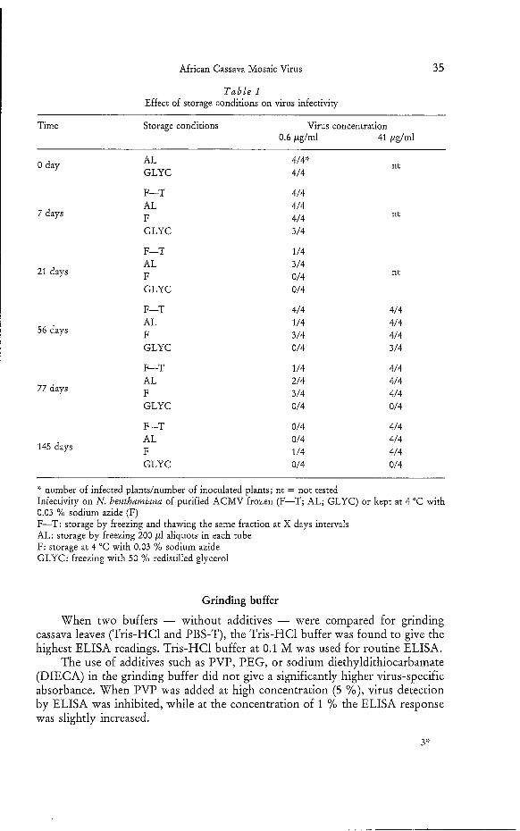
Purified ACMV particles could be stored for a long period without loss of infectivity or ELISA reactivity when kept frozen or at 4 OC, or when repeatedly frozen and thawed. N. benthamiana could still be infected when inoculated with the virus solution stored in these three conditions for 145 days. However, the virus stored in 50 Yo redistilled glycerol completely lost its infectivity after 20 days and no longer reacted in ELISA after about 35 days (Fig. 2 and Table 1).
Influence of crude plant extract on virus reactivity in ELISA
Virus detection was lowered when purified virus was diluted in the sap of healthy cassava leaves compared to the absorbance readings obtained when the virus was diluted in PBS buffer (data not shown). The sensitivity of virus detection was lower when the virus was diluted in extracts from older leaves compared to extracts of younger healthy leaves.
O D 405 nm
T
e--. O TIME (DAYS)
Fig. 2. ELISA response of purified virus after different periods of storage: The stability of the virus was tested by direct ELISA. Coating was done with 1 pg/ml IgG and the enzyme-conjugate was diluted 1/2,000. Before each experiment, virus was diluted to give a concentration range from 600 to
2 ng/ml. ELISA values are those obtained at 60 min with 66 ng/ml of virus. Buff: phosphate buffer
F-T: storage by freezing and thawing the same fraction at X days intervals AL: storage by freezing 200 p1 aliquots in each tube
F: storage at 4 "C with 0.03 % sodium azide GLYC: freezing with 50 % redistilled glycerol
O 14 28 42 56 70
African Cassava Mosaic Virus
T a b l e 1 Effect of storage conditions on virus infectivity
35
Time Storage conditions Virus concentration 0.6 pglml 41 &/ml
0 day AL GLYC
F-T AL F GLYC
7 days
F-T AL F GLYC
21 days
F-T AL F GLYC
56 days
F-T AL F GLYC
77 days
F-T
145 days AL F GLYC
4/4::.
4/4
4/4 4/4 4/4 3/4
1 /4 3/4 0/4 0/4
4/4 1/4 3/4 0/4
1 /4 2/4 3/4 O14
0/4 0/4 1 /4 0/4
nt
nt
nt
4/4 4/4 4/4 3/4
4/4 4/4 4/4 0/4
4/4 4/4 4/4 0/4
number of infected plandnumber of inoculated plants; nt = not tested Infectivity on N. benthamiana of purified ACMV frozen (F-T; AL; GLYC) or kept at 4 "C with 0.03 % sodium azide (F) F-T: storage by freezing and thawing the same fraction at X days intervals AL: storage by freezing 200 pl aliquots in each tube F: storage at 4 "C with 0.03 %O sodium azide GLYC: freezing with 50 %O redistilled glycerol
Grinding buffer
When two buffers - without additives - were compared for grinding cassava leaves (Tris-HC1 and PBS-T), the Tris-HC1 buffer was found to give the highest ELISA readings. Tris-HC1 buffer at 0.1 M was used for routine ELISA.
The use of additives such as PVP, PEG, or sodium diethyldithiocarbamate (DIECA) in the grinding buffer did not give a significantly higher virus-specific absorbance. When PVP was added at high concentration (5 Yo), virus detection by ELISA was inhibited, while at the concentration of 1 % the ELISA response was slightly increased.
3::

36
O.D.405nm
2 - -. -. -.
1.8
1,6
1.4
1,2
1
0.8
0.6
0.4
0.2
O
- *
-.
-. -. --
KOUNOUNGUISSA, GIVORD and WALTER
Direct ELISA
pH7.5
pH8.5
Healthy Dis.l/2 Dis. 1 /32 Healthy Dis. 1/2 Dis. 1/32
Fig. 3. Effect of p H of the grinding buffer on virus detection by direct and indirect ELISA: cassava leaves were ground and diluted 1/2 and 1/32 before addition on the plates using a 0.1 M Tris-HC1 buffer at p H 7.5, 8.0 or 8.5. O.D. readings at 40 min (direct ELISA) or 60 min (indirect ELISA) after
addition of substrate Dis. = infected leaves
When the p H of the grinding buffer was 8.0 to 8.5, ELISA absorbance values were higher than at p H 7.5 (Fig. 3).
In conclusion, the optimal grinding buffer was Tris-HC1 0.1 M, 1 YO PVP, p H 8.5 buffer.
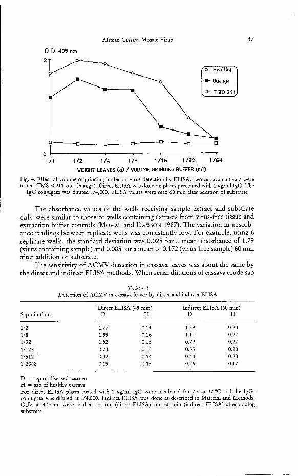
Because of the low concentration of viral antigen in cassava leaves of any cultivar tested here, it was important to determine the optimal conditions for grinding the leaves. The limit of detectability was obtained when one gram of cassava leaves was ground in 32 ml of grinding buffer (Fig. 4). While the detection was possible when one gram of leaves was ground in one milliliter of grinding buffer, the highest ELISA absorbance values were obtained by grinding one gram of cassava leaves in 2 to 4 ml of buffer.
Conditions for improving ACMV detection in cassava crude sap by ELISA
Antigen in cassava leaves could be detected when a dilution of 1/81,OOO of rabbit antiserum was used. When plates were coated directly with the antigen, detection was possible with rabbit antisera diluted at 1/27,000.
In direct ELISA, IgG used for coating the plates was diluted at 1 ,ug/ml and the IgG enzyme conjugate was at 1/4,000.
In the indirect ELISA, microplates were coated with 10 pg/ml IgY. In order to minimize the background absorbance reading observed with healthy samples, purified rabbit-IgG was used at 2 &ml and the goat anti-rabbit enzyme conjugate (GAR) was diluted at 1/2,000.
African Cassava Mosaic Virus
0 D 405nm
37
. 1 /1 1 /2 1 /4 1 / 8 1/16 1/32 1/64
WEIGHT LEAVES (q) / VOLUME GRINDING BUFFER (ml)
Fig. 4. Effect of volume of grinding buffer on virus detection by ELISA: two cassava cultivars were tested (TMS 30211 and Ouanga). Direct ELISA was done on plates precoated with 1 &ml IgG. The
IgG conjugate was diluted 1/4,000. ELISA values were read 60 min after addition of substrate
The absorbance values of the wells receiving sample extract and substrate only were similar to those of wells containing extracts from virus-free tissue and extraction buffer controls (MOWAT and DAWSON 1987). The variation in absorb- ance readings between replicate wells was consistently low. For example, using 6 replicate wells, the standard deviation was 0.025 for a mean absorbance of 1.79 (virus containing sample) and 0.005 for a mean of 0.172 (virus-free sample) 60 min after addition of substrate.
The sensitivity of ACMV detection in cassava leaves was about the same by the direct and indirect ELISA methods. When serial dilutions of cassava crude sap
Table 2 Detection of ACMV in cassava leaves by direct and indirect ELISA
Sap dilutions Direct ELISA (45 min) Indirect ELISA (60 min)
D H D H
1 /2 1.77 0.14 1.39 0.20 1/8 1.89 0.16 1.14 0.22 1/32 1.52 0.15 0.79 0.22 1/128 0.73 0.13 0.55 0.20 U512 0.32 0.14 0.40 0.20 1/2048 0.19 0.15 0.26 0.17
D = sap of diseased cassava H = sap of healthy cassava For direct ELISA plates coated with 1 &ml IgG were incubated for 2 h at 37 "C and the IgG- conjugate was diluted at 1/4,000. Indirect ELISA was done as described in Material and Methods. O.D. at 405 nm were read at 45 min (direct ELISA) and 60 min (indirect ELISA) after adding substrate.
38 KOUNOUNGUISSA, GIVORD and WALTER
were tested by the two methods, the detection end-point was the same: U512 (Table 2), although colour development occurred much faster by the direct method.
O D 405 nm
O 1 2 3 4 5 6
INCUBATION TIME OF IgG (Hours)
Fig. 5. Effect of incubation time with IgG used for coating the plates in direct ELISA: purified virus was incubated overnight at 4 "C; enzyme conjugate at 1/4,000 for 3 hours at 37 "C; readings of O.D.
at 405 nm were at 60 min after addition of substrate
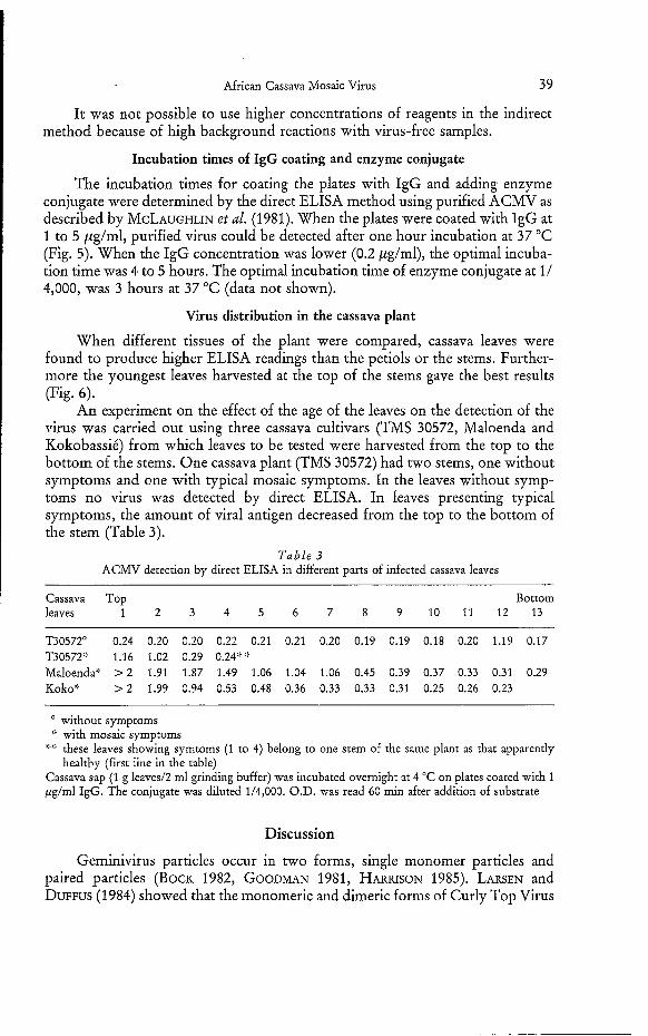
Tap L e m Petiol Stem Top L e a m Peti01 Stem Fig. 6 . ELISA response of different parts of cassava plants of two cultivars tested by direct ELISA. Plant tissues were crushed in a mortar using Tris-HC1 0.1 M buffer at pH 8.5 containing 1 % PVP (1 g/2 ml). Plates were coated with 1 pglml IgG and the conjugate was diluted 1/4,000. ELISA values
were read 60 min after adding substrate
African Cassava Mosaic Virus 39
It was not possible to use higher concentrations of reagents in the indirect method because of high background reactions with virus-free samples.
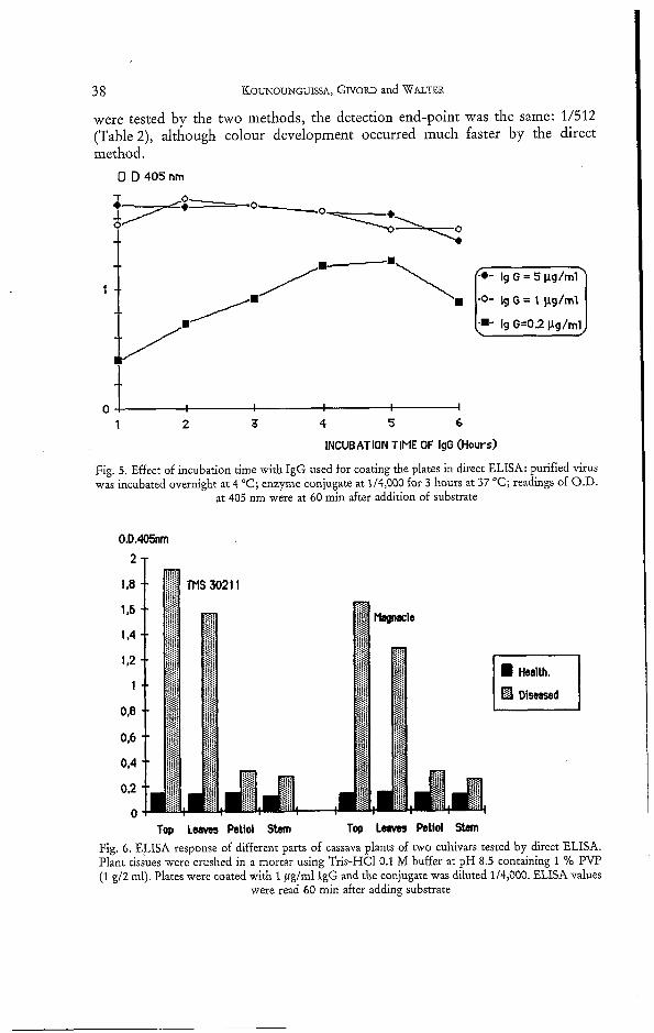
Incubation times of IgG coating and enzyme conjugate
The incubation times for coating the plates with IgG and adding enzyme conjugate were determined by the direct ELISA method using purified ACMV as described by MCLAUGHLIN et al. (1981). When the plates were coated with IgG at 1 to 5 ,ug/ml, purified virus could be detected after one hour incubation at 37 "C (Fig. 5). When the IgG concentration was lower (0.2 pglml), the optimal incuba- tion time was 4 to 5 hours. The optimal incubation time of enzyme conjugate at I / 4,000, was 3 hours at 37 "C (data not shown).
Virus distribution in the cassava plant
When different tissues of the plant were compared, cassava leaves were found to produce higher ELISA readings than the petiols or the stems. Further- more the youngest leaves harvested at the top of the stems gave the best results (Fig. 6).
An experiment on the effect of the age of the leaves on the detection of the virus was carried out using three cassava cultivars (TMS 30572, Maloenda and Kokobassié) from which leaves to be tested were harvested from the top to the bottom of the stems. One cassava plant (TMS 30572) had two stems, one without symptoms and one with typical mosaic symptoms. In the leaves without symp- toms no virus was detected by direct ELISA. In leaves presenting typical symptoms, the amount of viral antigen decreased from the top to the bottom of the stem (Table 3).
Table 3 ACMV detection by direct ELISA in different parts of infected cassava leaves
Cassava Top Bottom leaves 1 2 3 4 5 6 7 8 9 1 0 1 1 1 2 1 3
T30572" 0.24 0.20 0.20 0.22 0.21 0.21 0.20 0.19 0.19 0.18 0.20 1.19 0.17 T30572" 1.16 1.02 0.29 0.24't':. Maloenda- > 2 1.91 1.87 1.49 1.06 1.04 1.06 0.45 0.39 0.37 0.33 0.31 0.29 Koko': > 2 1.99 0.94 0.53 0.48 0.36 0.33 0.33 0.31 0.25 0.26 0.23
without symptoms with mosaic symptoms
healthy (first line in the table) Q;) these leaves showing symtoms (1 to 4) belong to one stem of the same plant as that apparently
Cassava sap (1 g leaved2 ml grinding buffer) was incubated overnight at 4 "C on plates coated with 1 pg/ml IgG. The conjugate was diluted 1/4,000. O.D. was read 60 min after addition of substrate
Discussion
Geminivirus particles occur in two forms, single monomer particles and paired particles (BOCK 1982, GOODMAN 1981, HARRISON 1985). LARSEN and DUFFUS (1984) showed that the monomeric and dimeric forms of Curly Top Virus
40 KOUNOUNGUISSA, GIVORD and WALTER
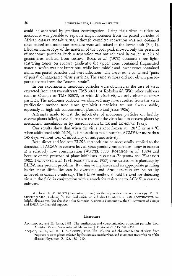
could be separated by gradient centrifugation. Using their virus purification method, it was possible to separate single monomer from the paired particles of African cassava mosaic virus, although complete separation was not obtained since paired and monomer particles were still mixed in the lower peak (Fig. I). Electron microscopy of the material of the upper peak showed only the presence of monomer particles. Such a separation was not achieved in earlier studies of geminivirus isolated from cassava. BOCK et al. (1978) obtained three light- scattering zones on sucrose gradients: the upper zone contained fragmented material which was not infectious, while both middle and lower zones contained numerous paired particles and were infectious. The lower zone contained “pairs of pairs” of aggregated virus particles. The same authors did not obtain paired- particle virus from the “coastal strain”.
In our experiments, monomer particles were obtained in the case of virus extracted from cassava cultivars TMS 30211 or Kokobassié. With other cultivars such as Ouanga or TMS 30572, or with M. glaziovii, we only obtained dimer particles. The monomer particles we observed may have resulted from the virus purification method used since geminivirus particles are not always stable, especially in high salt concentration (ABOUZID and JESKE 1986).
Attempts made to test the infectivity of monomer particles on healthy cassava plants failed, as did all trials to transmit the virus back to cassava plants by mechanical inoculation or by microinjection (DICK and LONGMAN 1985).
Our results show that when the virus is kept frozen at -20 “C or at 4 “C when additioned with NaN3, it is possible to stock purified ACMV for more than 145 days without loss of infectivity or antigenic activity.
Both direct and indirect ELISA methods can be successfully applied to the detection of ACMV in cassava leaves. Since geminivirus particles occur in cassava at a relatively low concentration (WALTER 1980, ROBINSON et al. 1984) and because of the presence of plant inhibitors in cassava (SEQUEIRA and HARRISON 1982, THOWENEL et al. 1984, FARGETTE et al. 1987) virus detection in plant sap by ELISA may present problems. By using young leaves and an appropriate grinding buffer these difficulties can be overcome and virus detection can be readily achieved in cassava crude sap. The ELISA method should be used for detecting virus in the field in conjunction with a search for resistance to ACMV in cassava cultivars.
We thank Dr. M. WURTZ (Biozentrum, Basel) for the help with electron microscopy, Mr. G. STOCKY (INRA, Colmar) for technical assistance and also Dr. M. H. V. VAN REGENMORTEL for helpful discussions. We also thank the European Economic Community, the Government of Congo and INRA for financial support.
Literature
ABOUZID, A., and H. JESKE, 1986: The purification and characterization of gemini particles from Abutilon Mosaic Virus infected Malvaceae. J. Phytopathol. 115, 344-353.
ADEJARE, G. O., and R. H. A. C O U ~ S , 1982: The isolation and characterization of virus from Nigerian cassava plants affected by the cassava mosaic virus, and attempted transmission of the disease. Phytopath. 2. 103, 198-210.

African Cassava Mosaic Virus 41
AVRAMEAS, S., 1969: Coupling of enzymes to proteins with glutaraldehyde. Use of the conjugates for
BOCK, K. R., 1982: Geminivirus diseases. Pl. Disease 66, 266-270. -- , E. J. GUTHRIE, and G. FIGUEIREDO, 1981: A strain of cassava latent virus occurring in coastal
the detection of antigens and antibodies. Immunochemistry 6, 43-52.
districts of Kenya. Ann. appl. Biol. 99, 151-159.
latent virus, a gemini virus. Ann. appl. Biol. 90, 361-367.
cassava latent viruses. Ann. appl. Biol. 85, 305-308.
immunosorbent assay for the detection of plant virus. J. Gen. Virol. 34, 4 7 5 4 8 3 .
in Togo. J. Plant Diseases and Protection 87, 607-620.
tura1 Journal 9, 211-214.
Appl. Agric. Colon. 9, 361.
by African cassava mosaic virus. Ann. appl. Biol. 110, 65-73.
-- , -- , and G. MEREDITH, 1978: Distribution, host range, properties and purification of cassava
-- , G. MEREDITH, and H. BARKER, 1977: RNA and protein components of maize streak and
CLARK, M. F., and A. N. ADAMS, 1977: Characteristics of the microplate method of enzyme linked
DENGEL, H. G., 1980: Investigations on the disease intensity/crop loss relationship of cassava mosaic
DICK, J. MCP., and K. A. LONGMAN, 1985: Techniques for injecting chemicals into trees. Arboricul-
DUFRENOY, J., and L. HEDIN, 1929: La mosàique'des feuilles de manioc au Cameroun. Rev. Bot.
FARGETTE, D., J.-C. THOUVENEL, and C. FAUQUET, 1987: Virus content of,leaves of cassava infected
GIBBS, A., and B. HARRISON, 1976: Plant Virology; the Principles. Edward ARNOLD (ed.). GOODMAN, R. M., 1981: Geminivirus. J. gen. Virol. 54, 9-21. HARDIE, G., and M. H. V. VAN REGENMORTEL, 1977: Isolation of specific antibody under conditions
HARRISON, B. D., 1985: Advances in geminivirus research. Ann. Rev. Phytopathology 23, 55-82. LARSEN, R. C., and J. E. DUFFUS, 1984: A simplified procedure for the purification of Curly Top
Virus and the isolation of its monomer and dimer particles. Phytopathology 74, 114-118. MCLAUGHLIN, M. R., O. W. BARNETT, P. M. BURROWS, and R. H. BAUM, 1981: Improved ELISA
conditions for detection of plant viruses. J. Virol. Meth. 3, 13-25. MOWAT, W. P., and S. DAWSON, 1987: Detection and identification of plant viruses by ELISA using
crude sap extracts and unfractionated antisera. J. Virol. Meth. 15, 233-247. POLSON, A., B. VON WECHMAR, and M. H. V. VAN REGENMORTEL, 1980: Isolation of viral IgY
antibodies from yolk of immunized hens. Immun. Commun. 9, 475-493. ROBERTS, I. M., D. J. ROBINSON, and B. D. HARRISON, 1984: Serological relationships and genome
homologies among geminiviruses. J. gen. Virol. 65, 1723-1730. ROBINSON, D. J., B. D. HARRISON, J. C. SEQUEIRA, and G. H. DUCAN, 1984: Detection of strains of
African cassava mosaic virus by nucleic acid hybridisation and some effects of temperature on their multiplication. Ann. appl. Biol. 105, 383-393.
SEQUEIRA, J. C., and B. D. HARRISON, 1982: Serological studies on cassava latent virus. Ann. appl. Biol. 101, 33-42 .
STOREY, H. H., 1936: Virus diseases of East African plants. East African Agric. J. 2, 34. _ _ , and R. F. W. NICHOLS, 1938: Studies on mosaic disease of cassava. Ann. appl. Biol. 25,
79&-806. THOMAS, J. E., P. R. MASSALSKI, and B. D. HARRISON, 1986: Production of monoclonal antibodies
to African cassava mosaic virus and differences in their reactivities with other whitefly- transmitted geminiviruses. J. gen. Virol. 67, 2739-2748.
THOWENEL, J.-C., D. FARGETTE, C. FAUQUET, and A. MONSABBAT, 1984: Serological diagnosis of African cassava mosaic by immunoenzymatic method. Proceed. of the sixth Inter. Trop. Root Crops Sympos. 21-23 Feb. 1983. Lima, Peru. pp. 353-356.
VAN REGENMORTEL, M. H. V., and J. BURCKARD, 1980: Detection of a wide spectrum of Tobacco Mosaic Virus strains by indirect enzyme-linked immunosorbent assays (ELISA). Virology
WALTER, B., 1980: Isolation and purification of a virus transmitted from mosaic diseased cassava in
WARBURG, O., 1894: Die Kulturpflanzen Usambaras. Mitt. Dtsch. Schutzgeb. 7, 131.
of low ionic strength. J. Immunol. Methods 15, 305-314.
106, 327-334.
the Ivory Coast. P1. Disease 64, 1040-1042.