Endoscopic Ultrasound in the Diagnosis and Staging of ... · Endoscopic Ultrasound in the Diagnosis...
11
GE Port J Gastroenterol. 2015;22(4):161---171 www.elsevier.pt/ge REVIEW ARTICLE Endoscopic Ultrasound in the Diagnosis and Staging of Pancreatic Cancer Bruno Gonc ¸alves, João Bruno Soares, Pedro Bastos ∗ Gastroenterology Department, Hospital de Braga, Braga, Portugal Received 17 March 2015; accepted 23 April 2015 Available online 2 July 2015 KEYWORDS Biopsy, Fine-Needle; Endosonography; Neoplasm Staging; Pancreatic Neoplasms Abstract Pancreatic cancer is one of the digestive cancers with the poorest prognosis, so an early and correct diagnosis is of utmost importance. With the development of new therapeutic options an accurate staging is essential. Endoscopic ultrasonography (EUS) has a major role in all stages of the management of these patients. EUS has a high accuracy in the diagnosis of pancreatic adenocarcinoma and the possibility to perform fine-needle aspiration/biopsy (FNA/FNB) increases the diagnostic yield of EUS. There is still no consensus on the several technical aspects of FNA, namely on the rapid on-site evaluation (ROSE), the diameter and type of needle, the number of passes and the use of stylet and suction. Contrast-enhanced EUS (CE-EUS) and EUS elastography (EUS-E) have been used in recent years as an adjunct to EUS-FNA. Given the higher sensitivity of these techniques a negative cytology by EUS-FNA should not exclude malignancy when CE-EUS and/or EUS-E are suggestive of pancreatic neoplasia. EUS remains one of the main methods in the staging of pancreatic adenocarcinoma, namely to further evaluate patients with non-metastatic disease that appears resectable on initial imaging. EUS is crucial for an accurate preoperative evaluation of pancreatic cancer which is essential to choose the correct management strategy. The possibility to obtain samples from suspicious lesions or lymph nodes, by means of EUS-guided fine-needle aspiration as well as the use of contrast-enhanced and elastography, makes EUS an ideal modality for the diagnosis and staging of pancreatic cancer. © 2015 Sociedade Portuguesa de Gastrenterologia. Published by Elsevier España, S.L.U. This is an open access article under the CC BY-NC-ND license (http://creativecommons. org/licenses/by-nc-nd/4.0/). PALAVRAS-CHAVE Biópsia por Agulha Fina; A Ecoendoscopia no Diagnóstico e Estadiamento do Cancro do Pâncreas Resumo O adenocarcinoma do pâncreas é uma das neoplasias digestivas com pior prognós- tico, sendo fundamental um diagnóstico correto e precoce. Com o desenvolvimento de novas ∗ Corresponding author. E-mail address: [email protected] (P. Bastos). http://dx.doi.org/10.1016/j.jpge.2015.04.007 2341-4545/© 2015 Sociedade Portuguesa de Gastrenterologia. Published by Elsevier España, S.L.U. This is an open access article under the CC BY-NC-ND license (http://creativecommons.org/licenses/by-nc-nd/4.0/).
Transcript of Endoscopic Ultrasound in the Diagnosis and Staging of ... · Endoscopic Ultrasound in the Diagnosis...

GE Port J Gastroenterol. 2015;22(4):161---171
www.elsevier.pt/ge
REVIEW ARTICLE
Endoscopic Ultrasound in the Diagnosis and Staging ofPancreatic Cancer
Bruno Goncalves, João Bruno Soares, Pedro Bastos ∗
Gastroenterology Department, Hospital de Braga, Braga, Portugal
Received 17 March 2015; accepted 23 April 2015Available online 2 July 2015
KEYWORDSBiopsy, Fine-Needle;Endosonography;Neoplasm Staging;Pancreatic Neoplasms
Abstract Pancreatic cancer is one of the digestive cancers with the poorest prognosis, so anearly and correct diagnosis is of utmost importance. With the development of new therapeuticoptions an accurate staging is essential. Endoscopic ultrasonography (EUS) has a major role inall stages of the management of these patients.
EUS has a high accuracy in the diagnosis of pancreatic adenocarcinoma and the possibility toperform fine-needle aspiration/biopsy (FNA/FNB) increases the diagnostic yield of EUS. There isstill no consensus on the several technical aspects of FNA, namely on the rapid on-site evaluation(ROSE), the diameter and type of needle, the number of passes and the use of stylet and suction.Contrast-enhanced EUS (CE-EUS) and EUS elastography (EUS-E) have been used in recent yearsas an adjunct to EUS-FNA. Given the higher sensitivity of these techniques a negative cytology byEUS-FNA should not exclude malignancy when CE-EUS and/or EUS-E are suggestive of pancreaticneoplasia. EUS remains one of the main methods in the staging of pancreatic adenocarcinoma,namely to further evaluate patients with non-metastatic disease that appears resectable oninitial imaging.
EUS is crucial for an accurate preoperative evaluation of pancreatic cancer which is essentialto choose the correct management strategy. The possibility to obtain samples from suspiciouslesions or lymph nodes, by means of EUS-guided fine-needle aspiration as well as the use ofcontrast-enhanced and elastography, makes EUS an ideal modality for the diagnosis and stagingof pancreatic cancer.© 2015 Sociedade Portuguesa de Gastrenterologia. Published by Elsevier España, S.L.U.This is an open access article under the CC BY-NC-ND license (http://creativecommons.org/licenses/by-nc-nd/4.0/).
PALAVRAS-CHAVEBiópsia por AgulhaFina;
A Ecoendoscopia no Diagnóstico e Estadiamento do Cancro do Pâncreas
Resumo O adenocarcinoma do pâncreas é uma das neoplasias digestivas com pior prognós-tico, sendo fundamental um diagnóstico correto e precoce. Com o desenvolvimento de novas
∗ Corresponding author.E-mail address: [email protected] (P. Bastos).
http://dx.doi.org/10.1016/j.jpge.2015.04.0072341-4545/© 2015 Sociedade Portuguesa de Gastrenterologia. Published by Elsevier España, S.L.U. This is an open access article under theCC BY-NC-ND license (http://creativecommons.org/licenses/by-nc-nd/4.0/).

162 B. Goncalves et al.
Estadiamento deNeoplasias;Ecoendoscopia;NeoplasiasPancreáticas
opcões terapêuticas é essencial um estadiamento preciso. A ecoendoscopia apresenta um papelrelevante em todas as fases da abordagem destes doentes.
A acuidade da ecoendoscopia no diagnóstico de adenocarcinoma pancreático é elevada.A possibilidade de realizacão de puncão aspirativa aumenta o potencial diagnóstico, nãohavendo ainda consenso relativamente a vários aspetos da técnica, nomeadamente em relacãoà presenca de citopatologista durante o procedimento, tipo e diâmetro de agulha, número depassagens e utilizacão estilete e aspiracão. Nos anos recentes tem-se assistido à utilizacão deecoendoscopia com contraste (CE-EUS) ou elastografia (EUS-E) como adjuvante da ecoendo-scopia. Estas técnicas apresentam elevada sensibilidade e uma citologia negativa não excluimalignidade se a CE-EUS e/ou EUS-E apresentarem características sugestivas de malignidade.A ecoendoscopia mantém-se um dos principais métodos no estadiamento do adenocarcinomapancreático, em especial na presenca de doenca não metastática que aparenta ser ressecávelnoutras técnicas imagiológicas.
A ecoendoscopia é fundamental na avaliacão pré-operatória do adenocarcinoma pancreáticoe na definicão da correta estratégia de tratamento. A possibilidade de obtencão de amostrasde lesões ou adenopatias suspeitas, através de puncão aspirativa, assim como a utilizacão decontraste e elastografia, fazem da ecoendoscopia uma técnica ideal no diagnóstico e estadia-mento.© 2015 Sociedade Portuguesa de Gastrenterologia. Publicado por Elsevier España, S.L.U.Este é um artigo Open Access sob a licença de CC BY-NC-ND (http://creativecommons.org/licenses/by-nc-nd/4.0/).
1. Introduction
Pancreatic neoplasia, particularly exocrine pancreatic can-cer, is a common cause of cancer-related death and issecond only to colorectal cancer as a cause of digestivecancer-related death.1 Due to its aggressive behavior andlate presentation, this disease has a poor prognosis with avery low five-year survival rate. Surgical resection offersthe only potential cure, but only 10---15% of patients arecandidates for pancreatectomy. Because of these reasonsit is of utmost importance to do an early and correct diag-nosis as well as the most precise staging before providingtherapeutic options.
This paper will discuss the role of endoscopic ultrasoundin the diagnosis and staging of pancreatic cancer.
2. How good is EUS guided FNA in thediagnosis of solid pancreatic lesions?
EUS-FNA is technically successful in 90---95% of procedures,with high sensitivity and specificity for malignancy. This isconfirmed by a recent meta-analysis that showed sensitivityof 85% and specificity of 98%.2 When patients with atypi-cal or suspicious cytology were reclassified as positive formalignancy, the sensitivity increased to 91%, with a slightdecrease in specificity. The negative predictive value of 64%reflects the important fact, that a negative result of EUS-FNA does not exclude malignancy with absolute certainty.
In spite of this excellent accuracy, there is some discus-sion if all pancreatic lesions should be sampled, particularlyin patients who are good surgical candidates with lesionsthat appear resectable. This is mainly due to the fact thatEUS-FNA can be hampered by the presence of stenosis or
other anatomical factors; its accuracy appears to be dimin-ished in the background of chronic pancreatitis3 and thereis some concern with needle tract seeding, even if this lastaspect has only been anecdotally reported.4 On the otherhand, performing EUS-FNA to all patients may be advanta-geous since it allows ruling out other types of malignancy,5
assists with surgical planning and confirms the diagnosis inpatients who want verification prior to surgery.
3. ROSE, needle, suction and passes
Several methodological features can contribute to the suc-cess of EUS guided FNA/FNB. These include the rapid on-siteevaluation (ROSE), the type of needle, the number of passesand the use of stylet and suction.
The role of ROSE and its relevance in EUS-FNA has beendescribed in a guideline published by the European Societyof Gastrointestinal Endoscopy.4 Its use is considered contro-versial even if a recent meta-analysis6 and various studieshave shown an improvement in adequacy rate.7,8 This ismainly due to the fact that ROSE only seems to improve theadequacy rate when it is below 90% and many recent stud-ies have reported adequacy rates superior to 90% withoutthe use of ROSE, indicating that, in high-volume centers,ROSE may not be indispensable. A valid alternative to thepresence of a cytopathologist is the preparation of slidesby a cytotechnician. This was retrospectively evaluated byAlsohaibani et al and the final diagnosis was higher in thegroup with on-site cytotechnologists preparing the slides(77% vs. 53%) and providing initial consideration about spec-imen adequacy.8
Unfortunately not all EUS-FNA performing units haveaccess to ROSE. In such cases it is of utmost importance
Endoscopic ultrasound in the diagnosis of pancreatic cancer 163
to optimize the FNA technique, namely the type of needleand the number of passes. The two most common needlesused to obtain cytology specimens are the 22- and 25-gaugeneedles. Several studies, including 4 randomized controlledtrials, have compared their cytological yield in the diagnosisof pancreatic malignancy.9---12 In general, these studies havenot found significant differences between both needles, butsuggest that 25 gauge could perform better in exceedinglyfibrotic lesions and those located in the head or uncinateprocess. In a recent meta-analysis, that included 8 studieswith 1292 patients, the sensitivity of the 25-gauge needlewas higher than the 22-gauge needle (93% vs. 85%) withsimilar specificity.13
Previous studies, using the 22-gauge needle, showed thatsensitivity improved with an increase in the number ofpasses.14 So, it was suggested that in absence of ROSE atleast 7 passes should be performed in pancreatic lesions.However recent advances in the EUS-FNA technique and thelater development of the 25-gauge needle should probablylead to a re-evaluation of this recommendation. In fact,a recent prospective study using the 25-gauge needle sug-gested that 4 passes may be sufficient to provide adequatecellularity in the absence of ROSE.15
It was previously believed that the use of a stylet duringEUS-FNA prevents clogging of the needle lumen by GI walltissue, which could limit the ability to aspirate cells from thetarget lesion. Hence, the use of the stylet is routine practicefor some endosonographers, even though its insertion andremoval is cumbersome and time consuming. Several stud-ies, including 3 randomized controlled trials have addressedthis subject.16---18 In none of these studies the use of styletwas associated with better specimen adequacy or improveddiagnostic yield for malignancy. In the study by Sahai et althere was inclusive a better quality of the specimen anddiminished bloodiness without the stylet.16 The stylet canalso be used to express the aspirate and this was evaluated ina recent randomized controlled trial. There were no differ-ences between reinsertion of the stylet or air flushing withregard to the number of diagnostic samples, overall accu-racy, cellularity and air-drying artifact.19 Altogether, thesedata does not support the use of a stylet during EUS-FNA.
A more controversial topic is the use of suction dur-ing EUS-FNA since quality data from appropriate trials arescarce. A randomized controlled trial by Lee et al19 thatfocused on 81 patients with solid pancreatic lesions revealeda higher accuracy in the group where suction was used(85.2% vs. 75.9%) despite the increased bloodiness. Thistrial used both the 22- and 25-gauge needle which can be aconfounding factor. The theoretical advantage of suction inpancreatic lesions is to improve sample cellularity since pan-creatic adenocarcinoma tends to be a fibrotic not denselycellular lesion.
In certain pancreatic lesions, such as lymphoma, neu-roendocrine tumors (NET) or autoimmune pancreatitis, itmight be helpful to perform a histological analysis. Thetechniques and methods to do so are beyond the scope ofthis paper and those interested are referred to an excellentreview by Panic et al.20 In a hand stroke, adequate histologicsamples can be obtained using 19- and 22-gauge ProCore orregular needles. On the other hand, the use of the EUS-Trucut needle has been hampered by technical difficultiesand low yield in the transduodenal route. Also, the 25-gauge
needle does not seem to be adequate to collect core sampleand the same applies to the more recent 25-gauge ProCoreneedle, as shown by a recent study in which a histologic corewas found in only 32% of 50 patients with solid pancreaticlesions.21 Interestingly, combining histologic and cytologicalanalysis may improve overall sensitivity.22
In summary, if one feels the need to improve the results ofEUS-FNA in solid pancreatic lesions, the first attitude shouldprobably be to contact the pathology department in orderto arrange an in-room cytopathologist or technician. If thatis not possible, a 25-gauge needle should be used, doing atleast 4 passes with suction. The acquisition of a fragment forhistological analysis should be considered, whether using adedicated FNB needle or a conventional one. Lastly, if EUS-FNA fails, repeating the procedure should be considered,since it has been demonstrated that this will improve thediagnostic rate.
4. Contrast-enhanced EUS andEUS-elastography
EUS provides high-resolution images of the pancreas andit is considered one of the most accurate methods for thediagnosis and staging of pancreatic neoplasia.23,24 Even withthe addition of tissue sampling via FNA, some limitationsexist in the differentiation of benign from malignant solidpancreatic lesions. EUS---FNA may be technically demandingand multiple punctures may be needed to obtain ade-quate cytological samples, which may be associated withfew but not insignificant complications such as acute iatro-genic pancreatitis, bleeding, and infection.25,26 In addition,EUS-FNA provides false-negative results up to 20---40% formalignancy, especially in patients with underlying chronicpancreatitis.2,27 Recent innovations in EUS that intendedto address these limitations include contrast-enhanced EUS(CE-EUS) and EUS elastography (EUS-E).28---31
4.1. Contrast-enhanced EUS (CE-EUS)
CE-EUS allows real-time evaluation of the microvasculatureof the lesion and the surrounding parenchyma through theuse of an intravenous ultrasound contrast agent (UCA)31---33
(Fig. 1). Second-generation UCAs, such as Sonovue and Son-azoid, are composed of a microsphere containing an inertgas.32 Compared with first-generation UCAs they have higherstability allowing real-time vascular images for a prolongedtime.32 CE-EUS can be performed by using color or powerDoppler as a generic signal intensifier or, more appropriately,by using a dedicated contrast harmonic-enhanced EUS.34
The evaluation of CE-EUS images can be done qualita-tively or quantitatively. In qualitative CE-EUS the enhance-ment of the lesion is compared with that of the surroundingparenchyma and classified as non-enhancement, hypo-enhancement, iso-enhancement and hyper-enhancement.35
This evaluation is easy to perform but it is very operatordependent. To increase interobserver agreement for CE-EUSrecent studies used quantification methods to obtain objec-tive results, such as uptake ratio index36 and time intensitycurve (TIC) analysis.37,38
First reports have shown that solid pancreatic lesionsmay have 3 main enhancement patterns: hypo-, iso- and

164 B. Goncalves et al.
Figure 1 Screen capture of video sequence of contrast-enhanced harmonic endoscopic ultrasonography (left panel). The videosequence also included a B-mode standard EUS image of the lesion of interest (right panel). This lesion has a hypo-enhancementpattern suggestive of pancreatic adenocarcinoma (this was confirmed by EUS-FNA).
hyper-enhancement.14---16 Pancreatic adenocarcinoma mostcommonly shows a hypo-enhancement pattern.14,15 Pseu-dotumoral chronic pancreatitis is usually iso-enhanced orhyper-enhanced,38 while neuroendocrine tumor (NET) iscommonly hyper-enhanced.39 Recently, three groups haveprospectively evaluated the performance of qualitativeCE-EUS in characterizing solid pancreatic lesions.39---41 Ade-nocarcinoma was diagnosed as a hypo-enhanced mass withhigh sensitivity (89---95%) and specificity (64---89%). A hyper-enhanced pattern of the mass on CE-EUS was 88---94%predictive of a lesion other than adenocarcinoma in the twofirst series, whereas a hyper-enhanced pattern diagnosedneuroendocrine tumor with a sensitivity and specificity of79% and 99%, respectively, in the last series.
The performance of quantitative CE-EUS in the diagnosisof pancreatic adenocarcinoma has been evaluated in threestudies.36---38 Seicean et al36 showed that the uptake ratioindex was significantly lower for pancreatic adenocarcinomathan for pseudotumoral chronic pancreatitis, with a sensitiv-ity of 80% and a specificity of 91%. Using time intensity curves(TICs), Matsubara et al37 revealed that the echo-intensityreduction rate from its peak at 1 min was greatest in pan-creatic adenocarcinoma, followed by chronic pancreatitis,autoimmune pancreatitis and NET. CE-EUS in combinationwith TIC increased the sensitivity, specificity and accuracy to96%, 93%, and 95%, respectively. Gheonea et al38 confirmedstatistically significant differences in TIC values when com-paring pseudotumoral chronic pancreatitis and pancreaticadenocarcinoma.
In a recent meta-analysis,42 the pooled sensitivity ofCE-EUS for the differential diagnosis of pancreatic adeno-carcinoma was 94% and the specificity was 89%. The AUROCwas 0.973. Besides characterization of pancreatic lesionsin general, CE-EUS may be particularly useful for detectionand characterization of small (<2 cm)39 or solid pancreaticlesions that are associated with biliary stents or chronicpancreatitis.41 CE-EUS has also been shown to be useful
for preoperative localization of NET’s,43 differentiation ofmural nodules in intraductal papillary mucinous neoplasmsand detection of malignant transformations44,45 and for pre-dicting and monitoring response of pancreatic cancer tochemotherapy.46,47
CE-EUS has however some limitations. As the componentshell and excipients of the UCAs are derived from macro-molecular substances, there is a risk of allergic reactions. Inaddition, UCAs should not be used in patients with unstableischemic heart disease.48 The differential diagnosis betweenpancreatic adenocarcinoma and pseudotumoral chronic pan-creatitis may also be particularly challenging since, similarlyto pancreatic adenocarcinoma, pseudotumoral chronic pan-creatitis may show hypo-enhancement at CE-EUS dependingon the degree of inflammation and fibrosis.35,39---41
4.2. EUS-elastography (EUS-E)
The basis for elastography is the fact that many differ-ent pathologic processes, including inflammation, fibrosis,and cancer, induce alterations in tissue stiffness. EUS-Eevaluates tissue stiffness through the application of slightcompression to the targeted tissue and recording the result-ing tissue displacement in the examined field.49 The imagesare obtained in real time, being overimposed as a transpar-ent color overlay on the usual EUS gray-scale images (Fig. 2).Elasticity is depicted using a color map, wherein hard tissueis shown in dark blue, medium hard tissue in cyan, tissuewith intermediate hardness in green, medium soft tissue inyellow and soft tissue in red. The region of interest (ROI)for the elastographic evaluation is manually selected. Opti-mally, the ROI includes the whole target lesion, as well assome surrounding tissues for reference. The first genera-tion of EUS elastography allowed only qualitative evaluation.Today, the second generation also allows quantitative eval-uation of tissue stiffness.50
Endoscopic ultrasound in the diagnosis of pancreatic cancer 165
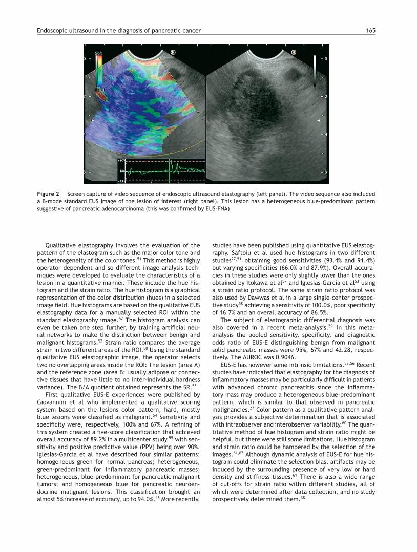
Figure 2 Screen capture of video sequence of endoscopic ultrasound elastography (left panel). The video sequence also includeda B-mode standard EUS image of the lesion of interest (right panel). This lesion has a heterogeneous blue-predominant patternsuggestive of pancreatic adenocarcinoma (this was confirmed by EUS-FNA).
Qualitative elastography involves the evaluation of thepattern of the elastogram such as the major color tone andthe heterogeneity of the color tones.51 This method is highlyoperator dependent and so different image analysis tech-niques were developed to evaluate the characteristics of alesion in a quantitative manner. These include the hue his-togram and the strain ratio. The hue histogram is a graphicalrepresentation of the color distribution (hues) in a selectedimage field. Hue histograms are based on the qualitative EUSelastography data for a manually selected ROI within thestandard elastography image.52 The histogram analysis caneven be taken one step further, by training artificial neu-ral networks to make the distinction between benign andmalignant histograms.52 Strain ratio compares the averagestrain in two different areas of the ROI.50 Using the standardqualitative EUS elastographic image, the operator selectstwo no overlapping areas inside the ROI: The lesion (area A)and the reference zone (area B; usually adipose or connec-tive tissues that have little to no inter-individual hardnessvariance). The B/A quotient obtained represents the SR.53
First qualitative EUS-E experiences were published byGiovannini et al who implemented a qualitative scoringsystem based on the lesions color pattern; hard, mostlyblue lesions were classified as malignant.54 Sensitivity andspecificity were, respectively, 100% and 67%. A refining ofthis system created a five-score classification that achievedoverall accuracy of 89.2% in a multicenter study,55 with sen-sitivity and positive predictive value (PPV) being over 90%.Iglesias-Garcia et al have described four similar patterns:homogeneous green for normal pancreas; heterogeneous,green-predominant for inflammatory pancreatic masses;heterogeneous, blue-predominant for pancreatic malignanttumors; and homogeneous blue for pancreatic neuroen-docrine malignant lesions. This classification brought analmost 5% increase of accuracy, up to 94.0%.56 More recently,
studies have been published using quantitative EUS elastog-raphy. Saftoiu et al used hue histograms in two differentstudies27,51 obtaining good sensitivities (93.4% and 91.4%)but varying specificities (66.0% and 87.9%). Overall accura-cies in these studies were only slightly lower than the onesobtained by Itokawa et al57 and Iglesias-Garcia et al53 usinga strain ratio protocol. The same strain ratio protocol wasalso used by Dawwas et al in a large single-center prospec-tive study58 achieving a sensitivity of 100.0%, poor specificityof 16.7% and an overall accuracy of 86.5%.
The subject of elastographic differential diagnosis wasalso covered in a recent meta-analysis.59 In this meta-analysis the pooled sensitivity, specificity, and diagnosticodds ratio of EUS-E distinguishing benign from malignantsolid pancreatic masses were 95%, 67% and 42.28, respec-tively. The AUROC was 0.9046.
EUS-E has however some intrinsic limitations.53,56 Recentstudies have indicated that elastography for the diagnosis ofinflammatory masses may be particularly difficult in patientswith advanced chronic pancreatitis since the inflamma-tory mass may produce a heterogeneous blue-predominantpattern, which is similar to that observed in pancreaticmalignancies.27 Color pattern as a qualitative pattern anal-ysis provides a subjective determination that is associatedwith intraobserver and interobserver variability.60 The quan-titative method of hue histogram and strain ratio might behelpful, but there were still some limitations. Hue histogramand strain ratio could be hampered by the selection of theimages.61,62 Although dynamic analysis of EUS-E for hue his-togram could eliminate the selection bias, artifacts may beinduced by the surrounding presence of very low or harddensity and stiffness tissues.61 There is also a wide rangeof cut-offs for strain ratio within different studies, all ofwhich were determined after data collection, and no studyprospectively determined them.38

166 B. Goncalves et al.
5. Point of view
Although CE-EUS and EUS-E will hardly replace EUS-FNA,they can be efficient complementary tools to differentiatesolid pancreatic lesions, mainly by reducing the number offalse negatives of EUS-FNA. Given the higher sensitivity ofCE-EUS and EUS-E a negative cytology by EUS-FNA should notbe considered as a benign lesion when CE-EUS and/or EUS-Eare suggestive of pancreatic adenocarcinoma. In these casesfurther cytological samples or surgery should be consideredinstead of follow-up. This has been evaluated in two studies.Napoleon et al40 observed that 4 out of 5 adenocarcinomaswith false-negative EUS-FNA findings were hypo-enhancedat CE-EUS. Additionally, CE-EUS revealed that all ductalcarcinomas with false-negative EUS-FNA results had hypo-enhancement in the study by Kitano et al.39 In addition,CE-EUS and EUS-E could guide FNA to choose an optimalpuncture site to improve the diagnostic accuracy. This hasbeen evaluated by Kitano et al63 who performed FNA guidedby CE-EUS on 39 patients with pancreatic tumors. No viablecells were found in areas with no enhancement, whereasadequate samples were obtained from 80% of sites with aheterogeneous vascular pattern and in all cases from areaswith a homogeneous pattern.
Based on the available data, we believe CE-EUS and EUS-E are ready for use in clinical practice as complementarytools for EUS-FNA. Nonetheless these techniques should beused only by endosonographers who have received propertraining, ideally in reference centers.
6. EUS in the staging of pancreatic cancer
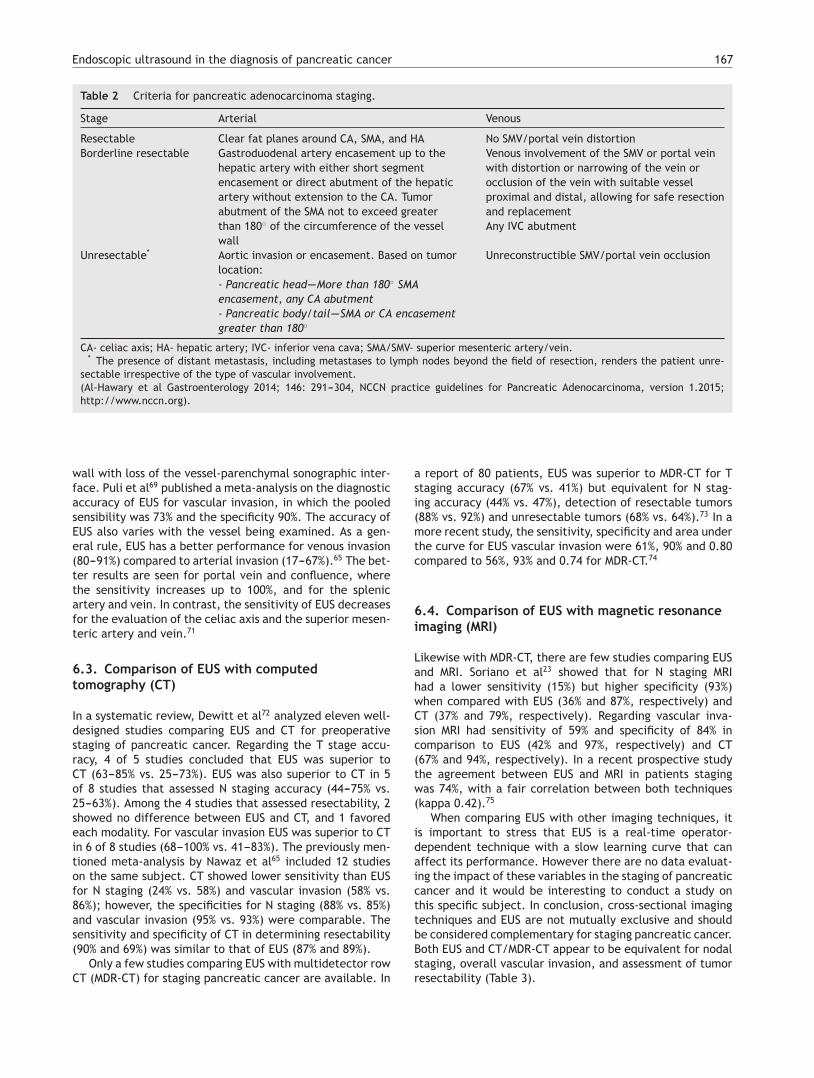
Treatment of pancreatic cancer is an evolving field andan accurate staging becomes increasingly important. Thestaging of pancreatic cancer is based on the tumor-node-metastasis (TNM) system (Table 1). From a clinical point ofview, the goal of preoperative staging is to identify 3 dif-ferent stages of pancreatic cancer and its treatment: (1)resectable tumor that should be referred directly for cura-tive surgery; (2) locally advanced/borderline tumor thatcan be considerer to neoadjuvant chemoradiation and (3)metastatic/unresectable tumor that should be directed topalliative chemotherapy. Current practice in expert centershas evolved to surgical excision of pancreatic adenocarci-noma regardless of extension to adjacent structures (suchas transverse colon, stomach, spleen, adrenal gland and kid-ney), since these structures can be resected along with theprimary tumor. On the other hand, the resectability statusdepends greatly on vascular invasion, and its criteria wererecently reviewed (Table 2).64
6.1. Overall performance
Multiple imaging modalities are available for staging pancre-atic cancer, and there is no evidence-based consensus on theoptimal preoperative imaging assessment of these patients.Several studies have been published assessing the accuracyof EUS for staging pancreatic cancer, and two recent meta-analyses evaluated the performance characteristics of EUSin this setting. In the first one, including 29 studies witha total of 1330 patients, the estimated pooled sensitivity
Table 1 TNM staging system for pancreaticadenocarcinoma.
Primary tumor (T)TX: Primary tumor cannot be assessedT0: No evidence of primary tumorTis: Carcinoma in situT1: Tumor limited to the pancreas, ≤2 cm in greatest
dimensionT2: Tumor limited to the pancreas, >2 cm in greatest
dimensionT3: Tumor extends beyond the pancreas but without
involvement of the celiac axis or the superior mesentericartery
T4: Tumor involves the celiac axis or the superiormesenteric artery (unresectable primary tumor)
Regional lymph nodes (N)NX: Regional lymph nodes cannot be assessedN0: No regional lymph node metastasisN1: Regional lymph node metastasis
Distant metastases (M)M0: No distant metastasisM1: Distant metastasisStage 0---Tis, N0, M0Stage IA---T1, N0, M0Stage IB---T2, N0, M0Stage IIA---T3, N0, M0Stage IIB---T1, N1, M0 or T2, N1, M0 or T3, N1, M0Stage III---T4, any N, M0Stage IV---any T, any N, M1
(AJCC Cancer Staging Manual. 7th ed. New York: Springer; 2010.p. 241).
and specificity was 69% and 81% for N staging; 85% and 91%for vascular invasion and 90% and 86% for resectability.65
In the second meta-analysis, including 20 studies with 726patients, the estimated pooled sensitivity, specificity andAUROC were 72%, 90% and 0.90 for early and intermediatedisease (T1 and T2); 90%, 72% and 0.90 for advanced disease(T3 and T4); 62%, 74% and 0.79 for N staging and 87%, 92%and 0.94 for vascular invasion.66 It seems that EUS has loweraccuracy, mostly less sensitivity, in staging the N status. Inthis sense, the use of EUS-FNA in addition to diagnostic EUShas the potential to increase the accuracy of nodal stagingup to 85%.67,68 At present, regarding EUS technologies, thereis not enough evidence to show a difference in the accuracybetween linear and radial probes.65,69 Keeping in mind thepossible need to perform EUS-FNA as a diagnostic and stagingadjuvant, the use of a linear probe could be more suitable.
6.2. Resectability and vascular invasion
As previously stated, the evaluation of vascular invasion isdeterminant for assessing tumor resectability. Several EUScriteria have been proposed to predict vascular invasion,however the most reliable are70: (1) peripancreatic venouscollaterals in an area of a mass that obliterates the normalanatomic location of a major vessel; (2) tumor within thevessel lumen and (3) abnormal vessel contour or irregular
Endoscopic ultrasound in the diagnosis of pancreatic cancer 167
Table 2 Criteria for pancreatic adenocarcinoma staging.
Stage Arterial Venous
Resectable Clear fat planes around CA, SMA, and HA No SMV/portal vein distortionBorderline resectable Gastroduodenal artery encasement up to the
hepatic artery with either short segmentencasement or direct abutment of the hepaticartery without extension to the CA. Tumorabutment of the SMA not to exceed greaterthan 180◦ of the circumference of the vesselwall
Venous involvement of the SMV or portal veinwith distortion or narrowing of the vein orocclusion of the vein with suitable vesselproximal and distal, allowing for safe resectionand replacementAny IVC abutment
Unresectable* Aortic invasion or encasement. Based on tumorlocation:- Pancreatic head----More than 180◦ SMAencasement, any CA abutment- Pancreatic body/tail----SMA or CA encasementgreater than 180◦
Unreconstructible SMV/portal vein occlusion
CA- celiac axis; HA- hepatic artery; IVC- inferior vena cava; SMA/SMV- superior mesenteric artery/vein.* The presence of distant metastasis, including metastases to lymph nodes beyond the field of resection, renders the patient unre-
sectable irrespective of the type of vascular involvement.(Al-Hawary et al Gastroenterology 2014; 146: 291---304, NCCN practice guidelines for Pancreatic Adenocarcinoma, version 1.2015;http://www.nccn.org).
wall with loss of the vessel-parenchymal sonographic inter-face. Puli et al69 published a meta-analysis on the diagnosticaccuracy of EUS for vascular invasion, in which the pooledsensibility was 73% and the specificity 90%. The accuracy ofEUS also varies with the vessel being examined. As a gen-eral rule, EUS has a better performance for venous invasion(80---91%) compared to arterial invasion (17---67%).65 The bet-ter results are seen for portal vein and confluence, wherethe sensitivity increases up to 100%, and for the splenicartery and vein. In contrast, the sensitivity of EUS decreasesfor the evaluation of the celiac axis and the superior mesen-teric artery and vein.71
6.3. Comparison of EUS with computedtomography (CT)
In a systematic review, Dewitt et al72 analyzed eleven well-designed studies comparing EUS and CT for preoperativestaging of pancreatic cancer. Regarding the T stage accu-racy, 4 of 5 studies concluded that EUS was superior toCT (63---85% vs. 25---73%). EUS was also superior to CT in 5of 8 studies that assessed N staging accuracy (44---75% vs.25---63%). Among the 4 studies that assessed resectability, 2showed no difference between EUS and CT, and 1 favoredeach modality. For vascular invasion EUS was superior to CTin 6 of 8 studies (68---100% vs. 41---83%). The previously men-tioned meta-analysis by Nawaz et al65 included 12 studieson the same subject. CT showed lower sensitivity than EUSfor N staging (24% vs. 58%) and vascular invasion (58% vs.86%); however, the specificities for N staging (88% vs. 85%)and vascular invasion (95% vs. 93%) were comparable. Thesensitivity and specificity of CT in determining resectability(90% and 69%) was similar to that of EUS (87% and 89%).
Only a few studies comparing EUS with multidetector rowCT (MDR-CT) for staging pancreatic cancer are available. In
a report of 80 patients, EUS was superior to MDR-CT for Tstaging accuracy (67% vs. 41%) but equivalent for N stag-ing accuracy (44% vs. 47%), detection of resectable tumors(88% vs. 92%) and unresectable tumors (68% vs. 64%).73 In amore recent study, the sensitivity, specificity and area underthe curve for EUS vascular invasion were 61%, 90% and 0.80compared to 56%, 93% and 0.74 for MDR-CT.74
6.4. Comparison of EUS with magnetic resonanceimaging (MRI)
Likewise with MDR-CT, there are few studies comparing EUSand MRI. Soriano et al23 showed that for N staging MRIhad a lower sensitivity (15%) but higher specificity (93%)when compared with EUS (36% and 87%, respectively) andCT (37% and 79%, respectively). Regarding vascular inva-sion MRI had sensitivity of 59% and specificity of 84% incomparison to EUS (42% and 97%, respectively) and CT(67% and 94%, respectively). In a recent prospective studythe agreement between EUS and MRI in patients stagingwas 74%, with a fair correlation between both techniques(kappa 0.42).75
When comparing EUS with other imaging techniques, itis important to stress that EUS is a real-time operator-dependent technique with a slow learning curve that canaffect its performance. However there are no data evaluat-ing the impact of these variables in the staging of pancreaticcancer and it would be interesting to conduct a study onthis specific subject. In conclusion, cross-sectional imagingtechniques and EUS are not mutually exclusive and shouldbe considered complementary for staging pancreatic cancer.Both EUS and CT/MDR-CT appear to be equivalent for nodalstaging, overall vascular invasion, and assessment of tumorresectability (Table 3).

168 B. Goncalves et al.
Tabl
e3
Glo
balp
erfo
rman
ceof
diff
eren
tim
agin
gm
etho
dsfo
rpa
ncre
atic
aden
ocar
cino
ma.
�
EUS
EUS
vs.
CTEU
Svs
.M
DR-
CTEU
Svs
.M
RI
Sens
itiv
ity
Spec
ifici
tyAU
CSe
nsit
ivit
ySp
ecifi
city
Accu
racy
Sens
itiv
ity
Spec
ifici
tyAc
cura
cySe
nsit
ivit
ySp
ecifi
city
Tst
agin
g72
---90
%72
---90
%0.
9063
---85
%vs
.25
---73
%67
vs.
41%
Nst
agin
g62
---69
%74
---81
%0.
7924
%vs
.58
%88
%vs
.85
%44
---75
%vs
.25
---63
%44
vs.
47%
36vs
.15
%87
vs.
97%
Vasc
ular
inva
sion
73---
87%
90---
92%
0.94
58%
vs.
86%
95%
vs.
93%
68---
100%
vs.
41---
83%
61vs
.56
%90
vs.
93%
0.8
vs.
0.74
*42
vs.
59%
97vs
.84
%
Veno
us80
---91
%Ar
teri
al17
---67
%67
%vs
.67
%10
0%vs
.90
%10
0vs
.60
%§
93vs
.92
%#
Rese
ctab
ility
90%
86%
87vs
.90
%89
%vs
.69%
63---
93%
vs.
60---
83%
88vs
.92
%
�Co
mpa
red
wit
hsu
rger
yor
clin
ical
follo
w-u
p.*
Area
unde
rth
ecu
rve.
§Po
siti
vepr
edic
tive
valu
e.#
Neg
ativ
epr
edic
tive
valu
e.
7. Conclusion
EUS is crucial for an accurate preoperative evaluation ofpancreatic cancer which is essential to choose the correctmanagement strategy.23,24 The possibility to obtain sam-ples, from suspicious lesions or lymph nodes, by meansof EUS-FNA, makes this procedure an ideal diagnosing andstaging modality for pancreatic cancer. EUS-FNA allows thediagnosis of solid pancreatic lesions other than ductal ade-nocarcinoma, staging of suspected or proven pancreaticcancer, and cytological/histological proof of unresectablepancreatic cancer.23,24 Nonetheless, EUS-FNA may providefalse-negative results for malignancy, especially for thepatients with underlying chronic pancreatitis.2,27 The diag-nostic yield of EUS-FNA may be improved by ROSE,6 correctchoice of the needle,13 the number of passes15 and repeti-tion of the procedure. Another option to raise the diagnosticyield of EUS-FNA is the addition of CE-EUS and/or EUS-E.Given the higher sensitivity of CE-EUS and EUS-E, in cases ofnegative cytology by EUS-FNA but in which CE-EUS and/orEUS-E is/are suggestive of pancreatic adenocarcinoma, fur-ther cytological samples or surgery should be consideredinstead of follow-up.39,40 In addition, CE-EUS and EUS-Ecould guide FNA to choose an optimal puncture site toimprove the diagnostic accuracy63.
For staging and assessment of resectability of pancreaticcancer, EUS is applied supplementary to CT.76 While the sec-ond should be the first choice in patients with suspectedpancreatic cancer, EUS may provide better assessment ofT staging and certain types of vascular invasion.77,78 ThusEUS plays a major role to further evaluate patients withnon-metastatic disease that appears resectable on initialimaging.77,78
Ethical disclosures
Protection of human and animal subjects. The authorsdeclare that no experiments were performed on humans oranimals for this study.
Confidentiality of data. The authors declare that no patientdata appear in this article.
Right to privacy and informed consent. The authorsdeclare that no patient data appear in this article.
Conflicts of interest
The authors have no conflicts of interest to declare.
References
1. Shaib YH, Davila JA, El-Serag HB. The epidemiology of pancre-atic cancer in the United States: changes below the surface.Aliment Pharmacol Ther. 2006;24:87---94.
2. Hewitt MJ, McPhail MJ, Possamai L, Dhar A, Vlavianos P, MonahanKJ. EUS-guided FNA for diagnosis of solid pancreatic neoplasms:a meta-analysis. Gastrointest Endosc. 2012;75:319---31.
3. Varadarajulu S, Tamhane A, Eloubeidi MA. Yield of EUS-guidedFNA of pancreatic masses in the presence or the absence

Endoscopic ultrasound in the diagnosis of pancreatic cancer 169
of chronic pancreatitis. Gastrointest Endosc. 2005;62:728---36,quiz 51, 53.
4. Dietrich CF, Jenssen C. Endoscopic ultrasound-guided samp-ling in gastroenterology: European society of gastroin-testinal endoscopy technical guidelines. Endosc Ultrasound.2013;2:117---22.
5. El H II, LeBlanc JK, Sherman S, Al-Haddad MA, Cote GA,McHenry L. Endoscopic ultrasound-guided biopsy of pancre-atic metastases: a large single-center experience. Pancreas.2013;42:524---30.
6. Schmidt RL, Witt BL, Matynia AP, Barraza G, Layfield LJ, AdlerDG. Rapid on-site evaluation increases endoscopic ultrasound-guided fine-needle aspiration adequacy for pancreatic lesions.Dig Dis Sci. 2013;58:872---82.
7. Iglesias-Garcia J, Dominguez-Munoz JE, Abdulkader I, Larino-Noia J, Eugenyeva E, Lozano-Leon A, et al. Influence ofon-site cytopathology evaluation on the diagnostic accu-racy of endoscopic ultrasound-guided fine needle aspiration(EUS-FNA) of solid pancreatic masses. Am J Gastroenterol.2011;106:1705---10.
8. Alsohaibani F, Girgis S, Sandha GS. Does onsite cytotechnologyevaluation improve the accuracy of endoscopic ultrasound-guided fine-needle aspiration biopsy. Can J Gastroenterol.2009;23:26---30.
9. Camellini L, Carlinfante G, Azzolini F, Iori V, Cavina M, SereniG, et al. A randomized clinical trial comparing 22G and 25Gneedles in endoscopic ultrasound-guided fine-needle aspirationof solid lesions. Endoscopy. 2011;43:709---15.
10. Lee JH, Stewart J, Ross WA, Anandasabapathy S, Xiao L, StaerkelG. Blinded prospective comparison of the performance of 22-gauge and 25-gauge needles in endoscopic ultrasound-guidedfine needle aspiration of the pancreas and peri-pancreaticlesions. Dig Dis Sci. 2009;54:2274---81.
11. Fabbri C, Polifemo AM, Luigiano C, Cennamo V, Baccarini P,Collina G, et al. Endoscopic ultrasound-guided fine needle aspi-ration with 22- and 25-gauge needles in solid pancreatic masses:a prospective comparative study with randomisation of needlesequence. Dig Liver Dis. 2011;43:647---52.
12. Siddiqui UD, Rossi F, Rosenthal LS, Padda MS, Murali-DharanV, Aslanian HR. EUS-guided FNA of solid pancreatic masses:a prospective, randomized trial comparing 22-gauge and 25-gauge needles. Gastrointest Endosc. 2009;70:1093---7.
13. Madhoun MF, Wani SB, Rastogi A, Early D, Gaddam S, TierneyWM, et al. The diagnostic accuracy of 22-gauge and 25-gaugeneedles in endoscopic ultrasound-guided fine needle aspira-tion of solid pancreatic lesions: a meta-analysis. Endoscopy.2013;45:86---92.
14. LeBlanc JK, Ciaccia D, Al-Assi MT, McGrath K, Imperiale T, TaoLC, et al. Optimal number of EUS-guided fine needle passesneeded to obtain a correct diagnosis. Gastrointest Endosc.2004;59:475---81.
15. Suzuki R, Irisawa A, Bhutani MS, Hikichi T, Takagi T, Sato A,et al. Prospective evaluation of the optimal number of 25-gaugeneedle passes for endoscopic ultrasound-guided fine-needleaspiration biopsy of solid pancreatic lesions in the absence ofan onsite cytopathologist. Dig Endosc. 2012;24:452---6.
16. Sahai AV, Paquin SC, Gariepy G. A prospective comparisonof endoscopic ultrasound-guided fine needle aspiration resultsobtained in the same lesion, with and without the needle stylet.Endoscopy. 2010;42:900---3.
17. Wani S, Gupta N, Gaddam S, Singh V, Ulusarac O, Romanas M,et al. A comparative study of endoscopic ultrasound guidedfine needle aspiration with and without a stylet. Dig Dis Sci.2011;56:2409---14.
18. Rastogi A, Wani S, Gupta N, Singh V, Gaddam S, ReddymasuS, et al. A prospective, single-blind, randomized, controlledtrial of EUS-guided FNA with and without a stylet. GastrointestEndosc. 2011;74:58---64.
19. Lee JK, Choi JH, Lee KH, Kim KM, Shin JU, Lee KT, et al. Aprospective, comparative trial to optimize sampling techniquesin EUS-guided FNA of solid pancreatic masses. GastrointestEndosc. 2013;77:745---51.
20. Panic N, Larghi A. Techniques for endoscopic ultrasound-guided fine-needle biopsy. Gastrointest Endosc Clin N Am.2014;24:83---107.
21. Iwashita T, Nakai Y, Samarasena JB, Park do H, Zhang Z, GuM, et al. High single-pass diagnostic yield of a new 25-gaugecore biopsy needle for EUS-guided FNA biopsy in solid pancreaticlesions. Gastrointest Endosc. 2013;77:909---15.
22. Moller K, Papanikolaou IS, Toermer T, Delicha EM, Sarbia M,Schenck U, et al. EUS-guided FNA of solid pancreatic masses:high yield of 2 passes with combined histologic---cytologic anal-ysis. Gastrointest Endosc. 2009;70:60---9.
23. Soriano A, Castells A, Ayuso C, Ayuso JR, de Caralt MT,Gines MA, et al. Preoperative staging and tumor resectabilityassessment of pancreatic cancer: prospective study compar-ing endoscopic ultrasonography, helical computed tomography,magnetic resonance imaging, and angiography. Am J Gastroen-terol. 2004;99:492---501.
24. Agarwal B, Abu-Hamda E, Molke KL, Correa AM, Ho L. Endoscopicultrasound-guided fine needle aspiration and multidetector spi-ral CT in the diagnosis of pancreatic cancer. Am J Gastroenterol.2004;99:844---50.
25. Eloubeidi MA, Tamhane A, Varadarajulu S, Wilcox CM. Frequencyof major complications after EUS-guided FNA of solid pancre-atic masses: a prospective evaluation. Gastrointest Endosc.2006;63:622---9.
26. Wang KX, Ben QW, Jin ZD, Du YQ, Zou DW, Liao Z,et al. Assessment of morbidity and mortality associated withEUS-guided FNA: a systematic review. Gastrointest Endosc.2011;73:283---90.
27. Puli SR, Bechtold ML, Buxbaum JL, Eloubeidi MA. How goodis endoscopic ultrasound-guided fine-needle aspiration in diag-nosing the correct etiology for a solid pancreatic mass? Ameta-analysis and systematic review. Pancreas. 2013;42:20---6.
28. Pedrosa MC, Barth BA, Desilets DJ, Kaul V, Kethu SR, PfauPR, et al. Enhanced ultrasound imaging. Gastrointest Endosc.2011;73:857---60.
29. Fusaroli P, Saftoiu A, Mancino MG, Caletti G, Eloubeidi MA. Tech-niques of image enhancement in EUS (with videos). GastrointestEndosc. 2011;74:645---55.
30. Popescu A, Saftoiu A. Can elastography replace fine needle aspi-ration. Endosc Ultrasound. 2014;3:109---17.
31. Kwek BE, Ang TL, Seo DW, Imazu H. Contrast-enhanced har-monic endoscopic ultrasonography of solid pancreatic lesions.Endosc Ultrasound. 2013;2:142---7.
32. Sanchez MV, Varadarajulu S, Napoleon B. EUS contrast agents:what is available, how do they work, and are they effective.Gastrointest Endosc. 2009;69:S71---7.
33. Jang SI, Lee DK. Contrast-enhanced endoscopic ultra-sonography: advance and current status. Ultrasonography.2014;33:161---9.
34. Claudon M, Cosgrove D, Albrecht T, Bolondi L, Bosio M, CalliadaF, et al. Guidelines and good clinical practice recommenda-tions for contrast enhanced ultrasound (CEUS) --- update 2008.Ultraschall Med. 2008;29:28---44.
35. Kitano M, Sakamoto H, Matsui U, Ito Y, Maekawa K, von SchrenckT, et al. A novel perfusion imaging technique of the pancreas:contrast-enhanced harmonic EUS (with video). GastrointestEndosc. 2008;67:141---50.
36. Seicean A, Badea R, Stan-Iuga R, Mocan T, Gulei I, PascuO. Quantitative contrast-enhanced harmonic endoscopic ultra-sonography for the discrimination of solid pancreatic masses.Ultraschall Med. 2010;31:571---6.
37. Matsubara H, Itoh A, Kawashima H, Kasugai T, Ohno E, IshikawaT, et al. Dynamic quantitative evaluation of contrast-enhanced

170 B. Goncalves et al.
endoscopic ultrasonography in the diagnosis of pancreatic dis-eases. Pancreas. 2011;40:1073---9.
38. Gheonea DI, Streba CT, Ciurea T, Saftoiu A. Quantitative lowmechanical index contrast-enhanced endoscopic ultrasound forthe differential diagnosis of chronic pseudotumoral pancreatitisand pancreatic cancer. BMC Gastroenterol. 2013;13:2.
39. Kitano M, Kudo M, Yamao K, Takagi T, Sakamoto H, Komaki T,et al. Characterization of small solid tumors in the pancreas: thevalue of contrast-enhanced harmonic endoscopic ultrasonogra-phy. Am J Gastroenterol. 2012;107:303---10.
40. Napoleon B, Alvarez-Sanchez MV, Gincoul R, Pujol B, LefortC, Lepilliez V, et al. Contrast-enhanced harmonic endoscopicultrasound in solid lesions of the pancreas: results of a pilotstudy. Endoscopy. 2010;42:564---70.
41. Fusaroli P, Spada A, Mancino MG, Caletti G. Contrast harmonicecho-endoscopic ultrasound improves accuracy in diagno-sis of solid pancreatic masses. Clin Gastroenterol Hepatol.2010;8:629---34, e1-2.
42. Gong TT, Hu DM, Zhu Q. Contrast-enhanced EUS for differentialdiagnosis of pancreatic mass lesions: a meta-analysis. Gastroin-test Endosc. 2012;76:301---9.
43. Ishikawa T, Itoh A, Kawashima H, Ohno E, Matsubara H, Itoh Y,et al. Usefulness of EUS combined with contrast-enhancementin the differential diagnosis of malignant versus benign andpreoperative localization of pancreatic endocrine tumors. Gas-trointest Endosc. 2010;71:951---9.
44. Yamashita Y, Ueda K, Itonaga M, Yoshida T, Maeda H, Maekita T,et al. Usefulness of contrast-enhanced endoscopic sonographyfor discriminating mural nodules from mucous clots in intraduc-tal papillary mucinous neoplasms: a single-center prospectivestudy. J Ultrasound Med. 2013;32:61---8.
45. Ohno E, Itoh A, Kawashima H, Ishikawa T, Matsubara H, ItohY, et al. Malignant transformation of branch duct-type intra-ductal papillary mucinous neoplasms of the pancreas based oncontrast-enhanced endoscopic ultrasonography morphologicalchanges: focus on malignant transformation of intraduc-tal papillary mucinous neoplasm itself. Pancreas. 2012;41:855---62.
46. Yamashita Y, Ueda K, Itonaga M, Yoshida T, Maeda H, MaekitaT, et al. Tumor vessel depiction with contrast-enhanced endo-scopic ultrasonography predicts efficacy of chemotherapy inpancreatic cancer. Pancreas. 2013;42:990---5.
47. Ohshima T, Yamaguchi T, Ishihara T, Yoshikawa M, KobayashiA, Sakaue N, et al. Evaluation of blood flow in pancre-atic ductal carcinoma using contrast-enhanced, wide-bandDoppler ultrasonography: correlation with tumor characteris-tics and vascular endothelial growth factor. Pancreas. 2004;28:335---43.
48. Raisinghani A, DeMaria AN. Physical principles of microbub-ble ultrasound contrast agents. Am J Cardiol. 2002;90:3J---7J.
49. Gao L, Parker KJ, Lerner RM, Levinson SF. Imaging of the elasticproperties of tissue --- a review. Ultrasound Med Biol. 1996;22:959---77.
50. Giovannini M. Endoscopic ultrasound elastography. Pancreatol-ogy. 2011;11 Suppl. 2:34---9.
51. Saftoiu A, Vilmann P, Gorunescu F, Janssen J, Hocke M, Larsen M,et al. Accuracy of endoscopic ultrasound elastography used fordifferential diagnosis of focal pancreatic masses: a multicenterstudy. Endoscopy. 2011;43:596---603.
52. Saftoiu A, Vilmann P, Gorunescu F, Gheonea DI, GorunescuM, Ciurea T, et al. Neural network analysis of dynamicsequences of EUS elastography used for the differential diagno-sis of chronic pancreatitis and pancreatic cancer. GastrointestEndosc. 2008;68:1086---94.
53. Iglesias-Garcia J, Larino-Noia J, Abdulkader I, Forteza J,Dominguez-Munoz JE. Quantitative endoscopic ultrasoundelastography: an accurate method for the differentiation
of solid pancreatic masses. Gastroenterology. 2010;139:1172---80.
54. Giovannini M, Hookey LC, Bories E, Pesenti C, Monges G, DelperoJR. Endoscopic ultrasound elastography: the first step towardsvirtual biopsy? Preliminary results in 49 patients. Endoscopy.2006;38:344---8.
55. Giovannini M, Thomas B, Erwan B, Christian P, Fabrice C,Benjamin E, et al. Endoscopic ultrasound elastography for eval-uation of lymph nodes and pancreatic masses: a multicenterstudy. World J Gastroenterol. 2009;15:1587---93.
56. Iglesias-Garcia J, Larino-Noia J, Abdulkader I, FortezaJ, Dominguez-Munoz JE. EUS elastography for thecharacterization of solid pancreatic masses. GastrointestEndosc. 2009;70:1101---8.
57. Itokawa F, Itoi T, Sofuni A, Kurihara T, Tsuchiya T, Ishii K, et al.EUS elastography combined with the strain ratio of tissue elas-ticity for diagnosis of solid pancreatic masses. J Gastroenterol.2011;46:843---53.
58. Dawwas MF, Taha H, Leeds JS, Nayar MK, Oppong KW. Diagnos-tic accuracy of quantitative EUS elastography for discriminatingmalignant from benign solid pancreatic masses: a prospective,single-center study. Gastrointest Endosc. 2012;76:953---61.
59. Mei M, Ni J, Liu D, Jin P, Sun L. EUS elastography for diagnosis ofsolid pancreatic masses: a meta-analysis. Gastrointest Endosc.2013;77:578---89.
60. Saftoiu A, Vilman P. Endoscopic ultrasound elastography --- anew imaging technique for the visualization of tissue elasticitydistribution. J Gastrointest Liver Dis. 2006;15:161---5.
61. Saftoiu A, Vilmann P, Ciurea T, Popescu GL, Iordache A, HassanH, et al. Dynamic analysis of EUS used for the differentiationof benign and malignant lymph nodes. Gastrointest Endosc.2007;66:291---300.
62. Jacobson BC. Pressed for an answer: has elastography finallycome to EUS. Gastrointest Endosc. 2007;66:301---3.
63. Kitano MS H, Komaki T, Kudo M. FNA guided by contrastenhanced harmonic EUS in pancreatic tumors. GastrointestEndosc. 2009;69:AB328.
64. Al-Hawary MM, Francis IR, Chari ST, Fishman EK, Hough DM, LuDS, et al. Pancreatic ductal adenocarcinoma radiology repor-ting template: consensus statement of the society of abdominalradiology and the american pancreatic association. Gastroen-terology. 2014;146:291e1---304e1.
65. Nawaz H, Fan CY, Kloke J, Khalid A, McGrath K, LandsittelD, et al. Performance characteristics of endoscopic ultrasoundin the staging of pancreatic cancer: a meta-analysis. JOP.2013;14:484---97.
66. Li JH, He R, Li YM, Cao G, Ma QY, Yang WB. Endoscopic ultra-sonography for tumor node staging and vascular invasion inpancreatic cancer: a meta-analysis. Dig Surg. 2014;31:297---305.
67. Chang KJ, Nguyen P, Erickson RA, Durbin TE, Katz KD. Theclinical utility of endoscopic ultrasound-guided fine-needleaspiration in the diagnosis and staging of pancreatic carcinoma.Gastrointest Endosc. 1997;45:387---93.
68. Shin HJ, Lahoti S, Sneige N. Endoscopic ultrasound-guided fine-needle aspiration in 179 cases: the M D. Anderson Cancer Centerexperience. Cancer. 2002;96:174---80.
69. Puli SR, Singh S, Hagedorn CH, Reddy J, Olyaee M. Diagnos-tic accuracy of EUS for vascular invasion in pancreatic andperiampullary cancers: a meta-analysis and systematic review.Gastrointest Endosc. 2007;65:788---97.
70. Snady H. EUS criteria for vascular invasion: analyzing the meta-analysis. Gastrointest Endosc. 2007;65:798---807.
71. Iglesias Garcia J, Larino Noia J, Dominguez Munoz JE. Endo-scopic ultrasound in the diagnosis and staging of pancreaticcancer. Rev Esp Enferm Dig. 2009;101:631---8.
72. Dewitt J, Devereaux BM, Lehman GA, Sherman S, Imperiale TF.Comparison of endoscopic ultrasound and computed tomogra-phy for the preoperative evaluation of pancreatic cancer: a

Endoscopic ultrasound in the diagnosis of pancreatic cancer 171
systematic review. Clin Gastroenterol Hepatol. 2006;4:717---25,quiz 664.
73. DeWitt J, Devereaux B, Chriswell M, McGreevy K, Howard T,Imperiale TF, et al. Comparison of endoscopic ultrasonogra-phy and multidetector computed tomography for detecting andstaging pancreatic cancer. Ann Intern Med. 2004;141:753---63.
74. Tellez-Avila FI, Chavez-Tapia NC, Lopez-Arce G, Franco-GuzmanAM, Sosa-Lozano LA, Alfaro-Lara R, et al. Vascular invasion inpancreatic cancer: predictive values for endoscopic ultrasoundand computed tomography imaging. Pancreas. 2012;41:636---8.
75. Shami VM, Mahajan A, Loch MM, Stella AC, Northup PG, WhiteGE, et al. Comparison between endoscopic ultrasound and
magnetic resonance imaging for the staging of pancreatic can-cer. Pancreas. 2011;40:567---70.
76. Seufferlein T, Bachet JB, Van Cutsem E, Rougier P. Pancreaticadenocarcinoma: ESMO-ESDO clinical practice guidelines fordiagnosis, treatment and follow-up. Ann Oncol. 2012;23 Suppl.7:vii33---40.
77. Saftoiu A, Vilmann P. Role of endoscopic ultrasound in thediagnosis and staging of pancreatic cancer. J Clin Ultrasound.2009;37:1---17.
78. Michl P, Pauls S, Gress TM. Evidence-based diagnosis and stag-ing of pancreatic cancer. Best Pract Res Clin Gastroenterol.2006;20:227---51.