Dominant ER Stress Inducing WFS1 Mutations …...Dominant ER Stress–Inducing WFS1 Mutations...
10
Dominant ER Stress–Inducing WFS1 Mutations Underlie a Genetic Syndrome of Neonatal/Infancy-Onset Diabetes, Congenital Sensorineural Deafness, and Congenital Cataracts Elisa De Franco, 1 Sarah E. Flanagan, 1 Takuya Yagi, 2 Damien Abreu, 2 Jana Mahadevan, 2 Matthew B. Johnson, 1 Garan Jones, 3 Fernanda Acosta, 4 Mphele Mulaudzi, 5 Ngee Lek, 6,7 Vera Oh, 6 Oliver Petz, 8 Richard Caswell, 1 Sian Ellard, 1,3 Fumihiko Urano, 2 and Andrew T. Hattersley 1 Diabetes 2017;66:2044–2053 | https://doi.org/10.2337/db16-1296 Neonatal diabetes is frequently part of a complex syndrome with extrapancreatic features: 18 genes causing syndromic neonatal diabetes have been identi- fied to date. There are still patients with neonatal diabetes who have novel genetic syndromes. We performed exome sequencing in a patient and his unrelated, unaffected parents to identify the genetic etiology of a syndrome characterized by neonatal diabetes, sensorineural deaf- ness, and congenital cataracts. Further testing was per- formed in 311 patients with diabetes diagnosed before 1 year of age in whom all known genetic causes had been excluded. We identified 5 patients, including the initial case, with three heterozygous missense mutations in WFS1 (4/5 confirmed de novo). They had diabetes diagnosed before 12 months (2 before 6 months) (5/5), sensorineural deaf- ness diagnosed soon after birth (5/5), congenital cataracts (4/5), and hypotonia (4/5). In vitro studies showed that these WFS1 mutations are functionally different from the known recessive Wolfram syndrome–causing mutations, as they tend to aggregate and induce robust endoplasmic reticulum stress. Our results establish specific dominant WFS1 mutations as a cause of a novel syndrome including neonatal/infancy-onset diabetes, congenital cataracts, and sensorineural deafness. This syndrome has a discrete pathophysiology and differs genetically and clinically from recessive Wolfram syndrome. Neonatal diabetes is diagnosed before 6 months of age and reflects a severe reduction in b-cell number or function. A genetic diagnosis is possible in 82% of patients with 23 ge- netic causes identified to date (1,2). Thirty-nine percent of patients with neonatal diabetes have a genetic etiology that results in the development of at least one extrapancreatic feature in addition to diabetes (1). This subtype is pheno- typically and genetically heterogeneous with mutations in 18 genes known to cause syndromic forms of neonatal di- abetes (1). Fifteen percent of patients with syndromic neo- natal diabetes do not have a mutation in one of the known etiological genes, suggesting undescribed novel genetic syndromes. The identi fication of novel etiological genes in patients with syndromic neonatal diabetes has been revolutionized by the introduction of next-generation sequencing. In outbred pedigrees, exome or whole-genome sequencing of an affected 1 Institute of Biomedical and Clinical Science, University of Exeter Medical School, Exeter, U.K. 2 Department of Medicine, Washington University School of Medicine in St. Louis, St. Louis, MO 3 Department of Molecular Genetics, Royal Devon & Exeter NHS Foundation Trust, Exeter, U.K. 4 Department of Pediatrics, Centro Médico Nacional 20 de Noviembre ISSSTE, Mexico City, Mexico 5 Department of Paediatrics and Child Health, University of Pretoria Medical School, Pretoria, South Africa 6 KK Women’s and Children’s Hospital, Singapore 7 Duke-NUS Medical School, National University of Singapore, Singapore 8 Praxis für Kinder-und Jugendmedizin, Diabetologische Schwerpunktpraxis, Coesfeld, Germany Corresponding authors: Andrew T. Hattersley, [email protected], and Fumihiko Urano, [email protected]. Received 25 October 2016 and accepted 23 April 2017. This article contains Supplementary Data online at http://diabetes .diabetesjournals.org/lookup/suppl/doi:10.2337/db16-1296/-/DC1. E.D.F. and S.E.F. contributed equally to this work. © 2017 by the American Diabetes Association. Readers may use this article as long as the work is properly cited, the use is educational and not for profit, and the work is not altered. More information is available at http://www.diabetesjournals .org/content/license. 2044 Diabetes Volume 66, July 2017 GENETICS/GENOMES/PROTEOMICS/METABOLOMICS
Transcript of Dominant ER Stress Inducing WFS1 Mutations …...Dominant ER Stress–Inducing WFS1 Mutations...
Dominant ER Stress–Inducing WFS1 Mutations Underlie aGenetic Syndrome of Neonatal/Infancy-Onset Diabetes,Congenital Sensorineural Deafness, and CongenitalCataractsElisa De Franco,1 Sarah E. Flanagan,1 Takuya Yagi,2 Damien Abreu,2 Jana Mahadevan,2
Matthew B. Johnson,1 Garan Jones,3 Fernanda Acosta,4 Mphele Mulaudzi,5 Ngee Lek,6,7 Vera Oh,6
Oliver Petz,8 Richard Caswell,1 Sian Ellard,1,3 Fumihiko Urano,2 and Andrew T. Hattersley1
Diabetes 2017;66:2044–2053 | https://doi.org/10.2337/db16-1296
Neonatal diabetes is frequently part of a complexsyndrome with extrapancreatic features: 18 genescausing syndromic neonatal diabetes have been identi-fied to date. There are still patients with neonatal diabeteswho have novel genetic syndromes. We performed exomesequencing in a patient and his unrelated, unaffectedparents to identify the genetic etiology of a syndromecharacterized by neonatal diabetes, sensorineural deaf-ness, and congenital cataracts. Further testing was per-formed in 311 patients with diabetes diagnosed before1 year of age in whom all known genetic causes had beenexcluded. We identified 5 patients, including the initial case,with three heterozygous missense mutations in WFS1 (4/5confirmed de novo). They had diabetes diagnosed before12 months (2 before 6 months) (5/5), sensorineural deaf-ness diagnosed soon after birth (5/5), congenital cataracts(4/5), and hypotonia (4/5). In vitro studies showed thatthese WFS1 mutations are functionally different from theknown recessive Wolfram syndrome–causing mutations,as they tend to aggregate and induce robust endoplasmicreticulum stress. Our results establish specific dominantWFS1mutations as a cause of a novel syndrome includingneonatal/infancy-onset diabetes, congenital cataracts,
and sensorineural deafness. This syndrome has a discretepathophysiology and differs genetically and clinically fromrecessive Wolfram syndrome.
Neonatal diabetes is diagnosed before 6 months of age andreflects a severe reduction in b-cell number or function. Agenetic diagnosis is possible in 82% of patients with 23 ge-netic causes identified to date (1,2). Thirty-nine percent ofpatients with neonatal diabetes have a genetic etiology thatresults in the development of at least one extrapancreaticfeature in addition to diabetes (1). This subtype is pheno-typically and genetically heterogeneous with mutations in18 genes known to cause syndromic forms of neonatal di-abetes (1). Fifteen percent of patients with syndromic neo-natal diabetes do not have a mutation in one of the knownetiological genes, suggesting undescribed novel geneticsyndromes.
The identification of novel etiological genes in patientswith syndromic neonatal diabetes has been revolutionized bythe introduction of next-generation sequencing. In outbredpedigrees, exome or whole-genome sequencing of an affected
1Institute of Biomedical and Clinical Science, University of Exeter Medical School,Exeter, U.K.2Department of Medicine, Washington University School of Medicine in St. Louis,St. Louis, MO3Department of Molecular Genetics, Royal Devon & Exeter NHS Foundation Trust,Exeter, U.K.4Department of Pediatrics, Centro Médico Nacional 20 de Noviembre ISSSTE,Mexico City, Mexico5Department of Paediatrics and Child Health, University of Pretoria MedicalSchool, Pretoria, South Africa6KK Women’s and Children’s Hospital, Singapore7Duke-NUS Medical School, National University of Singapore, Singapore8Praxis für Kinder-und Jugendmedizin, Diabetologische Schwerpunktpraxis,Coesfeld, Germany
Corresponding authors: Andrew T. Hattersley, [email protected], andFumihiko Urano, [email protected].
Received 25 October 2016 and accepted 23 April 2017.
This article contains Supplementary Data online at http://diabetes.diabetesjournals.org/lookup/suppl/doi:10.2337/db16-1296/-/DC1.
E.D.F. and S.E.F. contributed equally to this work.
© 2017 by the American Diabetes Association. Readers may use this article aslong as the work is properly cited, the use is educational and not for profit, and thework is not altered. More information is available at http://www.diabetesjournals.org/content/license.
2044 Diabetes Volume 66, July 2017
GENETIC
S/G
ENOMES/P
ROTEOMIC
S/M
ETABOLOMIC
S
case and their unaffected, unrelated parents is a powerfultool to identify de novo heterozygous mutations. We havesuccessfully used this approach to identify two noveletiological genes for syndromic neonatal diabetes in ourcohort (2,3).
Neonatal diabetes has not been described in patientswith recessive loss-of-function mutations in WFS1, the ge-netic cause of Wolfram syndrome, even though early-onsetdiabetes is a cardinal feature of this multisystem disease. Inthese patients, diabetes onset occurs in the first decade oflife (median age at onset 6 years, range 1–32 years [4–6]).Additional extrapancreatic features (optic atrophy, diabetesinsipidus, and hearing loss) usually develop sequentially be-tween the first and third decade of life.
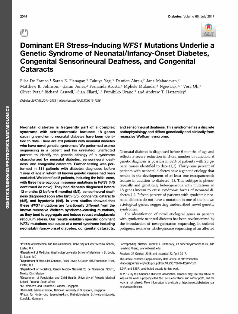
Heterozygous mutations in WFS1 have been found tocause less severe phenotypes than Wolfram syndrome–associated recessive mutations. In particular, dominant WFS1mutations have been reported as causing isolated adult-onset diabetes (7), isolated low-frequency hearing loss(8,9), optic atrophy and hearing impairment (10–12), andisolated congenital nuclear cataracts (13) (Fig. 1).
We used a trio-based exome sequencing strategy to identifydominant WFS1 mutations as the cause of a novel syn-drome characterized by permanent neonatal/infancy-onset
diabetes, congenital or early-onset sensorineural deafness,and congenital cataracts.
RESEARCH DESIGN AND METHODS
Genetic AnalysisWe performed exome sequencing on samples from anaffected proband and parents using SureSelect Human AllExon Kit V4 (Agilent). Paired-end sequencing was per-formed on an Illumina HiSeq2500, using 100 bp reads. Theresulting reads were aligned to the hg19 referencegenome with BWA. Variants were called with GATKUnifiedGenotyper and annotated using ANNOVAR andSeattleSeq Annotation server as previously described (3).
Replication studies were performed in a cohort of 311patients diagnosed with diabetes before 12 months of age inwhom mutations in the 22 known neonatal diabetes geneshad been excluded. All the patients in the cohort wereanalyzed using our targeted next-generation sequencing assaythat includes baits for WFS1 as previously described (14).
The bioinformatics tools SIFT, PolyPhen-2, and AlignGVGD were accessed through the Alamut software (In-teractive Biosoftware, Rouen, France) to predict the effectof novel variants on the WFS1 protein. If the variant wasdeemed likely pathogenic or of unknown significance,
Figure 1—Phenotypic spectrum caused by autosomal dominant and autosomal recessive WFS1 mutations. The figure includes WFS1 muta-tions reported as disease causing in the Human Gene Mutation Database that have a minor allele frequency <0.00447 in the ExAC database(maximum allele frequency possible for Wolfram syndrome–causing mutations, assuming a disease frequency of 1/100,000, as calculated by thealleleFrequencyApp [http://cardiodb.org/allelefrequencyapp/]).
diabetes.diabetesjournals.org De Franco and Associates 2045
mutation testing was performed in the patient (to confirmthe mutation) and the parents (when available) to assesswhether the mutation had arisen de novo. Microsatelliteanalysis of parent/proband trios using the PowerPlex kit(Promega, Southampton, U.K.) was used to confirm familyrelationships for the putative de novo mutations. In silicomodeling of the WFS1 p.Glu809Lys and p.Glu830Ala mu-tations was carried out using the SWISS-MODEL server(15). Additionally, as templates of only limited sequencesimilarity could be identified, the PredictProtein serverwas used to predict secondary structure and functional mo-tifs from primary sequence alone (16).
ImmunostainingCOS7 cells were transiently transfected with dominant andrecessive WFS1 mutations for 48 h. The cells were fixed in2% paraformaldehyde for 30 min at room temperature andthen permeabilized with 0.1% Triton X-100 for 2 min. Thefixed cells were washed with PBS/Tween 0.1%, blocked withImage-iT FX signal enhancer (Invitrogen) for 30 min, andincubated in primary antibody overnight at 4°C. The cellswere washed three times in PBS/Tween 0.1% and incubatedwith secondary antibody for 1 h at room temperature. Im-ages were obtained with an FSA 100 microscope (Olympus).Anti-FLAG antibody and anti-calnexin antibody wereobtained from Cell Signaling Technology (Danvers, MA)and Proteintech Group (Chicago, IL), respectively.
INS-1 cells were plated in eight-chamber slides (ThermoFisher Scientific) at a density of 50,000 cells per chamber andtransiently transfected with dominant and recessive GFP-tagged WFS1 mutant constructs for 48 h. The cells werefixed in 4% paraformaldehyde/PBS for 30 min at roomtemperature, washed three times with PBS, and then per-meabilized with 0.1% Triton X-100/PBS for 10 min at roomtemperature. Cells were blocked overnight in a 2% BSA so-lution of 0.1% Triton X-100/PBS and washed three timesthe next day with PBS. Slides were prepared with antifademounting medium containing DAPI (Vector Laboratories),and images were obtained with an FSA 100 microscope.
ImmunoblottingHeLa cells were transfected with full-length human WFS1cDNA tagged with a FLAG epitope, as well as p.Pro724Leu,p.Glu809Lys, p.Glu830Ala, and p.His313Tyr mutant WFS1cDNA. After transfection for 48 h, cells were lysed for15 min in ice-cold buffer (10 mmol/L Tris-HCl, 50 mmol/LHepes pH 7.4, 1% v/v Triton X-100, 1 mmol/L EDTA)containing protease inhibitors. Insoluble material was re-covered by centrifugation at 13,000 rpm for 15 min andsolubilized in 10 mmol/L Tris-HCl and 1% w/v sodiumdodecyl sulfate for 10 min at room temperature. Afterthe addition of 4 volumes of lysis buffer (20 mmol/L HepespH 7.4, 1% v/v Triton X-100, 150 mmol/L NaCl, 10% v/vglycerol, 1 mmol/L EDTA), samples were sonicated for 30 s.Lysates were separated using 4–20% linear gradient SDS-PAGE (Bio-Rad) and then electroblotted. Primary antibodiesused in this study were anti-FLAG (Cell Signaling Technol-ogy) and anti–a-tubulin (Cell Signaling Technology).
Luciferase Reporter AssayFor reporter assays, HeLa cells were cotransfected with theendoplasmic reticulum stress response element (ERSE) (ratpromoter GRP78)–luciferase construct (17) and variousconstructs as indicated. Prior to lysis at 24 h after trans-fection, cells were treated with or without 100 nmol/L ofthapsigargin (TG) for 8 h. Firefly luciferase activity wasmeasured using the Dual-Luciferase Reporter Assay System(Promega, Madison, WI) and normalized to Renilla lucifer-ase values of the cotransfected pRL-TK vector (Promega) tocontrol for differences in transfection efficiency. Statisticalanalysis of the data was performed by one-way ANOVAfollowed by Dunnett test using SPSS 22 (IBM).
RESULTS
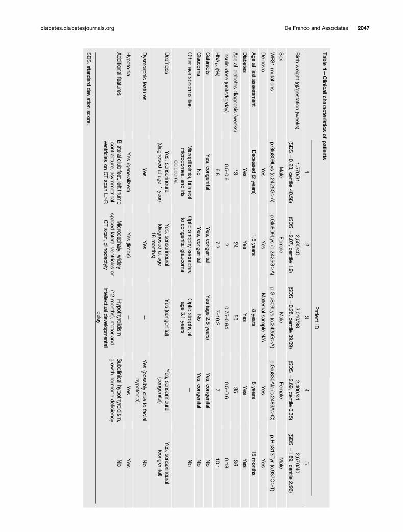
Genetic AnalysisWe performed exome sequencing analysis on a male patient(patient 1) (Table 1) born to unrelated parents who did nothave diabetes. He was diagnosed with neonatal diabetes atthe age of 13 weeks. He also had congenital cataracts, con-genital sensorineural deafness, and bilateral club feet. Thispatient died at the age of 2 years from sepsis.
Exome sequencing identified two de novo coding variantsin patient 1: WFS1 c.2425G.A p.Glu809Lys and ZNF513c.1516G.A p.Ala506Thr. The variant in ZNF513 was ex-cluded from further analysis as it was listed in the ExAC(Exome Aggregation Consortium) database (minor allele fre-quency 0.000008268) (Cambridge, MA; October 2016). TheWFS1 c.2425G.A p.Glu809Lys variant was not listed in59,030 exomes in ExAC and affects a highly conservedresidue located in the endoplasmic reticulum (ER) domainof the WFS1 protein.
As WFS1 recessive loss-of-function mutations usuallycause childhood-onset diabetes and parental carriers are un-affected, we hypothesized that the heterozygous p.Glu809Lysmutation was causing neonatal diabetes through a dominant-negative mechanism rather than haploinsufficiency. There-fore we investigated the presence of heterozygous missense(but not nonsense or frameshift) WFS1 mutations in a co-hort of 311 individuals with diabetes diagnosed in the firstyear of life in whom mutations in the known neonatal di-abetes genes had been excluded using our targeted next-generation sequencing assay (14).
We identified four additional patients harboring likelypathogenic variants inWFS1. Patients 2 and 3 were heterozy-gous for the same p.Glu809Lys variant identified in patient1. Patient 4 had a heterozygous c.2489A.C p.Glu830Alavariant and patient 5 carried the c.937C.T p.His313Tyrmutation. Both the p.Glu830Ala and p.His313Tyr variantsaffect highly conserved residues and are predicted to bepathogenic by in silico analysis. The p.Glu830Ala mutationaffects a residue located in the ER domain, whereas thep.His313Tyr involves an amino acid located in the firsttrans-membrane region of the WFS1 protein. These muta-tions were found to have arisen de novo in patients 1, 2, 4,and 5. Paternal inheritance was excluded in patient 3, butthe maternal sample was not available for testing.
2046 Congenital Syndrome Caused by WFS1 Mutations Diabetes Volume 66, July 2017
Table
1—Clinicalcharacteristics
ofpatients
Patient
ID
12
34
5
Birth
weight
(g)/gestation(weeks)
1,570/31(SDS20.23,
centile40.58)
2,500/40(SDS22.07,centile
1.9)3,010/38
(SDS20.28,centile
39.09)2,400/41
(SDS22.69,centile
0.35)2,670/40
(SDS21.89,
centile2.96)
Sex
Male
Female
Male
Female
Male
WFS
1mutations
p.Glu809Lys
(c.2425G.A)
p.Glu809Lys
(c.2425G.A)
p.Glu809Lys
(c.2425G.A)
p.Glu830A
la(c.2489A
.C)
p.His313Tyr
(c.937C.T)
Denovo
Yes
Yes
Maternalsam
pleN/A
Yes
Yes
Age
atlast
assessment
Deceased
(2years)
1.5years
8years
8years
15months
Diabetes
Yes
Yes
Yes
Yes
Yes
Age
atdiabetes
diagnosis(weeks)
1324
5035
36
Insulindose
(units/kg/day)0.5
–0.62
0.75–0.94
0.5–0.6
0.18
HbA
1c(%
)6.8
7.27–10.2
710.1
Cataracts
Yes,congenital
Yes,
congenitalYes
(age2.5
years)Yes,congenital
No
Glaucom
aNo
Yes,
congenitalNo
Yes,congenital
No
Other
eyeabnorm
alitiesMicropthalm
ia,bilateral
microcornea,
andiris
coloboma
Optic
atrophysecondary
tocongenitalglaucom
aOptic
atrophyat
age3.1
years—
No
Deafness
Yes,sensorineural
(diagnosedat
age1year)
Yes,
sensorineural(diagnosed
atage
18months)
Yes
(congenital)Yes,sensorineural(congenital)
Yes,
sensorineural(congenital)
Dysm
orphicfeatures
Yes
Yes
—Yes
(possiblydue
tofacial
hypotonia)No
Hypotonia
Yes
(generalized)Yes
(limbs)
—Yes
Yes
Additionalfeatures
Bilateralclub
feet,leftthumb
contracture,asym
metrical
ventricleson
CTscan
L.R
Microcephaly,
widely
spacedlateralventricles
onCTscan,
clinodactyly
Hypothyroidism
(12months),
motor
andintellectualdevelopm
entaldelay
Subclinicalhypothyroidism
,grow
thhorm
onede
ficiencyNo
SDS,standard
deviationscore.
diabetes.diabetesjournals.org De Franco and Associates 2047
Clinical FeaturesThe clinical features of the five patients are summarizedin Table 1. All five individuals had congenital sensorineu-ral deafness and early-onset diabetes diagnosed before12 months of age (range 13–51 weeks) that was treated witha full replacement dose of insulin. In all five patients, the birthweight was low (median standard deviation score 21.89[interquartile range22.07, 0.28], median centile 2.96), con-sistent with reduced insulin secretion in utero, a feature notdescribed in patients with “classic” Wolfram syndrome.
Congenital cataracts and additional eye abnormalitieswere present in four of five patients (patients 1, 2, 3, and4). Two of them also had congenital glaucoma (patients2 and 4), which had progressed to optic atrophy in patient2. Patient 1 did not have congenital glaucoma but wasdiagnosed with micropthalmia, bilateral microcornea, andiris coloboma. Patient 3 was diagnosed with optic atrophyat the age of 3.1 years.
Additional common clinical features included hypoto-nia (present in patients 1, 2, 4, and 5), hypothyroidism(diagnosed in patients 3 and 4), and structural neurologicaldefects identified by computed tomography (CT) scanning(in patients 1 and 2).
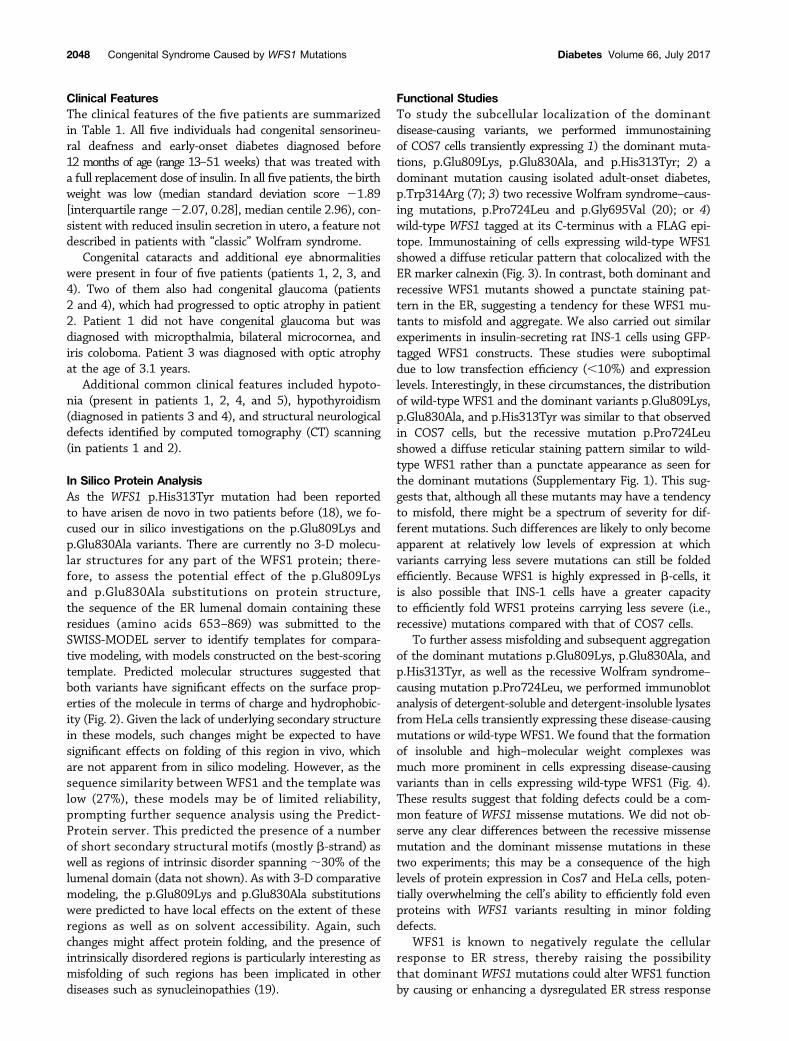
In Silico Protein AnalysisAs the WFS1 p.His313Tyr mutation had been reportedto have arisen de novo in two patients before (18), we fo-cused our in silico investigations on the p.Glu809Lys andp.Glu830Ala variants. There are currently no 3-D molecu-lar structures for any part of the WFS1 protein; there-fore, to assess the potential effect of the p.Glu809Lysand p.Glu830Ala substitutions on protein structure,the sequence of the ER lumenal domain containing theseresidues (amino acids 653–869) was submitted to theSWISS-MODEL server to identify templates for compara-tive modeling, with models constructed on the best-scoringtemplate. Predicted molecular structures suggested thatboth variants have significant effects on the surface prop-erties of the molecule in terms of charge and hydrophobic-ity (Fig. 2). Given the lack of underlying secondary structurein these models, such changes might be expected to havesignificant effects on folding of this region in vivo, whichare not apparent from in silico modeling. However, as thesequence similarity between WFS1 and the template waslow (27%), these models may be of limited reliability,prompting further sequence analysis using the Predict-Protein server. This predicted the presence of a numberof short secondary structural motifs (mostly b-strand) aswell as regions of intrinsic disorder spanning ;30% of thelumenal domain (data not shown). As with 3-D comparativemodeling, the p.Glu809Lys and p.Glu830Ala substitutionswere predicted to have local effects on the extent of theseregions as well as on solvent accessibility. Again, suchchanges might affect protein folding, and the presence ofintrinsically disordered regions is particularly interesting asmisfolding of such regions has been implicated in otherdiseases such as synucleinopathies (19).
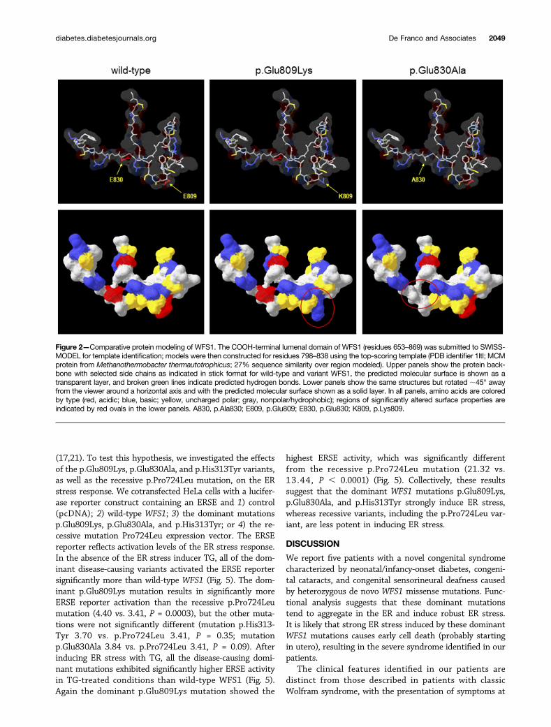
Functional StudiesTo study the subcellular localization of the dominantdisease-causing variants, we performed immunostainingof COS7 cells transiently expressing 1) the dominant muta-tions, p.Glu809Lys, p.Glu830Ala, and p.His313Tyr; 2) adominant mutation causing isolated adult-onset diabetes,p.Trp314Arg (7); 3) two recessive Wolfram syndrome–caus-ing mutations, p.Pro724Leu and p.Gly695Val (20); or 4)wild-type WFS1 tagged at its C-terminus with a FLAG epi-tope. Immunostaining of cells expressing wild-type WFS1showed a diffuse reticular pattern that colocalized with theER marker calnexin (Fig. 3). In contrast, both dominant andrecessive WFS1 mutants showed a punctate staining pat-tern in the ER, suggesting a tendency for these WFS1 mu-tants to misfold and aggregate. We also carried out similarexperiments in insulin-secreting rat INS-1 cells using GFP-tagged WFS1 constructs. These studies were suboptimaldue to low transfection efficiency (,10%) and expressionlevels. Interestingly, in these circumstances, the distributionof wild-type WFS1 and the dominant variants p.Glu809Lys,p.Glu830Ala, and p.His313Tyr was similar to that observedin COS7 cells, but the recessive mutation p.Pro724Leushowed a diffuse reticular staining pattern similar to wild-type WFS1 rather than a punctate appearance as seen forthe dominant mutations (Supplementary Fig. 1). This sug-gests that, although all these mutants may have a tendencyto misfold, there might be a spectrum of severity for dif-ferent mutations. Such differences are likely to only becomeapparent at relatively low levels of expression at whichvariants carrying less severe mutations can still be foldedefficiently. Because WFS1 is highly expressed in b-cells, itis also possible that INS-1 cells have a greater capacityto efficiently fold WFS1 proteins carrying less severe (i.e.,recessive) mutations compared with that of COS7 cells.
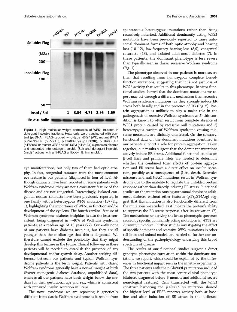
To further assess misfolding and subsequent aggregationof the dominant mutations p.Glu809Lys, p.Glu830Ala, andp.His313Tyr, as well as the recessive Wolfram syndrome–causing mutation p.Pro724Leu, we performed immunoblotanalysis of detergent-soluble and detergent-insoluble lysatesfrom HeLa cells transiently expressing these disease-causingmutations or wild-type WFS1. We found that the formationof insoluble and high–molecular weight complexes wasmuch more prominent in cells expressing disease-causingvariants than in cells expressing wild-type WFS1 (Fig. 4).These results suggest that folding defects could be a com-mon feature of WFS1 missense mutations. We did not ob-serve any clear differences between the recessive missensemutation and the dominant missense mutations in thesetwo experiments; this may be a consequence of the highlevels of protein expression in Cos7 and HeLa cells, poten-tially overwhelming the cell’s ability to efficiently fold evenproteins with WFS1 variants resulting in minor foldingdefects.
WFS1 is known to negatively regulate the cellularresponse to ER stress, thereby raising the possibilitythat dominant WFS1 mutations could alter WFS1 functionby causing or enhancing a dysregulated ER stress response
2048 Congenital Syndrome Caused by WFS1 Mutations Diabetes Volume 66, July 2017
(17,21). To test this hypothesis, we investigated the effectsof the p.Glu809Lys, p.Glu830Ala, and p.His313Tyr variants,as well as the recessive p.Pro724Leu mutation, on the ERstress response. We cotransfected HeLa cells with a lucifer-ase reporter construct containing an ERSE and 1) control(pcDNA); 2) wild-type WFS1; 3) the dominant mutationsp.Glu809Lys, p.Glu830Ala, and p.His313Tyr; or 4) the re-cessive mutation Pro724Leu expression vector. The ERSEreporter reflects activation levels of the ER stress response.In the absence of the ER stress inducer TG, all of the dom-inant disease-causing variants activated the ERSE reportersignificantly more than wild-type WFS1 (Fig. 5). The dom-inant p.Glu809Lys mutation results in significantly moreERSE reporter activation than the recessive p.Pro724Leumutation (4.40 vs. 3.41, P = 0.0003), but the other muta-tions were not significantly different (mutation p.His313-Tyr 3.70 vs. p.Pro724Leu 3.41, P = 0.35; mutationp.Glu830Ala 3.84 vs. p.Pro724Leu 3.41, P = 0.09). Afterinducing ER stress with TG, all the disease-causing domi-nant mutations exhibited significantly higher ERSE activityin TG-treated conditions than wild-type WFS1 (Fig. 5).Again the dominant p.Glu809Lys mutation showed the
highest ERSE activity, which was significantly differentfrom the recessive p.Pro724Leu mutation (21.32 vs.13.44, P , 0.0001) (Fig. 5). Collectively, these resultssuggest that the dominant WFS1 mutations p.Glu809Lys,p.Glu830Ala, and p.His313Tyr strongly induce ER stress,whereas recessive variants, including the p.Pro724Leu var-iant, are less potent in inducing ER stress.
DISCUSSION
We report five patients with a novel congenital syndromecharacterized by neonatal/infancy-onset diabetes, congeni-tal cataracts, and congenital sensorineural deafness causedby heterozygous de novo WFS1 missense mutations. Func-tional analysis suggests that these dominant mutationstend to aggregate in the ER and induce robust ER stress.It is likely that strong ER stress induced by these dominantWFS1 mutations causes early cell death (probably startingin utero), resulting in the severe syndrome identified in ourpatients.
The clinical features identified in our patients aredistinct from those described in patients with classicWolfram syndrome, with the presentation of symptoms at
Figure 2—Comparative protein modeling of WFS1. The COOH-terminal lumenal domain of WFS1 (residues 653–869) was submitted to SWISS-MODEL for template identification; models were then constructed for residues 798–838 using the top-scoring template (PDB identifier 1ltl; MCMprotein from Methanothermobacter thermautotrophicus; 27% sequence similarity over region modeled). Upper panels show the protein back-bone with selected side chains as indicated in stick format for wild-type and variant WFS1, the predicted molecular surface is shown as atransparent layer, and broken green lines indicate predicted hydrogen bonds. Lower panels show the same structures but rotated ;45° awayfrom the viewer around a horizontal axis and with the predicted molecular surface shown as a solid layer. In all panels, amino acids are coloredby type (red, acidic; blue, basic; yellow, uncharged polar; gray, nonpolar/hydrophobic); regions of significantly altered surface properties areindicated by red ovals in the lower panels. A830, p.Ala830; E809, p.Glu809; E830, p.Glu830; K809, p.Lys809.
diabetes.diabetesjournals.org De Franco and Associates 2049
such an early age being the major difference. All ourpatients were diagnosed with diabetes before 12 months ofage, with two patients being diagnosed in the neonatalperiod. In contrast, patients with genetically confirmedrecessive Wolfram syndrome develop insulin-dependentdiabetes at a median age at diagnosis of 6 years (range
1–32 years) (22). Diabetes is often the presenting featureof Wolfram syndrome, with optic atrophy and deafnessappearing months to years later (median age 11 and14 years, respectively). In contrast, our patients wereall diagnosed with sensorineural deafness soon afterbirth. Four out of five patients in our cohort also had
Figure 3—Double immunofluorescence staining of COS7 cells transiently transfected with dominant and recessive variants of WFS1. COS7cells were transiently transfected with 1) control (pcDNA3); 2) wild-type WFS1 tagged at its C-terminus with a FLAG epitope (WT); 3) thedominant mutations p.His313Tyr (H313Y), p.Glu809Lys (E809K), and p.Glu830Ala (E830A); 4) a dominant variant causing isolated adult-onsetdiabetes, p.Trp314Arg (W314R) (7); and 5) two recessive Wolfram syndrome–causing mutations, p.Pro724Leu (P724L) and p.Gly695Val (G695V)(20). The cells were stained with anti-FLAG (red fluorescence, left) and anti-calnexin (green fluorescence, center). Right panels: Merged imagesare shown. n = 4; magnification is 103.
2050 Congenital Syndrome Caused by WFS1 Mutations Diabetes Volume 66, July 2017
eye manifestations, but only two of them had optic atro-phy. In fact, congenital cataracts were the most commoneye feature in our patients (diagnosed in four of five). Al-though cataracts have been reported in some patients withWolfram syndrome, they are not a consistent feature of thedisease and are not congenital. Interestingly, isolated con-genital nuclear cataracts have been previously reported inone family with a heterozygous WFS1 mutation (13) (Fig.1), highlighting the importance of WFS1 in function and/ordevelopment of the eye lens. The fourth cardinal feature ofWolfram syndrome, diabetes insipidus, is also the least con-sistent, being diagnosed in ;40% of Wolfram syndromepatients, at a median age of 13 years (22). Currently noneof our patients have diabetes insipidus, but they are allyounger than the median age that this is diagnosed. Wetherefore cannot exclude the possibility that they mightdevelop this feature in the future. Clinical follow-up in thesepatients will be needed to establish the presence of anydevelopmental and/or growth delay. Another striking dif-ference between our patients and typical Wolfram syn-drome patients is the birth weight. Patients with classicWolfram syndrome generally have a normal weight at birth(Exeter monogenic diabetes database, unpublished data),whereas all our patients have birth weight below the me-dian for their gestational age and sex, which is consistentwith impaired insulin secretion in utero.
The novel syndrome we are reporting is geneticallydifferent from classic Wolfram syndrome as it results from
spontaneous heterozygous mutations rather than beingrecessively inherited. Additional dominantly acting WFS1mutations have been previously reported to cause auto-somal dominant forms of both optic atrophy and hearingloss (10–12), low-frequency hearing loss (8,9), congenitalcataracts (13), and isolated adult-onset diabetes (7). Inthese patients, the dominant phenotype is less severethan typically seen in classic recessive Wolfram syndrome(Fig. 1).
The phenotype observed in our patients is more severethan that resulting from homozygous complete loss-of-function mutations, suggesting that it is not just loss ofWFS1 activity that results in this phenotype. In vitro func-tional studies showed that the dominant mutations we re-port may act through a different mechanism than recessiveWolfram syndrome mutations, as they strongly induce ERstress both basally and in the presence of TG (Fig. 5). Pro-tein aggregation is unlikely to play a major role in thepathogenesis of recessive Wolfram syndrome as 1) this con-dition is known to often result from complete absence ofWFS1 protein caused by recessive null mutations and 2)heterozygous carriers of Wolfram syndrome–causing mis-sense mutations are clinically unaffected. On the contrary,functional data on the dominant mutations identified inour patients support a role for protein aggregation. Takentogether, our results suggest that the dominant mutationsactively induce ER stress. Additional functional studies onb-cell lines and primary islets are needed to determinewhether the combined toxic effects of protein aggrega-tion and ER stress have a direct effect on insulin secre-tion, possibly as a consequence of b-cell death. Recessivemissense and null WFS1 mutations result in Wolfram syn-drome due to the inability to regulate the unfolded proteinresponse rather than directly inducing ER stress. Functionalstudies on the mutation causing autosomal dominant adult-onset diabetes without other features (p.Trp314Arg) sug-gest that this mutation is also functionally different fromthe mutations we studied, as it impairs the protein’s abilityto suppress the ER stress response after its activation (7).The mechanisms underlying the broad phenotypic spectrumcaused by specific dominantly acting mutations inWFS1 arecurrently unknown. Further studies investigating the effectof specific dominant and recessiveWFS1mutations in othercell lines and animal models are needed to further our un-derstanding of the pathophysiology underlying this broadspectrum of disease.
The results of our functional studies suggest a directgenotype–phenotype correlation within the dominant mu-tations we report, which could be explained by the differ-ences in functional impact seen in the in vitro experiments.The three patients with the p.Glu809Lys mutation includedthe two patients with the most severe clinical phenotype(diabetes diagnosed before 6 months and additional severeneurological features). Cells transfected with the WFS1construct harboring the p.Glu809Lys mutation showedthe highest level of ERSE reporter activity both at base-line and after induction of ER stress in the luciferase
Figure 4—High–molecular weight complexes of WFS1 mutants indetergent-insoluble fractions. HeLa cells were transfected with con-trol (pcDNA), FLAG-tagged wild-type WFS1 (WT), mutant WFS1p.Pro724Leu (p.P724L), p.Glu809Lys (p.E809K), p.Glu830Ala(p.E830A), or mutantWFS1 p.His313Tyr (p.H313Y) expression plasmidand separated into detergent-soluble (Sol) and detergent-insoluble(Insol) fractions with anti-FLAG antibody. IB, immunoblot.
diabetes.diabetesjournals.org De Franco and Associates 2051
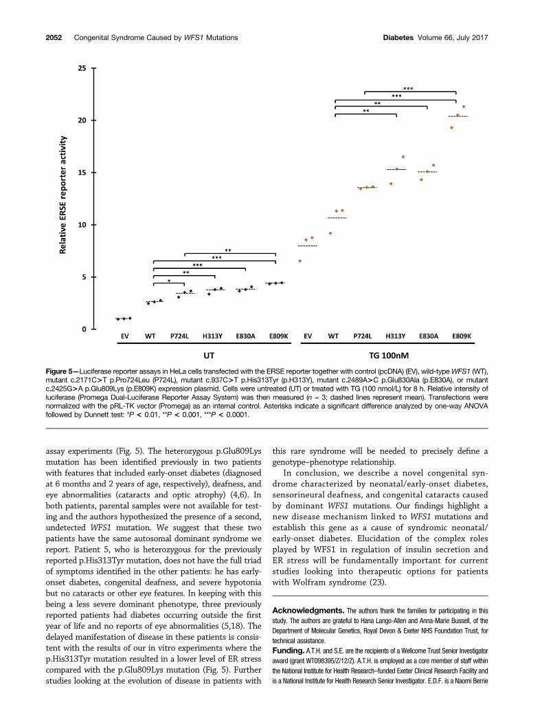
assay experiments (Fig. 5). The heterozygous p.Glu809Lysmutation has been identified previously in two patientswith features that included early-onset diabetes (diagnosedat 6 months and 2 years of age, respectively), deafness, andeye abnormalities (cataracts and optic atrophy) (4,6). Inboth patients, parental samples were not available for test-ing and the authors hypothesized the presence of a second,undetected WFS1 mutation. We suggest that these twopatients have the same autosomal dominant syndrome wereport. Patient 5, who is heterozygous for the previouslyreported p.His313Tyr mutation, does not have the full triadof symptoms identified in the other patients: he has early-onset diabetes, congenital deafness, and severe hypotoniabut no cataracts or other eye features. In keeping with thisbeing a less severe dominant phenotype, three previouslyreported patients had diabetes occurring outside the firstyear of life and no reports of eye abnormalities (5,18). Thedelayed manifestation of disease in these patients is consis-tent with the results of our in vitro experiments where thep.His313Tyr mutation resulted in a lower level of ER stresscompared with the p.Glu809Lys mutation (Fig. 5). Furtherstudies looking at the evolution of disease in patients with
this rare syndrome will be needed to precisely define agenotype–phenotype relationship.
In conclusion, we describe a novel congenital syn-drome characterized by neonatal/early-onset diabetes,sensorineural deafness, and congenital cataracts causedby dominant WFS1 mutations. Our findings highlight anew disease mechanism linked to WFS1 mutations andestablish this gene as a cause of syndromic neonatal/early-onset diabetes. Elucidation of the complex rolesplayed by WFS1 in regulation of insulin secretion andER stress will be fundamentally important for currentstudies looking into therapeutic options for patientswith Wolfram syndrome (23).
Acknowledgments. The authors thank the families for participating in thisstudy. The authors are grateful to Hana Lango-Allen and Anna-Marie Bussell, of theDepartment of Molecular Genetics, Royal Devon & Exeter NHS Foundation Trust, fortechnical assistance.Funding. A.T.H. and S.E. are the recipients of a Wellcome Trust Senior Investigatoraward (grant WT098395/Z/12/Z). A.T.H. is employed as a core member of staff withinthe National Institute for Health Research–funded Exeter Clinical Research Facility andis a National Institute for Health Research Senior Investigator. E.D.F. is a Naomi Berrie
Figure 5—Luciferase reporter assays in HeLa cells transfected with the ERSE reporter together with control (pcDNA) (EV), wild-typeWFS1 (WT),mutant c.2171C>T p.Pro724Leu (P724L), mutant c.937C>T p.His313Tyr (p.H313Y), mutant c.2489A>C p.Glu830Ala (p.E830A), or mutantc.2425G>A p.Glu809Lys (p.E809K) expression plasmid. Cells were untreated (UT) or treated with TG (100 nmol/L) for 8 h. Relative intensity ofluciferase (Promega Dual-Luciferase Reporter Assay System) was then measured (n = 3; dashed lines represent mean). Transfections werenormalized with the pRL-TK vector (Promega) as an internal control. Asterisks indicate a significant difference analyzed by one-way ANOVAfollowed by Dunnett test: *P < 0.01, **P < 0.001, ***P < 0.0001.
2052 Congenital Syndrome Caused by WFS1 Mutations Diabetes Volume 66, July 2017
Fellow in Diabetes Research. S.E.F. has a Sir Henry Dale Fellowship jointly funded bythe Wellcome Trust and the Royal Society (grant 105636/Z/14/Z). This work waspartly supported by grants from National Institutes of Health (DK020579 and UL1TR000448) to F.U.Author Contributions. E.D.F., S.E.F., M.B.J., and S.E. performed the geneticanalysis. E.D.F., S.E.F., and A.T.H. wrote the first draft of the manuscript. S.E.F., T.Y.,D.A., J.M., R.C., S.E., and F.U. participated in manuscript improvement. T.Y., D.A., andJ.M. participated in data acquisition. D.A., J.M., and F.U. participated in data analysis andinterpretation. J.M., S.E., F.U., and A.T.H. participated in study conception and design.G.J. and R.C. performed bioinformatic analysis. F.A., M.M., N.L., V.O., and O.P. collectedpatient samples and clinical data. A.T.H. analyzed the clinical data. All authors reviewedthe manuscript. A.T.H. is the guarantor of the clinical and genetic work and F.U. is theguarantor of the functional work, and both had full access to all the data in the study andtake responsibility for the integrity of the data and the accuracy of the data analysis.
References1. De Franco E, Flanagan SE, Houghton JA, et al. The effect of early, compre-hensive genomic testing on clinical care in neonatal diabetes: an international cohortstudy. Lancet 2015;386:957–9632. Flanagan SE, Haapaniemi E, Russell MA, et al. Activating germline mutations inSTAT3 cause early-onset multi-organ autoimmune disease. Nat Genet 2014;46:812–8143. Lango Allen H, Flanagan SE, Shaw-Smith C, et al.; International PancreaticAgenesis Consortium. GATA6 haploinsufficiency causes pancreatic agenesis in hu-mans. Nat Genet 2011;44:20–224. Matsunaga K, Tanabe K, Inoue H, et al. Wolfram syndrome in the Japanesepopulation; molecular analysis of WFS1 gene and characterization of clinical features.PLoS One 2014;9:e1069065. Rohayem J, Ehlers C, Wiedemann B, et al.; Wolfram Syndrome DiabetesWriting Group. Diabetes and neurodegeneration in Wolfram syndrome: a multicenterstudy of phenotype and genotype. Diabetes Care 2011;34:1503–15106. Chaussenot A, Rouzier C, Quere M, et al. Mutation update and uncommonphenotypes in a French cohort of 96 patients with WFS1-related disorders. Clin Genet2015;87:430–4397. Bonnycastle LL, Chines PS, Hara T, et al. Autosomal dominant diabetes arisingfrom a Wolfram syndrome 1 mutation. Diabetes 2013;62:3943–39508. Bespalova IN, Van Camp G, Bom SJ, et al. Mutations in the Wolfram syndrome1 gene (WFS1) are a common cause of low frequency sensorineural hearing loss.Hum Mol Genet 2001;10:2501–25089. Lesperance MM, Hall JW 3rd, San Agustin TB, Leal SM. Mutations in theWolfram syndrome type 1 gene (WFS1) define a clinical entity of dominant low-frequencysensorineural hearing loss. Arch Otolaryngol Head Neck Surg 2003;129:411–420
10. Eiberg H, Hansen L, Kjer B, et al. Autosomal dominant optic atrophy associatedwith hearing impairment and impaired glucose regulation caused by a missensemutation in the WFS1 gene. J Med Genet 2006;43:435–44011. Hogewind BF, Pennings RJ, Hol FA, et al. Autosomal dominant optic neuropathyand sensorineual hearing loss associated with a novel mutation of WFS1. Mol Vis2010;16:26–3512. Rendtorff ND, Lodahl M, Boulahbel H, et al. Identification of p.A684Vmissense mutation in the WFS1 gene as a frequent cause of autosomal domi-nant optic atrophy and hearing impairment. Am J Med Genet A 2011;155A:1298–131313. Berry V, Gregory-Evans C, Emmett W, et al. Wolfram gene (WFS1) mutationcauses autosomal dominant congenital nuclear cataract in humans. Eur J Hum Genet2013;21:1356–136014. Ellard S, Lango Allen H, De Franco E, et al. Improved genetic testing formonogenic diabetes using targeted next-generation sequencing. Diabetologia 2013;56:1958–196315. Biasini M, Bienert S, Waterhouse A, et al. SWISS-MODEL: modelling proteintertiary and quaternary structure using evolutionary information. Nucleic Acids Res2014;42:W252–25816. Yachdav G, Kloppmann E, Kajan L, et al. PredictProtein–an open resource foronline prediction of protein structural and functional features. Nucleic Acids Res2014;42:W337–34317. Fonseca SG, Ishigaki S, Oslowski CM, et al. Wolfram syndrome 1 gene neg-atively regulates ER stress signaling in rodent and human cells. J Clin Invest 2010;120:744–75518. Hansen L, Eiberg H, Barrett T, et al. Mutation analysis of the WFS1 gene inseven Danish Wolfram syndrome families; four new mutations identified. Eur J HumGenet 2005;13:1275–128419. Uversky VN, Oldfield CJ, Dunker AK. Intrinsically disordered proteins in humandiseases: introducing the D2 concept. Annu Rev Biophys 2008;37:215–24620. Inoue H, Tanizawa Y, Wasson J, et al. A gene encoding a transmembraneprotein is mutated in patients with diabetes mellitus and optic atrophy (Wolframsyndrome). Nat Genet 1998;20:143–14821. Fonseca SG, Fukuma M, Lipson KL, et al. WFS1 is a novel component of theunfolded protein response and maintains homeostasis of the endoplasmic reticulumin pancreatic beta-cells. J Biol Chem 2005;280:39609–3961522. de Heredia ML, Cleries R, Nunes V: Genotypic classification of patients withWolfram syndrome: insights into the natural history of the disease and correlationwith phenotype. Genet Med 2013;15:497–50623. Lu S, Kanekura K, Hara T, et al. A calcium-dependent protease as a potentialtherapeutic target for Wolfram syndrome. Proc Natl Acad Sci U S A 2014;111:E5292–E5301
diabetes.diabetesjournals.org De Franco and Associates 2053


![Inducing Patent Infringement - Law Review...2005] Inducing Patent Infringement 229 understanding, inducing infringement is a natural outgrowth of the common law principle of respondeat](https://static.fdocuments.net/doc/165x107/5f9608d795a783197246401f/inducing-patent-infringement-law-review-2005-inducing-patent-infringement.jpg)