Department of Microbiology 2020 Honours Programs in ... · 2020 Microbiology Honours Projects | 1...
27
2020 Honours Programs in Microbiology monash.edu/discovery-institute Department of Microbiology 2020 Honours Coordinator Associate Professor John Boyce Room: 215, 19 Innovation Walk Phone: +61 3 9902 9179 Email: [email protected] Deputy Coordinator Professor Julian Rood Room: 155, 19 Innovation Walk Phone: +61 3 9902 9157 Email: [email protected]
Transcript of Department of Microbiology 2020 Honours Programs in ... · 2020 Microbiology Honours Projects | 1...
2020 Honours Programs in Microbiology
monash.edu/discovery-institute
Department of Microbiology
2020 Honours CoordinatorAssociate Professor John BoyceRoom: 215, 19 Innovation Walk Phone: +61 3 9902 9179 Email: [email protected]
Deputy CoordinatorProfessor Julian RoodRoom: 155, 19 Innovation Walk Phone: +61 3 9902 9157 Email: [email protected]
2020 Microbiology Honours Projects | 1
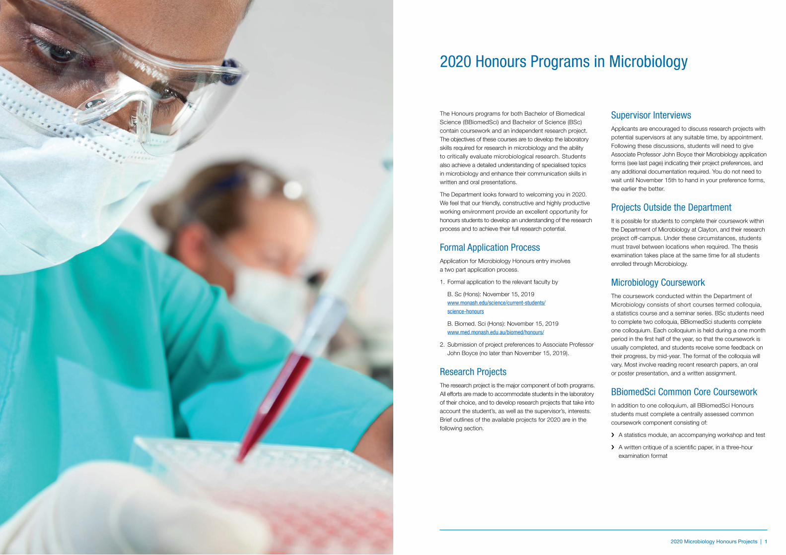
The Honours programs for both Bachelor of Biomedical Science (BBiomedSci) and Bachelor of Science (BSc) contain coursework and an independent research project. The objectives of these courses are to develop the laboratory skills required for research in microbiology and the ability to critically evaluate microbiological research. Students also achieve a detailed understanding of specialised topics in microbiology and enhance their communication skills in written and oral presentations.
The Department looks forward to welcoming you in 2020. We feel that our friendly, constructive and highly productive working environment provide an excellent opportunity for honours students to develop an understanding of the research process and to achieve their full research potential.
Formal Application ProcessApplication for Microbiology Honours entry involves a two part application process.
1. Formal application to the relevant faculty by
B. Sc (Hons): November 15, 2019 www.monash.edu/science/current-students/ science-honours
B. Biomed. Sci (Hons): November 15, 2019 www.med.monash.edu.au/biomed/honours/
2. Submission of project preferences to Associate Professor John Boyce (no later than November 15, 2019).
Research ProjectsThe research project is the major component of both programs. All efforts are made to accommodate students in the laboratory of their choice, and to develop research projects that take into account the student’s, as well as the supervisor’s, interests. Brief outlines of the available projects for 2020 are in the following section.
Supervisor InterviewsApplicants are encouraged to discuss research projects with potential supervisors at any suitable time, by appointment. Following these discussions, students will need to give Associate Professor John Boyce their Microbiology application forms (see last page) indicating their project preferences, and any additional documentation required. You do not need to wait until November 15th to hand in your preference forms, the earlier the better.
Projects Outside the DepartmentIt is possible for students to complete their coursework within the Department of Microbiology at Clayton, and their research project off-campus. Under these circumstances, students must travel between locations when required. The thesis examination takes place at the same time for all students enrolled through Microbiology.
Microbiology CourseworkThe coursework conducted within the Department of Microbiology consists of short courses termed colloquia, a statistics course and a seminar series. BSc students need to complete two colloquia, BBiomedSci students complete one colloquium. Each colloquium is held during a one month period in the first half of the year, so that the coursework is usually completed, and students receive some feedback on their progress, by mid-year. The format of the colloquia will vary. Most involve reading recent research papers, an oral or poster presentation, and a written assignment.
BBiomedSci Common Core CourseworkIn addition to one colloquium, all BBiomedSci Honours students must complete a centrally assessed common coursework component consisting of:
i A statistics module, an accompanying workshop and test
i A written critique of a scientific paper, in a three-hour examination format
2020 Honours Programs in Microbiology
2 | 2020 Microbiology Honours Projects 2020 Microbiology Honours Projects | 3
Literature surveyDuring first semester the students must submit a literature survey on their research project. The literature survey (which can be used as the basis for the introduction in the final report) allows the identification early in the year of those students who have problems with English expression so this can be addressed by directed English writing instruction. It also, of course, compels the students to become thoroughly conversant with their area of research.
Additional requirementsThe programs will commence on February 24, 2020 (mid year begins July 20) with a series of introductory lectures, before the students start work on their research projects. These lectures contain information on the course, departmental facilities and laboratory safety. In the second half of the year students may be given specific training in the presentation of written reports, and in oral presentation of their work. It is compulsory for students to attend the introductory lecture course, all departmental seminars, and any short courses on written and oral presentations.
AssessmentFinal assessment of the BSc Honours program follows the format:
Literature survey 7.5%
Research report/report review 60%
Seminar 7.5%
Microbiology coursework 25%
Final assessment of the BBiomedSci Honours program follows the format:
Literature survey 7.5%
Research report/report review 60%
Seminar 7.5%
Microbiology coursework 10%
Statistics Module 7.5%
Common written component 7.5%
EligibilityMonash BSc Students
Entry to the course is restricted to those students who have qualified for the award of the pass degree of BSc (all subjects completed before enrolment), and have an average of at least 70% in 24 points of relevant level-three science units. This generally includes at least 18 points of Microbiology units. Students studying combined Science degrees must be eligible for the award of BSc.
BSc Graduates of Other Universities
As for Monash students, applicants are required to have a BSc and distinction grades in Microbiology or closely related subjects. A certified copy of the applicant’s academic record and a statement to the effect that they have qualified for a pass degree are required as soon as they are available.
Monash BBiomedSci students
Students must have completed all requirements for the award of the pass degree of Bachelor of Biomedical Science offered at the Monash University. They must also have an average of 70% or higher in at least 24 points at third year level, with 12 points from third year core units.
BBiomedSci graduates from other universities
Students applying for admission based on a qualification other than the pass degree of Bachelor of Biomedical Science offered at Monash University will need to demonstrate that they have achieved an appropriate standard in studies comparable to 24 points of BBiomedSci subjects as stipulated above.
Part-time study and mid-year entryThe department prefers students to study on a full-time basis. However, it may be possible under special circumstance to complete the Honours degree in two consecutive years by doing the coursework and research project in separate years. It may also be possible to start the course mid-year. In both of these circumstances, the arrangements are made on an individual basis between applicants and supervisors.
Research Projects 2020
2020 Microbiology Honours Projects | 3
4 | 2020 Microbiology Honours Projects 2020 Microbiology Honours Projects | 5
Characterisation of the Acinetobacter baumannii Type VI Secretion SystemDr. Marina Harper and A/Professor John Boyce
Acinetobacter baumannii has been identified as one of the top three dangerous Gram-negative hospital pathogens as it can cause a range of life-threatening infections and most strains are now resistant to the majority of current antibiotics. We have characterised a type VI secretion system (T6SS) in A. baumannii that delivers antibacterial toxic proteins into other bacteria to give the cell a competitive advantage.
We are interested in defining the full complement of toxic effector proteins, determining how they kill other bacteria and characterising how the T6SS delivers these toxic proteins to the correct compartment of key competitor strains. This project will use a mix of PCR, directed mutagenesis, complementation, heterologous protein expression and protein-protein interaction approaches to identify novel toxin functions and identify crucial T6SS delivery determinants. A complete understanding of the A. baumannii T6SS, including the function of novel toxins and how these toxins are selected for targeted delivery, will allow us to genetically engineer commensal bacterial strains as live antibacterial delivery systems for the control of other multi-drug resistant pathogens.
Defining the Mechanisms of Pasteurella multocida Pathogenesis and Identifying Novel Virulence RegulatorsDr. Marina Harper, Thomas Smallman and A/Professor John Boyce
Pasteurella multocida is a Gram-negative bacterial pathogen that causes a range of diseases in humans, cattle, pigs and poultry. The animal diseases result in serious economic losses worldwide in food production industries. We are interested in understanding the molecular mechanisms of pathogenesis in this bacterium with an aim to developing new, more effective and widely applicable vaccines or antimicrobial drugs.
Recent work in our lab, using comparative genomics and transposon insertion site mutagenesis, has comprehensively defined the P. multocida genes essential for a range of virulence phenotypes, including growth in serum and production of the anti-phagocytic bacterial capsule. With this crucial data as a base, in this project we will use directed mutagenesis, complementation, whole-genome transcriptomic and proteomic techniques and established in vitro and in vivo assays to define the molecular mechanisms by which this important pathogen avoids killing by the host immune system and causes disease.
Associate Professor John BoyceEMAIL [email protected]
ONE VACANCYTELEPHONE +61 3 9902 9179
OFFICE Room 215, 19 Innovation Walk (Building 76)
WEB https://www.monash.edu/discovery-institute/boyce-lab
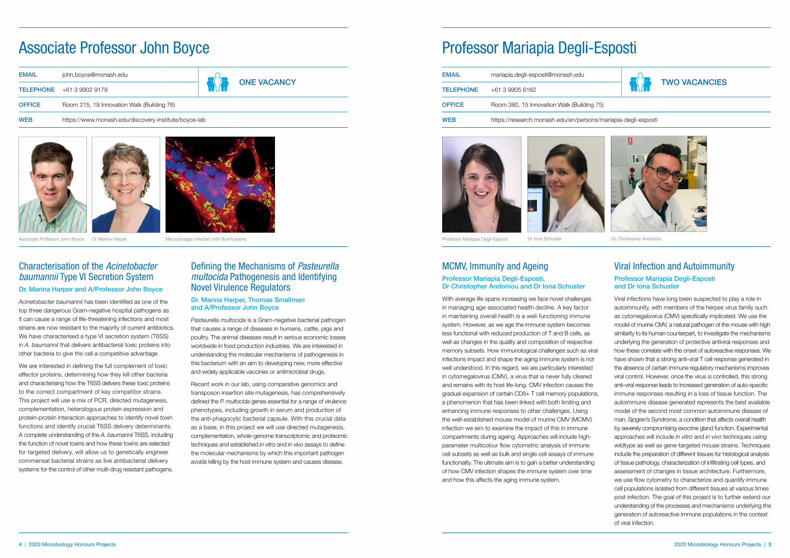
Associate Professor John Boyce Dr Marina Harper Macrophages infected with Burkholderia
MCMV, Immunity and AgeingProfessor Mariapia Degli-Esposti, Dr Christopher Andoniou and Dr Iona Schuster
With average life spans increasing we face novel challenges in managing age-associated health decline. A key factor in maintaining overall health is a well-functioning immune system. However, as we age the immune system becomes less functional with reduced production of T and B cells, as well as changes in the quality and composition of respective memory subsets. How immunological challenges such as viral infections impact and shape the aging immune system is not well understood. In this regard, we are particularly interested in cytomegalovirus (CMV), a virus that is never fully cleared and remains with its host life-long. CMV infection causes the gradual expansion of certain CD8+ T cell memory populations, a phenomenon that has been linked with both limiting and enhancing immune responses to other challenges. Using the well-established mouse model of murine CMV (MCMV) infection we aim to examine the impact of this in immune compartments during ageing. Approaches will include high-parameter multicolour flow cytometric analysis of immune cell subsets as well as bulk and single cell assays of immune functionality. The ultimate aim is to gain a better understanding of how CMV infection shapes the immune system over time and how this affects the aging immune system.
Viral Infection and Autoimmunity Professor Mariapia Degli-Esposti and Dr Iona Schuster
Viral infections have long been suspected to play a role in autoimmunity, with members of the herpes virus family such as cytomegalovirus (CMV) specifically implicated. We use the model of murine CMV, a natural pathogen of the mouse with high similarity to its human counterpart, to investigate the mechanisms underlying the generation of protective antiviral responses and how these correlate with the onset of autoreactive responses. We have shown that a strong anti-viral T cell response generated in the absence of certain immune regulatory mechanisms improves viral control. However, once the virus is controlled, this strong anti-viral response leads to increased generation of auto-specific immune responses resulting in a loss of tissue function. The autoimmune disease generated represents the best available model of the second most common autoimmune disease of man, Sjogren’s Syndrome, a condition that affects overall health by severely compromising exocrine gland function. Experimental approaches will include in vitro and in vivo techniques using wildtype as well as gene-targeted mouse strains. Techniques include the preparation of different tissues for histological analysis of tissue pathology, characterization of infiltrating cell types, and assessment of changes in tissue architecture. Furthermore, we use flow cytometry to characterize and quantify immune cell populations isolated from different tissues at various times post infection. The goal of this project is to further extend our understanding of the processes and mechanisms underlying the generation of autoreactive immune populations in the context of viral infection.
Professor Mariapia Degli-EspostiEMAIL [email protected]
TWO VACANCIESTELEPHONE +61 3 9905 6162
OFFICE Room 380, 15 Innovation Walk (Building 75)
WEB https://research.monash.edu/en/persons/mariapia-degli-esposti
Professor Mariapia Degli-Esposti Dr Iona Schuster Dr Christopher Andoniou
6 | 2020 Microbiology Honours Projects
Understanding Microbiome Interactions with the Innate Immune SystemSam Forster and Emily Gulliver
The innate immune system is capable of intricately detailed detection, differentiation and elimination of pathogenic bacteria. However, the vast majority of bacteria encountered by our innate immune system are beneficial to health. Indeed, over 500 species of these commensal bacteria, containing approximately 10,000 fold more genes than the human genome, exist in the human gastrointestinal tract alone.
Emerging research is demonstrating the importance of these bacterial communities in both maintaining health and causing or exacerbating disease. We have recently developed novel methods to grow the vast majority of bacteria from the gastrointestinal microbiota. This research has resulted in the discovery of hundreds of novel species which require further investigation. Combined with the established experimental and computational expertise in the analysis of innate immune signalling pathways, this project will include cutting-edge microbial culturing techniques, cell culture assays and advanced computational analysis to identify pro- and anti-inflammatory bacterial species.
Students interested in experimental or computational elements, will have the opportunity provided to develop skills in both areas.
tract, there are 100 trillion bacteria, representing more than 500 species, which are exposed to selection for antibiotic resistance during oral antibiotic treatment. The resistance mechanisms in these commensal bacteria remain largely undefined, despite representing a significant, hidden source of antibiotic resistance genes that could be transferred to pathogenic or other commensal bacterial species.
We have recently developed methods to culture the vast majority of the human gastrointestinal microbiota (Nature, 2016; Nature Biotechnology, 2019), which will provide an important resource to undertake these studies. This project will combine detailed genomic and metagenomic sequence analysis with in vitro microbiology to understand and monitor the diversity and distribution of antibiotic resistance within the human gastrointestinal microbiota. The opportunity also exists to focus the project to experimental or computational biology.
Dr Sam ForsterEMAIL [email protected]
ONE VACANCYTELEPHONE +61 3 8572 2753
OFFICE Room L2.52, Hudson Institute of Medical Research, 27-31 Wright St, Clayton
WEB https://hudson.org.au/research-group/microbiota-systems-biology/
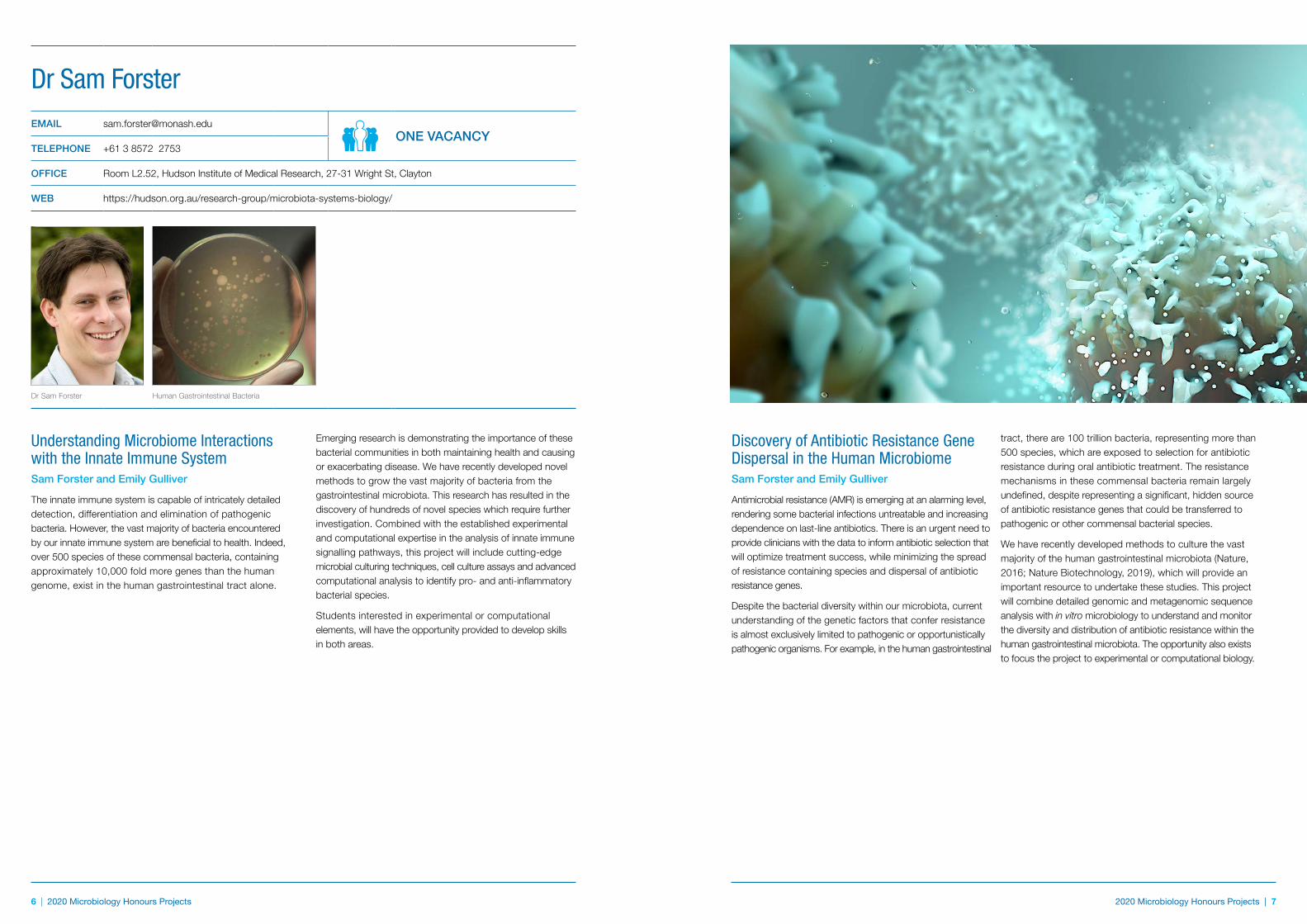
Dr Sam Forster Human Gastrointestinal Bacteria
Discovery of Antibiotic Resistance Gene Dispersal in the Human MicrobiomeSam Forster and Emily Gulliver
Antimicrobial resistance (AMR) is emerging at an alarming level, rendering some bacterial infections untreatable and increasing dependence on last-line antibiotics. There is an urgent need to provide clinicians with the data to inform antibiotic selection that will optimize treatment success, while minimizing the spread of resistance containing species and dispersal of antibiotic resistance genes.
Despite the bacterial diversity within our microbiota, current understanding of the genetic factors that confer resistance is almost exclusively limited to pathogenic or opportunistically pathogenic organisms. For example, in the human gastrointestinal
2019 Microbiology Honours Projects | 7 2020 Microbiology Honours Projects | 7
8 | 2020 Microbiology Honours Projects 2020 Microbiology Honours Projects | 9
Project 2Dr Philip Heraud and Professor Christian Doerig
Malaria was responsible for almost half a million deaths in 2015, the majority of which were children under 5 years old. The disease feeds into the cycle of poverty by reducing the growth of GDP by up to 1.3% per annum in some African countries. Increasing resistance to front line antimicrobials used to treat malaria is posing a real threat that could worsen this situation dramatically. Many potential anti-malarial drugs are known, such as the “malaria box” of 500 drugs with known anti-malarial action being offered up by GlaxoSmithKline company for research investigations. Unfortunately, the pace of development of new anti-malarials is slowed by the lack of knowledge of the precise mechanisms of action of even commonly employed agents such as choloroquine.
This project would use infrared spectroscopy to study the phenotypic response of the malaria Plasmodium organism to a number of antimalarial drugs with known and different modes of action. The aim would be to discover phenotypic signatures related to the different modes of action of the various drugs. If this is possible a drug discovery platform could be developed for rapid testing of potential new drugs that can classify them in terms of their modes of action thus speeding the development of new anti-malarial drugs.
Project 1Dr Philip Heraud, A/Professor Bayden Wood and Professor Anton Peleg
Resistance to antimicrobial drugs is a key issue in medicine with the rise of so-called ‘superbugs’ that are resistant to most or all antibiotics and are commonly observed in hospital settings threatening the lives of sick people. Methicillin-resistant Staphylococcus aureus (MRSA) is one of the most common organisms found in hospital and community infections, resistant to methicillin and all β-lactam antimicrobials. Vancomycin is a drug of choice for the treatment of serious MRSA infections; however, recently a number of vancomycin non-susceptible S. aureus strains have been reported because of the overuse of this drug.
Currently, point-of-care detection and characterization of methicillin and vancomycin resistant S. aureus strains is not available. The state of the art detection methods involve characterization by mass spectroscopy (MALDI-TOF); however, this relies on amplification of bacterial numbers from a serum sample using blood culturing methods taking at least 12 h to achieve. The delay in detection means that appropriate treatment is delayed, which can be critical in terms of patient outcomes.
Infrared spectroscopy (IRS) and Raman spectroscopy (RS) present as possible solutions in this area. Preliminary studies indicate that IRS and RS can provide a molecular fingerprint that is very sensitive to changes in phenotype related to antimicrobial resistance in S. aureus.
This project would explore whether different methicillin and vancomycin-resistant strains can be detected and classified using IRS and RS.
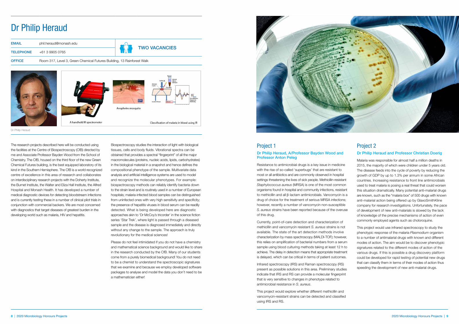
The research projects described here will be conducted using the facilities at the Centre of Biospectroscopy (CfB) directed by me and Associate Professor Bayden Wood from the School of Chemistry. The CfB, housed on the third floor of the new Green Chemical Futures building, is the best equipped laboratory of its kind in the Southern Hemisphere. The CfB is a world recognized centre of excellence in this area of research and collaborates on interdisciplinary research projects with the Doherty Institute, the Burnet Institute, the Walter and Eliza Hall Institute, the Alfred Hospital and Monash Health. It has developed a number of medical diagnostic devices for detecting bloodstream infections and is currently testing these in a number of clinical pilot trials in conjunction with commercial backers. We are most concerned with diagnostics that target diseases of greatest burden in the developing world such as malaria, HIV and hepatitis.
Biospectroscopy studies the interaction of light with biological tissues, cells and body fluids. Vibrational spectra can be obtained that provides a spectral “fingerprint” of all the major macromolecules (proteins, nucleic acids, lipids, carbohydrates) in the biological material in a snapshot and hence defines the compositional phenotype of the sample. Multivariate data analysis and artificial intelligence systems are used to model and recognize the molecular phenotypes. For example: biospectroscopy methods can reliably identify bacteria down to the strain level and is routinely used in a number of European hospitals; malaria-infected blood samples can be distinguished from uninfected ones with very high sensitivity and specificity; the presence of hepatitis viruses in blood serum can be readily detected. What is being developed here are diagnostic approaches akin to ‘Dr McCoy’s tricorder’ in the science fiction series ‘Star Trek’, where light is passed through a diseased sample and the disease is diagnosed immediately and directly without any change to the sample. The approach is truly revolutionary for the medical sciences!
Please do not feel intimidated if you do not have a chemistry and mathematical science background and would like to share in the research conducted by the CfB. Many of our students come from a purely biomedical background! You do not need to be a chemist to understand the spectroscopic signatures that we examine and because we employ developed software packages to analyse and model the data you don’t need to be a mathematician either!
Dr Philip HeraudEMAIL [email protected]
TWO VACANCIESTELEPHONE +61 3 9905 0765
OFFICE Room 317, Level 3, Green Chemical Futures Building, 13 Rainforest Walk
Dr Philip Heraud
10 | 2020 Microbiology Honours Projects 2020 Microbiology Honours Projects | 11
The Molecular Mechanisms by Which Helicobacter pylori Causes Stomach CancerHelicobacter pylori is a Gram-negative gastric bacterium that has co-evolved with humans for more than 50,000 years. It colonises the stomach of over 50% of the world’s population, making it one of the most prevalent human pathogens. It is a causative agent of severe gastric diseases including chronic gastritis, peptic ulcer and stomach cancer. H. pylori has been classified as a Group I (high-risk) carcinogen.
Highly virulent strains of H. pylori harbour a type IV secretion system (T4SS), a secretion machinery that functions as a “syringe” for injecting virulence proteins and peptidoglycan into the host cell. We discovered that CagL, a specialised adhesin present on the surface of the H. pylori T4SS, binds to the human integrin α5β1 receptor on stomach lining cells.This binding activates the T4SS and hence the secretion of virulence factors including the highly immunogenic and oncogenic protein, CagA, into stomach cells. ‘Injected’ CagA then interacts with host signalling molecules and triggers activation of a suite of host responses. Interestingly, our recent findings suggest that CagL can also directly modulate host cell functions. The precise mechanisms by which CagL functions both as a host-activated sensor of the H. pylori T4SS and as a direct activator of aberrant host responses remain to be fully understood.
Our team uses multi-disciplinary state-of-the-art approaches to study the molecular mechanism of H. pylori type IV secretion and H. pylori-host interactions. We aim to understand the molecular basis of how H. pylori induces stomach cancer, with the ultimate goal of providing knowledge for a better treatment and/or prevention of H. pylori-associated stomach diseases. Projects are available to address the following key questions:
i How does H. pylori trigger inflammation and carcinogenesis through the virulence functions of CagL and CagA?
i Can cagL and cagA genotypes predict gastric cancer risk and therefore help pinpoint cancer-prone patients for early treatment?
i How does CagL function as a host-activated sensor during type IV secretion?
i How does CagL and CagA modulate host cell signalling during chronic H. pylori infection?
i Can we utilise the type IV secretion system of H. pylori for delivery of therapeutic proteins?
The available honours projects will enable one to gain experience with the important techniques of molecular cloning and mutagenesis, bacterial culture, eukaryotic cell culture techniques, mouse infection models, CRISPR, RNAi, immunostaining, Western blotting, ELISA, confocal laser scanning microscopy, live cell imaging, etc. Someone who is enthusiastic in learning about the exciting secrets of bacteria- host interactions, infectious cancer biology and bacterial pathogenesis is strongly encouraged to apply.
Dr Terry Kwok-SchueleinEMAIL [email protected]
TWO VACANCIESTELEPHONE +61 3 9902 9216
OFFICE Room 231, level 2, 19 Innovation Walk (Building 76)
Dr Terry Kwok-Schuelein
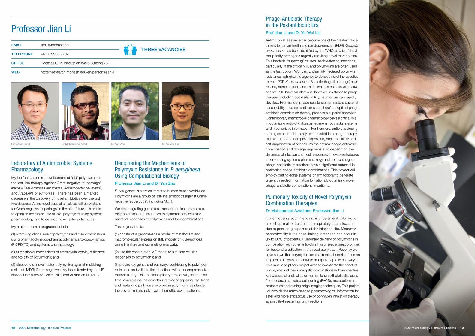
The oncogenic type IV secretion system (T4SS) of H. pylori (left) is activated upon H. pylori-host interaction (right).
10 | 2020 Microbiology Honours Projects
12 | 2020 Microbiology Honours Projects 2020 Microbiology Honours Projects | 13
Laboratory of Antimicrobial Systems PharmacologyMy lab focuses on re-development of ‘old’ polymyxins as the last-line therapy against Gram-negative ‘superbugs’ (namely Pseudomonas aeruginosa, Acinetobacter baumannii, and Klebsiella pneumoniae). There has been a marked decrease in the discovery of novel antibiotics over the last two decades. As no novel class of antibiotics will be available for Gram-negative ‘superbugs’ in the near future, it is crucial to optimise the clinical use of ‘old’ polymyxins using systems pharmacology and to develop novel, safer polymyxins.
My major research programs include:
(1) optimising clinical use of polymyxins and their combinations using pharmacokinetics/pharmacodynamics/toxicodynamics (PK/PD/TD) and systems pharmacology;
(2) elucidation of mechanisms of antibacterial activity, resistance, and toxicity of polymyxins; and
(3) discovery of novel, safer polymyxins against multidrug-resistant (MDR) Gram-negatives. My lab is funded by the US National Institutes of Health (NIH) and Australian NHMRC.
Deciphering the Mechanisms of Polymyxin Resistance in P. aeruginosa Using Computational BiologyProfessor Jian Li and Dr Yan Zhu
P. aeruginosa is a critical threat to human health worldwide. Polymyxins are a group of last-line antibiotics against Gram-negative ‘superbugs’, including MDR.
We are integrating genomics, transcriptomics, proteomics, metabolomics, and lipidomics to systematically examine bacterial responses to polymyxins and their combinations.
This project aims to:
(1) construct a genome-scale model of metabolism and macromolecular expression (ME model) for P. aeruginosa using literature and our multi-omics data;
(2) use the constructed ME model to simulate cellular responses to polymyxins; and
(3) predict key genes and pathways contributing to polymyxin resistance and validate their functions with our comprehensive mutant library. This multidisciplinary project will, for the first time, characterise the complex interplay of signaling, regulation and metabolic pathways involved in polymyxin resistance, thereby optimising polymyxin chemotherapy in patients.
Professor Jian LiEMAIL [email protected]
THREE VACANCIESTELEPHONE +61 3 9903 9702
OFFICE Room 220, 19 Innovation Walk (Building 76)
WEB https://research.monash.edu/en/persons/jian-li
Professor Jian Li
Phage-Antibiotic Therapy in the Postantibiotic EraProf Jian Li and Dr Yu-Wei Lin
Antimicrobial resistance has become one of the greatest global threats to human health and pandrug-resistant (PDR) Klebsiella pneumoniae has been identified by the WHO as one of the 3 top-priority pathogens urgently requiring novel therapeutics. This bacterial ‘superbug’ causes life-threatening infections, particularly in the critically ill, and polymyxins are often used as the last option. Worryingly, plasmid-mediated polymyxin resistance highlights the urgency to develop novel therapeutics to treat PDR K. pneumoniae. Bacteriophage (i.e. phage) have recently attracted substantial attention as a potential alternative against PDR bacterial infections; however, resistance to phage therapy (including cocktails) in K. pneumoniae can rapidly develop. Promisingly, phage resistance can restore bacterial susceptibility to certain antibiotics and therefore, optimal phage-antibiotic combination therapy provides a superior approach. Contemporary antimicrobial pharmacology plays a critical role in optimizing antibiotic dosage regimens, but lacks systems and mechanistic information. Furthermore, antibiotic dosing strategies cannot be easily extrapolated into phage therapy, mainly due to the complex disposition, host specificity and self-amplification of phages. As the optimal phage-antibiotic combination and dosage regimens also depend on the dynamics of infection and host responses, innovative strategies incorporating systems pharmacology and host-pathogen-phage-antibiotic interactions have a significant potential in optimising phage-antibiotic combinations. This project will employ cutting-edge systems pharmacology to generate urgently needed information for rationally optimising novel phage-antibiotic combinations in patients.
Pulmonary Toxicity of Novel Polymyxin Combination TherapiesDr Mohammad Azad and Professor Jian Li
Current dosing recommendations of parenteral polymyxins are suboptimal for treatment of respiratory tract infections due to poor drug exposure at the infection site. Moreover, nephrotoxicity is the dose-limiting factor and can occur in up to 60% of patients. Pulmonary delivery of polymyxins in combination with other antibiotics has offered a great promise for bacterial eradication in the respiratory tract. Recently we have shown that polymyxins localise in mitochondria of human lung epithelial cells and activate multiple apoptotic pathways. This multi-disciplinary project aims to investigate the effect of polymyxins and their synergistic combinations with another five key classes of antibiotics on human lung epithelial cells, using fluorescence activated cell sorting (FACS), metabolomics, proteomics and cutting-edge imaging techniques. This project will provide the much-needed pharmacological information for safer and more efficacious use of polymyxin inhalation therapy against life-threatening lung infections.
2020 Microbiology Honours Projects | 13
Dr Mohammad Azad Dr Yan Zhu Dr Yu-Wei Lin
14 | 2020 Microbiology Honours Projects 2020 Microbiology Honours Projects | 15
Mapping Diversity of Bacteriophage with Genomics and Structural BiologyDr Rhys Dunstan and Professor Trevor Lithgow
Bacteriophages (phages) dominate numerous ecosystems, and are most often isolated from water sources. Recently, phage discovery has been accelerated using new generation sequencing strategies: (i) viral metagenomics, in which complex phage populations are harvested en masse from environmental sources, and (ii) data mining archives of bacterial genome sequence information, to detect embedded prophage and phage-related sequences. As powerful as they are, these bioinformatics-based approaches do not yield virions for wet-lab analyses. Understanding the diversity present in the architecture and the biology of phages requires isolating and characterizing active virions.
This project aims to assess phage diversity through a classical environmental microbiology approach: using water samples collected from diverse locations around the world, the phages therein will be concentrated and plated on a lawn of bacteria. Attention will be focused on phages that infect the pathogen Klebsiella pneumoniae or a closely related plant commensal Klebsiella pseudopneumoniae that looks set to emerge as an important pathogen. Using a combination of electron microscopy to assess virion morphology, and bioinformatics for comparative genomics and protein identification, the project would classify, catalogue and compare the various phage isolated. Finally, a systematic assessment of cocktails of the various phage will be undertaken to determine killing efficacy for future therapeutic work.
Molecular Mechanisms of Bacteriophage ResistanceDr Rhys Dunstan and Professor Trevor Lithgow
By 2050 antimicrobial-resistant (AMR) infections will kill >10 million people every year. While new antibiotics will help slow this trend, discovery of adjunct treatments that will not further promote AMR is essential if we are to avoid the devastating impact of AMR superbugs. One such adjunct treatment is phage therapy: phages do not discriminate between antimicrobial-resistant and antimicrobial-sensitive bacteria: they kill both.
Phage therapy is a frontier medicine. Phage treatment of multidrug-resistant bacterial pathogens both reduces bacterial carriage and selects for phage-resistant bacteria that are less fit, less virulent and re-sensitized to antibiotics. Why it is that bacterial hosts must lose drug-resistant phenotypes in order to become phage-resistant remains unknown. Addressing this question is crucial if phage therapy is to become a wide-spread adjunct treatment for AMR infections.
Exploring the underlying molecular mechanisms in the evolution of phage-resistance is the focus of this project. The initial aim of the project is to characterize known and novel phages targeting multidrug-resistant Klebsiella pneumoniae. The project will then focus on phenotypic and molecular characterizations of the phage-resistant mutants of K. pneumoniae, both in terms of antimicrobial sensitivity and other growth-based and morphological aspects of their phenotypes. The project will involve the use of molecular technologies, microbial culturing, phenotyping and DNA sequencing.
Professor Trevor LithgowEMAIL [email protected]
ONE VACANCYTELEPHONE +61 3 9902 9217
OFFICE Room 318, 18 Innovation Walk; Room 252, 19 Innovation Walk
WEB https://www.monash.edu/discovery-institute/lithgow-lab/home
Professor Trevor Lithgow Dr Rhys Dunstan
16 | 2020 Microbiology Honours Projects 2020 Microbiology Honours Projects | 17
Analysis of Toxin Secretion in the Large Clostridial Toxin (LCT) Producing ClostridiaProfessor Dena Lyras, Dr Sheena McGowan and Dr Milena Awad
The LCT-producing clostridia are an important group of pathogens that cause severe disease in both humans and animals. In most cases the diseases caused by these organisms are at least partly mediated by the production of potent exotoxins known as the large clostridial toxins (LCTs), which are mono-glycosyltransferases that irreversibly inactivate members of the Rho family of small GTPases.
These toxins are structurally related and reside within a genomic region known as the Pathogenicity Locus (PaLoc) that also encodes several accessory proteins thought to be involved in the control of toxin production and secretion.
None of the LCTs have any recognisable signal peptides or export sequences, suggesting that they are secreted by a novel mechanism, potentially involving holin-like proteins. The aim of this project is to further define the mechanism by which the LCTs are secreted from the cell. This will be achieved through the construction of isogenic mutants in the LCT-producing clostridia and subsequent phenotypic testing, as well as through the purification and testing of proteins hypothesised to be involved in the export process.
Understanding the Role of Bacterial Structures in the Transfer of Antibiotic Resistance Genes During ConjugationProfessor Dena Lyras, Dr Yogitha Srikhanta and A/Professor Priscilla Johanesen
Antibiotics are a precious and diminishing resource. There is a desperate need to reduce or replace the use of antibiotics to treat bacterial infections, which is an important veterinary and medical pursuit in this new age of antibiotic-resistant “superbugs”. The treatment of bacterial infections in animals and humans has relied on the use of antibiotics. One consequence of the use of these drugs is antibiotic resistance, which is now one of our most serious global health threats. Bacteria can become resistant to antibiotics through lateral gene transfer of resistance genes, which are often located on mobile elements such as plasmids and transposons.
This project will focus on one such mechanism, conjugation, which is a process by which one bacterium transfers genetic material to another through direct cell-to-cell contact. Apart from the conjugation apparatus, very little is known about the role that bacterial structures play in conjugation. Here, we will examine the role of numerous structures in DNA transfer efficiency using molecular technology and microbial genetics, which may provide new targets and strategies through which the transfer of antibiotic resistance genes may be prevented.
Understanding the Host Immune Response to Clostridium difficile InfectionProfessor Dena Lyras, A/Professor Helen Abud, Dr Steven Mileto and Dr Melanie Hutton
Clostridium difficile is recognised as the major cause of nosocomial diarrhoea in Australian hospitals and in hospitals worldwide. Chronic colitis syndromes caused by this organism are a significant cause of morbidity in hospitals with control and treatment costs rapidly escalating. The recent emergence of hypervirulent strains has increased the severity of disease and hence the urgency with which the mechanism of disease needs to be understood. The pathogenesis of C. difficile-associated diseases involves the production of numerous toxins and other virulence factors. We have developed a mouse model of infection which closely mimics human infection. This project will use the mouse model of C. difficile infection to assess the host immune response to C. difficile infection, in particular using specific mutants of clinically relevant C. difficile strains. Our primary focus will be on the ability of C. difficile toxins to modulate specific immune responses during disease and we will extend these studies into a broader exploration of the pathways involved downstream of these responses, including the effect of C. difficile infection on repair capacity of the gut and stem cells.
Genetic and Phenotypic Analysis of Pathogens in Antibiotic-Associated Diarrhoeal DiseaseProfessor Dena Lyras, Dr Sarah Larcombe and Dr Grant Jenkin
The treatment of bacterial infections in humans and animals has largely relied on the use of antibiotics for over 70 years. One consequence of the use of these drugs is antibiotic resistance, which is now one of our most serious health threats worldwide. Another complication is antibiotic-associated diarrhoea (AAD), which results from the unintended disruption of the protective resident gut microbiota. This disruption can lead to opportunistic infection, subsequently leading to diarrhoeal disease. Using a multidisciplinary approach, and with the involvement of clinical colleagues, this project aims to gain new insights into the mechanisms of pathogenesis and the subversion of host processes by AAD-causing bacteria. Animal models of infection will be used, together with specific mutants, to study virulence factors and host interactions, allowing us to gain a mechanistic understanding of how these bacteria interact with, and damage, the host.
Professor Dena LyrasEMAIL [email protected]
TWO VACANCIESTELEPHONE +61 3 9902 9155
OFFICE Room 152, 19 Innovation Walk (Building 76)
Professor Dena Lyras Intestinal epithelium. Larcombe and Lyras
18 | 2020 Microbiology Honours Projects 2020 Microbiology Honours Projects | 19
Hijacking of Host Exosome Pathways by HCMVDr Rommel Mathias and Dr Yea Seul Shin
HCMV is a master at manipulating existing host pathways to benefit completion of the life cycle. It is known that maturing nucleocapsids bud into host membrane-derived structures to acquire the outer virion envelope. However, the origin of the membrane is unknown, and the precise molecular mechanisms remain elusive. In our lab, proteomic sequencing identified a strong enrichment of host exosome proteins in the virion. Exosomes are small nanovesicles (50-200 nm) secreted by almost all cell types. Therefore, we hypothesize that HCMV hijacks this pathway for viral egress.
We use CRISPR to knock-out various host exosome proteins in cells, and measure the functional impact to virions by assaying viral titre released from these ‘edited’ cells.
Biogenesis of the HCMV Viral Assembly ComplexDr Rommel Mathias and Dr Svenja Fritzlar
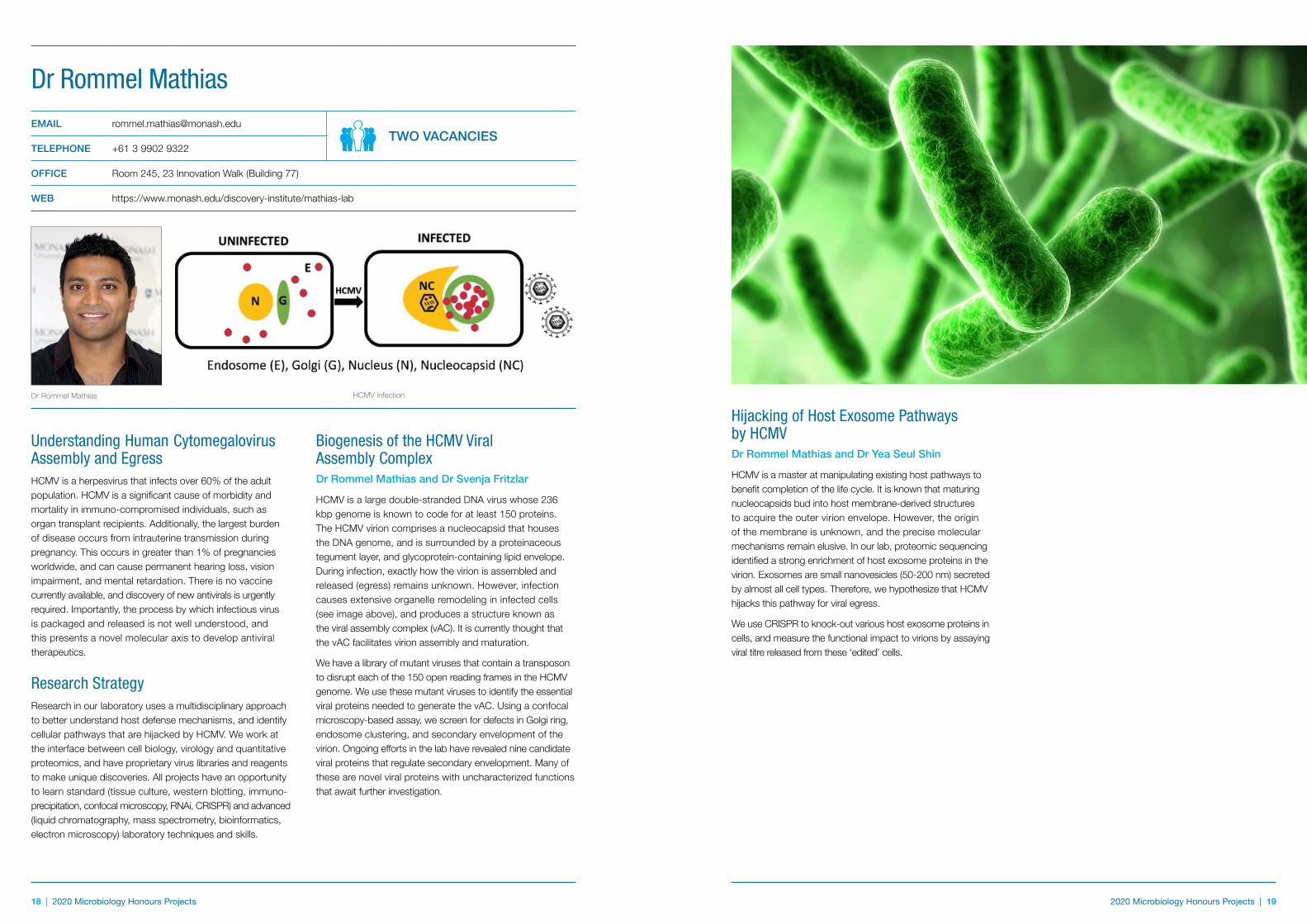
HCMV is a large double-stranded DNA virus whose 236 kbp genome is known to code for at least 150 proteins. The HCMV virion comprises a nucleocapsid that houses the DNA genome, and is surrounded by a proteinaceous tegument layer, and glycoprotein-containing lipid envelope. During infection, exactly how the virion is assembled and released (egress) remains unknown. However, infection causes extensive organelle remodeling in infected cells (see image above), and produces a structure known as the viral assembly complex (vAC). It is currently thought that the vAC facilitates virion assembly and maturation.
We have a library of mutant viruses that contain a transposon to disrupt each of the 150 open reading frames in the HCMV genome. We use these mutant viruses to identify the essential viral proteins needed to generate the vAC. Using a confocal microscopy-based assay, we screen for defects in Golgi ring, endosome clustering, and secondary envelopment of the virion. Ongoing efforts in the lab have revealed nine candidate viral proteins that regulate secondary envelopment. Many of these are novel viral proteins with uncharacterized functions that await further investigation.
Understanding Human Cytomegalovirus Assembly and EgressHCMV is a herpesvirus that infects over 60% of the adult population. HCMV is a significant cause of morbidity and mortality in immuno-compromised individuals, such as organ transplant recipients. Additionally, the largest burden of disease occurs from intrauterine transmission during pregnancy. This occurs in greater than 1% of pregnancies worldwide, and can cause permanent hearing loss, vision impairment, and mental retardation. There is no vaccine currently available, and discovery of new antivirals is urgently required. Importantly, the process by which infectious virus is packaged and released is not well understood, and this presents a novel molecular axis to develop antiviral therapeutics.
Research StrategyResearch in our laboratory uses a multidisciplinary approach to better understand host defense mechanisms, and identify cellular pathways that are hijacked by HCMV. We work at the interface between cell biology, virology and quantitative proteomics, and have proprietary virus libraries and reagents to make unique discoveries. All projects have an opportunity to learn standard (tissue culture, western blotting, immuno-precipitation, confocal microscopy, RNAi, CRISPR) and advanced (liquid chromatography, mass spectrometry, bioinformatics, electron microscopy) laboratory techniques and skills.
Dr Rommel MathiasEMAIL [email protected]
TWO VACANCIESTELEPHONE +61 3 9902 9322
OFFICE Room 245, 23 Innovation Walk (Building 77)
WEB https://www.monash.edu/discovery-institute/mathias-lab
Dr Rommel Mathias
HCMV infection
20 | 2020 Microbiology Honours Projects 2020 Microbiology Honours Projects | 21 20 | 2020 Microbiology Honours Projects
The McGowan laboratory is interested in characterising new drug targets. The lab has a strong research focus in the design of novel anti-malarial drugs as well as other parasitic and bacterial diseases. Primarily we are a structural microbiology laboratory using techniques in protein structural biology, biochemistry and molecular biology to analyse drug targets of interest. We use this mechanistic information to design inhibitors or analogues with potential applications in human medicine. The laboratory has close connections with both the Department of Biochemistry and the Monash Institute of Pharmaceutical Sciences (in Parkville).
The general projects are outlined below and interested students are encouraged to contact Sheena with any questions or to discuss further.
Penicillin Binding Proteins of Clostridioides difficileDr Sheena McGowan and Prof Dena Lyras
The human pathogen Clostridioides difficile produces spores as part of the bacteria’s mechanism of survival when confronted by antibiotics that lead to recurrent and debilitating infection particularly in hospital environments.We have discovered a set of penicillin binding proteins normally responsible for the biosynthesis of the bacterial cell wall that are also required for the formation of spore. This project will advance our understanding of a link between antibiotic use and sporulation, and provide a path to new drugs for prevention of sporulation.
Development of Phage Lysins as Novel AntimicrobialsDr Sheena McGowan
The growing problem of antibiotic resistance underlies the critical need to develop new treatments to prevent and control resistant bacterial infections. Exogenous application of bacteriophage lysins to dormant and actively growing cell cultures results in rapid cell death. Understanding the mechanism of action will allow the development of lysins as a next generation antimicrobial agent.
Antimicrobial Effectors from Acinetobacter baumanniiDr Sheena McGowan and A/Prof John Boyce
Acinetobacter baumannii is recognized as one of the three most important Gram-negative hospital-derived pathogens. A. baumannii isolates resistant to all available antibiotics have already been identified in patients and there is an urgent need to find new methods to control A. baumannii infections. Interestingly, A. baumannii encodes an arsenal of lethal toxins designed to directly kill competing bacteria and we believe that these toxins are a rich source of new antibacterial molecules. This project aims to characterise the structure and function of these novel toxins and assess their suitability as potential new antimicrobials.
Dr Sheena McGowanEMAIL [email protected]
TWO VACANCIESTELEPHONE +61 3 9902 9309
OFFICE Room 137, 19 Innovation Walk (Building 76)
WEB https://www.monash.edu/discovery-institute/mcgowan-lab/home
Dr Sheena McGowan
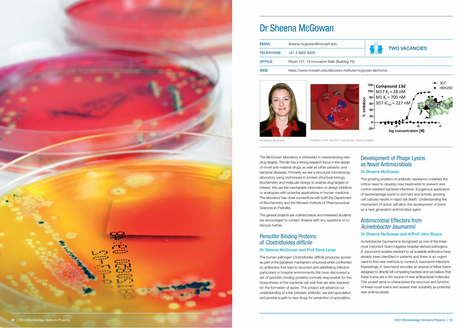
Compound 13d M17 Ki = 28 nM M1 Ki = 700 nM 3D7 IC50 = 227 nM
Inhibition of M1 and M17 starves the malaria parasite
22 | 2020 Microbiology Honours Projects 2020 Microbiology Honours Projects | 23
Super-Resolution Analysis of the Virus-Host InterfaceDr Greg Moseley and Dr Toby Bell
Viruses are experts at remodelling the infected cell, and can fundamentally alter cellular biology to transform host cells into efficient virus factories. Although molecular/ biochemical evidence indicates that certain viral proteins can functionally modify structures such as the mitochondria, cell membranes, nucleus, and cytoskeleton, understanding of the physical effects on these structures is limited due to the poor resolving power of standard cell imaging approaches. Using single molecule localization techniques to surpass the physical diffraction limit of visible light, we have developed methods to observe and quantify the effects of viral proteins on cellular structures at super-resolution, enabling us to directly measure viral remodelling of the subcellular environment. Using this approach, we demonstrated that virus protein targeting of the cytoskeleton correlates with the capacity to cause lethal disease in vivo. The project will apply state-of-the-art single molecule localization techniques such as 3D dSTORM to define viral effects on cellular structures in unprecedented detail; this will provide new insights into the ways that viruses co-opt cellular function to cause disease.
Why do Cytoplasmic RNA Viruses Target the Nucleolus?Dr Greg Moseley and Dr Stephen Rawlinson
Many diverse viral proteins have evolved independently to target the nucleolus but this phenomenon had been largely overlooked, particularly for RNA viruses that replicate within the cytoplasm. Following the development of advanced ‘systems-biology’ approaches to analyse nucleolar biology, it has become clear that the nucleolus, previously viewed solely as a factory for ribosome production, is in fact a complex, dynamic, and highly multifunctional machine that coordinates many critical cellular processes including immunity and cell survival. This has redefined our understanding of the nucleolus and suggests that the virus:nucleolar interface might represent a central hub for viral hijacking of cellular processes, important to viral replication and pathogenesis.
Using nucleolar proteins from the highly pathogenic RNA viruses rabies virus and Hendra virus, we are investigating in molecular detail the mechanisms by which viruses can reprogram the nucleolus to alter cellular biology. These studies are identifying for the first time specific nucleolar functions for RNA virus proteins. The project will advance this work, utilizing techniques including molecular biology, proteomics, confocal/super-resolution microscopy, virus replication and gene expression assays, and siRNA/CRISPR/ Cas gene knockout approaches to delineate the precise events underlying cellular dysfunction caused by virus- nucleolus interaction. [This project will be in collaboration with the CSIRO-AAHL PC4 high-containment laboratories in Geelong.]
Viral Reprogramming of Host Cell SignallingDr Greg Moseley and Dr Celine Deffrasnes
Central to the spread of pathogenic viruses is their capacity to interfere with host immunity, in particular the antiviral system mediated by cytokines such as the interferons. It is well known that many viruses target signalling by antiviral type I interferons to shut down the expression of interferon-stimulated genes. However, our recent work has indicated that the interaction of viruses with cytokine signalling pathways is much more complex and intricate than previously assumed.
In particular, we and our collaborators have found that rabies virus, the cause of c. 60,000 human deaths/year, interacts with multiple signalling pathways, including those initiated by interleukin-6 and interferon-a/ß using a number of mechanisms including viral interactions with and remodelling of cellular structures of the cytoskeleton and nucleus. Importantly, using mutagenic analysis and viral reverse genetics, we found that altering viral targeting of these pathways profoundly inhibits pathogenesis in vivo, indicative of critical roles in disease.
We are currently seeking to delineate the precise mechanisms by which viruses interfere with and modulate cellular pathways, not only to inhibit antiviral signalling, but also to reprogram specific signalling pathways toward ‘pro-viral’ responses, a novel concept in viral biology.
Can Rabies Cure Alzheimer’s?Dr Greg Moseley and Dr Celine Deffrasnes
Neuroinflammation is a major factor in human pathologies such as stroke, Alzheimer’s disease (AD), and traumatic brain injury (TBI). Viruses such as the lyssaviruses rabies and Australian bat lyssavirus, paramyxoviruses Nipah, Hendra and measles, and the filovirus Ebola, have evolved powerful mechanisms to shut down inflammatory signalling as part of their strategies for immune evasion. We aim to discover the molecular ‘tricks’ used by viruses to subvert host immunity, and to exploit these mechanisms to develop new methods to efficiently prevent the inappropriate immune responses underlying neuroinflammatory disorders.
We have made major advances in understanding how viruses achieve immune evasion, including defining the specific virus-host interactions involved, and the molecular basis of these interactions. Using this knowledge, and established models of stroke, TBI, AD and Parkinson’s disease, the project will investigate the potential of harnessing viral immune evasion to combat immune disorders.
Dr Greg MoseleyEMAIL [email protected]
TWO VACANCIESTELEPHONE +61 3 9905 1036
OFFICE Room 136, 19 Innovation Walk (Building 76)
Dr Greg Moseley
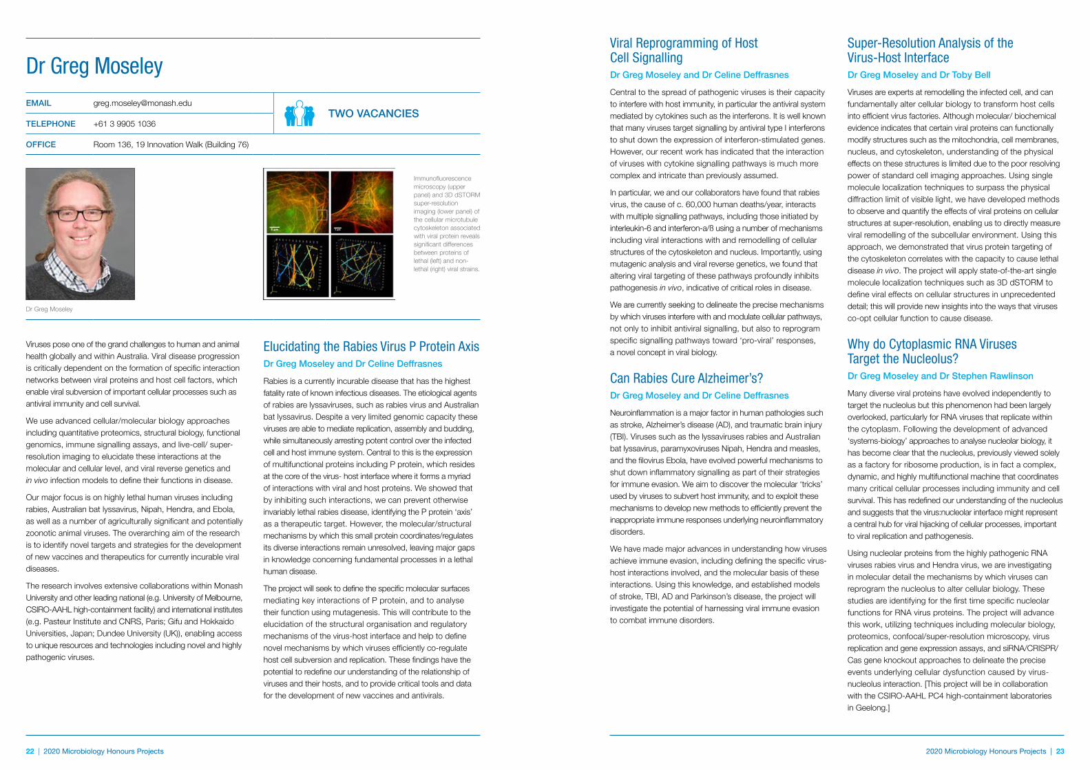
Immunofluorescence microscopy (upper panel) and 3D dSTORM super-resolution imaging (lower panel) of the cellular microtubule cytoskeleton associated with viral protein reveals significant differences between proteins of lethal (left) and non-lethal (right) viral strains.
Elucidating the Rabies Virus P Protein AxisDr Greg Moseley and Dr Celine Deffrasnes
Rabies is a currently incurable disease that has the highest fatality rate of known infectious diseases. The etiological agents of rabies are lyssaviruses, such as rabies virus and Australian bat lyssavirus. Despite a very limited genomic capacity these viruses are able to mediate replication, assembly and budding, while simultaneously arresting potent control over the infected cell and host immune system. Central to this is the expression of multifunctional proteins including P protein, which resides at the core of the virus- host interface where it forms a myriad of interactions with viral and host proteins. We showed that by inhibiting such interactions, we can prevent otherwise invariably lethal rabies disease, identifying the P protein ‘axis’ as a therapeutic target. However, the molecular/structural mechanisms by which this small protein coordinates/regulates its diverse interactions remain unresolved, leaving major gaps in knowledge concerning fundamental processes in a lethal human disease.
The project will seek to define the specific molecular surfaces mediating key interactions of P protein, and to analyse their function using mutagenesis. This will contribute to the elucidation of the structural organisation and regulatory mechanisms of the virus-host interface and help to define novel mechanisms by which viruses efficiently co-regulate host cell subversion and replication. These findings have the potential to redefine our understanding of the relationship of viruses and their hosts, and to provide critical tools and data for the development of new vaccines and antivirals.
Viruses pose one of the grand challenges to human and animal health globally and within Australia. Viral disease progression is critically dependent on the formation of specific interaction networks between viral proteins and host cell factors, which enable viral subversion of important cellular processes such as antiviral immunity and cell survival.
We use advanced cellular/molecular biology approaches including quantitative proteomics, structural biology, functional genomics, immune signalling assays, and live-cell/ super-resolution imaging to elucidate these interactions at the molecular and cellular level, and viral reverse genetics and in vivo infection models to define their functions in disease.
Our major focus is on highly lethal human viruses including rabies, Australian bat lyssavirus, Nipah, Hendra, and Ebola, as well as a number of agriculturally significant and potentially zoonotic animal viruses. The overarching aim of the research is to identify novel targets and strategies for the development of new vaccines and therapeutics for currently incurable viral diseases.
The research involves extensive collaborations within Monash University and other leading national (e.g. University of Melbourne, CSIRO-AAHL high-containment facility) and international institutes (e.g. Pasteur Institute and CNRS, Paris; Gifu and Hokkaido Universities, Japan; Dundee University (UK)), enabling access to unique resources and technologies including novel and highly pathogenic viruses.
24 | 2020 Microbiology Honours Projects 2020 Microbiology Honours Projects | 25
MECHANISMS OF PATHOGENESIS OF HOSPITAL-ACQUIRED ORGANISMS
Impact of Antibiotic Resistance on Immune Recognition of Staphylococcus aureusDr Jhih-Hang Jiang and Professor Anton Y. Peleg
S. aureus is one of the most common human bacterial pathogens, and is able to cause a wide range of life-threatening infections in the community and hospital setting. As a consequence of the rising rates of methicillin-resistant S. aureus (MRSA), agents such as vancomycin and daptomycin have been increasingly relied upon. Unfortunately, reduced susceptibility to these agents has now emerged. By using large-scale, whole-genome sequencing of clinical S. aureus isolates, whereby the first isolate is susceptible and the paired isolate is non-susceptible, we have been able to describe the genetic evolution of antibiotic resistance in patients. Interestingly, we have also identified, using both mammalian and non-mammalian model systems that these resistant strains have altered host-pathogen interactions, and appear to be more persistent.
Project 1The aim of this project is to characterise the mechanisms of MRSA adaptation and evasion to antibiotic and host innate immune attack. The work will comprehensively identify genetic mutations that confer a survival advantage to MRSA under daptomycin pressure in the context of an immune response. This will be achieved by exposing clinically relevant MRSA strains to both antibiotic and host immune selection pressure, and apply a novel sequencing approach to characterise the full repertoire of mutations in specific phospholipid biosynthesis genes known to be important for antibiotic resistance. The significance of the identified mutations will be assessed by making independent mutants using our well-developed targeted mutagenesis system. The impact of individual mutations on antibiotic resistance, staphylococcal virulence, bacterial membrane biogenesis and host immune responses, will be assessed. This project will combine exciting bacterial genetic techniques and advanced biochemical approaches together with novel infection model systems.
Project 2In collaboration with Professor Meredith O’Keeffe (Dept. of Biochemistry)
The aim of this project is to characterise the activation of pathogen recognition receptors by our paired susceptible and resistant clinical isolates. This will be achieved by studying one of the key first responders of our immune system; dendritic cells. Different dendritic cell types differ in their expression of pattern recognition receptors and hence the types of pathogens that they recognise. They also differ markedly in their subsequent innate response to pathogens, with discrete dendritic cell subsets specialised in the production of different cytokines and interferons. This project will focus on the differences in pathogen recognition and the subsequent immune activation between clinically important and drug-resistant S. aureus strains. Using established S. aureus mutants, we will also determine the impact of changes in bacterial surface characteristics on activation of pathogen recognition receptors.
Characterising Novel Virulence Mechanisms in the Emerging Hospital-Acquired Pathogen; Acinetobacter baumanniiA/Professor John Boyce, Dr Faye Morris and Professor Anton Peleg
Small RNA (sRNA) molecules play important roles in the regulation of a wide range of bacterial phenotypes including virulence. Together with Dr Gerald Murray we have previously determined which A. baumannii sRNA molecules are expressed in vivo during a mouse infection model. We predict that sRNAs expressed at high levels in vivo will have a role in regulating A. baumannii virulence factors. We have generated an assortment of individual sRNA mutants and in this project, we will select those that are highly expressed in vivo and complement the mutants by generating individual constructs expressing the relevant sRNA from different promoters. By comparing our repertoire of strains (ie sRNA mutants, complements and overexpression strains) we will analyse the effects on a range of virulence associated phenotypes (growth in human serum, biofilm formation, and mouse infection models), with a view to identify and confirm the sRNA specific targets using high-throughput proteomics and RNA sequencing of sRNA-mRNA duplexes. Where inactivation of an sRNA affects virulence, we will design and construct sRNA inhibitors and test these as novel antimicrobials.
Note: Working with animals is not compulsory for any of the advertised projects.
2020 Microbiology Honours Projects | 25
Professor Anton PelegEMAIL [email protected]
TWO VACANCIESTELEPHONE +61 3 9902 9159
OFFICE Room 153, 19 Innovation Walk (Building 76)
Professor Anton Peleg
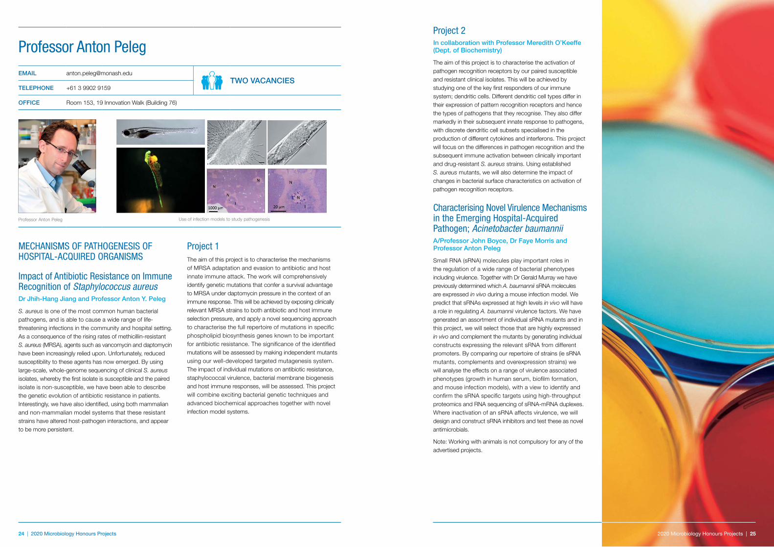
Use of infection models to study pathogenesis
26 | 2020 Microbiology Honours Projects 2020 Microbiology Honours Projects | 27
What is the mechanism of conjugation?
How are these plasmids maintained in the cell and replicated?
How is the conjugation process initiated?
Research in this laboratory has been addressing these questions. This project will focus on one aspect of these studies. The long-term objective is to build a functional model that explains the biology of the conjugative toxin plasmids of C. perfringens.
Dissecting Architecture of High Torque Bacterial Motor The bacterial flagellar motor is a remarkable nanoscale molecular engine. H. pylori evolved to be highly motile in the very viscous mucous layer of the stomach, and its flagellar motor is specialised for locomotion in viscous liquids – it produces a significantly higher torque (turning force) than, for example, enteric bacteria. Preliminary cryo-electron tomography reconstruction of this motor revealed a unique protein cage that supports a wider power-generating ring allowing it to sustain the larger torque. Our aim is to unravel the make-up of this cage and the structural basis for its ability to recruit more force-generating units.
How do Bacteria Sense Environmental Cues?Many bacteria are motile. Chemotaxis, mediated by chemoreceptors, plays an important role in bacterial survival and virulence. In this project, we shall investigate what ligands such receptors recognize and why some molecules are attractants and some repellents, how binding to the receptor leads to signalling, how mutations in the sensor domain affect ligand specificity and, building on this, how bacterial chemoreceptors can be redesigned to recognize and respond to non-native ligands for innovative applications in biotechnology and bioengineering.
Applications are welcome from students with a strong interest in structural biology, X-ray crystallography, the biology of H. pylori, or protein biochemistry.
FUNCTIONAL BIOLOGY OF BACTERIAL PATHOGENS
Conjugative Transfer, Replication and Maintenance of Plasmids from Clostridium perfringensDr Vicki Adams and Professor Julian Rood
Clostridium perfringens causes gas gangrene, food poisoning and non food-borne diarrhoea in humans as well as various life threatening diseases in domestic animals. Many of the toxins implicated in these diseases are located on large, closely related, conjugative plasmids and many C. perfringens strains carry more than one of these plasmids. Consequently, there is a lot of interest in furthering our understanding of C. perfringens plasmid biology.
Structural Studies of Virulence Factors of the Carcinogenic Bacterium Helicobacter pyloriHelicobacter pylori persistently colonize the epithelium of the stomach in roughly half of the world’s population. It is a causative agent of gastric and duodenal ulcers, mucosa- associated B-cell lymphoma and gastric adenocarcinoma.
Although it is a definitive carcinogen, there is no effective vaccine against this bacterium. Standard H. pylori eradication therapy now fails in up to 30%-40% of patients, mainly due to an increase in clarithromycin resistance. There is a clear demand for new strategies to fight H. pylori infections, strategies that involve new or unconventional targets for drug design. A key to success with this lies in strong basic knowledge of the molecular basis of bacterial virulence and survival. Our laboratory focuses on the mechanisms of acid acclimation, damage to gastric epithelial cells and motility and chemotaxis. We use in vitro molecular biophysics and crystallography techniques to investigate structure and dynamics of biomolecules and formulate hypotheses about molecular mechanisms which we then test in vivo using genetics, enzymology and cell biology methods.
Professor Julian RoodEMAIL [email protected]
ONE VACANCYTELEPHONE +61 3 9902 9157
OFFICE Room 155, 19 Innovation Walk (Building 76).
WEB www.med.monash.edu.au/microbiology/research/rood.html
Professor Julian Rood Dr Vicki Adams
Associate Professor Anna RoujeinikovaEMAIL [email protected]
TWO VACANCIESTELEPHONE +61 3 9902 9194
OFFICE Room 151, 19 Innovation Walk (Building 76)
Associate Professor Anna Roujeinikova
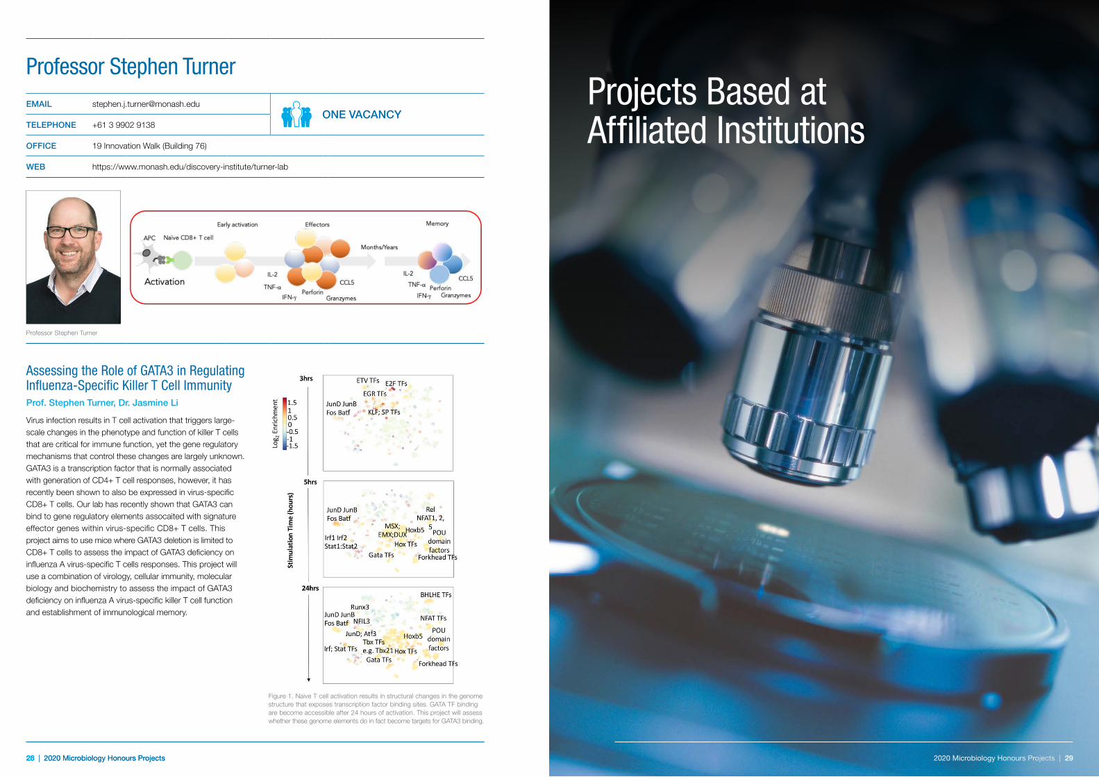
H. pylori CA-inhibitor complex
Chemoreceptor sensory domain
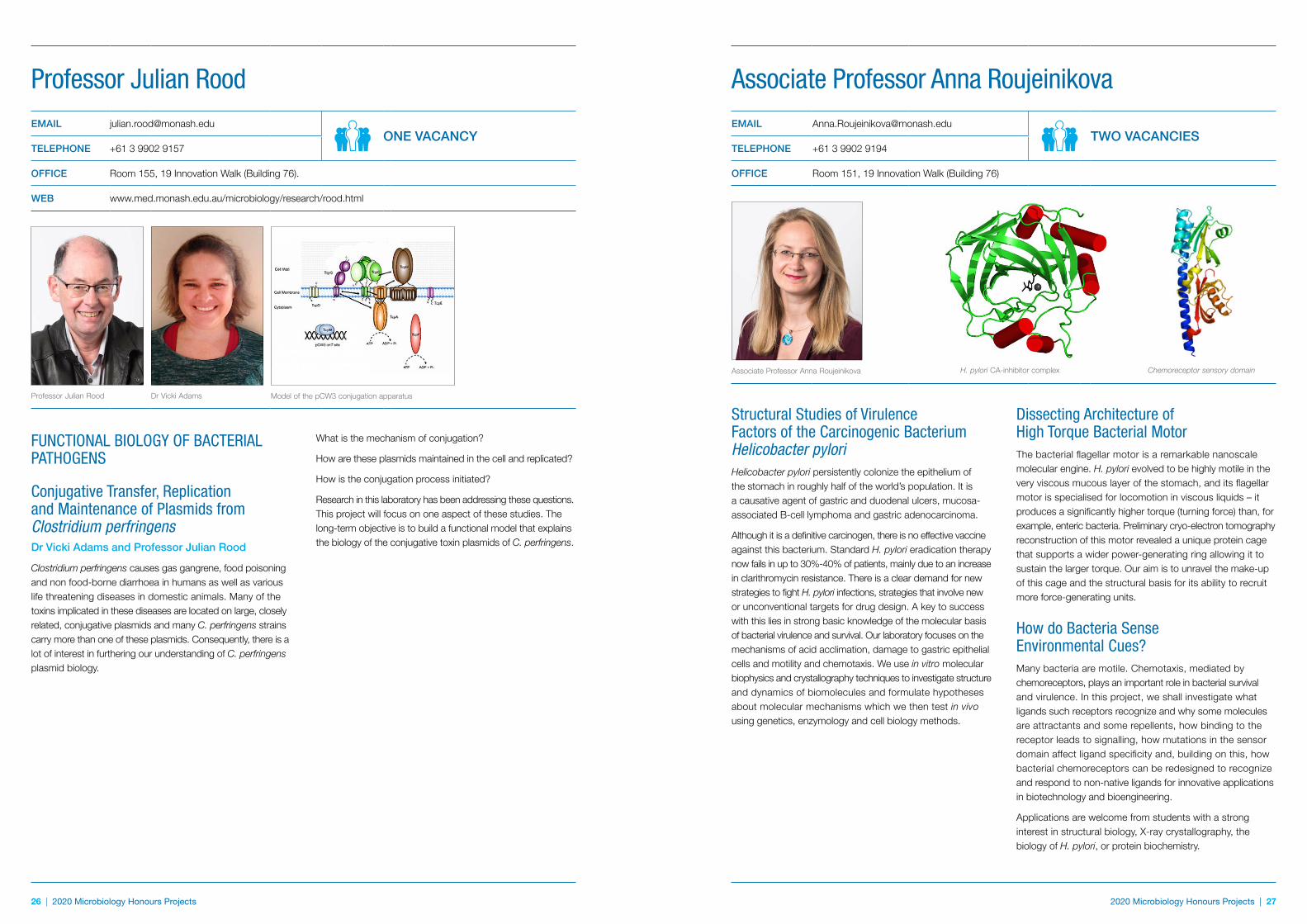
Model of the pCW3 conjugation apparatus
28 | 2020 Microbiology Honours Projects28 | 2020 Microbiology Honours Projects
Assessing the Role of GATA3 in Regulating Influenza-Specific Killer T Cell ImmunityProf. Stephen Turner, Dr. Jasmine Li
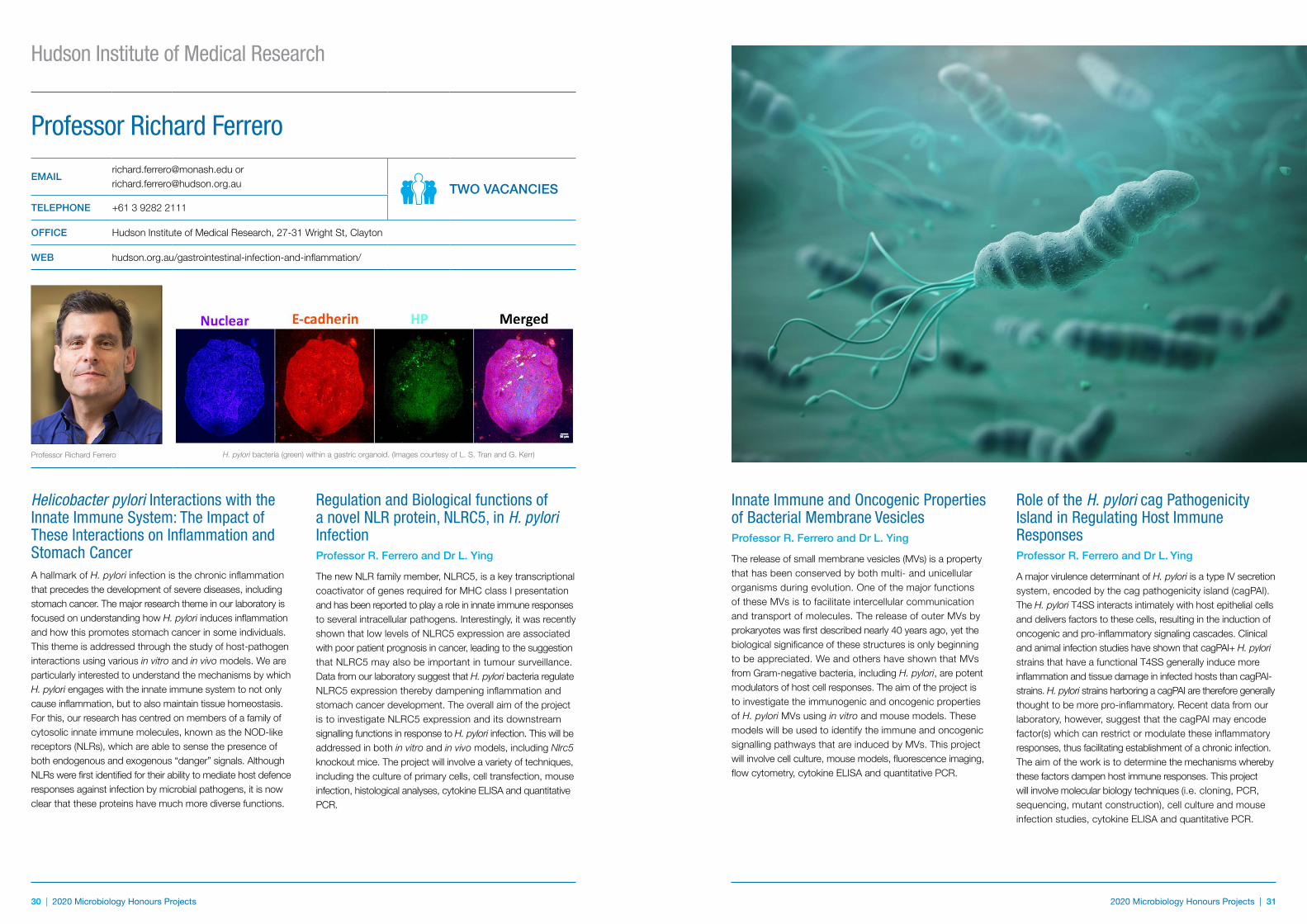
Virus infection results in T cell activation that triggers large-scale changes in the phenotype and function of killer T cells that are critical for immune function, yet the gene regulatory mechanisms that control these changes are largely unknown. GATA3 is a transcription factor that is normally associated with generation of CD4+ T cell responses, however, it has recently been shown to also be expressed in virus-specific CD8+ T cells. Our lab has recently shown that GATA3 can bind to gene regulatory elements assocaited with signature effector genes within virus-specific CD8+ T cells. This project aims to use mice where GATA3 deletion is limited to CD8+ T cells to assess the impact of GATA3 deficiency on influenza A virus-specific T cells responses. This project will use a combination of virology, cellular immunity, molecular biology and biochemistry to assess the impact of GATA3 deficiency on influenza A virus-specific killer T cell function and establishment of immunological memory.
Figure 1. Naive T cell activation results in structural changes in the genome structure that exposes transcription factor binding sites. GATA TF binding are become accessible after 24 hours of activation. This project will assess whether these genome elements do in fact become targets for GATA3 binding.
Professor Stephen TurnerEMAIL [email protected]
ONE VACANCYTELEPHONE +61 3 9902 9138
OFFICE 19 Innovation Walk (Building 76)
WEB https://www.monash.edu/discovery-institute/turner-lab
Professor Stephen Turner
Projects Based at Affiliated Institutions
2020 Microbiology Honours Projects | 29 2020 Microbiology Honours Projects | 29
30 | 2020 Microbiology Honours Projects 2020 Microbiology Honours Projects | 31
Helicobacter pylori Interactions with the Innate Immune System: The Impact of These Interactions on Inflammation and Stomach CancerA hallmark of H. pylori infection is the chronic inflammation that precedes the development of severe diseases, including stomach cancer. The major research theme in our laboratory is focused on understanding how H. pylori induces inflammation and how this promotes stomach cancer in some individuals. This theme is addressed through the study of host-pathogen interactions using various in vitro and in vivo models. We are particularly interested to understand the mechanisms by which H. pylori engages with the innate immune system to not only cause inflammation, but to also maintain tissue homeostasis. For this, our research has centred on members of a family of cytosolic innate immune molecules, known as the NOD-like receptors (NLRs), which are able to sense the presence of both endogenous and exogenous “danger” signals. Although NLRs were first identified for their ability to mediate host defence responses against infection by microbial pathogens, it is now clear that these proteins have much more diverse functions.
Regulation and Biological functions of a novel NLR protein, NLRC5, in H. pylori InfectionProfessor R. Ferrero and Dr L. Ying
The new NLR family member, NLRC5, is a key transcriptional coactivator of genes required for MHC class I presentation and has been reported to play a role in innate immune responses to several intracellular pathogens. Interestingly, it was recently shown that low levels of NLRC5 expression are associated with poor patient prognosis in cancer, leading to the suggestion that NLRC5 may also be important in tumour surveillance. Data from our laboratory suggest that H. pylori bacteria regulate NLRC5 expression thereby dampening inflammation and stomach cancer development. The overall aim of the project is to investigate NLRC5 expression and its downstream signalling functions in response to H. pylori infection. This will be addressed in both in vitro and in vivo models, including Nlrc5 knockout mice. The project will involve a variety of techniques, including the culture of primary cells, cell transfection, mouse infection, histological analyses, cytokine ELISA and quantitative PCR.
Hudson Institute of Medical Research
Professor Richard Ferrero
[email protected] or [email protected] TWO VACANCIES
TELEPHONE +61 3 9282 2111
OFFICE Hudson Institute of Medical Research, 27-31 Wright St, Clayton
WEB hudson.org.au/gastrointestinal-infection-and-inflammation/
Professor Richard Ferrero
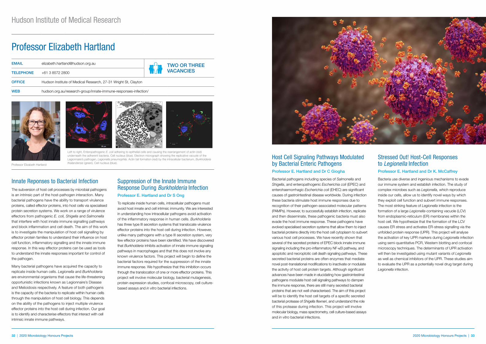
H. pylori bacteria (green) within a gastric organoid. (Images courtesy of L. S. Tran and G. Kerr)
Innate Immune and Oncogenic Properties of Bacterial Membrane VesiclesProfessor R. Ferrero and Dr L. Ying
The release of small membrane vesicles (MVs) is a property that has been conserved by both multi- and unicellular organisms during evolution. One of the major functions of these MVs is to facilitate intercellular communication and transport of molecules. The release of outer MVs by prokaryotes was first described nearly 40 years ago, yet the biological significance of these structures is only beginning to be appreciated. We and others have shown that MVs from Gram-negative bacteria, including H. pylori, are potent modulators of host cell responses. The aim of the project is to investigate the immunogenic and oncogenic properties of H. pylori MVs using in vitro and mouse models. These models will be used to identify the immune and oncogenic signalling pathways that are induced by MVs. This project will involve cell culture, mouse models, fluorescence imaging, flow cytometry, cytokine ELISA and quantitative PCR.
Role of the H. pylori cag Pathogenicity Island in Regulating Host Immune ResponsesProfessor R. Ferrero and Dr L. Ying
A major virulence determinant of H. pylori is a type IV secretion system, encoded by the cag pathogenicity island (cagPAI). The H. pylori T4SS interacts intimately with host epithelial cells and delivers factors to these cells, resulting in the induction of oncogenic and pro-inflammatory signaling cascades. Clinical and animal infection studies have shown that cagPAI+ H. pylori strains that have a functional T4SS generally induce more inflammation and tissue damage in infected hosts than cagPAI- strains. H. pylori strains harboring a cagPAI are therefore generally thought to be more pro-inflammatory. Recent data from our laboratory, however, suggest that the cagPAI may encode factor(s) which can restrict or modulate these inflammatory responses, thus facilitating establishment of a chronic infection. The aim of the work is to determine the mechanisms whereby these factors dampen host immune responses. This project will involve molecular biology techniques (i.e. cloning, PCR, sequencing, mutant construction), cell culture and mouse infection studies, cytokine ELISA and quantitative PCR.
32 | 2020 Microbiology Honours Projects 2020 Microbiology Honours Projects | 33
Innate Reponses to Bacterial Infection The subversion of host cell processes by microbial pathogens is an intrinsic part of the host-pathogen interaction. Many bacterial pathogens have the ability to transport virulence proteins, called effector proteins, into host cells via specialised protein secretion systems. We work on a range of virulence effectors from pathogenic E. coli, Shigella and Salmonella that interfere with host innate immune signalling pathways and block inflammation and cell death. The aim of this work is to investigate the manipulation of host cell signalling by effector protein families to understand their influence on host cell function, inflammatory signalling and the innate immune response. In this way effector proteins can be used as tools to understand the innate responses important for control of the pathogen.
Many bacterial pathogens have acquired the capacity to replicate inside human cells. Legionella and Burkholderia are environmental organisms that cause the life-threatening opportunistic infections known as Legionnaire’s Disease and Melioidosis respectively. A feature of both pathogens is the capacity of the bacteria to replicate within human cells through the manipulation of host cell biology. This depends on the ability of the pathogens to inject multiple virulence effector proteins into the host cell during infection. Our goal is to identify and characterise effectors that interact with cell intrinsic innate immune pathways.
Suppression of the Innate Immune Response During Burkholderia Infection Professor E. Hartland and Dr S Ong
To replicate inside human cells, intracellular pathogens must avoid host innate and cell intrinsic immunity. We are interested in understanding how intracellular pathogens avoid activation of the inflammatory response in human cells. Burkholderia has three type III secretion systems that translocate virulence effector proteins into the host cell during infection. However, unlike many pathogens with a type III secretion system, very few effector proteins have been identified. We have discovered that Burkholderia inhibits activation of innate immune signaling pathways in macrophages and that this does not involve any known virulence factors. This project will begin to define the bacterial factors required for the suppression of the innate immune response. We hypothesize that this inhibition occurs through the translocation of one or more effector proteins. This project will involve molecular biology, bacterial mutagenesis, protein expression studies, confocal microscopy, cell culture-based assays and in vitro bacterial infections.
Hudson Institute of Medical Research
Professor Elizabeth Hartland EMAIL [email protected] TWO OR THREE
VACANCIESTELEPHONE +61 3 8572 2800
OFFICE Hudson Institute of Medical Research, 27-31 Wright St, Clayton
WEB hudson.org.au/research-group/innate-immune-responses-infection/
Professor Elizabeth Hartland
Left to right, Enteropathogenic E. coli adhering to epithelial cells and causing the rearrangement of actin (red) underneath the adherent bacteria. Cell nucleus (blue). Electron micrograph showing the replicative vacuole of the Legionnaire’s pathogen, Legionella pneumophila. Actin tail formation (red) by the intracellular bacterium, Burkholderia thailandensis (green). Cell nucleus (blue).
Host Cell Signaling Pathways Modulated by Bacterial Enteric Pathogens Professor E. Hartland and Dr C Giogha
Bacterial pathogens including species of Salmonella and Shigella, and enteropathogenic Escherichia coli (EPEC) and enterohaemorrhagic Escherichia coli (EHEC) are significant causes of gastrointestinal disease worldwide. During infection these bacteria stimulate host immune responses due to recognition of their pathogen-associated molecular patterns (PAMPs). However, to successfully establish infection, replicate and then disseminate, these pathogenic bacteria must also evade the host immune response. These pathogens have evolved specialised secretion systems that allow them to inject bacterial proteins directly into the host cell cytoplasm to subvert various host cell processes. We have recently shown that several of the secreted proteins of EPEC block innate immune signaling including the pro-inflammatory NF-κB pathway, and apoptotic and necroptotic cell death signaling pathways. These secreted bacterial proteins are often enzymes that mediate novel post-translational modifications to inactivate or modulate the activity of host cell protein targets. Although significant advances have been made in elucidating how gastrointestinal pathogens modulate host cell signaling pathways to dampen the immune response, there are still many secreted bacterial proteins that are not well characterised. The aim of this project will be to identify the host cell targets of a specific secreted bacterial protease of Shigella flexneri, and understand the role of this protease during infection. This project will involve molecular biology, mass spectrometry, cell culture-based assays and in vitro bacterial infections.
Stressed Out! Host-Cell Responses to Legionella InfectionProfessor E. Hartland and Dr K. McCaffrey
Bacteria use diverse and ingenious mechanisms to evade our immune system and establish infection. The study of complex microbes such as Legionella, which reproduce inside our cells, allow us to identify novel ways by which they exploit cell function and subvert immune responses. The most striking feature of Legionella infection is the formation of a large Legionella-containing vacuole (LCV) from endoplasmic-reticulum (ER) membranes within the host cell. We hypothesize that the formation of the LCV causes ER stress and activates ER-stress signalling via the unfolded protein response (UPR). This project will analyse the activation of key UPR markers during Legionella infection using semi-quantitative PCR, Western blotting and confocal microscopy techniques. The determinants of UPR activation will then be investigated using mutant variants of Legionella as well as chemical inhibitors of the UPR. These studies aim to evaluate the UPR as a potentially novel drug target during Legionella infection.
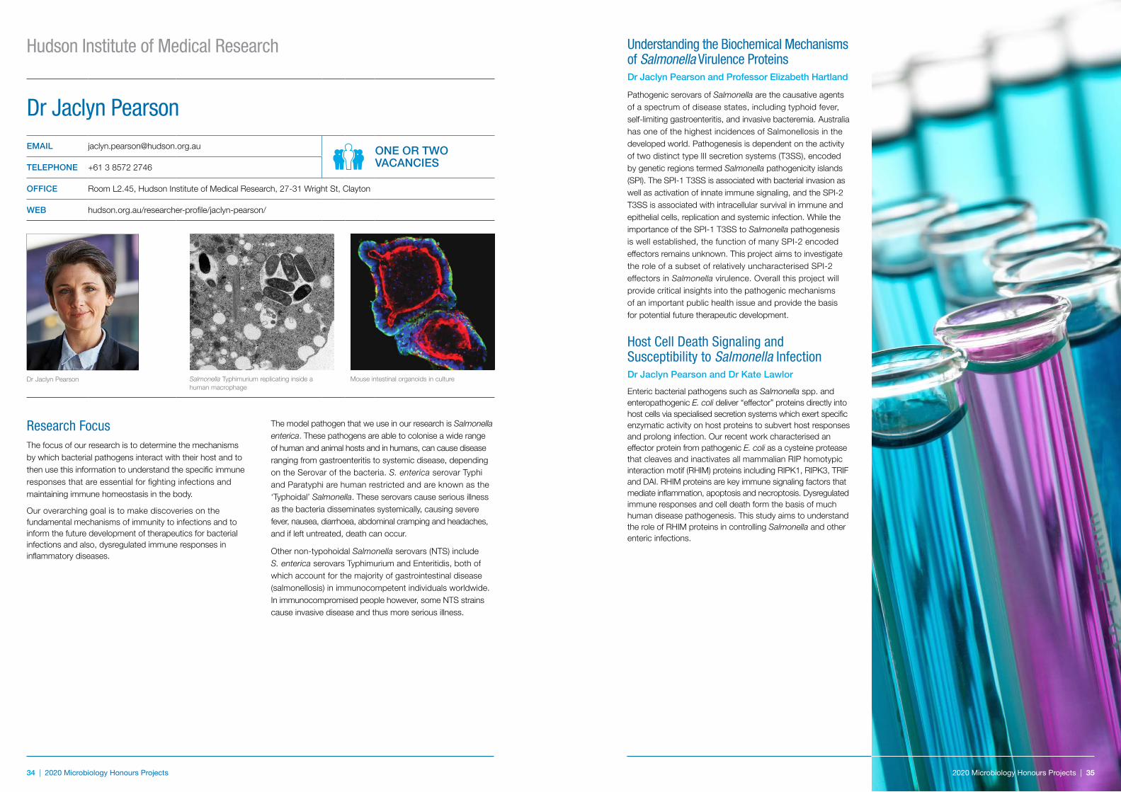
34 | 2020 Microbiology Honours Projects 2020 Microbiology Honours Projects | 35
Research FocusThe focus of our research is to determine the mechanisms by which bacterial pathogens interact with their host and to then use this information to understand the specific immune responses that are essential for fighting infections and maintaining immune homeostasis in the body.
Our overarching goal is to make discoveries on the fundamental mechanisms of immunity to infections and to inform the future development of therapeutics for bacterial infections and also, dysregulated immune responses in inflammatory diseases.
The model pathogen that we use in our research is Salmonella enterica. These pathogens are able to colonise a wide range of human and animal hosts and in humans, can cause disease ranging from gastroenteritis to systemic disease, depending on the Serovar of the bacteria. S. enterica serovar Typhi and Paratyphi are human restricted and are known as the ‘Typhoidal’ Salmonella. These serovars cause serious illness as the bacteria disseminates systemically, causing severe fever, nausea, diarrhoea, abdominal cramping and headaches, and if left untreated, death can occur.
Other non-typohoidal Salmonella serovars (NTS) include S. enterica serovars Typhimurium and Enteritidis, both of which account for the majority of gastrointestinal disease (salmonellosis) in immunocompetent individuals worldwide. In immunocompromised people however, some NTS strains cause invasive disease and thus more serious illness.
Dr Jaclyn PearsonEMAIL [email protected] ONE OR TWO
VACANCIESTELEPHONE +61 3 8572 2746
OFFICE Room L2.45, Hudson Institute of Medical Research, 27-31 Wright St, Clayton
WEB hudson.org.au/researcher-profile/jaclyn-pearson/
Dr Jaclyn Pearson
Salmonella Typhimurium replicating inside a human macrophage
Mouse intestinal organoids in culture
Hudson Institute of Medical Research Understanding the Biochemical Mechanisms of Salmonella Virulence ProteinsDr Jaclyn Pearson and Professor Elizabeth Hartland
Pathogenic serovars of Salmonella are the causative agents of a spectrum of disease states, including typhoid fever, self-limiting gastroenteritis, and invasive bacteremia. Australia has one of the highest incidences of Salmonellosis in the developed world. Pathogenesis is dependent on the activity of two distinct type III secretion systems (T3SS), encoded by genetic regions termed Salmonella pathogenicity islands (SPI). The SPI-1 T3SS is associated with bacterial invasion as well as activation of innate immune signaling, and the SPI-2 T3SS is associated with intracellular survival in immune and epithelial cells, replication and systemic infection. While the importance of the SPI-1 T3SS to Salmonella pathogenesis is well established, the function of many SPI-2 encoded effectors remains unknown. This project aims to investigate the role of a subset of relatively uncharacterised SPI-2 effectors in Salmonella virulence. Overall this project will provide critical insights into the pathogenic mechanisms of an important public health issue and provide the basis for potential future therapeutic development.
Host Cell Death Signaling and Susceptibility to Salmonella InfectionDr Jaclyn Pearson and Dr Kate Lawlor
Enteric bacterial pathogens such as Salmonella spp. and enteropathogenic E. coli deliver “effector” proteins directly into host cells via specialised secretion systems which exert specific enzymatic activity on host proteins to subvert host responses and prolong infection. Our recent work characterised an effector protein from pathogenic E. coli as a cysteine protease that cleaves and inactivates all mammalian RIP homotypic interaction motif (RHIM) proteins including RIPK1, RIPK3, TRIF and DAI. RHIM proteins are key immune signaling factors that mediate inflammation, apoptosis and necroptosis. Dysregulated immune responses and cell death form the basis of much human disease pathogenesis. This study aims to understand the role of RHIM proteins in controlling Salmonella and other enteric infections.
2020 Microbiology Honours Projects | 35
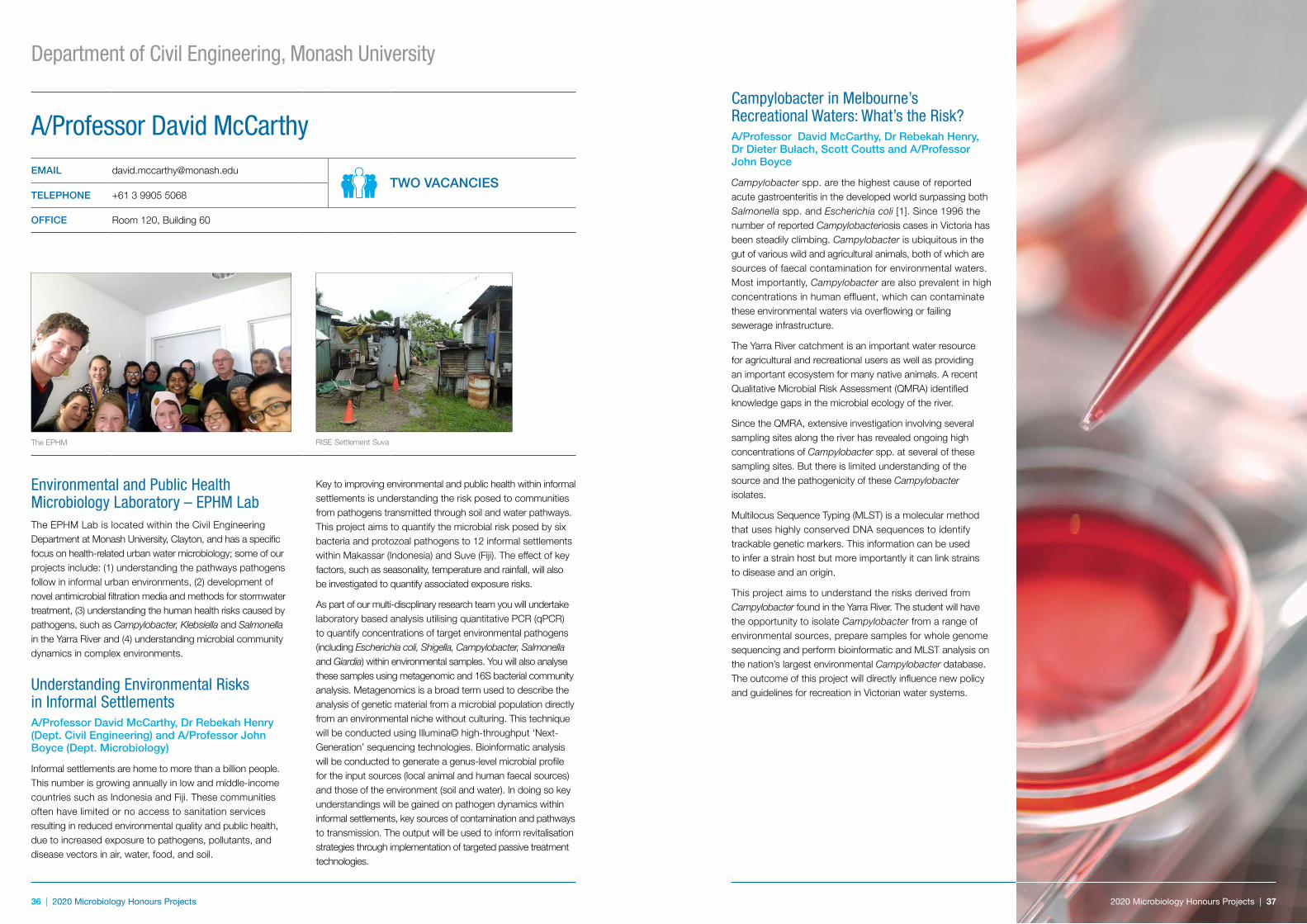
36 | 2020 Microbiology Honours Projects 2020 Microbiology Honours Projects | 37
Campylobacter in Melbourne’s Recreational Waters: What’s the Risk?A/Professor David McCarthy, Dr Rebekah Henry, Dr Dieter Bulach, Scott Coutts and A/Professor John Boyce
Campylobacter spp. are the highest cause of reported acute gastroenteritis in the developed world surpassing both Salmonella spp. and Escherichia coli [1]. Since 1996 the number of reported Campylobacteriosis cases in Victoria has been steadily climbing. Campylobacter is ubiquitous in the gut of various wild and agricultural animals, both of which are sources of faecal contamination for environmental waters. Most importantly, Campylobacter are also prevalent in high concentrations in human effluent, which can contaminate these environmental waters via overflowing or failing sewerage infrastructure.
The Yarra River catchment is an important water resource for agricultural and recreational users as well as providing an important ecosystem for many native animals. A recent Qualitative Microbial Risk Assessment (QMRA) identified knowledge gaps in the microbial ecology of the river.
Since the QMRA, extensive investigation involving several sampling sites along the river has revealed ongoing high concentrations of Campylobacter spp. at several of these sampling sites. But there is limited understanding of the source and the pathogenicity of these Campylobacter isolates.
Multilocus Sequence Typing (MLST) is a molecular method that uses highly conserved DNA sequences to identify trackable genetic markers. This information can be used to infer a strain host but more importantly it can link strains to disease and an origin.
This project aims to understand the risks derived from Campylobacter found in the Yarra River. The student will have the opportunity to isolate Campylobacter from a range of environmental sources, prepare samples for whole genome sequencing and perform bioinformatic and MLST analysis on the nation’s largest environmental Campylobacter database. The outcome of this project will directly influence new policy and guidelines for recreation in Victorian water systems.
Environmental and Public Health Microbiology Laboratory – EPHM LabThe EPHM Lab is located within the Civil Engineering Department at Monash University, Clayton, and has a specific focus on health-related urban water microbiology; some of our projects include: (1) understanding the pathways pathogens follow in informal urban environments, (2) development of novel antimicrobial filtration media and methods for stormwater treatment, (3) understanding the human health risks caused by pathogens, such as Campylobacter, Klebsiella and Salmonella in the Yarra River and (4) understanding microbial community dynamics in complex environments.
Understanding Environmental Risks in Informal SettlementsA/Professor David McCarthy, Dr Rebekah Henry (Dept. Civil Engineering) and A/Professor John Boyce (Dept. Microbiology)
Informal settlements are home to more than a billion people. This number is growing annually in low and middle-income countries such as Indonesia and Fiji. These communities often have limited or no access to sanitation services resulting in reduced environmental quality and public health, due to increased exposure to pathogens, pollutants, and disease vectors in air, water, food, and soil.
Key to improving environmental and public health within informal settlements is understanding the risk posed to communities from pathogens transmitted through soil and water pathways. This project aims to quantify the microbial risk posed by six bacteria and protozoal pathogens to 12 informal settlements within Makassar (Indonesia) and Suve (Fiji). The effect of key factors, such as seasonality, temperature and rainfall, will also be investigated to quantify associated exposure risks.
As part of our multi-discplinary research team you will undertake laboratory based analysis utilising quantitative PCR (qPCR) to quantify concentrations of target environmental pathogens (including Escherichia coli, Shigella, Campylobacter, Salmonella and Giardia) within environmental samples. You will also analyse these samples using metagenomic and 16S bacterial community analysis. Metagenomics is a broad term used to describe the analysis of genetic material from a microbial population directly from an environmental niche without culturing. This technique will be conducted using Illumina© high-throughput ‘Next- Generation’ sequencing technologies. Bioinformatic analysis will be conducted to generate a genus-level microbial profile for the input sources (local animal and human faecal sources) and those of the environment (soil and water). In doing so key understandings will be gained on pathogen dynamics within informal settlements, key sources of contamination and pathways to transmission. The output will be used to inform revitalisation strategies through implementation of targeted passive treatment technologies.
A/Professor David McCarthyEMAIL [email protected]
TWO VACANCIESTELEPHONE +61 3 9905 5068
OFFICE Room 120, Building 60
The EPHM RISE Settlement Suva
Department of Civil Engineering, Monash University
2020 Microbiology Honours Projects | 37
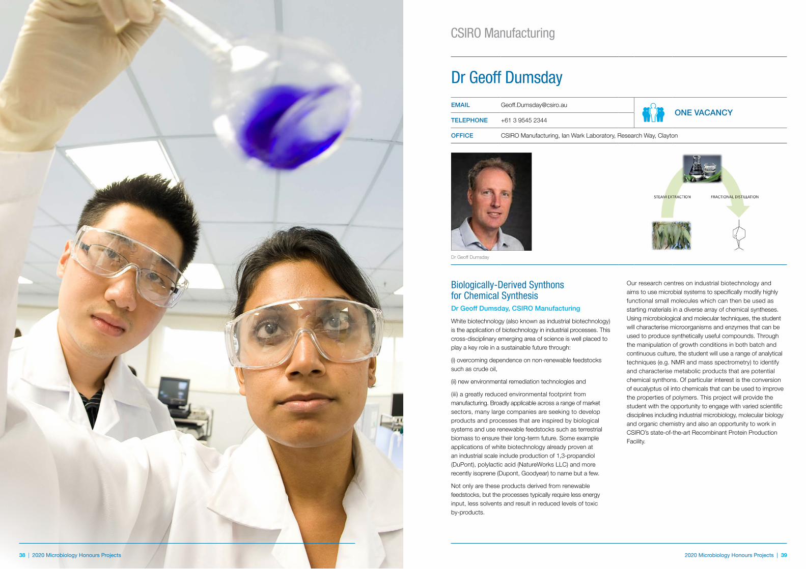
38 | 2020 Microbiology Honours Projects 2020 Microbiology Honours Projects | 39
CSIRO Manufacturing
38 | 2020 Microbiology Honours Projects
Biologically-Derived Synthons for Chemical SynthesisDr Geoff Dumsday, CSIRO Manufacturing
White biotechnology (also known as industrial biotechnology) is the application of biotechnology in industrial processes. This cross-disciplinary emerging area of science is well placed to play a key role in a sustainable future through:
(i) overcoming dependence on non-renewable feedstocks such as crude oil,
(ii) new environmental remediation technologies and
(iii) a greatly reduced environmental footprint from manufacturing. Broadly applicable across a range of market sectors, many large companies are seeking to develop products and processes that are inspired by biological systems and use renewable feedstocks such as terrestrial biomass to ensure their long-term future. Some example applications of white biotechnology already proven at an industrial scale include production of 1,3-propandiol (DuPont), polylactic acid (NatureWorks LLC) and more recently isoprene (Dupont, Goodyear) to name but a few.
Not only are these products derived from renewable feedstocks, but the processes typically require less energy input, less solvents and result in reduced levels of toxic by-products.
Our research centres on industrial biotechnology and aims to use microbial systems to specifically modify highly functional small molecules which can then be used as starting materials in a diverse array of chemical syntheses. Using microbiological and molecular techniques, the student will characterise microorganisms and enzymes that can be used to produce synthetically useful compounds. Through the manipulation of growth conditions in both batch and continuous culture, the student will use a range of analytical techniques (e.g. NMR and mass spectrometry) to identify and characterise metabolic products that are potential chemical synthons. Of particular interest is the conversion of eucalyptus oil into chemicals that can be used to improve the properties of polymers. This project will provide the student with the opportunity to engage with varied scientific disciplines including industrial microbiology, molecular biology and organic chemistry and also an opportunity to work in CSIRO’s state-of-the-art Recombinant Protein Production Facility.
Dr Geoff DumsdayEMAIL [email protected]
ONE VACANCYTELEPHONE +61 3 9545 2344
OFFICE CSIRO Manufacturing, Ian Wark Laboratory, Research Way, Clayton
Dr Geoff Dumsday
40 | 2020 Microbiology Honours Projects 2020 Microbiology Honours Projects | 41
Currently, very little is known about the interactions between malaria infection rates and patterns and malaria immunity in populations, and how these interact. Malaria control programs face the challenge that as malaria transmission declines, malaria immunity also declines, which places the population at higher risk of malaria transmission and rebound epidemics.
This project will investigate the impact of malaria immunity on malaria infection rates and transmission of malaria in populations. The student will analyse various parameters to define the patterns of infection and immunity, with a particular focus on defining the interaction between immunity and malaria transmission.
The findings of this project will be highly relevant to informing malaria elimination efforts and understanding the value of incorporating vaccines into elimination strategies. Skills acquired may include established high-throughput immunoassays and assays that quantify the functional activity of immune responses (e.g. flow cytometry, Fc-receptor mediated immunity, complement activation, western blots, ELISA, neutralisation assays). This could be expanded to include modelling of the interaction between infection and immunity, and how this may impact on malaria elimination and control. Interested students should contact Arzum, [email protected]
children from malaria or protect pregnant women and their developing babies from the devastating consequences of malaria in pregnancy.
The studies would particularly focus on using innovative approaches to understand how antibodies neutralise and clear malaria parasites in the blood, including interactions with monocytes/macrophages and dendritic cells, and identifying specific epitopes targeted by protective antibodies. Skills may involve assays of functional immunity, cell culture, isolation and analysis of immune cells, flow cytometry, western blotting, ELISA, and epitope mapping. The project will be tailored to best match the student’s interests and training background. Interested students should contact Arzum, [email protected]
Understanding Malaria Transmission and Immunity to inform Malaria EliminationMalaria transmission in populations involves interactions between infection rates and prevalence that drive transmission, and the presence of malaria immunity that has the potential to reduce transmission. Malaria immunity can act to reduce infection rates and levels of malaria parasitemia, and specific components of immunity can also function to directly block transmission of malaria. This is known as transmission-blocking immunity.
mediated immunity), flow cytometry, cell culture and protein expression. The project could also include bioinformatics, structural modelling of vaccine antigens, or modelling vaccine impact depending on the student’s interest. The specific activities and focus of the project will be refined to suit the interests and training background of the student. Interested students should contact Arzum, [email protected]
Discovering the Mechanisms and Targets of Immunity Against MalariaAntibodies are an important component of acquired immunity against malaria, as demonstrated in pivotal studies in which immunoglobulin G (IgG) from immune adults was transferred to malaria-infected children and resulted in clearance of infection. The mechanisms of protection and specific target epitopes of protective immunity are not well understood. In recent studies, we have begun to uncover important roles for antibodies that can directly inhibit host-cell infection, interact with immune cells to kill and clear malaria, or recruit complement to neutralise infection.
The aims of this project include identifying the key targets of specific mechanisms mediating immunity. The project may combine detailed studies of immune responses with clinical and population studies in Africa, Asia, and Papua New Guinea. It will examine how immune responses protect
Developing Vaccines Against MalariaMalaria is one of the world’s leading causes of death and illness, particularly among young children. There remains a strong need for highly effective vaccines to reduce the burden of malaria and progress towards eventual malaria elimination. To date, most vaccines have achieved only modest levels of efficacy, emphasising the need for novel approaches in vaccine design that can induce potent immune responses.
This project will focus on identifying key antigens and specific epitopes that are targets of protective immunity against malaria and understanding the mechanisms mediating immunity, which includes antibodies and cell-mediated responses.
This knowledge is crucial for the development of effective vaccines against malaria. The project will also involve using knowledge of immunity to malaria for informing vaccine design, and the expression and testing of novel vaccine candidates. These studies will use novel approaches in molecular biology, cell biology and immunology to address these aims, and will build on recent major advances generated from our malaria vaccine program.
The project will primarily involve laboratory-based research, including western blotting, imaging, standard immunoassays, functional immunoassays (e.g neutralisation assays, cell-
Professor James Beeson
[email protected]@burnet.edu.au TWO VACANCIES
TELEPHONE +61 3 9282 2111
OFFICE 85 Commercial Road, Melbourne (AMREP Campus)
Professor James Beeson
Immunofluorescence microscopy showing invasion of a human red blood cell by a malaria merozoite (green)
Phagocytosis of malaria merozoites by a human monocyte, an important mechanism of immunity, visualized using electron microscop
Burnet Institute
42 | 2020 Microbiology Honours Projects 2020 Microbiology Honours Projects | 43
Discovery of a New Drug Class for HIV Treatment and PreventionProfessor Gilda Tachedjian and Dr David Chalmers
There is a real threat that drug resistance, toxicity and intolerance will eventually lead to exhaustion of antiretroviral drug options for both HIV treatment and prevention, especially since there is little in the way of new drug classes in the pipeline. We have initiated a drug discovery program targeting HIV-1 reverse transcriptase (RT) to identify compounds that inhibit this essential enzyme. We are using an innovative and validated paradigm for drug discovery called fragment-based drug design (FBDD) that uses very small compounds called ‘fragments’ to find inhibitors with novel mechanisms of action against HIV-1 RT. Using FBDD means screening far fewer compounds than conventional drug screens since fragments are so small, and can be developed into more potent drugs. Two screens have identified promising fragments that inhibit HIV-1 RT. We have discovered fragments with mechanisms that are distinct from other drugs that inhibit HIV-1 RT in the clinic (La et al 2015 PNAS 112:6979) and some of these have been progressed to molecules that have low micromolar activity against wild-type and drug-resistant RT. The aim of this study is to progress one of the fragment hits into more potent RT inhibitors or drug leads. Compounds are structurally related to the fragment hit will be evaluated for their ability to bind and inhibit wild-type and drug-resistant RT, as well as inhibit HIV-1 replication. This study will identify leads for the development of a novel class of RT inhibitor for use in HIV treatment and prevention.
This is a joint project between the Tachedjian Lab and Monash Institute of Pharmaceutical Sciences.
Vaginal Microbiota and HIV SusceptibilityProfessor Gilda Tachedjian, Dr Anna Hearps and Dr Joshua Hayward
HIV transmission is modulated by the vaginal microbiota. Women colonised with optimal vaginal bacterial communities, typically dominated by beneficial Lactobacillus spp., have a decreased risk of acquiring and transmitting HIV compared to women colonised with non-optimal microbiota as exemplified by bacterial vaginosis (BV) a common but poorly understood condition in women of reproductive age (Tachedjian et al 2017 Res Microbiol 168:782). Non-optimal vaginal microbiota are characterised by a depletion of beneficial Lactobacillus spp. and high relative abundance or load of facultative and/or obligate anaerobes (McKinnon et al 2019 AIDS Res Hum Retroviruses 35:219). Non-optimal vaginal microbiota increase local pro-inflammatory cytokines, recruit activated HIV target cells and disrupt cervicovaginal epithelial barrier integrity that together drive increased HIV risk.
While studies have described the association between the vaginal microbiota and increased susceptibility to HIV, relatively little is known about the function of these microbiota through the action of their metabolites on the genital mucosa and directly on HIV and other sexually transmitted infections (STIs).
Major distinguishing features of women colonised with optimal vaginal microbiota compared to women with BV is a dramatic increase in the levels of lactic acid and depletion of short chain fatty acids (SCFAs) suggesting a role for these metabolites as effector molecules of vaginal bacteria (Aldunate et al 2015 Front Physiol 6:164). Projects are available to determine the direct anti-HIV mechanism of vaginal microbiota metabolites (Aldunate et al 2013 J Antimicrob Chemother 68:2015; Tyssen et al 2018 mSphere 3), and their immune modulatory and barrier promoting effects on cervicovaginal epithelial cells informed by RNAseq analyses. These studies are underpinning an exciting program at the Burnet to advance strategies to treat and prevent genital inflammation and consequently susceptibility to HIV, other STIs as well adverse reproductive health outcomes.
Professor Gilda TachedjianEMAIL [email protected]
UP TO TWO VACANCIESTELEPHONE +61 3 9282 2256
OFFICERetroviral Biology and Antivirals Laboratory (RBA) (Tachedjian Lab), Life Sciences DisciplineDisease Elimination and Health Security Programs, Burnet Institute
WEB www.burnet.edu.au/working_groups/21_tachedjian_laboratory
Tachedjian Laboratory Cell infected with HIV (green dots)
Endogenous retroviral sequences are present as a critical part of eukaryotic genomes and normally represent the fossil record of extinct viruses. Our analysis of the genome of bat species has revealed the presence of retroviral sequences, that at the amino-acid level, demonstrate homology to extant (currently existing) gammaretroviruses (Cui et al 2012 J Virol 86:4288) and betaretroviruses (Hayward et al 2013 Retrovirology 10:35) indicating that retroviruses have circulated in bats. However, whether infectious retroviruses are currently circulating in bats is unknown. We have discovered retroviral sequences in bat scat that we engineered into a replication-competent virus. This study will focus on characterising the ability of a class of bat restriction factors to inhibit the replication our engineered bat retrovirus.
This is a joint project between the Tachedjian Lab and CSIRO Australian Animal Health Laboratory.
Bat Antiviral Defenses Against VirusesProfessor Gilda Tachedjian and Dr Joshua Hayward
Bats are a major reservoir of viruses such as Hendra virus and Ebola virus that are pathogenic in humans but not in bats. The reason why bats can coexist with these viral pathogens is unknown. However, one explanation is that the coevolution of bats and their viruses have led them to a ‘peace treaty’, a biological equilibrium in which viral replication is regulated in such a way that both host and virus can co-exist without undue antagonism.
The role of the host’s immune system is to control infections and host restriction factors are the front line of the innate immune response that targets viruses. These factors have originally been discovered to restrict retroviruses from other mammalian species, such as HIV (Hayward et al 2018 Mol Biol Evol 35:1626), and some of these factors e.g. tetherin, can additionally restrict paramyxoviruses, coronaviruses and filoviruses. While bats are known to harbour many diverse viral species, retroviruses are yet to be isolated from bats.
Burnet Institute
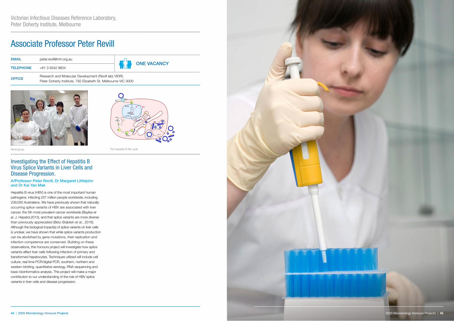
44 | 2020 Microbiology Honours Projects 2020 Microbiology Honours Projects | 45
Associate Professor Peter Revill
EMAIL [email protected] VACANCY
TELEPHONE +61 3 9342 9604
OFFICEResearch and Molecular Development (Revill lab) VIDRL Peter Doherty Institute, 792 Elizabeth St, Melbourne VIC 3000
Revill group
Nucleus
NTCP
ER
Golgi
Heparin sulphate
cccDNA
CorePolymerase
Viral RNAs
pgRNA
HBx
HBeAg
X
e
L SMSurface
proteins
Mature nucleocapsid
X
e
e
e
X
Secretion
Virion
Sub-viral particles
MVB
The hepatitis B life cycle
Investigating the Effect of Hepatitis B Virus Splice Variants in Liver Cells and Disease Progression. A/Professor Peter Revill, Dr Margaret Littlejohn and Dr Kai Yan Mak
Hepatitis B virus (HBV) is one of the most important human pathogens, infecting 257 million people worldwide, including 239,000 Australians. We have previously shown that naturally occurring splice variants of HBV are associated with liver cancer, the 5th most prevalent cancer worldwide (Bayliss et al, J. Hepatol,2013), and that splice variants are more diverse than previously appreciated (Betz-Stablein et al., 2016). Although the biological impact(s) of splice variants on liver cells is unclear, we have shown that while splice variants production can be abolished by gene mutations, their replication and infection competence are conserved. Building on these observations, this honours project will investigate how splice variants affect liver cells following infection of primary and transformed hepatocytes. Techniques utilized will include cell culture, real time PCR/digital PCR, southern, northern and western blotting, quantitative serology, RNA sequencing and basic bioinformatics analysis. This project will make a major contribution to our understanding of the role of HBV splice variants in liver cells and disease progression.
Victorian Infectious Diseases Reference Laboratory, Peter Doherty Institute, Melbourne
2020 Microbiology Honours Projects | 45

46 | 2020 Microbiology Honours Projects 2020 Microbiology Honours Projects | 47
Notes Monash Microbiology Honours 2020
Name .......................................................................................................................................................................
Address in December/January .................................................................................................................................
Mobile Phone (9 am – 5 pm) ...................................................................................................................................
Phone (Home) .........................................................................................................................................................
Email (Must be checked daily) ..................................................................................................................................
Project preferences (supervisor and brief project title):
1. .............................................................................................................................................................................
2. .............................................................................................................................................................................
3. .............................................................................................................................................................................
4. .............................................................................................................................................................................
5. .............................................................................................................................................................................
6. .............................................................................................................................................................................
7. .............................................................................................................................................................................
8. .............................................................................................................................................................................
9. .............................................................................................................................................................................
You MUST have talked to three potential supervisors before submitting this form.
Supervisor 1 Signature .............................................................................................................................................
Supervisor 2 Signature .............................................................................................................................................
Supervisor 3 Signature .............................................................................................................................................
#
Please return this form to Associate Professor John Boyce as soon as possible, but no later than Friday 15 November 2019
48 | 2020 Microbiology Honours Projects 2020 Microbiology Honours Projects | 49
Notes
Monash University reserves the right to alter information, procedures, fees and regulations contained in this document. Please check the Monash University website for updates (monash.edu). All information reflects prescriptions, policy and practice in force at time of publication. Produced by MPS Monash 311213. CRICOS Provider: Monash University 00008C. Published September 2019.
Further information
Associate Professor John Boyce Honours Coordinator Department of Microbiology School of Biomedical Sciences
Room 215 19 Innovation Walk Clayton Campus Monash University, VIC 3800
Telephone: +61 3 9902 9179 Email: [email protected]
Monash Biomedicine Discovery Institute 23 Innovation Walk Monash University Wellington Road Clayton, Victoria, 3800
Telephone: +61 3 9902 9400 Email: [email protected]
www.med.monash.edu
facebook.com/MonashBD
@MonashBDI
monash.edu/discovery-institute
![INDEX [cornerstone.ac.za]€¦ · Bachelor of Arts Honours in Community Development 158 Bachelor of Arts Honours in Psychology (Academic) 163 Bachelor of Arts Honours in Psychology](https://static.fdocuments.net/doc/165x107/5fc6aa834c59c236646cfc46/index-bachelor-of-arts-honours-in-community-development-158-bachelor-of-arts.jpg)