Canine Extrahepatic Biliary Tract Disease and...
13
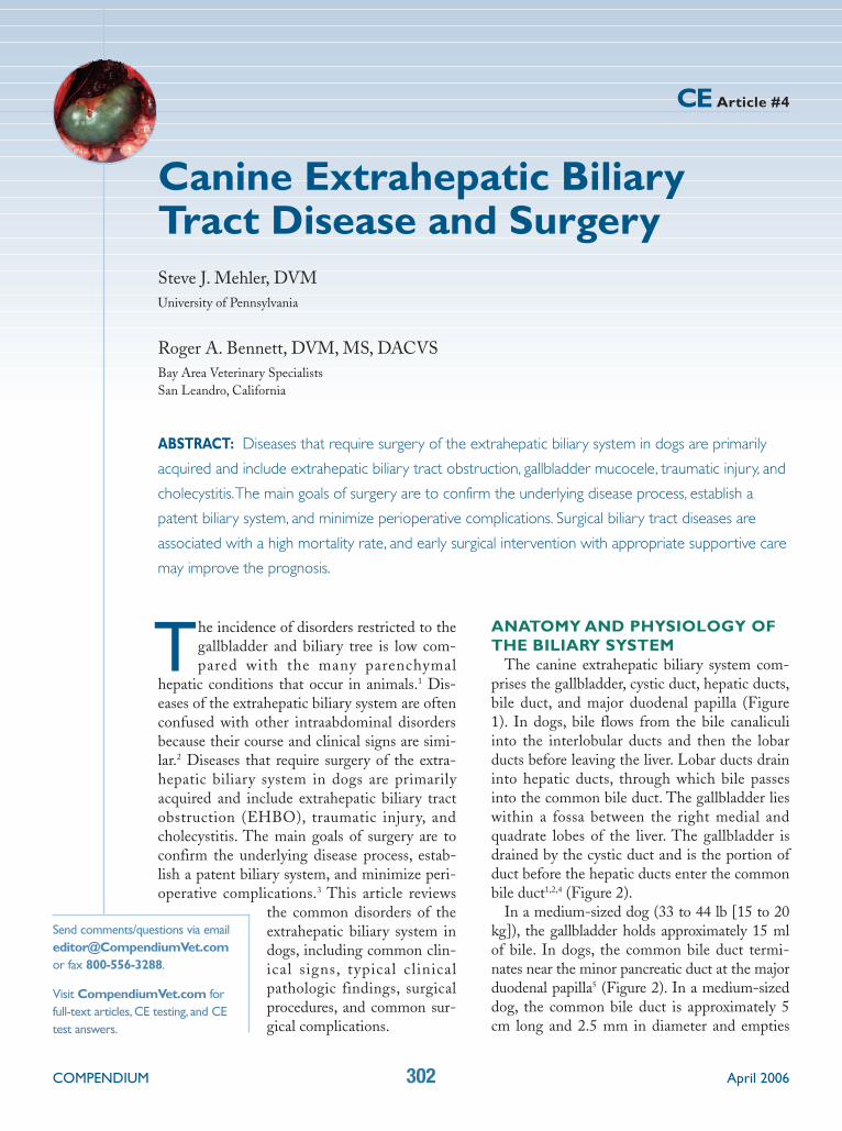
he incidence of disorders restricted to the gallbladder and biliary tree is low com- pared with the many parenchymal hepatic conditions that occur in animals. 1 Dis- eases of the extrahepatic biliary system are often confused with other intraabdominal disorders because their course and clinical signs are simi- lar. 2 Diseases that require surgery of the extra- hepatic biliary system in dogs are primarily acquired and include extrahepatic biliary tract obstruction (EHBO), traumatic injury, and cholecystitis. The main goals of surgery are to confirm the underlying disease process, estab- lish a patent biliary system, and minimize peri- operative complications. 3 This article reviews the common disorders of the extrahepatic biliary system in dogs, including common clin- ical signs, typical clinical pathologic findings, surgical procedures, and common sur- gical complications. ANATOMY AND PHYSIOLOGY OF THE BILIARY SYSTEM The canine extrahepatic biliary system com- prises the gallbladder, cystic duct, hepatic ducts, bile duct, and major duodenal papilla (Figure 1). In dogs, bile flows from the bile canaliculi into the interlobular ducts and then the lobar ducts before leaving the liver. Lobar ducts drain into hepatic ducts, through which bile passes into the common bile duct. The gallbladder lies within a fossa between the right medial and quadrate lobes of the liver. The gallbladder is drained by the cystic duct and is the portion of duct before the hepatic ducts enter the common bile duct 1,2,4 (Figure 2). In a medium-sized dog (33 to 44 lb [15 to 20 kg]), the gallbladder holds approximately 15 ml of bile. In dogs, the common bile duct termi- nates near the minor pancreatic duct at the major duodenal papilla 5 (Figure 2). In a medium-sized dog, the common bile duct is approximately 5 cm long and 2.5 mm in diameter and empties COMPENDIUM 302 April 2006 Canine Extrahepatic Biliary Tract Disease and Surgery Steve J. Mehler, DVM University of Pennsylvania Roger A. Bennett, DVM, MS, DACVS Bay Area Veterinary Specialists San Leandro, California T ABSTRACT: Diseases that require surgery of the extrahepatic biliary system in dogs are primarily acquired and include extrahepatic biliary tract obstruction, gallbladder mucocele, traumatic injury, and cholecystitis.The main goals of surgery are to confirm the underlying disease process, establish a patent biliary system, and minimize perioperative complications. Surgical biliary tract diseases are associated with a high mortality rate, and early surgical intervention with appropriate supportive care may improve the prognosis. Article # 4 CE Send comments/questions via email [email protected] or fax 800-556-3288. Visit CompendiumVet.com for full-text articles, CE testing, and CE test answers.
Transcript of Canine Extrahepatic Biliary Tract Disease and...
he incidence of disorders restricted to thegallbladder and biliary tree is low com-pared with the many parenchymal
hepatic conditions that occur in animals.1 Dis-eases of the extrahepatic biliary system are oftenconfused with other intraabdominal disordersbecause their course and clinical signs are simi-lar.2 Diseases that require surgery of the extra-hepatic biliary system in dogs are primarilyacquired and include extrahepatic biliary tractobstruction (EHBO), traumatic injury, andcholecystitis. The main goals of surgery are toconfirm the underlying disease process, estab-lish a patent biliary system, and minimize peri-operative complications.3 This article reviews
the common disorders of theextrahepatic biliary system indogs, including common clin-ical signs, typical c linicalpathologic findings, surgicalprocedures, and common sur-gical complications.
ANATOMY AND PHYSIOLOGY OFTHE BILIARY SYSTEM
The canine extrahepatic biliary system com-prises the gallbladder, cystic duct, hepatic ducts,bile duct, and major duodenal papilla (Figure1). In dogs, bile flows from the bile canaliculiinto the interlobular ducts and then the lobarducts before leaving the liver. Lobar ducts draininto hepatic ducts, through which bile passesinto the common bile duct. The gallbladder lieswithin a fossa between the right medial andquadrate lobes of the liver. The gallbladder isdrained by the cystic duct and is the portion ofduct before the hepatic ducts enter the commonbile duct1,2,4 (Figure 2).
In a medium-sized dog (33 to 44 lb [15 to 20kg]), the gallbladder holds approximately 15 mlof bile. In dogs, the common bile duct termi-nates near the minor pancreatic duct at the majorduodenal papilla5 (Figure 2). In a medium-sizeddog, the common bile duct is approximately 5cm long and 2.5 mm in diameter and empties
COMPENDIUM 302 April 2006
Canine Extrahepatic BiliaryTract Disease and SurgerySteve J. Mehler, DVMUniversity of Pennsylvania
Roger A. Bennett, DVM, MS, DACVSBay Area Veterinary SpecialistsSan Leandro, California
T
ABSTRACT: Diseases that require surgery of the extrahepatic biliary system in dogs are primarily
acquired and include extrahepatic biliary tract obstruction, gallbladder mucocele, traumatic injury, and
cholecystitis.The main goals of surgery are to confirm the underlying disease process, establish a
patent biliary system, and minimize perioperative complications. Surgical biliary tract diseases are
associated with a high mortality rate, and early surgical intervention with appropriate supportive care
may improve the prognosis.
Article #4CE
Send comments/questions via [email protected] fax 800-556-3288.
Visit CompendiumVet.com for full-text articles, CE testing, and CE test answers.

April 2006 COMPENDIUM
Canine Extrahepatic Biliary Tract Disease and Surgery 303CE
into the duodenum 2.5to 6 cm distal to thepylorus at the majorduodenal papilla aftercoursing intramurallyfor approximately 2cm.1,2 The blood sup-ply to the gallbladderand common bile ductis derived from the leftbranch of the properhepatic artery.
In dogs, the pH ofbile is usually greaterthan 6.2 Bile is acidi-fied by secretion ofhydrogen ions (H+)and not by reabsorp-tion of bicarbonateions (HCO3
-). Morethan 90% of biliarysolids are composed ofbile acids. Bile salts arekept in solution byincorporation intomicelles. Taurocholateand taurocholic acidare the primary saltand acid, respectively,in bile. Bile acids aresynthesized from cho-lesterol, conjugated inhepatocytes, and secreted continuously into bile canali-culi.6 Bile salts are formed within hepatocytes andsecreted as sodium ion (Na+) and potassium ion (K+) saltsof bile acids. They are secreted into the duodenum, reab-sorbed in the ileum, and then carried back to the liver forreexcretion. Ninety percent of bile is recirculated bilesalts.2 Bilirubin is the major bile pigment and a productof hemoprotein catabolism.6 The gallbladder reabsorbsmore than 50% of the calcium in bile. This maintainsfree calcium ions (Ca++) at a low concentration in bile,which is thought to protect against cholelith formation.1
Filling of the gallbladder with bile occurs continu-ously through hepatic secretion and passive gallbladderdistention. This is a low-flow, low-pressure system. Thesphincter of Oddi is the functional sphincter located atthe terminal portion of the common bile duct.1 Rhyth-mic contractions of the sphincter regulate duodenal bile
flow into spurts rather than continuous flow. Thesphincter functions as a one-way valve that can regulatepressure within the biliary tract and also provides resis-tance to the retrograde passage of duodenal contents orpancreatic secretions into the biliary tree.
Cholecystokinin is a hormone produced and secretedby the duodenal mucosa. It is the principal hormoneresponsible for stimulation of gallbladder contraction.Cholecystectomy, ileal resection, and a rise in cholestyr-amine increase the release of cholecystokinin.1,2,6 Motilinand the cholinergic pathway also stimulate gallbladdercontraction and bile flow.
Unconjugated bile acids are cytotoxic and induce tis-sue inflammation. They alter the permeability of vascu-lar structures within the peritoneum, resulting intransudation of fluid and then transmural migration ofenteric organisms into the peritoneal cavity. Althoughvirtually all bile acids derived from the biliary tree areconjugated, a bacterial infection or low pH within thebiliary tree can result in bile acid deconjugation.1,7
Bile salts enhance absorption of fat-soluble vitamins(i.e., vitamins A, D, E, and K). Decreased production,
Figure 2. Schematic drawing of the canine biliarysystem, including the gallbladder, hepatic ducts, cysticduct, common bile duct, and pancreatic ducts.
Figure 1. Anatomy of theextrahepatic biliary systemshowing the cystic duct (arrow),common bile duct (double-headed arrow), hepatic ducts(arrowheads), duodenum (D),and gallbladder (GB). (Courtesyof Dr. Phil Mayhew, University ofPennsylvania)
Illus
trat
ion
by F
elec
ia P
aras
GB
D
COMPENDIUM April 2006
Canine Extrahepatic Biliary Tract Disease and Surgery304 CE
inactivation of bile salts, or biliary obstruction can con-tribute to a clinically important decrease in vitaminK–dependent coagulation factors.2,6
CLINICAL SIGNS OF EXTRAHEPATICBILIARY DISEASE
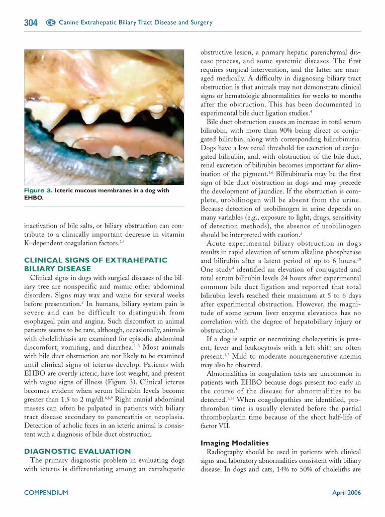
Clinical signs in dogs with surgical diseases of the bil-iary tree are nonspecific and mimic other abdominaldisorders. Signs may wax and wane for several weeksbefore presentation.2 In humans, biliary system pain issevere and can be difficult to distinguish fromesophageal pain and angina. Such discomfort in animalpatients seems to be rare, although, occasionally, animalswith cholelithiasis are examined for episodic abdominaldiscomfort, vomiting, and diarrhea.1–3 Most animalswith bile duct obstruction are not likely to be examineduntil clinical signs of icterus develop. Patients withEHBO are overtly icteric, have lost weight, and presentwith vague signs of illness (Figure 3). Clinical icterusbecomes evident when serum bilirubin levels becomegreater than 1.5 to 2 mg/dl.6,8,9 Right cranial abdominalmasses can often be palpated in patients with biliarytract disease secondary to pancreatitis or neoplasia.Detection of acholic feces in an icteric animal is consis-tent with a diagnosis of bile duct obstruction.
DIAGNOSTIC EVALUATIONThe primary diagnostic problem in evaluating dogs
with icterus is differentiating among an extrahepatic
obstructive lesion, a primary hepatic parenchymal dis-ease process, and some systemic diseases. The firstrequires surgical intervention, and the latter are man-aged medically. A difficulty in diagnosing biliary tractobstruction is that animals may not demonstrate clinicalsigns or hematologic abnormalities for weeks to monthsafter the obstruction. This has been documented inexperimental bile duct ligation studies.4
Bile duct obstruction causes an increase in total serumbilirubin, with more than 90% being direct or conju-gated bilirubin, along with corresponding bilirubinuria.Dogs have a low renal threshold for excretion of conju-gated bilirubin, and, with obstruction of the bile duct,renal excretion of bilirubin becomes important for elim-ination of the pigment.1,6 Bilirubinuria may be the firstsign of bile duct obstruction in dogs and may precedethe development of jaundice. If the obstruction is com-plete, urobilinogen will be absent from the urine.Because detection of urobilinogen in urine depends onmany variables (e.g., exposure to light, drugs, sensitivityof detection methods), the absence of urobilinogenshould be interpreted with caution.2
Acute experimental biliary obstruction in dogsresults in rapid elevation of serum alkaline phosphataseand bilirubin after a latent period of up to 6 hours.10
One study4 identified an elevation of conjugated andtotal serum bilirubin levels 24 hours after experimentalcommon bile duct ligation and reported that totalbilirubin levels reached their maximum at 5 to 6 daysafter experimental obstruction. However, the magni-tude of some serum liver enzyme elevations has nocorrelation with the degree of hepatobiliary injury orobstruction.1
If a dog is septic or necrotizing cholecystitis is pres-ent, fever and leukocytosis with a left shift are oftenpresent.1,2 Mild to moderate nonregenerative anemiamay also be observed.
Abnormalities in coagulation tests are uncommon inpatients with EHBO because dogs present too early inthe course of the disease for abnormalities to bedetected.1,11 When coagulopathies are identified, pro-thrombin time is usually elevated before the partialthromboplastin time because of the short half-life offactor VII.
Imaging ModalitiesRadiography should be used in patients with clinical
signs and laboratory abnormalities consistent with biliarydisease. In dogs and cats, 14% to 50% of choleliths are
Figure 3. Icteric mucous membranes in a dog withEHBO.
April 2006 COMPENDIUM
Canine Extrahepatic Biliary Tract Disease and Surgery 305CE
mineralized and, therefore, radiopaque.1,2,7,12,13 Gas withinthe biliary structures is likely due to emphysematouscholecystitis, cholangitis, choledochitis, or an abscess andwarrants prompt surgical and antimicrobial therapy.12,14
Abdominal ultrasonography is currently the most use-ful and practical technique for demonstrating gallblad-der and common bile duct dilation associated withobstruction in small animals. Ultrasonography is a sensi-tive and specific indicator of the cause of EHBO.15
Ultrasonographic findings of bile duct distention sec-ondary to obstruction may be identified in 62% to 100%of cases involving humans and dogs.13,16,17 Gallstones areinfrequently radiopaque but are readily identified withabdominal ultrasonography. The degree of biliary tractdilation in obstructed dogs and humans is variable.Therefore, duct size allows only a crude estimation ofthe duration of obstruction.4,9 It is important to notethat biliary obstruction can be diagnosed before theonset of clinical icterus with the use of abdominal ultra-sonography. Minimal intrahepatic ductule distention is asubtle abnormality but can be identified via ultrasonog-raphy as early as 4 hours after experimental biliaryocclusion; however, the absence of gallbladder dilationdoes not exclude EHBO because the gallbladder may becontracted because of inflammation.4,10
Oral, intravenous, and cholangiographic contrast stud-ies can be conducted. High serum bilirubin concentra-tions, hypoalbuminemia, icterus, hepatocellular disease,pancreatitis, peritonitis, biliary obstruction, cholecystitis,or concurrent sulfonamide and salicylate administrationmay cause decreased hepatic concentration of the con-trast, resulting in poor opacification of the extrahepaticbiliary system.3,9,12,13 Percutaneous transhepatic cholan-giography, endoscopic retrograde cholangiopancreatogra-phy, and cholecystocentesis are reliable techniques fordiagnosing EHBO in humans but are not routinely usedin dogs.1–4,9,12
Hepatobiliary scintigraphy can be a valuable diagnos-tic tool in differentiating EHBO from hepatocellulardisease.9 99mTc-DISHDA is used as the radio nucleo-tide, and high serum bilirubin concentrations at thetime of hepatobiliary scintigraphy do not interfere withthe diagnostic usefulness of the findings.
In humans, severe complications from vagal stimula-tion, which have progressed to cardiac arrest, have beendescribed in patients with acute cholecystitis undergoingvigorous diagnostic or therapeutic manipulation of thegallbladder. Similar problems have been observed indogs undergoing simple gallbladder aspiration.1,17,18
MicrobiologyPartial or complete EHBO can lead to bile stasis and
promote aerobic and anaerobic bacterial growth. Bile isthought to be sterile in normal dogs and cats.19,20 In hu-mans, infections of the biliary tree usually begin in thegallbladder and spread into the biliary system rather thanascend from the gut.1 In one study21 of dogs with bile peri-tonitis, 10 of 17 had positive cultures for bacteria in theirabdominal effusion, and multiple organisms were isolatedin eight of 10. The most common isolate was Escherichiacoli, followed by Enterococcus, Enterobacter, Klebsiella, Strep-tococcus, Pseudomonas, Bacteroides, and Acinetobacter spp. Inanother study16 of nine dogs with EHBO, six of nine cul-tures were positive for aerobic bacteria, which included E.coli as well as Enterobacter, Enterococcus, Staphylococcus,Micrococcus, and Streptococcus spp. In four of six dogs, amixed population of bacteria was isolated.
In one report7 of 29 dogs with cholelithiasis, 20 dogshad cultures of a cholelith or bile and 14 of 20 had posi-tive growth. The most common organisms grown were E.coli as well as Streptococcus, Enterococcus, and Klebsiella spp.
EXTRAHEPATIC CAUSES OF BILIARYTRACT OBSTRUCTION
The incidence of biliary tract obstruction is much lowerin dogs than in humans because intraluminal extrahepaticobstruction due to cholelithiasis is uncommon indogs.3,4,19 A number of other disorders cause EHBO indogs; mechanical obstructions of the duct due to neoplas-tic diseases of the liver, gallbladder, bile ducts, pancreas,gastrointestinal (GI) tract, or lymphatic tissue are com-mon.1,3–5,22 Abscesses, granulomas, and fibrosis secondaryto trauma or, more commonly, inflammation from pan-creatitis can also cause extraluminal obstruction.4,5,22,23
Pancreatic disease is the most common cause ofEHBO in dogs.5 Scar tissue can form in or around thecommon bile duct, or the duct can be compressed byfibrotic or inflamed pancreatic tissue, pancreatic cysts, orpancreatic abscesses. In a study24 evaluating dogs under-going surgery of the extrahepatic biliary tract, 17 of 60cases were caused by EHBO. Five of these were due toneoplasia, and 12 were secondary to pancreatitis.
Intraluminal obstruction is less common but may becaused by cholelithiasis, choledocholithiasis, or inspis-sated bile.3,5 It has been reported that biliary obstructionlasting over 6 weeks can lead to biliary cirrhosis.1 In thepresence of complete bile duct obstruction, bile maybecome colorless (white bile) because of reduced secre-tion of bilirubin and increased production of mucin.1,13,25

COMPENDIUM April 2006
Canine Extrahepatic Biliary Tract Disease and Surgery306 CE
CholelithiasisUntil recently, cholelithiasis has been considered an
uncommon disease in dogs and cats (Figure 4). It hasbeen reported2,6,26 that dogs with cholelithiasis andcholedocholithiasis account for less than 1% of patientswith liver disease. Cholelithiasis is a condition of senior,female small-breed dogs and is often an incidental find-ing at necropsy or via radiology.7 As a clinical problem,cholelithiasis has a high incidence in miniature schnau-zers and miniature poodles.4 Although most choledo-choliths are believed to form in the gallbladder, someform in the ducts27,28 (Figure 5). Calcium salts are themajor component of pigmented gallstones, and theavailability of ionized calcium may be important in gall-stone formation in dogs.5
It is believed that choleliths are typically clinicallysilent because they are common, incidental postmortemfindings.1,5–7,26 As many as 75% of reported cases ofcholelithiasis have been diagnosed at necropsy with noreported association with clinical signs.26 Clinical signsassociated with cholelithiasis are thought to be morecommonly related to cholecystitis. The prevalence ofcholelithiasis causing cholecystitis is estimated to be lessthan 1% of dogs with biliary disease.13 Choleliths havebeen associated with vomiting, anorexia, icterus, fever,and abdominal pain.1,2,5–7,13,25
Gallstones are readily diagnosed using abdominalultrasonography.1,7 There is great variation in the size,shape, number, and composition of choleliths. The com-position is cholesterol; a combination of bilirubin, cal-
cium carbonate, calcium phosphate, and glycoproteins;or both.2,7,13,25 Cholesterol stones are large, white, andlightweight and occur in animals on high-cholesteroldiets.26 Canine cholelithiasis may be rare because ofdecreased concentrations of cholesterol in canine bilecompared with those in human bile. Pigment stones areyellow, dark brown, or black stones that vary in weightand fragility and are more common in dogs. The causeof pigment stone formation is most likely related toinfectious cholecystitis. The mechanism of formation isthought to be similar to that of urinary calculi. Solidparticles of dead cells or inspissated material act as anidus for crystallization.1,7,25,26 In addition, some con-stituents of bile are resorbed during biliary stasis moreeasily than others, leaving highly desiccated residueswithin the gallbladder.
Pancreatic DiseasesMortality rates in dogs with pancreatitis that under-
went biliary tract surgery were 50% to 100%.2,24 Earlydetection and aggressive management of pancreaticnecrosis or abscessation was shown to improve survival(Figure 6).
There have been a few reports29–32 of pancreatic pseudo-cysts causing EHBO in dogs. These pseudocysts form asa result of pancreatic duct disruption from acute orchronic pancreatitis or trauma (Figure 7). CT has beenused in differentiating between pancreatic pseudocystsand pancreatic abscesses, although ultrasonography hasalso been useful in diagnosing and clinically monitoring
Figure 4. Removal of a large cholelith from thegallbladder of a dog.
Figure 5. Removal of a choledocholith from acholedochotomy incision.

April 2006 COMPENDIUM
Canine Extrahepatic Biliary Tract Disease and Surgery 307CE
pancreatic pseudocysts.29,33 Treatment involves drainingthe pseudocyst into the GI tract and omentalization ofthe pseudocyst. Other conditions associated with thepancreas that cause EHBO are pancreatic neoplasia,abscesses, cysts, and granulomas. These causes are lesscommon than obstruction caused by chronic pancreatitis.
Neoplastic DiseasesPrimary neoplasms of the gallbladder and common
bile duct are rare in domestic animals, comprising 2% ofall canine hepatic tumors.2,22,25 Up to 20% of animalswith EHBO have neoplasia of the pancreas or the bileduct.22 Adenomas of the gallbladder are benign localizedtumors originating from the glandular epithelium. Thetumor projects into the lumen and does not infiltratethe deeper parts of the gallbladder wall.25 Adenocarcino-mas of the gallbladder invade the wall, form incompletetubules and undifferentiated cells, undergo necrosis, andmay extend into the liver and adjacent peritoneum.Metastasis to the lungs and perineural lymphatics hasbeen reported.
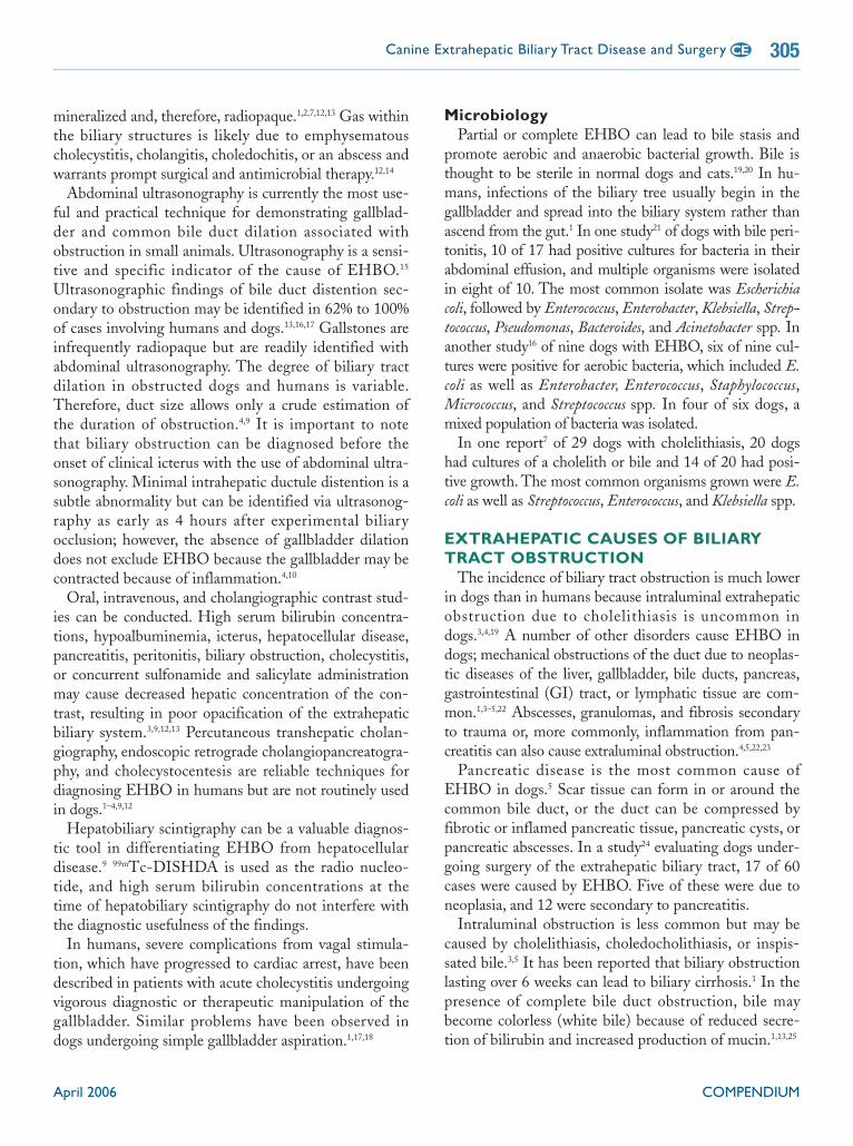
MucoceleA biliary mucocele is distention of a bile-containing
structure or cavity due to inappropriate accumulation ofmucus2,16,34–37 (Figure 8). Biliary mucoceles occur in dogsolder than 6 years of age and have no breed or sexpredilection.2 EHBO does not appear to play a primaryrole in gallbladder mucocele formation, and no inflam-matory or infectious cause has been associated with gall-bladder mucoceles.36 Biliary stasis, decreased gallbladder
motility, and altered absorption of water from the gall-bladder lumen predispose animals to biliary sludge for-mation. Biliary sludge may be a precipitating factor forthe development of canine biliary mucoceles.
There are reports16,36,37 of a 50% to 60% incidence ofgallbladder rupture in patients with biliary mucocelesand a 13% to 80% incidence of a histopathologic diag-nosis of gallbladder wall necrosis. Ruptured mucocelescontain semisolidified bile and leak slowly, causing acutebut local chemical, sterile peritonitis. In some dogs withruptured mucoceles, the solidified mucocele can befound intact, floating freely in the abdomen.
In most dogs with gallbladder mucoceles, the diseaseis contained within the gallbladder and does not involvethe hepatic ducts or the common bile duct. In somecases, gelatinous biliary material is present within thecommon bile duct and is easily dislodged or flushedthrough the duodenal papilla (Figure 9). Dogs with bil-iary mucoceles that undergo cholecystectomy and sur-vive the immediate perioperative period have anexcellent prognosis.36,37
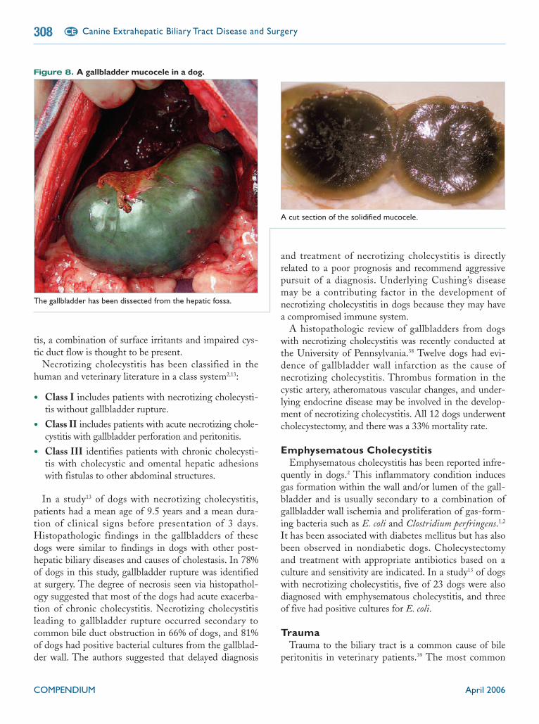
Necrotizing CholecystitisNecrotizing cholecystitis has reportedly been a com-
mon cause of gallbladder rupture in dogs.21 Impairmentof the cystic arterial circulation by occlusion, bacterialinfection, or cystic duct obstruction from choleliths,neoplasia, or an adjacent inflammatory process can leadto cholecystitis.1 In most cases of necrotizing cholecysti-
Figure 6. Necrotizing pancreatitis in a dog. The arrow ispointing at the duodenum.
Figure 7. Pancreatic pseudocyst causing partial EHBOin a dog.
COMPENDIUM April 2006
Canine Extrahepatic Biliary Tract Disease and Surgery308 CE
tis, a combination of surface irritants and impaired cys-tic duct flow is thought to be present.
Necrotizing cholecystitis has been classified in thehuman and veterinary literature in a class system2,13:
• Class I includes patients with necrotizing cholecysti-tis without gallbladder rupture.
• Class II includes patients with acute necrotizing chole-cystitis with gallbladder perforation and peritonitis.
• Class III identifies patients with chronic cholecysti-tis with cholecystic and omental hepatic adhesionswith fistulas to other abdominal structures.
In a study13 of dogs with necrotizing cholecystitis,patients had a mean age of 9.5 years and a mean dura-tion of clinical signs before presentation of 3 days.Histopathologic findings in the gallbladders of thesedogs were similar to findings in dogs with other post-hepatic biliary diseases and causes of cholestasis. In 78%of dogs in this study, gallbladder rupture was identifiedat surgery. The degree of necrosis seen via histopathol-ogy suggested that most of the dogs had acute exacerba-tion of chronic cholecystitis. Necrotizing cholecystitisleading to gallbladder rupture occurred secondary tocommon bile duct obstruction in 66% of dogs, and 81%of dogs had positive bacterial cultures from the gallblad-der wall. The authors suggested that delayed diagnosis
and treatment of necrotizing cholecystitis is directlyrelated to a poor prognosis and recommend aggressivepursuit of a diagnosis. Underlying Cushing’s diseasemay be a contributing factor in the development ofnecrotizing cholecystitis in dogs because they may havea compromised immune system.
A histopathologic review of gallbladders from dogswith necrotizing cholecystitis was recently conducted atthe University of Pennsylvania.38 Twelve dogs had evi-dence of gallbladder wall infarction as the cause ofnecrotizing cholecystitis. Thrombus formation in thecystic artery, atheromatous vascular changes, and under-lying endocrine disease may be involved in the develop-ment of necrotizing cholecystitis. All 12 dogs underwentcholecystectomy, and there was a 33% mortality rate.
Emphysematous CholecystitisEmphysematous cholecystitis has been reported infre-
quently in dogs.2 This inflammatory condition inducesgas formation within the wall and/or lumen of the gall-bladder and is usually secondary to a combination ofgallbladder wall ischemia and proliferation of gas-form-ing bacteria such as E. coli and Clostridium perfringens.1,2
It has been associated with diabetes mellitus but has alsobeen observed in nondiabetic dogs. Cholecystectomyand treatment with appropriate antibiotics based on aculture and sensitivity are indicated. In a study13 of dogswith necrotizing cholecystitis, five of 23 dogs were alsodiagnosed with emphysematous cholecystitis, and threeof five had positive cultures for E. coli.
TraumaTrauma to the biliary tract is a common cause of bile
peritonitis in veterinary patients.39 The most common
Figure 8. A gallbladder mucocele in a dog.
The gallbladder has been dissected from the hepatic fossa.
A cut section of the solidified mucocele.

April 2006 COMPENDIUM
Canine Extrahepatic Biliary Tract Disease and Surgery 309CE
traumatic cause is being hit by a car, but gunshot wounds,bite wounds, and other penetrating abdominal woundshave also been reported as causes.22,39 Coexisting injuriesinvolving other major abdominal organs usually do notoccur. Blunt trauma causes rupture of the common bileduct more frequently than rupture of the gallbladder.Rupture has also been reported in the distal common bileduct, the cystic duct, and hepatic ducts. The most com-mon site of common bile duct rupture is just distal to theentrance of the last hepatic duct.2,5,40,41 The pathogenesisof bile duct rupture from trauma includes the short cysticduct, rapid gallbladder emptying, and a simultaneousshearing force applied to the duct.
Recognition of biliary tract trauma is most oftendelayed, and clinical signs result from ensuing bile peri-tonitis.22 Bile peritonitis is usually the cause of death inpatients with traumatic extrahepatic biliary tract rupture.
Bile PeritonitisBile peritonitis, or bilious ascites, is the inflammatory
response of the peritoneum to the presence of bile. Bileperitonitis is caused by rupture of the extrahepatic bil-iary system. The poorly vascularized fundus is the areamost susceptible to rupture in the canine gallblad-der.7,41–43 Inspissated bile may cause focal or localizedperitonitis rather than generalized bile peritonitis. If thebilirubin concentration of the abdominal effusion isgreater than twice that of the serum concentration, it isdiagnostic of bile peritonitis.21 There is a 27% to 45%survival rate for dogs with septic bile peritonitis and an87% to 100% survival rate for dogs with sterile bile peri-tonitis.21,24 Humans with sterile biliary effusion mayhave vague symptoms that last for an average of 30 daysbefore surgical treatment and have a mortality rate ofless than 10%. Humans with septic bile peritonitis havean acute presentation with abdominal pain and shockand a mortality rate of greater than 20%.44
Clinical signs associated with bile peritonitis includevomiting, anorexia, diarrhea, weight loss, icterus,abdominal distention, fever, and abdominal pain. Theonset of clinical signs in dogs with a ruptured biliarytract and the degree of peritonitis present depend on thevolume of liquid bile and the concentration of bile salts.Many studies1,2,13,21,45 have identified a prolonged period(i.e., 3 to 30 days) from rupture to presentation for bileperitonitis. Previous studies13,45 have reported that a pro-longed clinical course and delayed treatment of bileperitonitis lead to a higher mortality rate. Other stud-ies7,21 have contradicted this finding.
Bile acids are toxic to tissues and cause permeabilitychanges and tissue necrosis, which encourage bacterialgrowth. Sources of bacteria are thought to be endoge-nous anaerobic bacteria from the liver and intestine aswell as hematogenous bacteria. The toxicity of bile acidsdepends not only on the amount of bile in the peritonealcavity but also on the concentration of bile acids in thebile.22 Bile acids cause erythrocyte lysis, electrolyteimbalance, hypoproteinemia, anemia, and marked dehy-dration due to the alkaline pH and hyperosmolality ofbile acids. Bile peritonitis is reportedly more lethal indogs than in humans because of the higher content oftaurocholic acid in canine bile. Taurocholic acid is con-sidered much more toxic than the glycocholic acid com-ponent that predominates in human bile.
Survivors of biliary surgery for bile peritonitis have asignificantly lower leukocyte count (mean: 20,608/µl)compared with nonsurvivors (mean: 35,712/µl). Thesame trend was observed in dogs with circulating bands(survivors had a mean count of 686/µl, and nonsurvivorshad a mean of 4,852/µl).21
Figure 9. Gelatinous biliary material within thecommon bile duct of a dog at necropsy. The dog also had agallbladder mucocele.
COMPENDIUM April 2006
Canine Extrahepatic Biliary Tract Disease and Surgery310 CE
SURGERYThree surgical approaches to the extrahepatic biliary
system of dogs have been described. Ventral midlineceliotomy is most commonly performed. This approachcan be extended through the sternebrae or paracostallyon the right side. A third approach involves a thoraco-tomy through the right seventh or eighth intercostalspace.46,47 In many cases of biliary obstruction secondaryto hepatic neoplasm, it is necessary not only to use a bilererouting procedure but also to remove some of the liver.It has been reported48 that up to 40% of the liver can beresected without major complications in dogs withobstructive jaundice.
Many animals requiring surgery of the extrahepaticbiliary tract are nutritionally compromised. The poten-tial for long hospital stays, concurrent or developingpancreatitis, and underlying systemic disease frequentlynecessitates the placement of distal duodenal or jejunalfeeding tubes during laparotomy.
GallbladderCholecystectomy is preferred over cholecystotomy for
stone removal because cholecystectomy removes thereservoir for subsequent stone accumulation and mayhave a lower morbidity and mortality rate than doescholecystotomy.1,6,7 The gallbladder wall does not sealwell immediately after cholecystocentesis or cholecysto-tomy. A double-layer closure and/or placing an omentalpatch over the incision in the gallbladder is typicallyindicated. Regardless of underlying disease requiringcholecystectomy, it is important to evaluate and flush the
bile ducts to remove stones, debris, or sludge that maylead to reobstruction of the extrahepatic biliary tract.
DuctsPrimary repair of ruptured hepatic ducts, the cystic
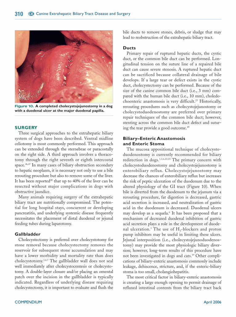
duct, or the common bile duct can be performed. Lon-gitudinal tension on the suture line of a repaired bileduct can cause severe stenosis. A ruptured hepatic ductcan be sacrificed because collateral drainage of biledevelops. If a large tear or defect exists in the cysticduct, cholecystectomy can be performed. Because of thesize of the canine common bile duct (i.e., 3 mm) com-pared with the human bile duct (i.e., 10 mm), choledo-choenteric anastomosis is very difficult.23 Historically,rerouting procedures such as cholecystojejunostomy orcholecystoduodenostomy are preferred over primaryrepair techniques of the common bile duct; however,stenting across the common bile duct defect and sutur-ing the tear provide a good outcome.49
Biliary–Enteric Anastomosis and Enteric Stoma
The mucosa appositional technique of cholecysto-duodenostomy is currently recommended for biliaryredirection in dogs.1,3,6,22,23 The primary concern withcholecystoduodenostomy and cholecystojejunostomy isenterobiliary reflux. Cholecystojejunostomy maydecrease the chances of enterobiliary reflux but increasesthe risk of peptic ulceration of the duodenum due to thealtered physiology of the GI tract (Figure 10). Whenbile is diverted from the duodenum to the jejunum via arerouting procedure, fat digestion is decreased, gastricacid secretion is increased, and neutralization of gastricacid in the duodenum is decreased. Duodenal ulcersmay develop as a sequela.6 It has been proposed that amechanism of decreased duodenal inhibition of gastricacid secretion plays a role in the development of duode-nal ulceration.1 The use of H2-blockers and protonpump inhibitors may be useful in limiting these ulcers.Jejunal interposition (i.e., cholecystojejunoduodenos-tomy) may provide the most physiologic biliary diver-sion; however, long-term results of this procedure havenot been investigated in dogs and cats.19 Other compli-cations of biliary-enteric anastomosis commonly includeleakage, dehiscence, stricture, and, if the enteric-biliarystoma is too small, cholangiohepatitis.
The most critical factor in biliary-enteric anastomosisis creating a large enough opening to permit drainage ofrefluxed intestinal contents from the biliary tract back
Figure 10. A completed cholecystojejunostomy in a dogwith a duodenal ulcer at the major duodenal papilla.

into the intestine. The cholecystoenterotomy openingshould be 2.5 to 4 cm long to minimize postoperativeproblems such as cholangitis associated with inadequatedraining of refluxed intestinal contents. Clinical mani-festations of cholangitis are rare when an adequatelysized stoma is created.1,3,23 Cholangiohepatitis occurswhen duodenal or jejunal contents are allowed to sit inthe gallbladder for long periods and ascending infectionoccurs. Creating an incision in the gallbladder from thefundus to the beginning of the cystic duct and a corre-sponding incision of equal length in the intestine gener-ally creates a stoma of adequate length. Bacterial growthwithin the biliary tract is expected during the initialpostoperative period; however, clinical cholangitis usu-ally does not develop unless the biliary tree becomesobstructed or the stoma is too small.
A liver biopsy should be performed during surgery forbiliary disease. Histopathologic evaluation of the liverparenchyma may provide useful information regardingthe underlying disease process, its effect on the liver,long-term management of the patient, and prognosis.6
Biliary Stents and TubesBiliary decompression by means of a cholecystostomy
tube may be indicated in critically ill patients until they
are stable enough for a more complicated rerouting pro-cedure.6 It is important to remember that treatment ofbiliary tract sepsis without biliary tract decompression isineffective because no antibiotics achieve adequate levelsin the biliary tree in the presence of EHBO.1
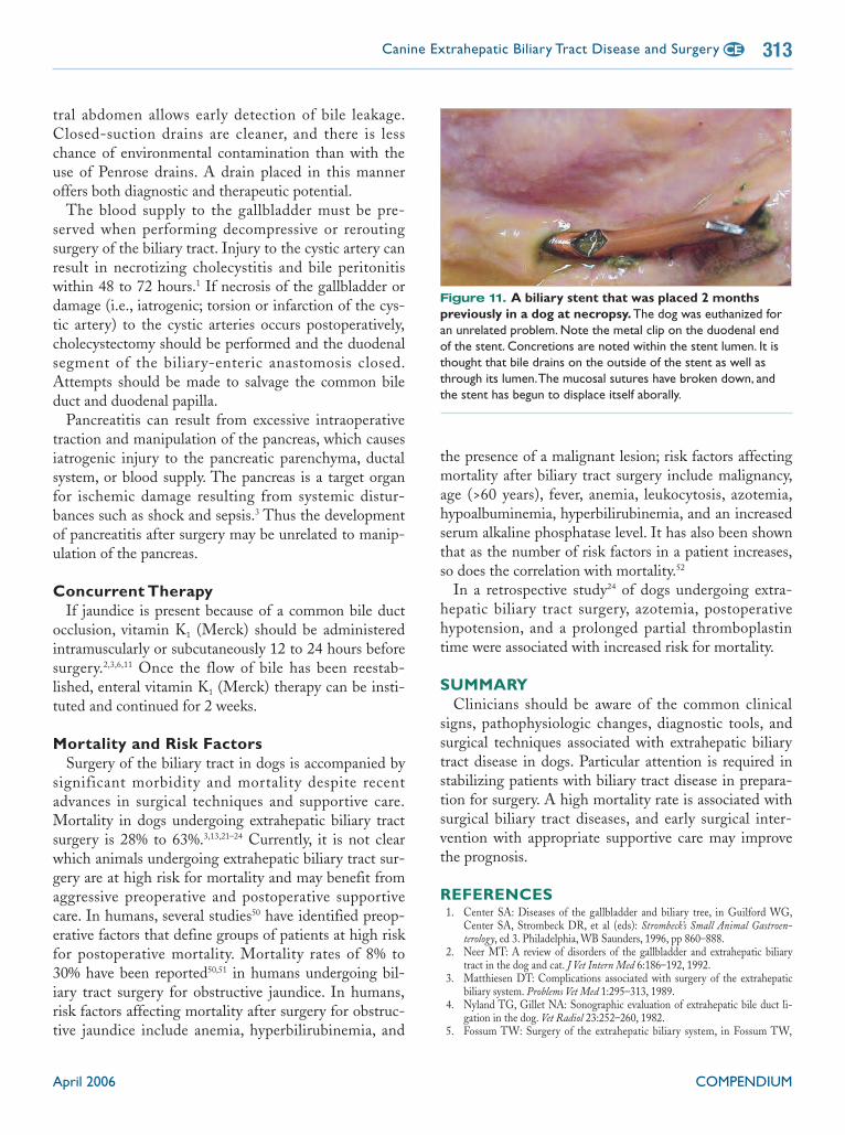
In a recent report,49 eight dogs had a choledochal tubestent placed to relieve obstructions of the common bileduct and duodenal papilla due to extraluminal compres-sion. A duodenotomy was created and the stent (i.e., redrubber catheter) placed up the duodenal papilla and intothe common bile duct. The stent was then sutured inplace to the mucosa of the duodenum with one or twostay sutures of an absorbable material. A hemostatic clipwas placed in the lumen of the duodenal side of thestent to allow radiographic assessment of the position ofthe stent (Figure 11). Theoretically, after the suturebreaks, the stent is passed in the feces. Discharge of thestent in the eight dogs was unpredictable; one dogrequired endoscopic removal of the stent duringendoscopy for small intestinal biopsies. Complicationsof choledochal stenting include obstruction of the stentwith bile concretions and promotion of stricture forma-tion. Stricture formation can occur because the red rub-ber catheters may incite an inflammatory response.Intraluminal tubes may interfere with normal biledrainage, promoting cholangitis.
Perioperative ComplicationsExcessive bleeding can occur following blunt dissec-
tion and retraction of the gallbladder from the hepaticfossa, particularly in dogs with a coagulopathy.3 Assess-ments of coagulation factors and platelet deficiency ordysfunction should be conducted preoperatively. In dogswith hemorrhage from the hepatic fossa, a hemostaticagent can be placed in the fossa or an omental pediclecan be sutured over the area. In dogs with potentialbleeding diathesis, freeing the gallbladder from the fossacan be partially or completely avoided if a duodenal orjejunal loop can be anatomically positioned adjacent tothe gallbladder and biliary-enteric anastomosis can besuccessfully performed with minimal tension on thesutures.
Dehiscence of a biliary-enteric anastomosis can occurwith improper suture placement, ischemic injury to thegallbladder, or excessive tension across the anastomosissite. An early postoperative diagnosis of bile leakage canbe difficult to make because of the insidious onset ofclinical signs associated with bile peritonitis. Placing aclosed-suction drain intraoperatively in the cranioven-
COMPENDIUM April 2006
Canine Extrahepatic Biliary Tract Disease and Surgery312 CE
tral abdomen allows early detection of bile leakage.Closed-suction drains are cleaner, and there is lesschance of environmental contamination than with theuse of Penrose drains. A drain placed in this manneroffers both diagnostic and therapeutic potential.
The blood supply to the gallbladder must be pre-served when performing decompressive or reroutingsurgery of the biliary tract. Injury to the cystic artery canresult in necrotizing cholecystitis and bile peritonitiswithin 48 to 72 hours.1 If necrosis of the gallbladder ordamage (i.e., iatrogenic; torsion or infarction of the cys-tic artery) to the cystic arteries occurs postoperatively,cholecystectomy should be performed and the duodenalsegment of the biliary-enteric anastomosis closed.Attempts should be made to salvage the common bileduct and duodenal papilla.
Pancreatitis can result from excessive intraoperativetraction and manipulation of the pancreas, which causesiatrogenic injury to the pancreatic parenchyma, ductalsystem, or blood supply. The pancreas is a target organfor ischemic damage resulting from systemic distur-bances such as shock and sepsis.3 Thus the developmentof pancreatitis after surgery may be unrelated to manip-ulation of the pancreas.
Concurrent TherapyIf jaundice is present because of a common bile duct
occlusion, vitamin K1 (Merck) should be administeredintramuscularly or subcutaneously 12 to 24 hours beforesurgery.2,3,6,11 Once the flow of bile has been reestab-lished, enteral vitamin K1 (Merck) therapy can be insti-tuted and continued for 2 weeks.
Mortality and Risk FactorsSurgery of the biliary tract in dogs is accompanied by
significant morbidity and mortality despite recentadvances in surgical techniques and supportive care.Mortality in dogs undergoing extrahepatic biliary tractsurgery is 28% to 63%.3,13,21–24 Currently, it is not clearwhich animals undergoing extrahepatic biliary tract sur-gery are at high risk for mortality and may benefit fromaggressive preoperative and postoperative supportivecare. In humans, several studies50 have identified preop-erative factors that define groups of patients at high riskfor postoperative mortality. Mortality rates of 8% to30% have been reported50,51 in humans undergoing bil-iary tract surgery for obstructive jaundice. In humans,risk factors affecting mortality after surgery for obstruc-tive jaundice include anemia, hyperbilirubinemia, and
the presence of a malignant lesion; risk factors affectingmortality after biliary tract surgery include malignancy,age (>60 years), fever, anemia, leukocytosis, azotemia,hypoalbuminemia, hyperbilirubinemia, and an increasedserum alkaline phosphatase level. It has also been shownthat as the number of risk factors in a patient increases,so does the correlation with mortality.52
In a retrospective study24 of dogs undergoing extra-hepatic biliary tract surgery, azotemia, postoperativehypotension, and a prolonged partial thromboplastintime were associated with increased risk for mortality.
SUMMARYClinicians should be aware of the common clinical
signs, pathophysiologic changes, diagnostic tools, andsurgical techniques associated with extrahepatic biliarytract disease in dogs. Particular attention is required instabilizing patients with biliary tract disease in prepara-tion for surgery. A high mortality rate is associated withsurgical biliary tract diseases, and early surgical inter-vention with appropriate supportive care may improvethe prognosis.
REFERENCES1. Center SA: Diseases of the gallbladder and biliary tree, in Guilford WG,
Center SA, Strombeck DR, et al (eds): Strombeck’s Small Animal Gastroen-terology, ed 3. Philadelphia, WB Saunders, 1996, pp 860–888.
2. Neer MT: A review of disorders of the gallbladder and extrahepatic biliarytract in the dog and cat. J Vet Intern Med 6:186–192, 1992.
3. Matthiesen DT: Complications associated with surgery of the extrahepaticbiliary system. Problems Vet Med 1:295–313, 1989.
4. Nyland TG, Gillet NA: Sonographic evaluation of extrahepatic bile duct li-gation in the dog. Vet Radiol 23:252–260, 1982.
5. Fossum TW: Surgery of the extrahepatic biliary system, in Fossum TW,
April 2006 COMPENDIUM
Canine Extrahepatic Biliary Tract Disease and Surgery 313CE
Figure 11. A biliary stent that was placed 2 monthspreviously in a dog at necropsy. The dog was euthanized foran unrelated problem. Note the metal clip on the duodenal endof the stent. Concretions are noted within the stent lumen. It isthought that bile drains on the outside of the stent as well asthrough its lumen.The mucosal sutures have broken down, andthe stent has begun to displace itself aborally.

Hedlund CS, Hulse DA, et al (eds): Small Animal Surgery, ed 2. St. Louis,Mosby, 2002, pp 475–486.
6. Martin RA: Biliary obstruction/stones, in Bojrab MJ (ed): Disease Mechanisms inSmall Animal Surgery, ed 2. Philadelphia, Lea and Febiger, 1993, pp 306–310.
7. Kirpensteijn J, Fingland RB, Ulrich T, et al: Cholelithiasis in dogs: 29 cases.JAVMA 202:1137–1142, 1993.
8. Fossum TW, Willard MD: Diseases of the gallbladder and extrahepatic bil-iary system, in Ettinger SJ, Feldman EC (eds): Textbook of Veterinary InternalMedicine: Diseases of the Dog and Cat, ed 4. Philadelphia, WB Saunders, 1995,pp 1393–1398.
9. Boothe HW, Boothe DM, Komkov A, Hightower D: Use of hepatobiliaryscintigraphy in the diagnosis of extrahepatic biliary obstruction in dogs andcats: 25 cases (1982–1989). JAVMA 201:134–141, 1992.
10. Zeman RK, Taylor KJW, Rosenfield AT, et al: Acute experimental biliaryobstruction in the dog: Sonographic findings and clinical implications. Am JRoentgenol 136:965–967, 1981.
11. Mount ME: Proteins induced by vitamin K absence or antagonists (PIKVA),in Kirk RW (ed): Current Veterinary Therapy IX: Small Animal Practice.Philadelphia, WB Saunders, 1986.
12. Smith SA, Biller DS, Kraft SL, et al: Diagnostic imaging of biliary obstruc-tion. Compend Contin Educ Pract Vet 20:1225–1234, 1998.
13. Church ME, Matthiesen DT: Surgical treatment of 23 dogs with necrotizingcholecystitis. JAAHA 24:305–310, 1993.
14. Partington BP, Biller DS: Hepatic imaging with radiology and ultrasound.Vet Clin North Am Small Anim Pract 25(2):305–335, 1995.
15. Blackbourne LH, Earnhardt RC, Sistrom CL, et al: The sensitivity and roleof ultrasound in the evaluation of biliary obstruction. Am Surg60(9):683–690, 1994.
16. Besso JG, Wrigley RH, Gliatto JM, Webster CRL: Ultrasonographic appear-ance and clinical findings in 14 dogs with gallbladder mucocele. Vet RadiolUltrasound 41:261–271, 2000.
17. Rivers BJ, Walter PA, Johnston GR, et al: Acalculous cholecystitis in fourcanine cases: Ultrasonographic findings and use of ultrasonographic-guided,percutaneous cholecystocentesis in diagnosis. JAAHA 33:207–214, 1997.
18. van Sonnenberg E, Wittich GR, Casola G, et al: Diagnostic and therapeuticpercutaneous gallbladder procedures. Radiology 160:23–26, 1986.
19. Fahie MA, Martin RA: EHBO: A retrospective study of 45 cases(1983–1993). JAAHA 31:478–482, 1995.
20. Musgrove JE, Grindlay JH, Karlsson AG: Intestinal biliary reflux after anas-tomosis of the common bile duct to duodenum or jejunum. Arch Surg64:579–589, 1952.
21. Ludwig LL, McLoughlin MA, Graves TK, Crisp MS: Surgical treatment ofbile peritonitis in 24 dogs and 2 cats: A retrospective study (1987–1994). VetSurg 26:90–98, 1997.
22. Martin RA, MacCoy DM, Harvey HJ: Surgical management of extrahepaticbiliary tract disease: A report of eleven cases. JAAHA 22:301–307, 1986.
23. Matthiesen DT, Rosin E: Common bile duct obstruction secondary tochronic fibrosing pancreatitis: Treatment by use of cholecystoduodenostomyin the dog. JAVMA 189:1443–1446, 1986.
24. Mehler SJ, Mayhew PD, Drobatz K, Holt DE: Variables associated with out-come in dogs undergoing extrahepatic biliary surgery: 60 cases (1988–2002).Vet Surg 33:644–649, 2004.
25. Jones TC, Hunt RD, King NW: Veterinary Pathology, ed 6. Philadelphia, WBSaunders, 1995, pp 1103–1105.
26. Schall WD, Chapman WL, Finco DR, et al: Cholelithiasis in dogs. JAVMA163:469–472, 1973.
27. Way LW, Sleisenger MH: Biliary obstruction, cholangitis, and choledo-cholithiasis, in Gastrointestinal Disease: Pathophysiology, Diagnosis, Manage-ment, ed 4. Philadelphia, WB Saunders, 1989.
28. Cantwell HD, Blevins WE, Hanika-Rebar C, Godshalk CP: Radiopaquehepatic and lobar duct choleliths in a dog. JAAHA 19, 1983.
29. Marchevsky AM, Yovich JC, Wyatt KM: Pancreatic pseudocyst causingextrahepatic biliary obstruction in a dog. Aust Vet J 78:99–101, 2000.
30. Bellenger CR, llkiw JE, Malik R: Cystogastrostomy in the treatment of pan-creatic pseudocyst/abscess in two dogs. Vet Rec 125:181–184, 1989.
31. Rutgers C, Herring DS, Orton EC: Pancreatic pseudocyst associated withacute pancreatitis in a dog: Ultrasonographic diagnosis. JAAHA 21:411–416,1984.
32. Bellenger CR, Farrow BRH, Allen GS, Cooper NA: Pancreatic pseudocyst ina dog. Aust Vet Pract 13:67–68, 1983.
33. Reber HA, Way LW: Pancreas, in Way LW (ed): Current Surgical Diagnosisand Treatment, ed 9. Norwalk, CT, Appleton and Lange, 1991, pp 558–584.
34. Dorland’s Illustrated Medical Dictionary, ed 28. Philadelphia, WB Saunders,1994.
35. Newell SM, Selcer BA, Mahaffey MB, et al: Gallbladder mucocele causingbiliary obstruction in two dogs: Ultrasonographic, scintigraphic, and patho-logical findings. JAAHA 31:467–472, 1995.
36. Pike FS, Berg J, King NW, et al: Gallbladder mucocele in dogs: 30 cases(2000–2002). JAVMA 224:1615–1621, 2004.
37. Worley DR, Hottinger HA, Lawrence HJ: Surgical management of gallblad-der mucoceles in dogs: 22 cases (1999–2003). JAVMA 225:1418–1422, 2004.
38. Holt DE, Mehler SJ, Mayhew PD, Hendrick MJ: Canine gallbladder infarc-tion: 12 cases (1993–2003). Vet Pathol 41:416–418, 2004.
39. Watkins PE, Pearson H, Denny HR: Traumatic rupture of the bile duct inthe dog: A report of seven cases. J Small Anim Pract 24:731–740, 1983.
40. Roslyn J, Busuttil RW: Perforation of the gallbladder: A frequently misman-aged condition. Am J Surg 137:307–312, 1979.
41. Parchman MB, Flanders JA: Extrahepatic biliary tract rupture: Evaluation ofthe relationship between the site of rupture and the cause of rupture in 15dogs. Cornell Vet 80:267–272, 1990.
42. Matthiesen DT, Lammerding J: Gallbladder rupture and bile peritonitis sec-ondary to cholelithiasis and cholecystitis in a dog. JAVMA 184:1282–1283,1984.
43. Harris SJ, Simpson JW, Thoday KL: Obstructive cholelithiasis and gallblad-der rupture in a dog. J Small Anim Pract 25:661–667, 1984.
44. Ibragimov ET, Ordabecov SO, Ongarbaev SZ: Diagnosis and treatment ofpostoperative biliary peritonitis. Khirurgiia (Mosk) 1:86–88, 1992.
45. Felice PR, Trowbridge PE, Terrara JJ: Evolving changes in the pathogenesisand treatment of the perforated gallbladder. Am J Surg 149:466–473, 1985.
46. Blass CE: Surgery of the extrahepatic biliary tract. Compend Contin EducPract Vet 5:801–808, 1983.
47. Rosenthal JJ, Kipnis RM: Cholecystectomy in the dog: A comparison ofapproaches. Vet Med Small Anim Clin 66:351–354, 1971.
48. Mizumoto R, Kawarada Y, Yamawaki T, et al: Resectability and functionalreserve of the liver with obstructive jaundice in dogs. Am J Surg 137:768–772,1979.
49. Mayhew PD, Richardson RW, Mehler SJ, et al: Choledochal tube stentingfor decompression of extrahepatic biliary obstruction in dogs [abstract]. ProcAnnu Meet Am Coll Vet Surg, 2004.
50. Pitt HA, Cameron JL, Postier RG, Gadacz TR: Factors affecting mortality inbiliary tract surgery. Am J Surg 141:66–72, 1981.
51. Dixon JM, Armstrong CP, Duffy SW, Davies GC: Factors affecting morbid-ity and mortality after surgery for obstructive jaundice. Gut 24:845–852, 1983.
52. Pitt HA, Cameron JL, Postier RG, Gadacz TR: Factors affecting mortality inbiliary tract surgery. Am J Surg 141:66–72, 1981.
1. The extrahepatic biliary system does not includethea. gallbladder. d. hepatic ducts.b. interlobular ducts. e. common bile duct.c. cystic duct.
COMPENDIUM April 2006
Canine Extrahepatic Biliary Tract Disease and Surgery314 CE
ARTICLE #4 CE TESTThis article qualifies for 2 contact hours of continuingeducation credit from the Auburn University College ofVeterinary Medicine. Paid subscribers may purchaseindividual CE tests or sign up for our annual CE program.Those who wish to apply this credit to fulfill state relicensurerequirements should consult their respective stateauthorities regarding the applicability of this program.To participate, fill out the test form inserted at the end of this issue or take CE tests online and get real-time scores at CompendiumVet.com.
CE
2. Bile salts enhance absorption of fat-soluble vita-mins, which do not include vitamina. A. d. E.b. B. e. K.c. D.
3. Clinical signs of icterus develop at serum totalbilirubin levels as low as _____ mg/dl.a. 1 to 1.5 d. 8 to 9b. 1.5 to 2 e. 10 to 10.5c. 4 to 4.5
4. Bilirubinuria may be the first sign of bile ductobstruction in dogs and may precede the devel-opment of jaundice because dogs a. have a low renal threshold for excretion of unconju-
gated bilirubin.b. are unable to deposit conjugated bilirubin in their
mucous membranes.c. are predisposed to renal failure from hyperbilirubine-
mia.d. have a low renal threshold for excretion of conju-
gated bilirubin.e. cannot unconjugate bilirubin.
5. Which statement is incorrect?a. Hepatobiliary scintigraphy can be a valuable diagnos-
tic tool in differentiating EHBO from hepatocellulardisease.
b. Biliary obstruction can be diagnosed before the onsetof clinical icterus with the use of abdominal ultra-sonography.
c. Bile is thought to be sterile in normal dogs and cats.d. Cholelithiasis always occurs in a sterile environment.e. Infection of the extrahepatic biliary tract commonly
involves multiple organisms.
6. Reported causes of extraluminal EHBO do notincludea. pancreatitis.b. pancreatic pseudocyst.c. pancreatic tumors.d. portal vein infarction.e. hepatic neoplasia.
7. Which statement regarding cholelithiasis isincorrect?a. Cholelithiasis is a condition of senior, female small-
breed dogs and is often an incidental finding atnecropsy or via radiology.
b. Choleliths have been associated with vomiting,anorexia, icterus, fever, or abdominal pain.
c. Choleliths commonly form in the lobar ducts andtravel into the gallbladder or common bile duct.
d. Pigment stones are yellow, dark brown, or black; varyin weight and fragility; and are common in dogs withcholelithiasis.
e. Canine cholelithiasis may be rare because ofdecreased concentrations of cholesterol in caninebile compared with those in human bile.
8. Which statement regarding gallbladder mucoce-les is incorrect?a. Biliary mucoceles occur in dogs older than 6 years of
age and have no breed or sex predilection.b. In some cases, gelatinous biliary material is present
within the common bile duct and needs to be flushedout.
c. Gallbladder mucoceles occur secondary to neoplasticchanges in the hepatic ductule system.
d. There are reports of a 50% to 60% incidence of gall-bladder rupture in patients with biliary mucoceles.
e. Dogs with biliary mucoceles that undergo cholecys-tectomy and survive the immediate perioperativeperiod have an excellent prognosis.
9. Which statement regarding bile peritonitis isincorrect?a. The pathogenesis of bile duct rupture from trauma
includes the short cystic duct, rapid gallbladder emp-tying, and a simultaneous shearing force applied tothe duct.
b. The poorly vascularized fundus is the area most sus-ceptible to rupture in the canine gallbladder.
c. The presence of bile in the peritoneum causes imme-diate, rapid, and severe peritonitis.
d. Dogs with septic bile peritonitis have a higher mor-tality than do dogs with sterile bile peritonitis.
e. Bile peritonitis is usually the cause of death in patientswith traumatic extrahepatic biliary tract rupture.
10. Complications of biliary-enteric anastomosis donot includea. stricture.b. dehiscence.c. enterobiliary reflux.d. cholecystojejunal intussusception.e. hemorrhage from the hepatic fossa.
April 2006 Test answers available to paid subscribers at CompendiumVet.com COMPENDIUM
Canine Extrahepatic Biliary Tract Disease and Surgery 315CE